User login
AVAHO
div[contains(@class, 'header__large-screen')]
div[contains(@class, 'read-next-article')]
div[contains(@class, 'nav-primary')]
nav[contains(@class, 'nav-primary')]
section[contains(@class, 'footer-nav-section-wrapper')]
footer[@id='footer']
div[contains(@class, 'main-prefix')]
section[contains(@class, 'nav-hidden')]
div[contains(@class, 'ce-card-content')]
nav[contains(@class, 'nav-ce-stack')]

Breast cancer patients getting unnecessary scans against recommendations
PHOENIX – Despite clear guidance on lack of benefit and potential harms, many patients with early-stage breast cancers at low metastasis risk have undergone imaging tests for staging, recent retrospective studies have shown.
Nearly one-third of early-stage breast cancer patients received the unnecessary and potentially harmful interventions in one of the two studies presented at a symposium on quality care sponsored by the American Society of Clinical Oncology (ASCO).
Low-risk patients in that study were more likely to undergo imaging if they were younger or had triple-negative disease, among other factors, said researcher Brett Barlow, of the University of Alabama at Birmingham.
Physicians should be further educated about the low-risk nature of early-stage breast cancer, even in subgroups that are perceived to be higher risk, Mr. Barlow said in an interview.
“I think we can reassure physicians that these patients will do well and that these guidelines are based on good data,” he said “There could potentially be an element of distrust in these guidelines in these higher-risk patients, and that may be what’s driving some of these extra tests.”
According to Mr. Barlow, imaging low-risk patients is inconsistent with guidance from Choosing Wisely, an initiative designed to promote discussions between clinicians and patients about medical tests or procedures that are unnecessary.
As part of that initiative, ASCO recommended that PET, CT, and radionuclide bone scans should not be performed for staging of early-stage breast cancer at low risk for metastasis.
There is a lack of evidence that demonstrates a benefit for those imaging modalities in patients with newly identified ductal carcinoma in situ (DCIS) or clinical stage I or II disease, the society said at the time.
Unnecessary imaging can result in unnecessary invasive procedures, overtreatment, radiation exposure, and misdiagnosis, the society said in the guidance, which was published in 2012.
Despite the guidance, 262 out of 872 patients with stage 0-II breast cancer (30%) seen during 2013-2015 underwent imaging, according to results of the single-center retrospective cohort study Mr. Barlow and his coauthors described in a poster presentation.
The median age of the patients who underwent unnecessary imaging was 55 years versus 60 years for patients who did not, according to the researchers.
Risk of inappropriate imaging was increased in younger patients, those with triple-negative disease versus those with any hormone receptor–positive disease, those with higher-stage breast cancer, and those without Medicare insurance, investigators found.
Although it’s unclear whether there were any formal, institution-level efforts to promote the ASCO recommendations during the 2013-2015 period, it was “definitely a topic of debate at the time,” Mr. Barlow said.
“Something we hope to evaluate further is whether we have improved,” he said. “It’s important to set a baseline and see how we did in this area. We look forward to reevaluating that in a few years to see.”
In a separate study, investigators reviewed records from Mount Sinai Health System in New York and found that unnecessary scans were performed in 19% of patients diagnosed with stage I-II breast cancer during 2014-2015.
No cases of metastatic disease were found in 733 patients included in the study, and 43% had false-positive findings, according to Ana I. Velazquez Manana, MD, MS, of Mount Sinai Beth Israel Foundation, New York, and her coinvestigators.
Imaging increased costs by $4,480 per patient, according to the investigators, who found in multivariate analysis that the unnecessary scans were associated with young age, presence of T2 tumor, positive lymph nodes, and triple-negative disease.
“Further educational efforts are needed to avoid unnecessary scans in patients with early-stage breast cancer,” the researchers wrote in an abstract describing the results.
Mr. Barlow reported no disclosures, while one coauthor reported disclosures related to Carevive Systems, Genentech/Roche, Medscape, Pack Health, and Pfizer. Dr. Velazquez Manana and her coauthors had no relationships to disclose, and their study was funded by the Medical Student Rotation for Underrepresented Populations.
SOURCE: Barlow B et al. Quality Care Symposium, Abstract 51. Velazquez Manana AI et al. Quality Care Symposium, Abstract 52.
PHOENIX – Despite clear guidance on lack of benefit and potential harms, many patients with early-stage breast cancers at low metastasis risk have undergone imaging tests for staging, recent retrospective studies have shown.
Nearly one-third of early-stage breast cancer patients received the unnecessary and potentially harmful interventions in one of the two studies presented at a symposium on quality care sponsored by the American Society of Clinical Oncology (ASCO).
Low-risk patients in that study were more likely to undergo imaging if they were younger or had triple-negative disease, among other factors, said researcher Brett Barlow, of the University of Alabama at Birmingham.
Physicians should be further educated about the low-risk nature of early-stage breast cancer, even in subgroups that are perceived to be higher risk, Mr. Barlow said in an interview.
“I think we can reassure physicians that these patients will do well and that these guidelines are based on good data,” he said “There could potentially be an element of distrust in these guidelines in these higher-risk patients, and that may be what’s driving some of these extra tests.”
According to Mr. Barlow, imaging low-risk patients is inconsistent with guidance from Choosing Wisely, an initiative designed to promote discussions between clinicians and patients about medical tests or procedures that are unnecessary.
As part of that initiative, ASCO recommended that PET, CT, and radionuclide bone scans should not be performed for staging of early-stage breast cancer at low risk for metastasis.
There is a lack of evidence that demonstrates a benefit for those imaging modalities in patients with newly identified ductal carcinoma in situ (DCIS) or clinical stage I or II disease, the society said at the time.
Unnecessary imaging can result in unnecessary invasive procedures, overtreatment, radiation exposure, and misdiagnosis, the society said in the guidance, which was published in 2012.
Despite the guidance, 262 out of 872 patients with stage 0-II breast cancer (30%) seen during 2013-2015 underwent imaging, according to results of the single-center retrospective cohort study Mr. Barlow and his coauthors described in a poster presentation.
The median age of the patients who underwent unnecessary imaging was 55 years versus 60 years for patients who did not, according to the researchers.
Risk of inappropriate imaging was increased in younger patients, those with triple-negative disease versus those with any hormone receptor–positive disease, those with higher-stage breast cancer, and those without Medicare insurance, investigators found.
Although it’s unclear whether there were any formal, institution-level efforts to promote the ASCO recommendations during the 2013-2015 period, it was “definitely a topic of debate at the time,” Mr. Barlow said.
“Something we hope to evaluate further is whether we have improved,” he said. “It’s important to set a baseline and see how we did in this area. We look forward to reevaluating that in a few years to see.”
In a separate study, investigators reviewed records from Mount Sinai Health System in New York and found that unnecessary scans were performed in 19% of patients diagnosed with stage I-II breast cancer during 2014-2015.
No cases of metastatic disease were found in 733 patients included in the study, and 43% had false-positive findings, according to Ana I. Velazquez Manana, MD, MS, of Mount Sinai Beth Israel Foundation, New York, and her coinvestigators.
Imaging increased costs by $4,480 per patient, according to the investigators, who found in multivariate analysis that the unnecessary scans were associated with young age, presence of T2 tumor, positive lymph nodes, and triple-negative disease.
“Further educational efforts are needed to avoid unnecessary scans in patients with early-stage breast cancer,” the researchers wrote in an abstract describing the results.
Mr. Barlow reported no disclosures, while one coauthor reported disclosures related to Carevive Systems, Genentech/Roche, Medscape, Pack Health, and Pfizer. Dr. Velazquez Manana and her coauthors had no relationships to disclose, and their study was funded by the Medical Student Rotation for Underrepresented Populations.
SOURCE: Barlow B et al. Quality Care Symposium, Abstract 51. Velazquez Manana AI et al. Quality Care Symposium, Abstract 52.
PHOENIX – Despite clear guidance on lack of benefit and potential harms, many patients with early-stage breast cancers at low metastasis risk have undergone imaging tests for staging, recent retrospective studies have shown.
Nearly one-third of early-stage breast cancer patients received the unnecessary and potentially harmful interventions in one of the two studies presented at a symposium on quality care sponsored by the American Society of Clinical Oncology (ASCO).
Low-risk patients in that study were more likely to undergo imaging if they were younger or had triple-negative disease, among other factors, said researcher Brett Barlow, of the University of Alabama at Birmingham.
Physicians should be further educated about the low-risk nature of early-stage breast cancer, even in subgroups that are perceived to be higher risk, Mr. Barlow said in an interview.
“I think we can reassure physicians that these patients will do well and that these guidelines are based on good data,” he said “There could potentially be an element of distrust in these guidelines in these higher-risk patients, and that may be what’s driving some of these extra tests.”
According to Mr. Barlow, imaging low-risk patients is inconsistent with guidance from Choosing Wisely, an initiative designed to promote discussions between clinicians and patients about medical tests or procedures that are unnecessary.
As part of that initiative, ASCO recommended that PET, CT, and radionuclide bone scans should not be performed for staging of early-stage breast cancer at low risk for metastasis.
There is a lack of evidence that demonstrates a benefit for those imaging modalities in patients with newly identified ductal carcinoma in situ (DCIS) or clinical stage I or II disease, the society said at the time.
Unnecessary imaging can result in unnecessary invasive procedures, overtreatment, radiation exposure, and misdiagnosis, the society said in the guidance, which was published in 2012.
Despite the guidance, 262 out of 872 patients with stage 0-II breast cancer (30%) seen during 2013-2015 underwent imaging, according to results of the single-center retrospective cohort study Mr. Barlow and his coauthors described in a poster presentation.
The median age of the patients who underwent unnecessary imaging was 55 years versus 60 years for patients who did not, according to the researchers.
Risk of inappropriate imaging was increased in younger patients, those with triple-negative disease versus those with any hormone receptor–positive disease, those with higher-stage breast cancer, and those without Medicare insurance, investigators found.
Although it’s unclear whether there were any formal, institution-level efforts to promote the ASCO recommendations during the 2013-2015 period, it was “definitely a topic of debate at the time,” Mr. Barlow said.
“Something we hope to evaluate further is whether we have improved,” he said. “It’s important to set a baseline and see how we did in this area. We look forward to reevaluating that in a few years to see.”
In a separate study, investigators reviewed records from Mount Sinai Health System in New York and found that unnecessary scans were performed in 19% of patients diagnosed with stage I-II breast cancer during 2014-2015.
No cases of metastatic disease were found in 733 patients included in the study, and 43% had false-positive findings, according to Ana I. Velazquez Manana, MD, MS, of Mount Sinai Beth Israel Foundation, New York, and her coinvestigators.
Imaging increased costs by $4,480 per patient, according to the investigators, who found in multivariate analysis that the unnecessary scans were associated with young age, presence of T2 tumor, positive lymph nodes, and triple-negative disease.
“Further educational efforts are needed to avoid unnecessary scans in patients with early-stage breast cancer,” the researchers wrote in an abstract describing the results.
Mr. Barlow reported no disclosures, while one coauthor reported disclosures related to Carevive Systems, Genentech/Roche, Medscape, Pack Health, and Pfizer. Dr. Velazquez Manana and her coauthors had no relationships to disclose, and their study was funded by the Medical Student Rotation for Underrepresented Populations.
SOURCE: Barlow B et al. Quality Care Symposium, Abstract 51. Velazquez Manana AI et al. Quality Care Symposium, Abstract 52.
REPORTING FROM THE QUALITY CARE SYMPOSIUM
Key clinical point: Many patients with early breast cancers at low metastasis risk received imaging tests for staging despite ASCO recommendations against such testing.
Major finding: In two studies, 30% and 19% of low-risk breast cancer patients underwent imaging for staging.
Study details: Two single-center, retrospective cohort studies that included 872 and 733 patients, respectively.
Disclosures: In one study, researchers reported disclosures related to Carevive Systems, Genentech/Roche, Medscape, Pack Health, and Pfizer. The second study was funded by the Medical Student Rotation for Underrepresented Populations.
Source: Barlow B et al. Quality Care Symposium, Abstract 51; Velazquez Manana AI et al. Quality Care Symposium, Abstract 52.
Can VA Increase Access to Clinical Trials for Veterans?
The 14th annual meeting of the Association of VA Hematology/Oncology is set to begin near Chicago on Friday, September 28, 2018. The conference is expected to bring together hundreds of VA hematology and oncology care providers from across the US Department of Veterans Affairs (VA) and other federal health care systems. Helping VA health care providers increase access to clinical trials is expected to be a prominent theme of the meeting.
Subramanyeswara Rao Arekapudi, MD, MPH, FACP, of the VA Central California Health Care System (VACCHCS) will provide an update on an effort to improve clinicaltrials.gov search functionality. As reported earlier this year, through AVAHO, VACCHCS oncologists and a team at the Richmond VA Medical Center (VAMC) in Virginia are working with the National Institutes of Health to create a VA-specific cancer clinical trials’ filter at the website Clinicaltrials.gov.
Holly Carlson, RN, MSN, OCN, will also present on the development of a program in Des Moines, Iowa, to connect veterans with clinical trials at local and regional facilities. The program provides a framework for coordinating care between VA and non-VA oncology teams to ensure veterans can be included in open clinical trials without sacrificing their current care within the VA system.
According to Col (ret) Rick Starrs, CEO of the National Association of Veterans' Research and Education Foundations (NAVREF), these are just a few of the many efforts underway to help increase access to clinical trials for veterans with cancer. Starrs will also be delivering a keynote address at the conference on opportunities for veterans in clinical trials. Formed in 1992, NAVREF is a nonprofit organization of research and education foundations affiliated with VA medical centers that facilitate research and education.
Starrs will discuss NAVREF’s role in playing a “matchmaking role” in connecting VA researchers and clinical trial sponsors. NAVREF also has helped the VA implement a $50 million agreement with the Prostate Cancer Foundation that expands clinical research for prostate cancer in the VA. “Our main goal is to bring more clinical trials to veterans and VAMCs,” Starrs told Federal Practitioner. According to Starrs, NAVREF has partnered with the AVAHO clinical research committee as well as other stakeholders throughout the VA to improve capabilities and increase “predictability” for research partners.
The 14th annual meeting of the Association of VA Hematology/Oncology is set to begin near Chicago on Friday, September 28, 2018. The conference is expected to bring together hundreds of VA hematology and oncology care providers from across the US Department of Veterans Affairs (VA) and other federal health care systems. Helping VA health care providers increase access to clinical trials is expected to be a prominent theme of the meeting.
Subramanyeswara Rao Arekapudi, MD, MPH, FACP, of the VA Central California Health Care System (VACCHCS) will provide an update on an effort to improve clinicaltrials.gov search functionality. As reported earlier this year, through AVAHO, VACCHCS oncologists and a team at the Richmond VA Medical Center (VAMC) in Virginia are working with the National Institutes of Health to create a VA-specific cancer clinical trials’ filter at the website Clinicaltrials.gov.
Holly Carlson, RN, MSN, OCN, will also present on the development of a program in Des Moines, Iowa, to connect veterans with clinical trials at local and regional facilities. The program provides a framework for coordinating care between VA and non-VA oncology teams to ensure veterans can be included in open clinical trials without sacrificing their current care within the VA system.
According to Col (ret) Rick Starrs, CEO of the National Association of Veterans' Research and Education Foundations (NAVREF), these are just a few of the many efforts underway to help increase access to clinical trials for veterans with cancer. Starrs will also be delivering a keynote address at the conference on opportunities for veterans in clinical trials. Formed in 1992, NAVREF is a nonprofit organization of research and education foundations affiliated with VA medical centers that facilitate research and education.
Starrs will discuss NAVREF’s role in playing a “matchmaking role” in connecting VA researchers and clinical trial sponsors. NAVREF also has helped the VA implement a $50 million agreement with the Prostate Cancer Foundation that expands clinical research for prostate cancer in the VA. “Our main goal is to bring more clinical trials to veterans and VAMCs,” Starrs told Federal Practitioner. According to Starrs, NAVREF has partnered with the AVAHO clinical research committee as well as other stakeholders throughout the VA to improve capabilities and increase “predictability” for research partners.
The 14th annual meeting of the Association of VA Hematology/Oncology is set to begin near Chicago on Friday, September 28, 2018. The conference is expected to bring together hundreds of VA hematology and oncology care providers from across the US Department of Veterans Affairs (VA) and other federal health care systems. Helping VA health care providers increase access to clinical trials is expected to be a prominent theme of the meeting.
Subramanyeswara Rao Arekapudi, MD, MPH, FACP, of the VA Central California Health Care System (VACCHCS) will provide an update on an effort to improve clinicaltrials.gov search functionality. As reported earlier this year, through AVAHO, VACCHCS oncologists and a team at the Richmond VA Medical Center (VAMC) in Virginia are working with the National Institutes of Health to create a VA-specific cancer clinical trials’ filter at the website Clinicaltrials.gov.
Holly Carlson, RN, MSN, OCN, will also present on the development of a program in Des Moines, Iowa, to connect veterans with clinical trials at local and regional facilities. The program provides a framework for coordinating care between VA and non-VA oncology teams to ensure veterans can be included in open clinical trials without sacrificing their current care within the VA system.
According to Col (ret) Rick Starrs, CEO of the National Association of Veterans' Research and Education Foundations (NAVREF), these are just a few of the many efforts underway to help increase access to clinical trials for veterans with cancer. Starrs will also be delivering a keynote address at the conference on opportunities for veterans in clinical trials. Formed in 1992, NAVREF is a nonprofit organization of research and education foundations affiliated with VA medical centers that facilitate research and education.
Starrs will discuss NAVREF’s role in playing a “matchmaking role” in connecting VA researchers and clinical trial sponsors. NAVREF also has helped the VA implement a $50 million agreement with the Prostate Cancer Foundation that expands clinical research for prostate cancer in the VA. “Our main goal is to bring more clinical trials to veterans and VAMCs,” Starrs told Federal Practitioner. According to Starrs, NAVREF has partnered with the AVAHO clinical research committee as well as other stakeholders throughout the VA to improve capabilities and increase “predictability” for research partners.
Cirrhosis study finds no link between screening, liver cancer mortality
In a case-control study of patients with cirrhosis, screening for hepatocellular carcinoma up to 4 years prior to diagnosis was not associated with lower mortality.

Similar proportions of cases and controls underwent screening with abdominal ultrasonography, serum alpha-fetoprotein (AFP) testing, or both, reported Andrew M. Moon, MD, MPH, of the University of North Carolina at Chapel Hill, and his associates. “There was also no difference in receipt of these screening tests within 1, 2, or 3 years prior to the index date,” they wrote. The report was published in Gastroenterology. The findings “[suggest] that either these screening tests or the currently available treatments [for liver cancer], or both, are suboptimal and need to be improved.”
Because cirrhosis significantly increases the risk of hepatocellular carcinoma, the American Association for the Study of Liver Diseases, the European Association for the Study of the Liver, and the Asian Pacific Association for the Study of the Liver recommend screening cirrhotic patients every 6 months with abdominal ultrasonography with or without concomitant serum AFP. But nonliver societies have not endorsed this approach, citing a lack of high-quality data. One problem is that studies have compared patients whose liver cancer was diagnosed by screening with those diagnosed after they became symptomatic, which creates a lead-time bias that inherently favors screening, Dr. Moon and his associates noted.
To help fill the evidence gap, they identified 238 patients from the Veterans Affairs health care system who had died of hepatocellular carcinoma between 2013 and 2015 and who had been diagnosed with cirrhosis at least 4 years beforehand. They compared these cases with an equal number of patients with cirrhosis who had been in VA care for a similar amount of time and had not died of hepatocellular carcinoma. Cases and controls were matched by etiology of cirrhosis, year that cirrhosis was diagnosed, race, age, sex, Model for End-Stage Liver Disease score, and VA medical center. The researchers identified screening tests by reviewing blinded medical charts.
There were no significant differences in the proportions of cases and controls who underwent screening ultrasonography (52.9% versus 54.2%, respectively), screening serum AFP (74.8% versus 73.5%), either test (81.1% versus 79.4%), or both tests (46.6% versus 48.3%) within 4 years of the index date or the matched control. The result was similar after potential confounders were controlled for and when examining shorter time frames of 1, 2, and 3 years.
It was unlikely that these results reflect delayed diagnosis of liver cancer or a lack of treatment within the VA system, the experts wrote. A total of 51.3% of cases were diagnosed with Milan criteria, which exceeds the proportion in the national Surveillance, Epidemiology, and End Results registry, they noted. None of the fatal cases underwent liver transplantation, but 66.8% received other treatments for liver cancer.
Funders included the National Institutes of Health and the Veterans Affairs Clinical Science Research & Development. The investigators reported having no conflicts of interest.
SOURCE: Moon AM et al. Gastroenterology. 2018 Jul 5. doi: 10.1053/j.gastro.2018.06.079.
In a case-control study of patients with cirrhosis, screening for hepatocellular carcinoma up to 4 years prior to diagnosis was not associated with lower mortality.
Similar proportions of cases and controls underwent screening with abdominal ultrasonography, serum alpha-fetoprotein (AFP) testing, or both, reported Andrew M. Moon, MD, MPH, of the University of North Carolina at Chapel Hill, and his associates. “There was also no difference in receipt of these screening tests within 1, 2, or 3 years prior to the index date,” they wrote. The report was published in Gastroenterology. The findings “[suggest] that either these screening tests or the currently available treatments [for liver cancer], or both, are suboptimal and need to be improved.”
Because cirrhosis significantly increases the risk of hepatocellular carcinoma, the American Association for the Study of Liver Diseases, the European Association for the Study of the Liver, and the Asian Pacific Association for the Study of the Liver recommend screening cirrhotic patients every 6 months with abdominal ultrasonography with or without concomitant serum AFP. But nonliver societies have not endorsed this approach, citing a lack of high-quality data. One problem is that studies have compared patients whose liver cancer was diagnosed by screening with those diagnosed after they became symptomatic, which creates a lead-time bias that inherently favors screening, Dr. Moon and his associates noted.
To help fill the evidence gap, they identified 238 patients from the Veterans Affairs health care system who had died of hepatocellular carcinoma between 2013 and 2015 and who had been diagnosed with cirrhosis at least 4 years beforehand. They compared these cases with an equal number of patients with cirrhosis who had been in VA care for a similar amount of time and had not died of hepatocellular carcinoma. Cases and controls were matched by etiology of cirrhosis, year that cirrhosis was diagnosed, race, age, sex, Model for End-Stage Liver Disease score, and VA medical center. The researchers identified screening tests by reviewing blinded medical charts.
There were no significant differences in the proportions of cases and controls who underwent screening ultrasonography (52.9% versus 54.2%, respectively), screening serum AFP (74.8% versus 73.5%), either test (81.1% versus 79.4%), or both tests (46.6% versus 48.3%) within 4 years of the index date or the matched control. The result was similar after potential confounders were controlled for and when examining shorter time frames of 1, 2, and 3 years.
It was unlikely that these results reflect delayed diagnosis of liver cancer or a lack of treatment within the VA system, the experts wrote. A total of 51.3% of cases were diagnosed with Milan criteria, which exceeds the proportion in the national Surveillance, Epidemiology, and End Results registry, they noted. None of the fatal cases underwent liver transplantation, but 66.8% received other treatments for liver cancer.
Funders included the National Institutes of Health and the Veterans Affairs Clinical Science Research & Development. The investigators reported having no conflicts of interest.
SOURCE: Moon AM et al. Gastroenterology. 2018 Jul 5. doi: 10.1053/j.gastro.2018.06.079.
In a case-control study of patients with cirrhosis, screening for hepatocellular carcinoma up to 4 years prior to diagnosis was not associated with lower mortality.
Similar proportions of cases and controls underwent screening with abdominal ultrasonography, serum alpha-fetoprotein (AFP) testing, or both, reported Andrew M. Moon, MD, MPH, of the University of North Carolina at Chapel Hill, and his associates. “There was also no difference in receipt of these screening tests within 1, 2, or 3 years prior to the index date,” they wrote. The report was published in Gastroenterology. The findings “[suggest] that either these screening tests or the currently available treatments [for liver cancer], or both, are suboptimal and need to be improved.”
Because cirrhosis significantly increases the risk of hepatocellular carcinoma, the American Association for the Study of Liver Diseases, the European Association for the Study of the Liver, and the Asian Pacific Association for the Study of the Liver recommend screening cirrhotic patients every 6 months with abdominal ultrasonography with or without concomitant serum AFP. But nonliver societies have not endorsed this approach, citing a lack of high-quality data. One problem is that studies have compared patients whose liver cancer was diagnosed by screening with those diagnosed after they became symptomatic, which creates a lead-time bias that inherently favors screening, Dr. Moon and his associates noted.
To help fill the evidence gap, they identified 238 patients from the Veterans Affairs health care system who had died of hepatocellular carcinoma between 2013 and 2015 and who had been diagnosed with cirrhosis at least 4 years beforehand. They compared these cases with an equal number of patients with cirrhosis who had been in VA care for a similar amount of time and had not died of hepatocellular carcinoma. Cases and controls were matched by etiology of cirrhosis, year that cirrhosis was diagnosed, race, age, sex, Model for End-Stage Liver Disease score, and VA medical center. The researchers identified screening tests by reviewing blinded medical charts.
There were no significant differences in the proportions of cases and controls who underwent screening ultrasonography (52.9% versus 54.2%, respectively), screening serum AFP (74.8% versus 73.5%), either test (81.1% versus 79.4%), or both tests (46.6% versus 48.3%) within 4 years of the index date or the matched control. The result was similar after potential confounders were controlled for and when examining shorter time frames of 1, 2, and 3 years.
It was unlikely that these results reflect delayed diagnosis of liver cancer or a lack of treatment within the VA system, the experts wrote. A total of 51.3% of cases were diagnosed with Milan criteria, which exceeds the proportion in the national Surveillance, Epidemiology, and End Results registry, they noted. None of the fatal cases underwent liver transplantation, but 66.8% received other treatments for liver cancer.
Funders included the National Institutes of Health and the Veterans Affairs Clinical Science Research & Development. The investigators reported having no conflicts of interest.
SOURCE: Moon AM et al. Gastroenterology. 2018 Jul 5. doi: 10.1053/j.gastro.2018.06.079.
FROM GASTROENTEROLOGY
Key clinical point: Among patients with cirrhosis, screening for hepatocellular carcinoma was not associated with reductions in liver cancer mortality.
Major finding: Similar proportions of cases and controls were screened by abdominal ultrasonography, serum alpha-fetoprotein, or both up to 4 years before the index date and even after researchers controlled for relevant confounders.
Study details: A matched case-control study of 476 patients from the Veterans Affairs health care system.
Disclosures: Funders included the National Institutes of Health and the Veterans Affairs Clinical Science Research & Development. The investigators reported no conflicts of interest.
Source: Moon AM et al. Gastroenterology. 2018 Jul 5. doi: 10.1053/j.gastro.2018.06.079.
Real-world clues for optimal sequencing of CLL novel agents
NEW YORK – Although optimal sequencing strategies in chronic lymphocytic leukemia are still unclear, real-world data suggest an alternate kinase inhibitor or venetoclax is the best approach for a patient who has received ibrutinib or idelalisib, according to John N. Allan, MD, of Cornell University, New York.
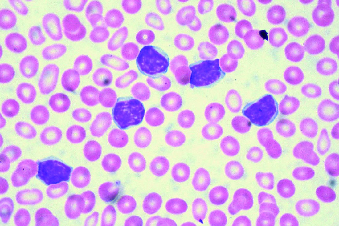
“I think for the most part, there’s enough evidence,” Dr. Allan said at the annual congress on Hematologic Malignancies held by the National Comprehensive Cancer Network.
“If you had one to two lines of therapy, it still favors the novel agents rather than the chemotherapy arms in all these studies,” said Dr. Allan, referring to some of the pivotal trials supporting approval of novel agents in chronic lymphocytic leukemia (CLL). “The earlier we get to these drugs, I believe, the better.”
While venetoclax after ibrutinib is supported by multiple studies, “vice versa is unknown, but there’s seemingly no reason to think it wouldn’t work – different mechanisms of actions, different pathways,” Dr. Allan said.
What is clear, he added, is that retreating those patients with chemoimmunotherapy is not optimal.
In support of that, he cited a multicenter retrospective analysis, which is believed to be the largest real-world experience to date of novel agents in CLL looking at post–kinase inhibitor salvage strategies (Ann Oncol. 2017 May 1;28[5]:1050-6).
Using an alternate kinase inhibitor or venetoclax resulted in superior progression-free survival versus chemoimmunotherapy at the time of initial kinase inhibitor failure in that study, which looked at treatment strategies and outcomes for 683 patients.
Ibrutinib appeared to be superior to idelalisib as a first kinase inhibitor, with significantly better progression-free survival in both frontline and relapsed/refractory settings, and in both complex karyotype and del17p patients, according to the report. Additionally, the response rate to venetoclax seemed superior to that of idelalisib in patients who discontinued ibrutinib because of progression or toxicity.
All of that supports the need for trials to test various sequencing strategies and establish clear treatment algorithms, according to Dr. Allan. “Optimal sequencing is unknown, but real-world data gives us some idea.”
For relapsed/refractory patients, ibrutinib, idelalisib, and venetoclax all have lengthened responses, improved survival, and are approved by the Food and Drug Administration, he added, noting that the toxicity profiles vary and must be understood when dosing and prescribing these agents.
More novel treatments are on the way. On Sept. 24, just days after Dr. Allan’s NCCN presentation, the FDA granted approval to duvelisib for adults with relapsed or refractory CLL or small lymphocytic lymphoma following two or more previous lines of therapy.
Dr. Allan reported financial disclosures related to AbbVie, Acerta Pharma, Genentech, Pharmacyclics, Sunesis Pharmaceuticals, and Verastem Oncology.
NEW YORK – Although optimal sequencing strategies in chronic lymphocytic leukemia are still unclear, real-world data suggest an alternate kinase inhibitor or venetoclax is the best approach for a patient who has received ibrutinib or idelalisib, according to John N. Allan, MD, of Cornell University, New York.
“I think for the most part, there’s enough evidence,” Dr. Allan said at the annual congress on Hematologic Malignancies held by the National Comprehensive Cancer Network.
“If you had one to two lines of therapy, it still favors the novel agents rather than the chemotherapy arms in all these studies,” said Dr. Allan, referring to some of the pivotal trials supporting approval of novel agents in chronic lymphocytic leukemia (CLL). “The earlier we get to these drugs, I believe, the better.”
While venetoclax after ibrutinib is supported by multiple studies, “vice versa is unknown, but there’s seemingly no reason to think it wouldn’t work – different mechanisms of actions, different pathways,” Dr. Allan said.
What is clear, he added, is that retreating those patients with chemoimmunotherapy is not optimal.
In support of that, he cited a multicenter retrospective analysis, which is believed to be the largest real-world experience to date of novel agents in CLL looking at post–kinase inhibitor salvage strategies (Ann Oncol. 2017 May 1;28[5]:1050-6).
Using an alternate kinase inhibitor or venetoclax resulted in superior progression-free survival versus chemoimmunotherapy at the time of initial kinase inhibitor failure in that study, which looked at treatment strategies and outcomes for 683 patients.
Ibrutinib appeared to be superior to idelalisib as a first kinase inhibitor, with significantly better progression-free survival in both frontline and relapsed/refractory settings, and in both complex karyotype and del17p patients, according to the report. Additionally, the response rate to venetoclax seemed superior to that of idelalisib in patients who discontinued ibrutinib because of progression or toxicity.
All of that supports the need for trials to test various sequencing strategies and establish clear treatment algorithms, according to Dr. Allan. “Optimal sequencing is unknown, but real-world data gives us some idea.”
For relapsed/refractory patients, ibrutinib, idelalisib, and venetoclax all have lengthened responses, improved survival, and are approved by the Food and Drug Administration, he added, noting that the toxicity profiles vary and must be understood when dosing and prescribing these agents.
More novel treatments are on the way. On Sept. 24, just days after Dr. Allan’s NCCN presentation, the FDA granted approval to duvelisib for adults with relapsed or refractory CLL or small lymphocytic lymphoma following two or more previous lines of therapy.
Dr. Allan reported financial disclosures related to AbbVie, Acerta Pharma, Genentech, Pharmacyclics, Sunesis Pharmaceuticals, and Verastem Oncology.
NEW YORK – Although optimal sequencing strategies in chronic lymphocytic leukemia are still unclear, real-world data suggest an alternate kinase inhibitor or venetoclax is the best approach for a patient who has received ibrutinib or idelalisib, according to John N. Allan, MD, of Cornell University, New York.
“I think for the most part, there’s enough evidence,” Dr. Allan said at the annual congress on Hematologic Malignancies held by the National Comprehensive Cancer Network.
“If you had one to two lines of therapy, it still favors the novel agents rather than the chemotherapy arms in all these studies,” said Dr. Allan, referring to some of the pivotal trials supporting approval of novel agents in chronic lymphocytic leukemia (CLL). “The earlier we get to these drugs, I believe, the better.”
While venetoclax after ibrutinib is supported by multiple studies, “vice versa is unknown, but there’s seemingly no reason to think it wouldn’t work – different mechanisms of actions, different pathways,” Dr. Allan said.
What is clear, he added, is that retreating those patients with chemoimmunotherapy is not optimal.
In support of that, he cited a multicenter retrospective analysis, which is believed to be the largest real-world experience to date of novel agents in CLL looking at post–kinase inhibitor salvage strategies (Ann Oncol. 2017 May 1;28[5]:1050-6).
Using an alternate kinase inhibitor or venetoclax resulted in superior progression-free survival versus chemoimmunotherapy at the time of initial kinase inhibitor failure in that study, which looked at treatment strategies and outcomes for 683 patients.
Ibrutinib appeared to be superior to idelalisib as a first kinase inhibitor, with significantly better progression-free survival in both frontline and relapsed/refractory settings, and in both complex karyotype and del17p patients, according to the report. Additionally, the response rate to venetoclax seemed superior to that of idelalisib in patients who discontinued ibrutinib because of progression or toxicity.
All of that supports the need for trials to test various sequencing strategies and establish clear treatment algorithms, according to Dr. Allan. “Optimal sequencing is unknown, but real-world data gives us some idea.”
For relapsed/refractory patients, ibrutinib, idelalisib, and venetoclax all have lengthened responses, improved survival, and are approved by the Food and Drug Administration, he added, noting that the toxicity profiles vary and must be understood when dosing and prescribing these agents.
More novel treatments are on the way. On Sept. 24, just days after Dr. Allan’s NCCN presentation, the FDA granted approval to duvelisib for adults with relapsed or refractory CLL or small lymphocytic lymphoma following two or more previous lines of therapy.
Dr. Allan reported financial disclosures related to AbbVie, Acerta Pharma, Genentech, Pharmacyclics, Sunesis Pharmaceuticals, and Verastem Oncology.
EXPERT ANALYSIS FROM NCCN HEMATOLOGIC MALIGNANCIES CONGRESS
Health Care Disparities Dominate at AVAHO Meeting
More than 500 VA hematologists, oncologists, pharmacists, nurses, social workers, and cancer registrars came to the 14th annual Association of VA Hematology/Oncology meeting held outside of Chicago, September 28 through September 30. Mark Klein, MD, succeeded Rusty Crawford, BPharm, as president and William Wachsman, MD, PhD, was named president-elect.
A key focus of the meeting was health care disparities and providing cancer care to underserved populations. An entire track of the meeting was devoted to patients with serious mental illness (SMI), including discussions of the risk of suicide Also a focus was the challenge of providing optimal treatments for patients who may not be adherent because they do not have access to support networks or steady housing.
According to Jessica B. Geller, PhD, MS, and Jessica L. Brand, PhD, a patient with a SMI diagnosis should not preclude considering aggressive treatment but requires a close level of care. Cancer care of patients with SMI requires close collaboration with mental health and pharmacy providers to manage these complex patients. According to Elizabeth Holyman, PsyD, care providers should give serious attention to complaints despite difficult behavior, treat pain, use active listening and provide interpersonal supports, and assume people with SMI have similar needs to the non-SMI.
For both patients with SMI and the entire veteran population, a cancer diagnosis can raise the risk of suicide, explained Catherine Rotolo, LISW-S. Suicidal ideation occurs in nearly 9% of people with cancer, most frequently in the first 3 months after diagnosis, and remains a significant cause of mortality for cancer patients. For patients with cancer, the rate of suicide—already elevated for veterans—is twice that of the baseline rate.
In a keynote address, Lisa Margolies, executive director of the LGBT Cancer Network, cautioned care providers not only to provide a welcoming environment, but also avoid making any assumptions about sexual behavior. “Do ask, we want to tell,” she told attendees, “Make it a part of your regular questions.” According to Margolies, there are more than 1 million LGBT veterans; they use the VA at higher rates, and the best estimate is that there are about 150,000 transgender veterans. According to Margolies, many do not tell their health care providers about their identity; 24% of lesbian, gay, bisexual, and transgender (LGBT) adults withheld information about sexual practices; 9.4% of men who identified as straight had sex with another man within the previous year. In another survey, more than 77% of lesbians had at least 1 sexual experience with a man—8% in the previous year. Margolies’ key point: avoid assumptions about a patient’s identity and simply take a comprehensive medical history so that you better understand the individual in front of you.
Another track of sessions focused on the challenges women face receiving care in the VA. Many female veterans, especially survivors of military sexual trauma (MST), are not comfortable going to the VA for care or being around groups of male veterans. Col (ret) Mona Pearl Treyball, PhD, RN, pointed out that up to 70% of women veterans experienced MST; most never reported it. That point was reinforced by Chad Hamilton, MD, who noted that among veteran women “Significantly higher proportion of users, compared to nonusers reported avoiding VA because of past sexual trauma (19% of users vs 8% of nonusers).
Yeun-hee Anna Park reported on the High Risk Breast Cancer Screening Program, a 10-site pilot project to assess breast cancer risk. The quality improvement program sought to enhance screening for high-risk breast cancer and increase use of chemoprevention and genetic counseling in accordance with national guidelines. In the pilot, women veterans were at increased risk of breast cancer compared with that of the general population (46% vs 13%, respectively), based on a high rate of prior breast biopsies or positive family history. Moreover, posttraumatic stress disorder rates were nearly 3 times the national average. In the program, use of chemoprevention was nearly 2 times the national average.
Female patients undergoing treatment for cancer also face distinct risks related to fertility, bone health, and vasomotor symptoms of menopause. According to Tyler Fenton, PharmD, some cancer treatment approaches involving chemotherapy, radiation, and/or surgery carry a risk of ovarian failure and the accompanying symptoms of premature menopause. Dr. Fenton noted, that menopausal symptoms such as hot flashes are reported to occur in as many as 73% of breast cancer survivors, and 42% of female patients with cancer of reproductive age may develop premature ovarian failure as a result of their chemotherapy.
More than 500 VA hematologists, oncologists, pharmacists, nurses, social workers, and cancer registrars came to the 14th annual Association of VA Hematology/Oncology meeting held outside of Chicago, September 28 through September 30. Mark Klein, MD, succeeded Rusty Crawford, BPharm, as president and William Wachsman, MD, PhD, was named president-elect.
A key focus of the meeting was health care disparities and providing cancer care to underserved populations. An entire track of the meeting was devoted to patients with serious mental illness (SMI), including discussions of the risk of suicide Also a focus was the challenge of providing optimal treatments for patients who may not be adherent because they do not have access to support networks or steady housing.
According to Jessica B. Geller, PhD, MS, and Jessica L. Brand, PhD, a patient with a SMI diagnosis should not preclude considering aggressive treatment but requires a close level of care. Cancer care of patients with SMI requires close collaboration with mental health and pharmacy providers to manage these complex patients. According to Elizabeth Holyman, PsyD, care providers should give serious attention to complaints despite difficult behavior, treat pain, use active listening and provide interpersonal supports, and assume people with SMI have similar needs to the non-SMI.
For both patients with SMI and the entire veteran population, a cancer diagnosis can raise the risk of suicide, explained Catherine Rotolo, LISW-S. Suicidal ideation occurs in nearly 9% of people with cancer, most frequently in the first 3 months after diagnosis, and remains a significant cause of mortality for cancer patients. For patients with cancer, the rate of suicide—already elevated for veterans—is twice that of the baseline rate.
In a keynote address, Lisa Margolies, executive director of the LGBT Cancer Network, cautioned care providers not only to provide a welcoming environment, but also avoid making any assumptions about sexual behavior. “Do ask, we want to tell,” she told attendees, “Make it a part of your regular questions.” According to Margolies, there are more than 1 million LGBT veterans; they use the VA at higher rates, and the best estimate is that there are about 150,000 transgender veterans. According to Margolies, many do not tell their health care providers about their identity; 24% of lesbian, gay, bisexual, and transgender (LGBT) adults withheld information about sexual practices; 9.4% of men who identified as straight had sex with another man within the previous year. In another survey, more than 77% of lesbians had at least 1 sexual experience with a man—8% in the previous year. Margolies’ key point: avoid assumptions about a patient’s identity and simply take a comprehensive medical history so that you better understand the individual in front of you.
Another track of sessions focused on the challenges women face receiving care in the VA. Many female veterans, especially survivors of military sexual trauma (MST), are not comfortable going to the VA for care or being around groups of male veterans. Col (ret) Mona Pearl Treyball, PhD, RN, pointed out that up to 70% of women veterans experienced MST; most never reported it. That point was reinforced by Chad Hamilton, MD, who noted that among veteran women “Significantly higher proportion of users, compared to nonusers reported avoiding VA because of past sexual trauma (19% of users vs 8% of nonusers).
Yeun-hee Anna Park reported on the High Risk Breast Cancer Screening Program, a 10-site pilot project to assess breast cancer risk. The quality improvement program sought to enhance screening for high-risk breast cancer and increase use of chemoprevention and genetic counseling in accordance with national guidelines. In the pilot, women veterans were at increased risk of breast cancer compared with that of the general population (46% vs 13%, respectively), based on a high rate of prior breast biopsies or positive family history. Moreover, posttraumatic stress disorder rates were nearly 3 times the national average. In the program, use of chemoprevention was nearly 2 times the national average.
Female patients undergoing treatment for cancer also face distinct risks related to fertility, bone health, and vasomotor symptoms of menopause. According to Tyler Fenton, PharmD, some cancer treatment approaches involving chemotherapy, radiation, and/or surgery carry a risk of ovarian failure and the accompanying symptoms of premature menopause. Dr. Fenton noted, that menopausal symptoms such as hot flashes are reported to occur in as many as 73% of breast cancer survivors, and 42% of female patients with cancer of reproductive age may develop premature ovarian failure as a result of their chemotherapy.
More than 500 VA hematologists, oncologists, pharmacists, nurses, social workers, and cancer registrars came to the 14th annual Association of VA Hematology/Oncology meeting held outside of Chicago, September 28 through September 30. Mark Klein, MD, succeeded Rusty Crawford, BPharm, as president and William Wachsman, MD, PhD, was named president-elect.
A key focus of the meeting was health care disparities and providing cancer care to underserved populations. An entire track of the meeting was devoted to patients with serious mental illness (SMI), including discussions of the risk of suicide Also a focus was the challenge of providing optimal treatments for patients who may not be adherent because they do not have access to support networks or steady housing.
According to Jessica B. Geller, PhD, MS, and Jessica L. Brand, PhD, a patient with a SMI diagnosis should not preclude considering aggressive treatment but requires a close level of care. Cancer care of patients with SMI requires close collaboration with mental health and pharmacy providers to manage these complex patients. According to Elizabeth Holyman, PsyD, care providers should give serious attention to complaints despite difficult behavior, treat pain, use active listening and provide interpersonal supports, and assume people with SMI have similar needs to the non-SMI.
For both patients with SMI and the entire veteran population, a cancer diagnosis can raise the risk of suicide, explained Catherine Rotolo, LISW-S. Suicidal ideation occurs in nearly 9% of people with cancer, most frequently in the first 3 months after diagnosis, and remains a significant cause of mortality for cancer patients. For patients with cancer, the rate of suicide—already elevated for veterans—is twice that of the baseline rate.
In a keynote address, Lisa Margolies, executive director of the LGBT Cancer Network, cautioned care providers not only to provide a welcoming environment, but also avoid making any assumptions about sexual behavior. “Do ask, we want to tell,” she told attendees, “Make it a part of your regular questions.” According to Margolies, there are more than 1 million LGBT veterans; they use the VA at higher rates, and the best estimate is that there are about 150,000 transgender veterans. According to Margolies, many do not tell their health care providers about their identity; 24% of lesbian, gay, bisexual, and transgender (LGBT) adults withheld information about sexual practices; 9.4% of men who identified as straight had sex with another man within the previous year. In another survey, more than 77% of lesbians had at least 1 sexual experience with a man—8% in the previous year. Margolies’ key point: avoid assumptions about a patient’s identity and simply take a comprehensive medical history so that you better understand the individual in front of you.
Another track of sessions focused on the challenges women face receiving care in the VA. Many female veterans, especially survivors of military sexual trauma (MST), are not comfortable going to the VA for care or being around groups of male veterans. Col (ret) Mona Pearl Treyball, PhD, RN, pointed out that up to 70% of women veterans experienced MST; most never reported it. That point was reinforced by Chad Hamilton, MD, who noted that among veteran women “Significantly higher proportion of users, compared to nonusers reported avoiding VA because of past sexual trauma (19% of users vs 8% of nonusers).
Yeun-hee Anna Park reported on the High Risk Breast Cancer Screening Program, a 10-site pilot project to assess breast cancer risk. The quality improvement program sought to enhance screening for high-risk breast cancer and increase use of chemoprevention and genetic counseling in accordance with national guidelines. In the pilot, women veterans were at increased risk of breast cancer compared with that of the general population (46% vs 13%, respectively), based on a high rate of prior breast biopsies or positive family history. Moreover, posttraumatic stress disorder rates were nearly 3 times the national average. In the program, use of chemoprevention was nearly 2 times the national average.
Female patients undergoing treatment for cancer also face distinct risks related to fertility, bone health, and vasomotor symptoms of menopause. According to Tyler Fenton, PharmD, some cancer treatment approaches involving chemotherapy, radiation, and/or surgery carry a risk of ovarian failure and the accompanying symptoms of premature menopause. Dr. Fenton noted, that menopausal symptoms such as hot flashes are reported to occur in as many as 73% of breast cancer survivors, and 42% of female patients with cancer of reproductive age may develop premature ovarian failure as a result of their chemotherapy.
Outpatient lenalidomide/rituximab yields long-term MCL remission
After 5 years, the combination of lenalidomide and rituximab as first-line therapy for mantle cell lymphoma (MCL) continues to show durable responses with manageable toxicities, long-term results from a phase 2 clinical trial show.
After a median follow-up of 64 months, 21 of 33 patients with initial responses remained in durable, minimal residual disease (MRD)-negative remission following induction with lenalidomide and rituximab and maintenance with those same two agents for at least 3 years.
The patients with durable responses included five who opted to discontinue maintenance after 3 years, reported Jia Ruan, MD, PhD, of Cornell University, New York, and her colleagues.
“Our long-term data provide proof of concept that an outpatient-based induction and maintenance strategy free of conventional chemotherapy is effective, safe, and feasible as first-line therapy for MCL,” they wrote. Their report was published in Blood.
In the multicenter, phase 2 single-arm study, 38 patients with untreated MCL were enrolled and treated with lenalidomide 20 mg daily on days 1-21 of each 28-day cycle for 12 cycles during induction, followed by dose reduction to 15 mg during the maintenance phase. Patients also received standard dose rituximab 375 mg/m2 weekly for 4 weeks during cycle 1, then once every other cycle.
Patients remained on treatment until disease progression, unacceptable toxicities, or study withdrawal. Patients who remained in remission after 3 years, based on routine surveillance CT scans, had the option to discontinue maintenance.
Of the original 38 patients enrolled, 36 were evaluable for response, including 23 with a complete response (CR) and 10 with a partial response.
At the 64-month median follow-up, neither the median progression-free survival (PFS) nor duration of response had been reached.
Overall, 21 of the 33 patients with responses (64%) had ongoing responses, including six patients with responses beyond 6 years.
Estimated 3-year and 5-year PFS rates were 80.3% and 63.9%, respectively. Respective estimated 3- and 5-year overall survival rates were 89.5% and 77.4%.
Mantle cell lymphoma international prognostic index (MIPI) scores were not associated with either response or PFS rates, but patients with high-risk MIPI scores were significantly more likely to have worse overall survival (P = .04).
Grade 3 or greater hematologic toxicities included neutropenia in 42% of patients in both induction and maintenance, anemia in 8% and 3%, thrombocytopenia in 11% and 5%, and febrile neutropenia in 3% and 5%.
Secondary primary malignancies occurred in six patients. These included five noninvasive skin cancers requiring only local therapy without the need for study interruption. Two patients, including one with a skin cancer, died from the secondary malignancies, including one from Merkel cell carcinoma and one from pancreatic cancer.
“The efficacy and survival outcome observed in our study compared favorably to those reported with lenalidomide either as single agent, or in combination with rituximab in relapsed and refractory setting, lending support for prioritizing novel agents such as lenalidomide early in the treatment sequence, to compare to conventional chemotherapy-based approach,” the investigators wrote.
The study was supported in part by Celgene Corporation, a Clinical Translational Science Center grant, and the Lymphoma Foundation. Dr. Ruan has received research support and been a consultant for Celgene, and other coauthors reported research support and consultant relationships with the company.
SOURCE: Ruan J et al. Blood. 2018 Sep 4. doi: 10.1182/blood-2018-07-859769.
After 5 years, the combination of lenalidomide and rituximab as first-line therapy for mantle cell lymphoma (MCL) continues to show durable responses with manageable toxicities, long-term results from a phase 2 clinical trial show.
After a median follow-up of 64 months, 21 of 33 patients with initial responses remained in durable, minimal residual disease (MRD)-negative remission following induction with lenalidomide and rituximab and maintenance with those same two agents for at least 3 years.
The patients with durable responses included five who opted to discontinue maintenance after 3 years, reported Jia Ruan, MD, PhD, of Cornell University, New York, and her colleagues.
“Our long-term data provide proof of concept that an outpatient-based induction and maintenance strategy free of conventional chemotherapy is effective, safe, and feasible as first-line therapy for MCL,” they wrote. Their report was published in Blood.
In the multicenter, phase 2 single-arm study, 38 patients with untreated MCL were enrolled and treated with lenalidomide 20 mg daily on days 1-21 of each 28-day cycle for 12 cycles during induction, followed by dose reduction to 15 mg during the maintenance phase. Patients also received standard dose rituximab 375 mg/m2 weekly for 4 weeks during cycle 1, then once every other cycle.
Patients remained on treatment until disease progression, unacceptable toxicities, or study withdrawal. Patients who remained in remission after 3 years, based on routine surveillance CT scans, had the option to discontinue maintenance.
Of the original 38 patients enrolled, 36 were evaluable for response, including 23 with a complete response (CR) and 10 with a partial response.
At the 64-month median follow-up, neither the median progression-free survival (PFS) nor duration of response had been reached.
Overall, 21 of the 33 patients with responses (64%) had ongoing responses, including six patients with responses beyond 6 years.
Estimated 3-year and 5-year PFS rates were 80.3% and 63.9%, respectively. Respective estimated 3- and 5-year overall survival rates were 89.5% and 77.4%.
Mantle cell lymphoma international prognostic index (MIPI) scores were not associated with either response or PFS rates, but patients with high-risk MIPI scores were significantly more likely to have worse overall survival (P = .04).
Grade 3 or greater hematologic toxicities included neutropenia in 42% of patients in both induction and maintenance, anemia in 8% and 3%, thrombocytopenia in 11% and 5%, and febrile neutropenia in 3% and 5%.
Secondary primary malignancies occurred in six patients. These included five noninvasive skin cancers requiring only local therapy without the need for study interruption. Two patients, including one with a skin cancer, died from the secondary malignancies, including one from Merkel cell carcinoma and one from pancreatic cancer.
“The efficacy and survival outcome observed in our study compared favorably to those reported with lenalidomide either as single agent, or in combination with rituximab in relapsed and refractory setting, lending support for prioritizing novel agents such as lenalidomide early in the treatment sequence, to compare to conventional chemotherapy-based approach,” the investigators wrote.
The study was supported in part by Celgene Corporation, a Clinical Translational Science Center grant, and the Lymphoma Foundation. Dr. Ruan has received research support and been a consultant for Celgene, and other coauthors reported research support and consultant relationships with the company.
SOURCE: Ruan J et al. Blood. 2018 Sep 4. doi: 10.1182/blood-2018-07-859769.
After 5 years, the combination of lenalidomide and rituximab as first-line therapy for mantle cell lymphoma (MCL) continues to show durable responses with manageable toxicities, long-term results from a phase 2 clinical trial show.
After a median follow-up of 64 months, 21 of 33 patients with initial responses remained in durable, minimal residual disease (MRD)-negative remission following induction with lenalidomide and rituximab and maintenance with those same two agents for at least 3 years.
The patients with durable responses included five who opted to discontinue maintenance after 3 years, reported Jia Ruan, MD, PhD, of Cornell University, New York, and her colleagues.
“Our long-term data provide proof of concept that an outpatient-based induction and maintenance strategy free of conventional chemotherapy is effective, safe, and feasible as first-line therapy for MCL,” they wrote. Their report was published in Blood.
In the multicenter, phase 2 single-arm study, 38 patients with untreated MCL were enrolled and treated with lenalidomide 20 mg daily on days 1-21 of each 28-day cycle for 12 cycles during induction, followed by dose reduction to 15 mg during the maintenance phase. Patients also received standard dose rituximab 375 mg/m2 weekly for 4 weeks during cycle 1, then once every other cycle.
Patients remained on treatment until disease progression, unacceptable toxicities, or study withdrawal. Patients who remained in remission after 3 years, based on routine surveillance CT scans, had the option to discontinue maintenance.
Of the original 38 patients enrolled, 36 were evaluable for response, including 23 with a complete response (CR) and 10 with a partial response.
At the 64-month median follow-up, neither the median progression-free survival (PFS) nor duration of response had been reached.
Overall, 21 of the 33 patients with responses (64%) had ongoing responses, including six patients with responses beyond 6 years.
Estimated 3-year and 5-year PFS rates were 80.3% and 63.9%, respectively. Respective estimated 3- and 5-year overall survival rates were 89.5% and 77.4%.
Mantle cell lymphoma international prognostic index (MIPI) scores were not associated with either response or PFS rates, but patients with high-risk MIPI scores were significantly more likely to have worse overall survival (P = .04).
Grade 3 or greater hematologic toxicities included neutropenia in 42% of patients in both induction and maintenance, anemia in 8% and 3%, thrombocytopenia in 11% and 5%, and febrile neutropenia in 3% and 5%.
Secondary primary malignancies occurred in six patients. These included five noninvasive skin cancers requiring only local therapy without the need for study interruption. Two patients, including one with a skin cancer, died from the secondary malignancies, including one from Merkel cell carcinoma and one from pancreatic cancer.
“The efficacy and survival outcome observed in our study compared favorably to those reported with lenalidomide either as single agent, or in combination with rituximab in relapsed and refractory setting, lending support for prioritizing novel agents such as lenalidomide early in the treatment sequence, to compare to conventional chemotherapy-based approach,” the investigators wrote.
The study was supported in part by Celgene Corporation, a Clinical Translational Science Center grant, and the Lymphoma Foundation. Dr. Ruan has received research support and been a consultant for Celgene, and other coauthors reported research support and consultant relationships with the company.
SOURCE: Ruan J et al. Blood. 2018 Sep 4. doi: 10.1182/blood-2018-07-859769.
FROM BLOOD
Key clinical point:
Major finding: After 64-months of median follow-up, 21 of 33 patients with initial responses remained in remission.
Study details: Five-year follow-up of a phase 2 single arm trial of lenalidomide/rituximab induction and maintenance in 38 patients with mantle cell lymphoma.
Disclosures: The study was supported in part by Celgene Corporation, a Clinical Translational Science Center grant, and the Lymphoma Foundation. Dr. Ruan has received research support and been a consultant for Celgene, and other coauthors reported research support and consultant relationships with the company.
Source: Ruan J et al. Blood. 2018 Sep 4. doi: 10.1182/blood-2018-07-859769.
Neoadjuvant combo yields high pathologic response in NSCLC
TORONTO – Neoadjuvant combined chemotherapy and immunotherapy (CT-IO) yielded high pathologic response rates for locally advanced, potentially resectable non-small cell lung cancer in the phase 2 multicenter study Nadim study.
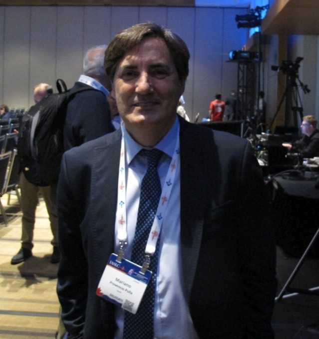
All 30 adults with stage IIIA N2 non-small cell lung cancer (NSCLC) who enrolled in the ongoing single-arm, open-label study and have undergone surgery to date, had resectable tumors following neoadjuvant treatment, Mariano Provencio, MD, of Hospital Puerta de Hierro, Madrid, Spain, reported at the World Conference on Lung Cancer.
The complete and partial response rates following the treatment were 10% and 60%, respectively, and the remaining patients had stable disease, Dr. Provencio said, noting that an additional 10 enrolled patients have completed treatment and are awaiting resection, and another 3 are still completing treatment.
The overall major pathologic response rate was 80%, including 75% with complete remission, he said at the conference, which was sponsored by the International Association for the Study of Lung Cancer.
“With a median follow-up of 4 months, no patients suffered any recurrence,” he said. “We have been unable to identify any significant factor related to the response, but we still do not have information about biomarkers.”
The study participants had a median age of 64 years, most (73%) were men, and all were current or former smokers. All received the planned neoadjuvant doses, which included 3 cycles of intravenous nivolumab at a dose of 360 mg every 3 weeks, plus 200 mg/m2 of paclitaxel and carboplatin at AUC 6 every 3 weeks.
“The most important toxicity was hematological toxicity related to chemotherapy; there were very few episodes of non-hematological toxicity,” he said.
Tumors were then assessed, and surgery–lobectomy in 90% of cases–was performed in the 3rd or 4th week after day 21 of the third neoadjuvant treatment cycle.
“There were no intraoperative complications, and 7 patients experienced some postoperative complications, but there was no postoperative mortality,” he said.
Adjuvant treatment included 240 mg of intravenous nivolumab every 2 weeks for 4 months and then 480 mg every 4 weeks for 8 months after resection.
The primary end-point of the study is 24-month progression-free survival.
CT-IO has a high response rate and longer survival in unselected patients with metastatic NSCLC, but there were previously no data about this combination in the neoadjuvant setting, Dr. Provencio said.
The findings of this ongoing study are limited by their preliminary nature and the related lack of information about overall survival and biomarkers, Dr. Provencio said.
“But the higher complete remission and major pathological response rates make this a promising future treatment in stage 3 [NSCLC,] he said.
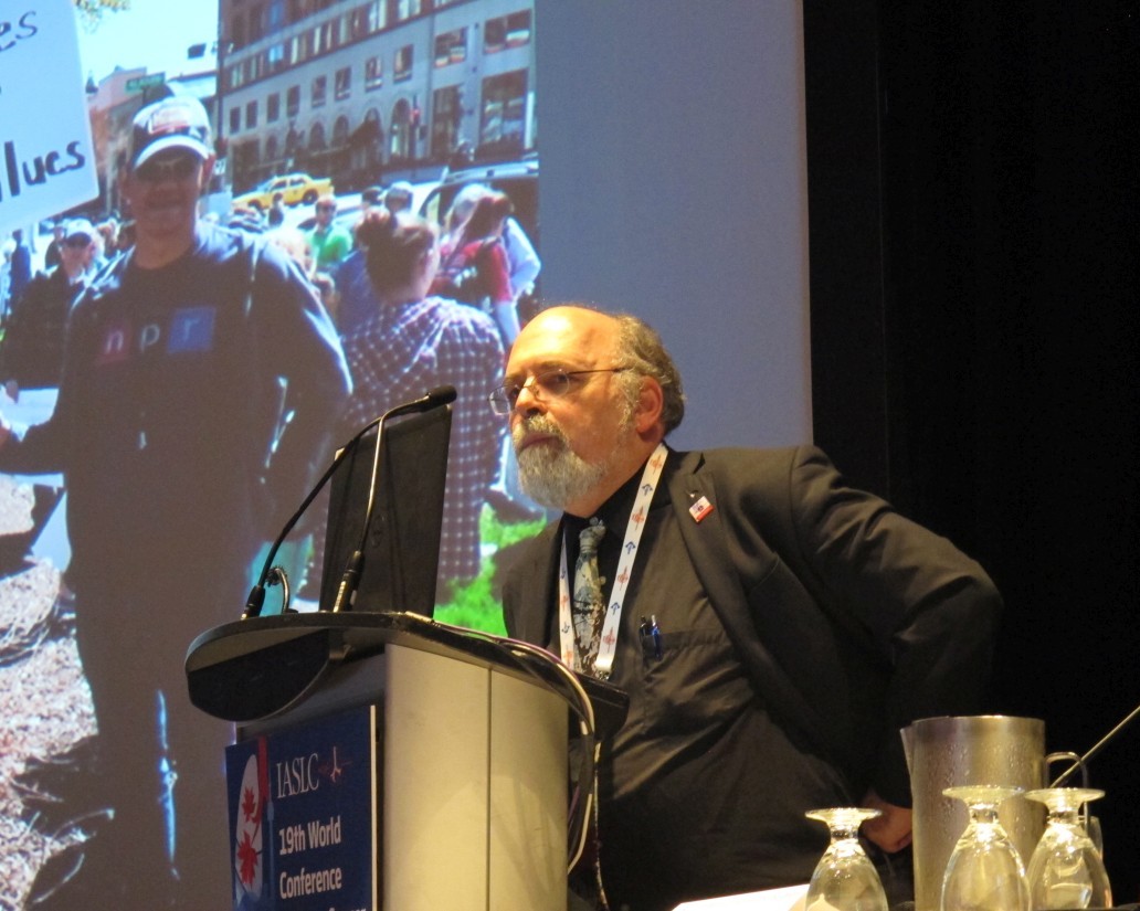
Indeed, the Nadim study thus far has shown “an amazing result that needs to be verified ... [there is] no prospective proof yet in this context that major pathologic response is parlayed with improved [progression-free survival] or [overall survival],” said discussant Corey Langer, MD, professor of medicine and director of thoracic oncology at the University of Pennsylvania, Philadelphia. “We need to track the late sequelae and [central nervous system] recurrences.”
Dr. Provencio reported relationships with AstraZeneca, Bristol-Meyers Squibb, Fabre, Merck, Pierre, Roche, and Bristol-Meyers Squibb. Dr. Langer reported relationships with numerous companies and organizations.
SOURCE: Provencio, Mario et al., WCLC 2018 Abstract OA01.05.
TORONTO – Neoadjuvant combined chemotherapy and immunotherapy (CT-IO) yielded high pathologic response rates for locally advanced, potentially resectable non-small cell lung cancer in the phase 2 multicenter study Nadim study.
All 30 adults with stage IIIA N2 non-small cell lung cancer (NSCLC) who enrolled in the ongoing single-arm, open-label study and have undergone surgery to date, had resectable tumors following neoadjuvant treatment, Mariano Provencio, MD, of Hospital Puerta de Hierro, Madrid, Spain, reported at the World Conference on Lung Cancer.
The complete and partial response rates following the treatment were 10% and 60%, respectively, and the remaining patients had stable disease, Dr. Provencio said, noting that an additional 10 enrolled patients have completed treatment and are awaiting resection, and another 3 are still completing treatment.
The overall major pathologic response rate was 80%, including 75% with complete remission, he said at the conference, which was sponsored by the International Association for the Study of Lung Cancer.
“With a median follow-up of 4 months, no patients suffered any recurrence,” he said. “We have been unable to identify any significant factor related to the response, but we still do not have information about biomarkers.”
The study participants had a median age of 64 years, most (73%) were men, and all were current or former smokers. All received the planned neoadjuvant doses, which included 3 cycles of intravenous nivolumab at a dose of 360 mg every 3 weeks, plus 200 mg/m2 of paclitaxel and carboplatin at AUC 6 every 3 weeks.
“The most important toxicity was hematological toxicity related to chemotherapy; there were very few episodes of non-hematological toxicity,” he said.
Tumors were then assessed, and surgery–lobectomy in 90% of cases–was performed in the 3rd or 4th week after day 21 of the third neoadjuvant treatment cycle.
“There were no intraoperative complications, and 7 patients experienced some postoperative complications, but there was no postoperative mortality,” he said.
Adjuvant treatment included 240 mg of intravenous nivolumab every 2 weeks for 4 months and then 480 mg every 4 weeks for 8 months after resection.
The primary end-point of the study is 24-month progression-free survival.
CT-IO has a high response rate and longer survival in unselected patients with metastatic NSCLC, but there were previously no data about this combination in the neoadjuvant setting, Dr. Provencio said.
The findings of this ongoing study are limited by their preliminary nature and the related lack of information about overall survival and biomarkers, Dr. Provencio said.
“But the higher complete remission and major pathological response rates make this a promising future treatment in stage 3 [NSCLC,] he said.
Indeed, the Nadim study thus far has shown “an amazing result that needs to be verified ... [there is] no prospective proof yet in this context that major pathologic response is parlayed with improved [progression-free survival] or [overall survival],” said discussant Corey Langer, MD, professor of medicine and director of thoracic oncology at the University of Pennsylvania, Philadelphia. “We need to track the late sequelae and [central nervous system] recurrences.”
Dr. Provencio reported relationships with AstraZeneca, Bristol-Meyers Squibb, Fabre, Merck, Pierre, Roche, and Bristol-Meyers Squibb. Dr. Langer reported relationships with numerous companies and organizations.
SOURCE: Provencio, Mario et al., WCLC 2018 Abstract OA01.05.
TORONTO – Neoadjuvant combined chemotherapy and immunotherapy (CT-IO) yielded high pathologic response rates for locally advanced, potentially resectable non-small cell lung cancer in the phase 2 multicenter study Nadim study.
All 30 adults with stage IIIA N2 non-small cell lung cancer (NSCLC) who enrolled in the ongoing single-arm, open-label study and have undergone surgery to date, had resectable tumors following neoadjuvant treatment, Mariano Provencio, MD, of Hospital Puerta de Hierro, Madrid, Spain, reported at the World Conference on Lung Cancer.
The complete and partial response rates following the treatment were 10% and 60%, respectively, and the remaining patients had stable disease, Dr. Provencio said, noting that an additional 10 enrolled patients have completed treatment and are awaiting resection, and another 3 are still completing treatment.
The overall major pathologic response rate was 80%, including 75% with complete remission, he said at the conference, which was sponsored by the International Association for the Study of Lung Cancer.
“With a median follow-up of 4 months, no patients suffered any recurrence,” he said. “We have been unable to identify any significant factor related to the response, but we still do not have information about biomarkers.”
The study participants had a median age of 64 years, most (73%) were men, and all were current or former smokers. All received the planned neoadjuvant doses, which included 3 cycles of intravenous nivolumab at a dose of 360 mg every 3 weeks, plus 200 mg/m2 of paclitaxel and carboplatin at AUC 6 every 3 weeks.
“The most important toxicity was hematological toxicity related to chemotherapy; there were very few episodes of non-hematological toxicity,” he said.
Tumors were then assessed, and surgery–lobectomy in 90% of cases–was performed in the 3rd or 4th week after day 21 of the third neoadjuvant treatment cycle.
“There were no intraoperative complications, and 7 patients experienced some postoperative complications, but there was no postoperative mortality,” he said.
Adjuvant treatment included 240 mg of intravenous nivolumab every 2 weeks for 4 months and then 480 mg every 4 weeks for 8 months after resection.
The primary end-point of the study is 24-month progression-free survival.
CT-IO has a high response rate and longer survival in unselected patients with metastatic NSCLC, but there were previously no data about this combination in the neoadjuvant setting, Dr. Provencio said.
The findings of this ongoing study are limited by their preliminary nature and the related lack of information about overall survival and biomarkers, Dr. Provencio said.
“But the higher complete remission and major pathological response rates make this a promising future treatment in stage 3 [NSCLC,] he said.
Indeed, the Nadim study thus far has shown “an amazing result that needs to be verified ... [there is] no prospective proof yet in this context that major pathologic response is parlayed with improved [progression-free survival] or [overall survival],” said discussant Corey Langer, MD, professor of medicine and director of thoracic oncology at the University of Pennsylvania, Philadelphia. “We need to track the late sequelae and [central nervous system] recurrences.”
Dr. Provencio reported relationships with AstraZeneca, Bristol-Meyers Squibb, Fabre, Merck, Pierre, Roche, and Bristol-Meyers Squibb. Dr. Langer reported relationships with numerous companies and organizations.
SOURCE: Provencio, Mario et al., WCLC 2018 Abstract OA01.05.
REPORTING FROM WCLC 2018
Key clinical point: Neoadjuvant CT-IO yielded high pathologic response rates for stage IIIA N2 NSCLC.
Major finding: The major pathologic response rate was 80%, including 10% with a complete response.
Study details: A phase 2, single-arm, open-label study of 30 patients.
Disclosures: Dr. Provencio reported relationships with AstraZeneca, Bristol-Meyers Squibb, Fabre, Merck, Pierre, Roche, and Bristol-Meyers Squibb. Dr. Langer reported relationships with numerous companies and organizations.
Source: Provencio, Mario et al., WCLC 2018 Abstract OA01.05.
Disappointing Results From a Cetuximab Study
Cetuximab plus radiation therapy has worse outcomes than the current standard of radiation and cisplatin for patients with human papillomavirus-positive (HPV+) oropharyngeal cancer. In fact, the researchers, who reported preliminary findings from a phase 3 study funded by the National Cancer Institute (NCI), were “surprised by the loss of tumor control with cetuximab.”
Cetuximab with radiation is an accepted standard of care, especially for patients who cannot tolerate cisplatin, and it is approved for patients with head and neck cancer, including oropharyngeal cancer. Researchers and the NCI are looking for more ways to “de-escalate” therapies for cancers that have a good prognosis, such as HPV+ cancer of the oropharynx. The goal of this trial was to find an alternative to cisplatin that would control the cancer as effectively but with fewer side effects.
The researchers enrolled 849 patients to randomly receive either cetuximab or cisplatin with radiation. The third, final interim analysis, after a median follow-up of 4.5 years, found that overall survival on the cetuximab arm was significantly inferior to the cisplatin arm. Moreover, serious adverse events were similar in both groups, although toxic side effects were more common in the cisplatin arm.
The study is the first randomized clinical trial specifically designed for patients with HPV+ oropharyngeal cancer, and “it establishes cisplatin with radiation as the standard of care.”
Cetuximab plus radiation therapy has worse outcomes than the current standard of radiation and cisplatin for patients with human papillomavirus-positive (HPV+) oropharyngeal cancer. In fact, the researchers, who reported preliminary findings from a phase 3 study funded by the National Cancer Institute (NCI), were “surprised by the loss of tumor control with cetuximab.”
Cetuximab with radiation is an accepted standard of care, especially for patients who cannot tolerate cisplatin, and it is approved for patients with head and neck cancer, including oropharyngeal cancer. Researchers and the NCI are looking for more ways to “de-escalate” therapies for cancers that have a good prognosis, such as HPV+ cancer of the oropharynx. The goal of this trial was to find an alternative to cisplatin that would control the cancer as effectively but with fewer side effects.
The researchers enrolled 849 patients to randomly receive either cetuximab or cisplatin with radiation. The third, final interim analysis, after a median follow-up of 4.5 years, found that overall survival on the cetuximab arm was significantly inferior to the cisplatin arm. Moreover, serious adverse events were similar in both groups, although toxic side effects were more common in the cisplatin arm.
The study is the first randomized clinical trial specifically designed for patients with HPV+ oropharyngeal cancer, and “it establishes cisplatin with radiation as the standard of care.”
Cetuximab plus radiation therapy has worse outcomes than the current standard of radiation and cisplatin for patients with human papillomavirus-positive (HPV+) oropharyngeal cancer. In fact, the researchers, who reported preliminary findings from a phase 3 study funded by the National Cancer Institute (NCI), were “surprised by the loss of tumor control with cetuximab.”
Cetuximab with radiation is an accepted standard of care, especially for patients who cannot tolerate cisplatin, and it is approved for patients with head and neck cancer, including oropharyngeal cancer. Researchers and the NCI are looking for more ways to “de-escalate” therapies for cancers that have a good prognosis, such as HPV+ cancer of the oropharynx. The goal of this trial was to find an alternative to cisplatin that would control the cancer as effectively but with fewer side effects.
The researchers enrolled 849 patients to randomly receive either cetuximab or cisplatin with radiation. The third, final interim analysis, after a median follow-up of 4.5 years, found that overall survival on the cetuximab arm was significantly inferior to the cisplatin arm. Moreover, serious adverse events were similar in both groups, although toxic side effects were more common in the cisplatin arm.
The study is the first randomized clinical trial specifically designed for patients with HPV+ oropharyngeal cancer, and “it establishes cisplatin with radiation as the standard of care.”
Fatal toxicities from checkpoint inhibitors vary by agent
Fatal adverse events are uncommon with immune checkpoint inhibitors (ICI), but they are not unknown, and it’s important to recognize rare, potentially lethal side effects with these agents, investigators say.
A systematic review and meta-analysis of records from academic medical centers and from a pharmacovigilance database found that fatal side adverse events varied in nature and severity according to the agent and immune checkpoint targeted, reported Daniel Y. Wang, MD, of Vanderbilt University Medical Center, Nashville, Tenn., and his colleagues.
“This study underscores that the risk of death associated with complications of ICI therapy is real, but within or well below fatality rates for common oncologic interventions,” they wrote in JAMA Oncology.
In their study, rates of fatal toxic effects associated with immune checkpoint inhibitors ranged from 0.3% to 1.3%. In contrast, the investigators noted, platinum doublet chemotherapy is associated with a 0.9% rate of fatal toxicities, targeted therapies with angiogenesis inhibitors or tyrosine kinase inhibitors have had fatal toxicity rates up to 4%, and allogeneic hematopoietic stem cell transplants are associated with a fatal adverse event rate of approximately 15%. In addition, the death rate associated with complex oncologic surgeries such as the Whipple procedure or esophagectomy ranges from 1% to 10%.
The authors queried the World Health Organization pharmacovigilance database, called Vigilyze, which contains data on more than 16 million adverse drug reactions; records from patients treated with checkpoint inhibitors in seven academic centers in the United States, Germany, and Australia; and published clinical trials.
They combed through the data to identify fatal toxic effects associated with the anti-programmed death 1/ligand-1 (PD-1/PD-L1) inhibitors nivolumab (Opdivo), pembrolizumab (Keytruda), atezolizumab (Tecentriq), avelumab (Bavencio), and durvalumab (Imfinzi), and with the cytotoxic T-lymphocyte antigen-4 (CTLA-4) inhibitors ipilimumab (Yervoy) and tremelimumab (investigational).
They found in the Vigilyze data that of 193 deaths attributed to CTLA-4 inhibitors, 70% were from colitis, 16% from hepatitis, 8% from pneumonitis, and the remainder from other causes. In contrast, the most common fatal toxicities associated with PD-1/PD-L1 inhibitors were pneumonitis in 35%, hepatitis in 22%, colitis in 17%, and the remainder from other causes.
With the two classes of ICIs used in combination, primarily for treatment of malignant melanoma, colitis accounted for 37% of toxic fatal events, followed by myocarditis in 25%, hepatitis in 22%, pneumonitis in 14%, and others.
The events occurred early in the course of therapy, at a median of 14.5 days from the start of therapy for combination treatment, and 40 days each for anti-PD-1/PD-L1 and anti-CTLA-4 agents.
The complication with highest fatality rate was myocarditis, which killed 39.7% of affected patients. In contrast, endocrine events and colitis were fatal in only 2%-5% of cases. The fatality rates associated with toxic effects in other organ systems ranged from 10% to 17%.
A review of data on 3,545 patients treated with immune checkpoint inhibitors at the seven academic centers found a 0.6% fatality rate, with cardiac and neurologic events accounting for 43% of the deaths.
The investigators also conducted a meta-analysis of data from 112 clinical trials with a total of 19,217 patients treated with ICIs. In these studies, anti-PD-1 agents were associated with a 0.36% rate of fatal toxicities, anti-PD-L1 agents were associated with a 0.38% rate, anti-CTLA-4 agents were linked to a 1.08% rate, and combined PD-1/PD-L1 and CTLA-4 therapy was associated with a 1.23% fatal toxicity rate.
“Each database largely validated the patterns of death from distinct regimens with a few exceptions. Intriguingly, our retrospective analysis at large, experienced academic centers suggested that neurologic and cardiac toxic effects comprised nearly half of deaths. Thus, we surmise that more optimized treatment of these events is urgently needed. In addition, persistent deaths from colitis and pneumonitis, events with standardized treatment algorithms and clear symptoms, suggests that patient and physician education remains a critical objective,” the investigators wrote.
SOURCE: Wang DY et al. JAMA Oncol. 2018 Sept 13. doi: 10.1001/jamaoncol.2018.3923.
Fatal adverse events are uncommon with immune checkpoint inhibitors (ICI), but they are not unknown, and it’s important to recognize rare, potentially lethal side effects with these agents, investigators say.
A systematic review and meta-analysis of records from academic medical centers and from a pharmacovigilance database found that fatal side adverse events varied in nature and severity according to the agent and immune checkpoint targeted, reported Daniel Y. Wang, MD, of Vanderbilt University Medical Center, Nashville, Tenn., and his colleagues.
“This study underscores that the risk of death associated with complications of ICI therapy is real, but within or well below fatality rates for common oncologic interventions,” they wrote in JAMA Oncology.
In their study, rates of fatal toxic effects associated with immune checkpoint inhibitors ranged from 0.3% to 1.3%. In contrast, the investigators noted, platinum doublet chemotherapy is associated with a 0.9% rate of fatal toxicities, targeted therapies with angiogenesis inhibitors or tyrosine kinase inhibitors have had fatal toxicity rates up to 4%, and allogeneic hematopoietic stem cell transplants are associated with a fatal adverse event rate of approximately 15%. In addition, the death rate associated with complex oncologic surgeries such as the Whipple procedure or esophagectomy ranges from 1% to 10%.
The authors queried the World Health Organization pharmacovigilance database, called Vigilyze, which contains data on more than 16 million adverse drug reactions; records from patients treated with checkpoint inhibitors in seven academic centers in the United States, Germany, and Australia; and published clinical trials.
They combed through the data to identify fatal toxic effects associated with the anti-programmed death 1/ligand-1 (PD-1/PD-L1) inhibitors nivolumab (Opdivo), pembrolizumab (Keytruda), atezolizumab (Tecentriq), avelumab (Bavencio), and durvalumab (Imfinzi), and with the cytotoxic T-lymphocyte antigen-4 (CTLA-4) inhibitors ipilimumab (Yervoy) and tremelimumab (investigational).
They found in the Vigilyze data that of 193 deaths attributed to CTLA-4 inhibitors, 70% were from colitis, 16% from hepatitis, 8% from pneumonitis, and the remainder from other causes. In contrast, the most common fatal toxicities associated with PD-1/PD-L1 inhibitors were pneumonitis in 35%, hepatitis in 22%, colitis in 17%, and the remainder from other causes.
With the two classes of ICIs used in combination, primarily for treatment of malignant melanoma, colitis accounted for 37% of toxic fatal events, followed by myocarditis in 25%, hepatitis in 22%, pneumonitis in 14%, and others.
The events occurred early in the course of therapy, at a median of 14.5 days from the start of therapy for combination treatment, and 40 days each for anti-PD-1/PD-L1 and anti-CTLA-4 agents.
The complication with highest fatality rate was myocarditis, which killed 39.7% of affected patients. In contrast, endocrine events and colitis were fatal in only 2%-5% of cases. The fatality rates associated with toxic effects in other organ systems ranged from 10% to 17%.
A review of data on 3,545 patients treated with immune checkpoint inhibitors at the seven academic centers found a 0.6% fatality rate, with cardiac and neurologic events accounting for 43% of the deaths.
The investigators also conducted a meta-analysis of data from 112 clinical trials with a total of 19,217 patients treated with ICIs. In these studies, anti-PD-1 agents were associated with a 0.36% rate of fatal toxicities, anti-PD-L1 agents were associated with a 0.38% rate, anti-CTLA-4 agents were linked to a 1.08% rate, and combined PD-1/PD-L1 and CTLA-4 therapy was associated with a 1.23% fatal toxicity rate.
“Each database largely validated the patterns of death from distinct regimens with a few exceptions. Intriguingly, our retrospective analysis at large, experienced academic centers suggested that neurologic and cardiac toxic effects comprised nearly half of deaths. Thus, we surmise that more optimized treatment of these events is urgently needed. In addition, persistent deaths from colitis and pneumonitis, events with standardized treatment algorithms and clear symptoms, suggests that patient and physician education remains a critical objective,” the investigators wrote.
SOURCE: Wang DY et al. JAMA Oncol. 2018 Sept 13. doi: 10.1001/jamaoncol.2018.3923.
Fatal adverse events are uncommon with immune checkpoint inhibitors (ICI), but they are not unknown, and it’s important to recognize rare, potentially lethal side effects with these agents, investigators say.
A systematic review and meta-analysis of records from academic medical centers and from a pharmacovigilance database found that fatal side adverse events varied in nature and severity according to the agent and immune checkpoint targeted, reported Daniel Y. Wang, MD, of Vanderbilt University Medical Center, Nashville, Tenn., and his colleagues.
“This study underscores that the risk of death associated with complications of ICI therapy is real, but within or well below fatality rates for common oncologic interventions,” they wrote in JAMA Oncology.
In their study, rates of fatal toxic effects associated with immune checkpoint inhibitors ranged from 0.3% to 1.3%. In contrast, the investigators noted, platinum doublet chemotherapy is associated with a 0.9% rate of fatal toxicities, targeted therapies with angiogenesis inhibitors or tyrosine kinase inhibitors have had fatal toxicity rates up to 4%, and allogeneic hematopoietic stem cell transplants are associated with a fatal adverse event rate of approximately 15%. In addition, the death rate associated with complex oncologic surgeries such as the Whipple procedure or esophagectomy ranges from 1% to 10%.
The authors queried the World Health Organization pharmacovigilance database, called Vigilyze, which contains data on more than 16 million adverse drug reactions; records from patients treated with checkpoint inhibitors in seven academic centers in the United States, Germany, and Australia; and published clinical trials.
They combed through the data to identify fatal toxic effects associated with the anti-programmed death 1/ligand-1 (PD-1/PD-L1) inhibitors nivolumab (Opdivo), pembrolizumab (Keytruda), atezolizumab (Tecentriq), avelumab (Bavencio), and durvalumab (Imfinzi), and with the cytotoxic T-lymphocyte antigen-4 (CTLA-4) inhibitors ipilimumab (Yervoy) and tremelimumab (investigational).
They found in the Vigilyze data that of 193 deaths attributed to CTLA-4 inhibitors, 70% were from colitis, 16% from hepatitis, 8% from pneumonitis, and the remainder from other causes. In contrast, the most common fatal toxicities associated with PD-1/PD-L1 inhibitors were pneumonitis in 35%, hepatitis in 22%, colitis in 17%, and the remainder from other causes.
With the two classes of ICIs used in combination, primarily for treatment of malignant melanoma, colitis accounted for 37% of toxic fatal events, followed by myocarditis in 25%, hepatitis in 22%, pneumonitis in 14%, and others.
The events occurred early in the course of therapy, at a median of 14.5 days from the start of therapy for combination treatment, and 40 days each for anti-PD-1/PD-L1 and anti-CTLA-4 agents.
The complication with highest fatality rate was myocarditis, which killed 39.7% of affected patients. In contrast, endocrine events and colitis were fatal in only 2%-5% of cases. The fatality rates associated with toxic effects in other organ systems ranged from 10% to 17%.
A review of data on 3,545 patients treated with immune checkpoint inhibitors at the seven academic centers found a 0.6% fatality rate, with cardiac and neurologic events accounting for 43% of the deaths.
The investigators also conducted a meta-analysis of data from 112 clinical trials with a total of 19,217 patients treated with ICIs. In these studies, anti-PD-1 agents were associated with a 0.36% rate of fatal toxicities, anti-PD-L1 agents were associated with a 0.38% rate, anti-CTLA-4 agents were linked to a 1.08% rate, and combined PD-1/PD-L1 and CTLA-4 therapy was associated with a 1.23% fatal toxicity rate.
“Each database largely validated the patterns of death from distinct regimens with a few exceptions. Intriguingly, our retrospective analysis at large, experienced academic centers suggested that neurologic and cardiac toxic effects comprised nearly half of deaths. Thus, we surmise that more optimized treatment of these events is urgently needed. In addition, persistent deaths from colitis and pneumonitis, events with standardized treatment algorithms and clear symptoms, suggests that patient and physician education remains a critical objective,” the investigators wrote.
SOURCE: Wang DY et al. JAMA Oncol. 2018 Sept 13. doi: 10.1001/jamaoncol.2018.3923.
FROM JAMA ONCOLOGY
Key clinical point: Clinicians should be aware of the potential fatal toxic effects associated with immune checkpoint inhibitors.
Major finding: Cardiac and neurologic toxicities accounted for approximately 43% of all toxicity related deaths.
Study details: Systematic review from WHO data on fatal adverse events, data on 3,545 patients treated in academic medical centers, and a meta-analysis from 112 trials involving 19,217 patients treated with immune checkpoint inhibitors.
Disclosures: The study was supported by the French National Alliance for Life and Health Sciences, “Plan Cancer 2014-2019,” the National Institutes of Health, the James C. Bradford Jr. Melanoma Fund, and the Melanoma Research Foundation. Corresponding author Douglas B Johnson, MD, disclosed serving on advisory boards for Array Biopharma, Bristol Myers Squibb, Incyte, Merck, Novartis, and Navigate BP, and research funding from Bristol Myers Squibb and Incyte. Four other coauthors reported similar relationships.
Source: Wang DY et al. JAMA Oncol. 2018 Sept 13. doi: 10.1001/jamaoncol.2018.3923.
Proteasomal Degradation of AMPK-alpha 1 Sensitizes Braf Inhibitor-Resistant Melanoma Cells to Arginine Deprivation
Background: Melanomas harboring BRAF mutation (V600E) is known to recur following treatment with BRAF inhibitors (BRAFi) despite a high initial response rate. While the addition of MEK inhibitor can increase both response rate and duration of response. Relapsed is still inevitable. We have found that both BRAFi-resistant melanoma (BR) and BRAFi/ MEKi resistant (BMR) cells are vulnerable to arginine depletion. This is due to the fact that they cannot readily undergo autophagy upon arginine deprivation and unable to turn on argininosuccinate synthetase 1 (ASS1) to synthesize arginine. In this report, we have studied the underlying mechanisms.
Methods: 4 pairs of melanoma cell lines (parental and its BRAFi/MEKi resistant variants): A375/A375BMR, Skmel28/Skmel28BMR, A2058/A2058BMR, and MEL1220/Mel120BMR) were used for the study. MEL1220 was established in our laboratory. BMR resistant cell line was generated by continuous exposure of parental to BRAF inhibitor (Vemurafenib) and MEK inhibitor (Trametinib).
Results/Discussion: We demonstrated that downregulation of AMPK- 1 is a major factor contributing to impairment of autophagy. In addition, decreased AMPK- 1 expression also results in metabolic reprogramming in BMR cells toward more dependency on arginine and less on glycolysis evidenced by upregulated arginine transporter CAT-2 and downregulated glucose transporter GLUT1. Notably, when naïve melanoma cells become BMR cells by long-term exposure to BRAFi/MEKi, a stepwise degradation of AMPK- 1 is initiated via ubiquitin-proteasome system (UPS). Using proteomic analysis, we discovered that a novel E3 ligase, RING finger 44 (RNF44), can interact with AMPK- 1 and promote AMPK- 1 degradation in both BR (BRAF resistance) and BMR cells. Elevated RNF44 and downregulated AMPK- 1 expressions are also found in both BR BMR cells. Elevated RNF44 and downregulated AMPK- 1 expressions are also found in both BR and BMR and tumor samples from BR and BMR patients.
Conclusions: Our results suggest that BRAFi resistance-driven proteasomal degradation of AMPK- 1 is crucial to govern vulnerability to arginine deprivation in melanoma cells; therefore, depleting exogenous arginine can be considered as a novel salvage treatment for melanoma patients who fail BRAF/MEK inhibitors. Furthermore arginine deprivation also results in an increase PD-L1 expression which make them more sensitive to check point inhibitors (supported by VA Merit Review Award (1BX003328) to Savaraj and 1R01CA152197 to Kuo, Feun and Savaraj.
Background: Melanomas harboring BRAF mutation (V600E) is known to recur following treatment with BRAF inhibitors (BRAFi) despite a high initial response rate. While the addition of MEK inhibitor can increase both response rate and duration of response. Relapsed is still inevitable. We have found that both BRAFi-resistant melanoma (BR) and BRAFi/ MEKi resistant (BMR) cells are vulnerable to arginine depletion. This is due to the fact that they cannot readily undergo autophagy upon arginine deprivation and unable to turn on argininosuccinate synthetase 1 (ASS1) to synthesize arginine. In this report, we have studied the underlying mechanisms.
Methods: 4 pairs of melanoma cell lines (parental and its BRAFi/MEKi resistant variants): A375/A375BMR, Skmel28/Skmel28BMR, A2058/A2058BMR, and MEL1220/Mel120BMR) were used for the study. MEL1220 was established in our laboratory. BMR resistant cell line was generated by continuous exposure of parental to BRAF inhibitor (Vemurafenib) and MEK inhibitor (Trametinib).
Results/Discussion: We demonstrated that downregulation of AMPK- 1 is a major factor contributing to impairment of autophagy. In addition, decreased AMPK- 1 expression also results in metabolic reprogramming in BMR cells toward more dependency on arginine and less on glycolysis evidenced by upregulated arginine transporter CAT-2 and downregulated glucose transporter GLUT1. Notably, when naïve melanoma cells become BMR cells by long-term exposure to BRAFi/MEKi, a stepwise degradation of AMPK- 1 is initiated via ubiquitin-proteasome system (UPS). Using proteomic analysis, we discovered that a novel E3 ligase, RING finger 44 (RNF44), can interact with AMPK- 1 and promote AMPK- 1 degradation in both BR (BRAF resistance) and BMR cells. Elevated RNF44 and downregulated AMPK- 1 expressions are also found in both BR BMR cells. Elevated RNF44 and downregulated AMPK- 1 expressions are also found in both BR and BMR and tumor samples from BR and BMR patients.
Conclusions: Our results suggest that BRAFi resistance-driven proteasomal degradation of AMPK- 1 is crucial to govern vulnerability to arginine deprivation in melanoma cells; therefore, depleting exogenous arginine can be considered as a novel salvage treatment for melanoma patients who fail BRAF/MEK inhibitors. Furthermore arginine deprivation also results in an increase PD-L1 expression which make them more sensitive to check point inhibitors (supported by VA Merit Review Award (1BX003328) to Savaraj and 1R01CA152197 to Kuo, Feun and Savaraj.
Background: Melanomas harboring BRAF mutation (V600E) is known to recur following treatment with BRAF inhibitors (BRAFi) despite a high initial response rate. While the addition of MEK inhibitor can increase both response rate and duration of response. Relapsed is still inevitable. We have found that both BRAFi-resistant melanoma (BR) and BRAFi/ MEKi resistant (BMR) cells are vulnerable to arginine depletion. This is due to the fact that they cannot readily undergo autophagy upon arginine deprivation and unable to turn on argininosuccinate synthetase 1 (ASS1) to synthesize arginine. In this report, we have studied the underlying mechanisms.
Methods: 4 pairs of melanoma cell lines (parental and its BRAFi/MEKi resistant variants): A375/A375BMR, Skmel28/Skmel28BMR, A2058/A2058BMR, and MEL1220/Mel120BMR) were used for the study. MEL1220 was established in our laboratory. BMR resistant cell line was generated by continuous exposure of parental to BRAF inhibitor (Vemurafenib) and MEK inhibitor (Trametinib).
Results/Discussion: We demonstrated that downregulation of AMPK- 1 is a major factor contributing to impairment of autophagy. In addition, decreased AMPK- 1 expression also results in metabolic reprogramming in BMR cells toward more dependency on arginine and less on glycolysis evidenced by upregulated arginine transporter CAT-2 and downregulated glucose transporter GLUT1. Notably, when naïve melanoma cells become BMR cells by long-term exposure to BRAFi/MEKi, a stepwise degradation of AMPK- 1 is initiated via ubiquitin-proteasome system (UPS). Using proteomic analysis, we discovered that a novel E3 ligase, RING finger 44 (RNF44), can interact with AMPK- 1 and promote AMPK- 1 degradation in both BR (BRAF resistance) and BMR cells. Elevated RNF44 and downregulated AMPK- 1 expressions are also found in both BR BMR cells. Elevated RNF44 and downregulated AMPK- 1 expressions are also found in both BR and BMR and tumor samples from BR and BMR patients.
Conclusions: Our results suggest that BRAFi resistance-driven proteasomal degradation of AMPK- 1 is crucial to govern vulnerability to arginine deprivation in melanoma cells; therefore, depleting exogenous arginine can be considered as a novel salvage treatment for melanoma patients who fail BRAF/MEK inhibitors. Furthermore arginine deprivation also results in an increase PD-L1 expression which make them more sensitive to check point inhibitors (supported by VA Merit Review Award (1BX003328) to Savaraj and 1R01CA152197 to Kuo, Feun and Savaraj.