User login
Guided TMS Not Effective for Hallucinations
PARIS – Transcranial magnetic stimulation guided by functional MRI reduced auditory hallucinations in patients with schizophrenia, but so did a sham treatment in a randomized, controlled study of 62 patients.
The findings were "an unpleasant surprise," Dr. Iris E.C. Sommer reported at the annual congress of the European College of Neuropsychopharmacology.
Even when the fMRI-guided group and the standard TMS group were pooled, results were not significantly better than with placebo
The severity of auditory hallucinations decreased by about 40% after 15 sessions of 20-minute treatments in each of three groups. One group received 1 Hz of transcranial magnetic stimulation (TMS) at 90% of the individual motor threshold, guided by functional MRI (fMRI) to the area of brain activity during hallucinations. A second group underwent nonguided fMRI aimed at the left temporoparietal cortex, and the third group had a placebo TMS coil placed perpendicular to the head.
The results contradict a meta-analysis of previous studies of TMS for hallucinations that reported a mean halving of hallucinations using 1 Hz TMS treatments (J. Clin. Psychiatry 2010;71:873-84). The earlier studies in the meta-analysis might have had a positive bias toward new technology, suggested Dr. Sommer of the department of psychiatry at University Medical Center Utrecht (the Netherlands).
The current study’s results are supported by three other studies that were not included in the meta-analysis, including one Canadian study with more than 100 patients, she added.
Dr. Sommer and her associates had hoped that guiding TMS via fMRI would make the treatment more effective. First, they used fMRI to localize brain activity during hallucinations and used the results to calculate hallucination "hot spots" in the brain. They repeated these steps in 33 patients because "fMRI is notorious for having low test-retest reliability," she said. The mean distance between hot spots in the first and second scans was less than 2 cm for all brain regions, averaging 1.4 cm, "which is large but still doable for TMS," she said.
The results suggest that fMRI guidance for hallucinations in single patients is feasible for TMS and possibly other symptoms with on/off states, such as obsessions, compulsions, tics, dystonia, or tardive dyskinesia.
However, the 1-HzTMS stimulation protocol was not effective enough for hallucinations, Dr. Sommer said. Even when the fMRI-guided group and the standard TMS group were pooled, results were not significantly better than with placebo (Biol. Psychiatry 2011;69:450-6).
It’s possible that results might differ with more effective TMS coils or other TMS stimulation paradigms such as burst frequencies.
Auditory hallucinations affect 70%-90% of patients with schizophrenia, and about 8% will have treatment-resistant auditory hallucinations. This represents 0.08% of the general population and can greatly affect quality of life and patients’ risks if they are hearing commands like, "You must burn down the library," she said.
Dr. Sommer said she has no relevant conflicts of interest.
FROM THE ANNUAL CONGRESS OF THE EUROPEAN COLLEGE OF NEUROPSYCHO- PHARMACOLOGY
Major Finding: Transcranial magnetic stimulation guided by fMRI was no more effective than conventional TMS or sham TMS in reducing auditory hallucinations.
Data Source: Randomized, controlled trial in 62 patients with schizophrenia and auditory hallucinations.
Disclosures: Dr. Sommer said she has no relevant conflicts of interest.
Age, Location of Bruises Flag Child Abuse
SAN FRANCISCO – The location of bruising and the age of a child can help hone clinical suspicion of child abuse, thanks to a study identifying these predictors.
Because of this study, clinicians now have a better way of raising the topic with parents, which is always a difficult scenario, Dr. Robert Sidbury said.

Instead of physicians having to say that they need to discuss the possibility the bruises might be from nonaccidental trauma, they can now can say, "I’ve got this paper that says bruises in this certain location in a child of this age make me have to check this out," he said at the Women’s and Pediatric Dermatology Seminar, sponsored by Skin Disease Education Foundation (SDEF). "To me, that sounds different."
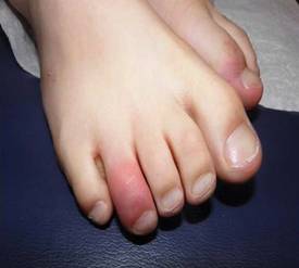
The study compared the characteristics of bruises on 95 infants aged 0-48 months seen in a pediatric ICU, 53 of whom had accidental trauma and 42 of whom were victims of abuse. Bruising on the torso, ear, or neck ("Think TEN," he suggested) in a child younger than 4 years of age increased the possibility of abuse (Pediatrics 2010;125:67-74).
"Does that mean a child can’t fall and bruise an ear? Of course not," said Dr. Sidbury, chief of dermatology at Seattle Children’s Hospital. "It is one thing to add to the list when we’re doing an assessment of the interaction with the parent, interaction with the child, [and] any other signs of trauma – all the things we go through" when considering the possibility of abuse.
"Remember, if they can’t cruise, they can’t bruise." Also, bruising anywhere on an infant younger than 4 months of age was suggestive of abuse. "Remember, if they can’t cruise, they can’t bruise," he said. "Is that evidence of abuse? It is not. Is it something we should pay attention to? I think it is."
Bruising on multiple sites was not in the study’s model, but also is suggestive of child abuse, Dr. Sidbury added.
He described seeing a 2-month-old patient with multiple linear, angulated bruises, some of them in the TEN locations. "The index of suspicion was high, and sadly, this was absolutely a case of abuse," he said.
The more data like this that can be gathered, the easier it will make the physician’s job when assessing a child that might be a victim of abuse.
"It is a wrenching issue," he said. "It is wrenching if it is abuse, and it is equally wrenching if you falsely raise the specter of abuse."
Dr. Sidbury said he had no relevant conflicts of interest.
SDEF and this news organization are owned by Elsevier.
SAN FRANCISCO – The location of bruising and the age of a child can help hone clinical suspicion of child abuse, thanks to a study identifying these predictors.
Because of this study, clinicians now have a better way of raising the topic with parents, which is always a difficult scenario, Dr. Robert Sidbury said.
Instead of physicians having to say that they need to discuss the possibility the bruises might be from nonaccidental trauma, they can now can say, "I’ve got this paper that says bruises in this certain location in a child of this age make me have to check this out," he said at the Women’s and Pediatric Dermatology Seminar, sponsored by Skin Disease Education Foundation (SDEF). "To me, that sounds different."
The study compared the characteristics of bruises on 95 infants aged 0-48 months seen in a pediatric ICU, 53 of whom had accidental trauma and 42 of whom were victims of abuse. Bruising on the torso, ear, or neck ("Think TEN," he suggested) in a child younger than 4 years of age increased the possibility of abuse (Pediatrics 2010;125:67-74).
"Does that mean a child can’t fall and bruise an ear? Of course not," said Dr. Sidbury, chief of dermatology at Seattle Children’s Hospital. "It is one thing to add to the list when we’re doing an assessment of the interaction with the parent, interaction with the child, [and] any other signs of trauma – all the things we go through" when considering the possibility of abuse.
"Remember, if they can’t cruise, they can’t bruise." Also, bruising anywhere on an infant younger than 4 months of age was suggestive of abuse. "Remember, if they can’t cruise, they can’t bruise," he said. "Is that evidence of abuse? It is not. Is it something we should pay attention to? I think it is."
Bruising on multiple sites was not in the study’s model, but also is suggestive of child abuse, Dr. Sidbury added.
He described seeing a 2-month-old patient with multiple linear, angulated bruises, some of them in the TEN locations. "The index of suspicion was high, and sadly, this was absolutely a case of abuse," he said.
The more data like this that can be gathered, the easier it will make the physician’s job when assessing a child that might be a victim of abuse.
"It is a wrenching issue," he said. "It is wrenching if it is abuse, and it is equally wrenching if you falsely raise the specter of abuse."
Dr. Sidbury said he had no relevant conflicts of interest.
SDEF and this news organization are owned by Elsevier.
EXPERT ANALYSIS FROM THE SDEF WOMEN’S & PEDIATRIC DERMATOLOGY SEMINAR
Case Reports Link ADHD Meds to Skin Eruptions
SAN FRANCISCO – Cutaneous reactions are starting to be seen in children on stimulant medications for attention deficit hyperactivity disorder.
Methylphenidate hydrochloride (Ritalin) was associated with acute generalized exanthematous pustulosis (AGEP) in a recent report of a 9-year-old boy being treated for ADHD (Arch. Dermatol. 2011;147:872-3). Dextroamphetamine plus amphetamine (Adderall) was associated with pernio in a separate report of a 9-year-old girl also being treated for ADHD (J. Am. Acad. Dermatol. 2011;64:1218-9).
Before the report of AGEP and Ritalin use, "I had not heard of it related to stimulant therapy," Dr. Robert Sidbury said at the SDEF Women’s and Pediatric Dermatology Seminar, sponsored by Skin Disease Education Foundation (SDEF).
One could be skeptical about a single report of AGEP associated with Ritalin, he acknowledged, but in the case of pernio and stimulants, "I’ve now had three cases of kids who developed this reaction to stimulant therapy," he said. "I believe this to be true."
Both have dramatic presentations that can alarm parents and physicians. "It’s nice to know you can just link it to a certain medication," said Dr. Sidbury, chief of dermatology at Seattle Children’s Hospital. "That can cause a lot of relief."
AGEP is a shower of very small pustules that can be related to a variety of medications. The child in the first report had been taking Ritalin for 6 weeks. Pathology showed sterile intracorneal pustules with mixed eosinophilic infiltrate. A lymphocyte transformation test was positive for AGEP. The patient stopped Ritalin, was treated with topical and systemic steroids for the AGEP, and "did just fine," he said.
Previously, Ritalin has been associated with other cutaneous reactions including morbilliform eruption, urticaria, and even alopecia.
Pernio, also known as chilblain, is a distinctive vasoactive reaction with asymptomatic erythema progressing to blue discoloration of a toe. Its dramatic presentation – "blue toes kind of coming out of nowhere" – can make physicians worry about potential vasculitis, connective disease, or other problems, Dr. Sidbury said.
The second published report described a child who had been taking extended-release Adderall for ADHD for 6 months, was otherwise well, and had no known triggers for pernio such as exposure to cold. The pernio resolved 4 weeks after discontinuing the medication.
Pernio traditionally has been described in Wisconsin hunters with wet socks and cold (but not extremely cold) exposure of the extremities. "In a susceptible population, you get almost a Raynaud’s-type phenomenon," he said. In the case of pernio from stimulants, patients don’t get the classic red, white, and blue cutaneous changes described in the hunters, "you just get a very classic blue-looking toe."
Mechanistically, it makes sense that stimulants could have this sort of reaction in susceptible individuals, he added.
Dr. Sidbury said he had no relevant conflicts of interest.
SDEF and this news organization are owned by Elsevier.
EXPERT ANALYSIS FROM THE SDEF WOMEN'S & PEDIATRIC DERMATOLOGY SEMINAR
Survival Slips After VAD Bridge to Heart Transplant
PARIS – Worse long-term outcomes after high-emergency heart transplants in patients bridged to transplant with cardiac assist devices seem to be related to more complex transplant procedures for those patients rather than differences in patients or donors, a registry study suggests.
The study compared data on 107 patients who had been supported with short-term ventricular assist devices (VADs) and 597 patients supported with conventional therapy before high-emergency heart transplantation (defined by United Network for Organ Sharing status 1 criteria). Data came from 15 centers in the Spanish National Heart Transplant Registry in 2000-2009.

In the postoperative period, rates of primary graft failure and major bleeding were higher among VAD patients than among the conventional group (39% vs. 22%, and 33% vs. 23%, respectively). Furthermore, 22% in the VAD group and 14% in the conventional group required cardiac reoperation. These differences between groups were statistically significant, Dr. Eduardo Barge-Caballero and his associates reported in a press briefing at the annual congress of the European Society of Cardiology.
There was a trend toward higher risk of in-hospital death after surgery in the VAD group (36%) compared with the conventional group (27%), but this did not reach statistical significance. Approximately 60% of patients in the VAD group survived a year after transplantation compared with about 70% of patients in the conventional group.
Long-term survival rates differed significantly between groups by the first year after heart transplantation after adjustment for confounding variables, but the differences were not significant at 10 years of follow-up, said Dr. Barge-Caballero of Hospital Universitario A Coruña, Spain.
Concerns about potential adverse impacts of short-term VAD on outcomes after high-emergency heart transplantation have been cited previously. It has been suggested that these adverse impacts may have been caused by the VAD patients being in worse clinical condition, undergoing more difficult procedures with bleeding complications and infections caused by removal of the VAD, receiving less-desirable donor hearts, or being at a higher risk of rejection.
The current study, which compared the clinical characteristics of donor hearts and recipients in the VAD and conventional groups, concluded that donors were similar between groups and recipients were "not drastically different" between groups, Dr. Barge-Caballero said.
Two kinds of VADs were implanted: Forty-nine patients got extracorporeal continuous-flow devices, and 58 patients received paracorporeal pulsatile-flow devices. There was a trend toward a higher risk of bleeding with the paracorporeal pulsatile-flow devices, but there were not enough patients in each group to show a significant difference, he said.
Patients supported by VAD were significantly younger than were those receiving conventional bridge therapy (48 vs. 51 years, respectively) and were more likely to be female (37% vs. 17%) and to have had previous cardiac surgery (53% vs. 20%). Before VAD implantation, patients in the VAD group received higher doses of intravenous inotropes than did patients in the conventional group.
Measures of end-organ function such as creatinine and bilirubin were similar between groups. Donor organs were similar between groups in age, cold ischemia time, and other characteristics.
The surgical bypass time was significantly longer in the VAD group (156 minutes) than in the conventional group (133 minutes), which may reflect more complicated heart transplantation surgery in the VAD group, he suggested.
Patients spent a mean of only 5 days on bridge support with VAD or conventional bridge therapy, including intra-aortic balloon pump and/or IV inotropes, invasive mechanical ventilation, and dialysis.
Stable patients awaiting high-emergency heart transplantation in countries with short waiting lists should not routinely get VADs, Dr. Barge-Caballero suggested. Because of their risks, VADs should be reserved for critically ill patients who deteriorate when conventional therapy does not provide adequate peripheral perfusion to avoid irreversible end-organ damage.
"Routine implantation of a short-term VAD is not a good option for all patients undergoing a high-emergency heart transplantation," Dr. Barge-Caballero said. "It’s evident that if the patient is really not doing well, has severe hemodynamic instability, [and] is not well with conventional support, we have to implant a VAD. But it’s not, in our opinion, a good option for all patients."
Dr. Barge-Caballero said the investigators had no relevant conflicts of interest.
This is a very, very intriguing finding. Whether patients who are listed for transplantation should or should not get a VAD is a very critical decision.
There are some differences between practices in the Unites States and Europe. In the United States, cardiologists would rather embrace the idea of putting the patient on a VAD as a bridge to transplantation. These findings are challenging that view.
When artificial hearts came to our clinic 10 years ago, the other specialties would come to our hospital and be intrigued. Now it’s almost routine in many centers worldwide, but it’s fascinating. Spain is one of the leaders in Europe. This Spanish study has large numbers of patients. That’s why this is very important information.
Dr. Frank Ruschitzka is a cardiologist at the University of Zurich. He presented these comments as comoderator of the press briefing.
PARIS – Worse long-term outcomes after high-emergency heart transplants in patients bridged to transplant with cardiac assist devices seem to be related to more complex transplant procedures for those patients rather than differences in patients or donors, a registry study suggests.
The study compared data on 107 patients who had been supported with short-term ventricular assist devices (VADs) and 597 patients supported with conventional therapy before high-emergency heart transplantation (defined by United Network for Organ Sharing status 1 criteria). Data came from 15 centers in the Spanish National Heart Transplant Registry in 2000-2009.
In the postoperative period, rates of primary graft failure and major bleeding were higher among VAD patients than among the conventional group (39% vs. 22%, and 33% vs. 23%, respectively). Furthermore, 22% in the VAD group and 14% in the conventional group required cardiac reoperation. These differences between groups were statistically significant, Dr. Eduardo Barge-Caballero and his associates reported in a press briefing at the annual congress of the European Society of Cardiology.
There was a trend toward higher risk of in-hospital death after surgery in the VAD group (36%) compared with the conventional group (27%), but this did not reach statistical significance. Approximately 60% of patients in the VAD group survived a year after transplantation compared with about 70% of patients in the conventional group.
Long-term survival rates differed significantly between groups by the first year after heart transplantation after adjustment for confounding variables, but the differences were not significant at 10 years of follow-up, said Dr. Barge-Caballero of Hospital Universitario A Coruña, Spain.
Concerns about potential adverse impacts of short-term VAD on outcomes after high-emergency heart transplantation have been cited previously. It has been suggested that these adverse impacts may have been caused by the VAD patients being in worse clinical condition, undergoing more difficult procedures with bleeding complications and infections caused by removal of the VAD, receiving less-desirable donor hearts, or being at a higher risk of rejection.
The current study, which compared the clinical characteristics of donor hearts and recipients in the VAD and conventional groups, concluded that donors were similar between groups and recipients were "not drastically different" between groups, Dr. Barge-Caballero said.
Two kinds of VADs were implanted: Forty-nine patients got extracorporeal continuous-flow devices, and 58 patients received paracorporeal pulsatile-flow devices. There was a trend toward a higher risk of bleeding with the paracorporeal pulsatile-flow devices, but there were not enough patients in each group to show a significant difference, he said.
Patients supported by VAD were significantly younger than were those receiving conventional bridge therapy (48 vs. 51 years, respectively) and were more likely to be female (37% vs. 17%) and to have had previous cardiac surgery (53% vs. 20%). Before VAD implantation, patients in the VAD group received higher doses of intravenous inotropes than did patients in the conventional group.
Measures of end-organ function such as creatinine and bilirubin were similar between groups. Donor organs were similar between groups in age, cold ischemia time, and other characteristics.
The surgical bypass time was significantly longer in the VAD group (156 minutes) than in the conventional group (133 minutes), which may reflect more complicated heart transplantation surgery in the VAD group, he suggested.
Patients spent a mean of only 5 days on bridge support with VAD or conventional bridge therapy, including intra-aortic balloon pump and/or IV inotropes, invasive mechanical ventilation, and dialysis.
Stable patients awaiting high-emergency heart transplantation in countries with short waiting lists should not routinely get VADs, Dr. Barge-Caballero suggested. Because of their risks, VADs should be reserved for critically ill patients who deteriorate when conventional therapy does not provide adequate peripheral perfusion to avoid irreversible end-organ damage.
"Routine implantation of a short-term VAD is not a good option for all patients undergoing a high-emergency heart transplantation," Dr. Barge-Caballero said. "It’s evident that if the patient is really not doing well, has severe hemodynamic instability, [and] is not well with conventional support, we have to implant a VAD. But it’s not, in our opinion, a good option for all patients."
Dr. Barge-Caballero said the investigators had no relevant conflicts of interest.
FROM THE ANNUAL CONGRESS OF THE EUROPEAN SOCIETY OF CARDIOLOGY
Major Finding: Patients whose bridge therapy to heart transplantation was a VAD had higher rates of primary graft failure (39% vs. 22%), major bleeding (33% vs. 23%), and cardiac reoperations (22% vs. 14%), and lower 1-year survival than did patients who had conventional treatment.
Data Source: Registry data on 704 patients undergoing high-emergency heart transplantation at 15 Spanish centers in 2000-2009.
Disclosures: Dr. Barge-Caballero said the investigators had no relevant conflicts of interest.
Vertebroplasty 'Benefits' May Be Placebo Effect
SAN FRANCISCO – Vertebroplasty worked no better than sham surgery to reduce pain and disability from vertebral fracture, according to data from recent randomized, controlled trials that put nonsurgical therapies firmly in the first line of treatment.
Osteoporotic vertebral fractures should be treated aggressively with antiresorptive or anabolic therapy for at least 6–12 weeks before considering surgery, Dr. Douglas C. Bauer said at a meeting on osteoporosis sponsored by the University of California, San Francisco. Optimize medical therapy, physical therapy, and other options that might be appropriate such as adding calcitonin or referring the patient for a facet joint injection, he said.
Even after all that, clinicians should consider kyphoplasty before resorting to vertebroplasty, said Dr. Bauer, who is professor of medicine and of epidemiology and biostatistics at the university.
Findings from one unblinded, randomized trial suggests that kyphoplasty may reduce pain and disability, compared with conservative care initially, though the difference in results is less apparent 1 year after surgery.
Despite data from numerous uncontrolled studies suggesting that vertebroplasty also lessens pain and improves function, findings from two well-designed controlled trials “raised a brouhaha” and surprised investigators by showing vertebroplasty to have no benefit, “suggesting that a very commonly done procedure is not helpful,” he said. It's unclear whether the uncontrolled trial results were due to an extended placebo effect or some other factor.
In kyphoplasty, surgeons insert a balloon device to reduce the cervical fracture, remove the balloon, and replace it with cement. Vertebroplasty injects cement only, without the balloon, and does not attempt to increase vertebral height. Both are minimally invasive surgeries that usually are performed under general anesthesia but can be done using local anesthesia, often with conscious sedation.
The unblinded trial of kyphoplasty randomized 149 patients to kyphoplasty and 151 to usual nonsurgical care. “The patients were typical of who we see with vertebral fracture,” Dr. Bauer said. One month after surgery, scores on the Short Form-36 (SF-36) Physical Component Summary had risen from 26 at baseline in both groups to 27 in the kyphoplasty group and 33 in the control group, a significant difference between groups (Lancet 2009;373:1016-24).
Follow-up continued out to 3, 6, and 12 months after surgery, and results were significantly better in the kyphoplasty group at all time points for the SF-36 Physical Component, patient-reported Visual Analog Scale (VAS) scores for back pain, and the number of days of limited activity in the previous 2 weeks.
Although statistically significant, some of the differences between groups were more clinically significant than others. The self-reported VAS pain scores, for example, differed between groups by only 1 point on a 10-point scale at 12 months. The kyphoplasty group, however, enjoyed an average of 60 fewer days of limited activity during those 12 months, compared with the control group, he said.
At 24 months, only the difference in pain scores remained statistically significant between groups (J. Bone Miner. Res. 2011;26:1627-37).
More trials of kyphoplasty are needed before the surgery becomes widespread, Dr. Bauer said.
A separate uncontrolled trial that randomized 202 patients to vertebroplasty or usual care similarly found statistically greater improvements in the vertebroplasty group in VAS pain scores at 1 month (a decrease of 5 points) and 1 year (a 6-point drop), compared with usual care (a 3- and 4-point drop, respectively). Patients in the surgery arm also reported less narcotic use (Lancet 2010;376:1085-92).
The two well-designed controlled trials of vertebroplasty contradict other findings, however. Patients were taken to the operating room before randomization. The control group received sham surgery that included needle insertions in their backs and the breaking of a vial of chemicals to disperse a chemical smell. Outcomes assessors were blinded to randomization.
In one study of 71 patients, scores for back pain decreased significantly in both the real and sham surgery groups, but outcomes did not differ significantly between groups at any time point out to 6 months (N. Engl. J. Med. 2009;361:557-68).
In the other study of 131 patients, both groups showed immediate improvements in disability and pain scores but no outcomes differed significantly between groups at 1 month (N. Engl. J. Med. 2009;361:569-79).
While it's conceivable that the benefits reported for vertebroplasty and kyphoplasty in uncontrolled studies are due to an extended placebo effect, the likelihood that the placebo effect would last for as much as 24 months of follow-up is unclear, Dr. Bauer said.
Some have suggested that the sham-surgery studies included a harder-to-treat population by accepting patients with vertebral fractures up to 1 year in duration, but a subsequent analysis of data limited to fractures of less than 6 weeks duration found no change in the overall results.
Case series have shown that anesthetic or steroid injections alone can reduce vertebral fracture pain, which may explain the improvement in pain scores in both the real and sham-surgery groups in the vertebroplasty trials, he suggested.
There also may be a difference between the two surgeries that produce different results from kyphoplasty or vertebroplasty. Randomized controlled trials comparing the two are underway.
Dr. Bauer has received research funding from Amgen and Novartis.
CT myelogram shows an epidural hematoma and cord compression associated with a vertebral fracture.
Source Courtesy Dr. Victor Jaramillo/Dr. Aravind Pothineni
Expert Analysis from a Meeting on Osteoporosis
Bariatric Surgery Can Lead to Bone Loss
SAN FRANCISCO – Bariatric surgery can be beneficial for obese people, but it also can lead to significant bone loss.
The limited data so far suggest that decreased bone mineral density after bariatric surgery increases the risk for fracture, Dr. Anne Schafer said at the meeting.
The extent of bone loss within a year after Roux-en-Y gastric bypass can be equivalent to “what you would expect in the first 5 years of menopause” in some women, said Dr. Schafer of the division of endocrinology at the University of California, San Francisco.
A 2011 study not yet published by the Mayo Clinic, Rochester, Minn., compared fracture rates in 277 patients undergoing bariatric surgery with local age- and sex-matched fracture rates. The surgeries occurred in 1985–2004, and 94% were gastric bypasses. The retrospective chart study found 138 fractures in 82 patients since the surgery, with a standardized incidence ratio of 2.1 for any fracture and 1.9 for fractures of the hip, spine, wrist, or arm after bariatric surgery, she said.
Dr. Schafer incorporated her own clinical experience with recommendations from the Endocrine Society and from Tufts University in advising clinicians to take the following steps in managing patients undergoing bariatric surgery.
Prior to surgery, check serum 25-hydroxyvitamin D (25[OH]D) levels and prescribe preoperative treatment to augment vitamin D in patients with low levels.
After surgery, all patients should take two multivitamins per day to make sure their micronutrient needs are met.
After malabsorptive bariatric surgery, such as gastric bypass, patients also should take calcium supplements, though there aren't enough data to pinpoint the best dose or to identify which patients might most need it, Dr. Schafer said. She recommended 1,200–2,000 mg/day (preferably in citrate form) after malabsorptive surgery and possibly after restrictive bariatric surgery such as adjustable gastric banding.
Based on the preoperative vitamin D level, prescribe 800–2,000 IU/day of vitamin D3 supplementation after malabsorptive surgery and possibly after restrictive bariatric surgery. “I've had people who need more” than that dose range, she added.
For postoperative surveillance, check calcium homeostasis laboratory tests every 6 months for the first 2 years and then annually after malabsorptive surgery and possibly after any bariatric surgery. The tests include calcium, albumin, phosphate, creatinine, 24(OH)D, and parathyroid hormone.
If the parathyroid hormone level is high, but the 25(OH)D level is low, treat with vitamin D supplementation. If the parathyroid hormone level is high and the 25(OH)D level is ideal, check the patient's 24-hour urinary calcium, and if that is low, increase calcium intake.
Because some of the etiology of bariatric surgery–induced bone loss may be the preferential loss of lean mass over fat mass, or changes in fat distribution, encourage patients to consume protein and to exercise, she said.
The Endocrine Society recommends dual-energy x-ray absorptiometry (DXA) at baseline and annually in people undergoing malabsorptive bariatric surgery. No data show that such monitoring improves outcomes, “but I do think that you should consider it for any people who can fit on the DXA scan before the operation,” Dr. Schafer said. The weight limit for the scanner is about 275–350 pounds.
Dr. Schafer also advises a DXA scan 1–2 years postoperatively. Incorporate those results into “your clinical judgment and other risk factors like age or prior history of fractures to set up an individualized plan for monitoring bone density from there.”
High body mass index has been associated with high bone mineral density, and either voluntary or involuntary weight loss is associated with bone loss and increased fracture risk. Bariatric surgery leads to loss of bone mass for many reasons, she said, including nutritional deficiencies from malabsorption, the body's signals about decreased skeletal loading with weight loss, and changes in fat-secreted hormone.
Most of the data on bone loss after bariatric surgery are for Roux-en-Y gastric bypass, which induces early and sustained increases in bone turnover and decreases in bone mineral density. Fewer data are available on other procedures, but some studies suggest that another malabsorptive procedure, biliopancreatic diversion, may produce effects similar to those of gastric bypass, and that adjustable gastric banding may have less of an impact on bone, she said.
For gastric bypass, one study of 15 patients showed an 8% decrease in total hip bone mineral density within 9 months (J. Clin. Endocrinol. Metab. 2004;89:1061-5). Femoral neck bone density decreased by 9% within 1 year of gastric bypass in a separate study of 23 patients (J. Clin. Endocrinol. Metab. 2008;93:3735-40). A third study of 42 patients showed a 7% decrease in spine bone density and a 10% decrease in total hip bone density ayear after gastric bypass (Obes. Surg. 2009;19:41-6).
Vitamin D deficiency can be a problem after the surgery because many patients have low vitamin D levels beforehand, some of the surgeries are designed to create malabsorption, and patients eat less food and different kinds of food after surgery. In the worst cases, patients develop secondary hyperparathyroidism or bone loss, and there have been case reports of osteomalacia.
All the studies used DXA scans to assess bone density after bariatric surgery, but DXA assessment may be biased in the setting of marked weight loss because of changes in soft tissue surrounding the bones. “We need nonbiased methods of assessing bone mineral density” for future studies of bariatric surgery's effects, she said.
Dr. Schafer had no disclosures.
To view a video interview with Dr. Schafer, scan the QR code with your smartphone.
Expert Analysis from a Meetingon Osteoporosis Sponsored by the University of California, San Francisco
Denosumab's Effect Greater in Selected Patients
SAN FRANCISCO – The pivotal clinical trial of denosumab showed a 20% decrease in nonvertebral fractures compared with placebo treatment, but a new subgroup analysis shows the protective effect is significantly higher in patients with femoral neck osteoporosis.
The preplanned subgroup analysis of data from the FREEDOM trial found that denosumab decreased nonvertebral fractures by 35% in patients with a femoral neck bone mineral density T score of −2.5 or lower and by only 3% in patients with higher femoral neck T scores, compared with patients in those subgroups who received placebo, Dr. Steven R. Cummings said at the meeting.
The report of a 20% reduction in nonvertebral fractures in the overall trial for denosumab “underestimates its efficacy for those patients that we're most interested in treating with this drug – those with osteoporosis,” he said at the conference, sponsored by the University of California, San Francisco.
The findings have been submitted for publication. The analysis is one of several preplanned subgroup analyses being conducted, though this one is “the most interesting result for clinical care,” said Dr. Cummings, emeritus professor of medicine and epidemiology and biostatistics at the university.
The original FREEDOM study (Fracture Reduction Evaluation of Denosumab in Osteoporosis Every 6 Months) enrolled 7,808 postmenopausal women aged 60–80 years with osteoporosis to receive every 6 months either a subcutaneous injection of denosumab (60 mg) or placebo along with daily calcium and vitamin D supplements.
All of the subjects had bone mineral density T scores that were less than −2.5 but not less than −4.0 at the lumbar spine or total hip.
At 36 months, denosumab was associated with reductions of 68% in vertebral fracture and 40% in hip fracture (N. Engl. J. Med. 2009;361:756–65).
The FREEDOM results were the basis of the Food and Drug Administration's approval of denosumab in June 2010.
Data for 2,343 patients who continued denosumab for another 2 years and 2,207 patients who switched from placebo to denosumab in an ongoing extension of the trial suggest that the incidence of nonvertebral fractures continues to decline in the first 5 years of denosumab use. The 5-year results have been submitted for publication, he said.
For nonvertebral fractures, the incidence decreased from 2.6% in the denosumab group in the first year of the FREEDOM trial to 2.1% in year 2 and 2.2% in year 3. Nonvertebral fractures were seen in 1.4% of patients in year 4 and 1.1% of patients in year 5, extension study data show.
Similar rates were seen for vertebral fractures.
Dr. Cummings has been a consultant to Amgen Pharmaceuticals, which markets denosumab; to Merck, which markets alendronate; and to Eli Lilly & Company.
EXPERT ANALYSIS FROM A MEETING ON OSTEOPOROSIS
Physician Smoking Clouds Stop-Smoking Message
Somewhere between 12% and 24% of people working in health care smoke, according to new data from the U.S. Centers for Disease Control and Prevention. In surveys of working adults, the smoking rate in the "health care and social assistance" industry was 16%, lower than the 19% rate for U.S. adults as a whole. Stratified by occupation, 12% of health-care practitioners and technical workers and 24% of health-care support workers smoked.
I’m always a bit flabbergasted when I see attendees smoking outside of medical conferences that I cover. There’s always a handful of them, but if I walk quickly enough I can get through the smoke and into the conference center without gasping for air.

When I covered the European Society of Cardiology Congress recently, however, the experience was breathtaking. Literally. Everyone walked the same route from the train station to the one main entrance, with smokers trying to squeeze in a pungent French smoke between the two.
Dr. Nicolas Danchin of Paris presented a study on how to improve mortality rates in patients who are on optimal medical therapy after having a heart attack, and one of the strategies he emphasized was the need for physicians to be “relentless” in encouraging patients (and colleagues?) to stop smoking. After two weeks of dining at Parisian cafes and restaurants with clouds of smoke wafting in from the outside tables, I can appreciate the challenges ahead for cardiologists who champion smoking cessation.
Among Europeans, approximately 30% of French residents smoked in 2005, and 17 European countries had rates that high or higher. In the U.S., a 21% smoking rate in 2005 declined only slightly to the 19% rate in 2010, the CDC says.
Granted, the U.S. is years ahead of Europe in regulations and programs to limit smoking. California banned smoking in workplaces in 1995. France followed suit in 2007.
The U.S. efforts are paying off. The California Department of Public Health conducted stings and found that only 6% of stores sold cigarettes to minors in 2011, the lowest rate in 15 years, the Bay Citizen reports. The CDC also recently reported that lung cancer incidences declined for men in 35 states and for women in 6 states from 1999 to 2008. The greatest drops were seen in Western states with the lowest smoking prevalence and highest ratios of former smokers to ever smokers.
The payoffs for stop-smoking efforts are clear. But the relatively stagnant U.S. smoking rate, the higher European rates, and the still impressive percentages of health care workers who smoke show how far we have yet to go.
n --by Sherry Boschert (@sherryboschert on Twitter)
Somewhere between 12% and 24% of people working in health care smoke, according to new data from the U.S. Centers for Disease Control and Prevention. In surveys of working adults, the smoking rate in the "health care and social assistance" industry was 16%, lower than the 19% rate for U.S. adults as a whole. Stratified by occupation, 12% of health-care practitioners and technical workers and 24% of health-care support workers smoked.
I’m always a bit flabbergasted when I see attendees smoking outside of medical conferences that I cover. There’s always a handful of them, but if I walk quickly enough I can get through the smoke and into the conference center without gasping for air.
When I covered the European Society of Cardiology Congress recently, however, the experience was breathtaking. Literally. Everyone walked the same route from the train station to the one main entrance, with smokers trying to squeeze in a pungent French smoke between the two.
Dr. Nicolas Danchin of Paris presented a study on how to improve mortality rates in patients who are on optimal medical therapy after having a heart attack, and one of the strategies he emphasized was the need for physicians to be “relentless” in encouraging patients (and colleagues?) to stop smoking. After two weeks of dining at Parisian cafes and restaurants with clouds of smoke wafting in from the outside tables, I can appreciate the challenges ahead for cardiologists who champion smoking cessation.
Among Europeans, approximately 30% of French residents smoked in 2005, and 17 European countries had rates that high or higher. In the U.S., a 21% smoking rate in 2005 declined only slightly to the 19% rate in 2010, the CDC says.
Granted, the U.S. is years ahead of Europe in regulations and programs to limit smoking. California banned smoking in workplaces in 1995. France followed suit in 2007.
The U.S. efforts are paying off. The California Department of Public Health conducted stings and found that only 6% of stores sold cigarettes to minors in 2011, the lowest rate in 15 years, the Bay Citizen reports. The CDC also recently reported that lung cancer incidences declined for men in 35 states and for women in 6 states from 1999 to 2008. The greatest drops were seen in Western states with the lowest smoking prevalence and highest ratios of former smokers to ever smokers.
The payoffs for stop-smoking efforts are clear. But the relatively stagnant U.S. smoking rate, the higher European rates, and the still impressive percentages of health care workers who smoke show how far we have yet to go.
n --by Sherry Boschert (@sherryboschert on Twitter)
Algorithm Helps Differentiate MI From Noncoronary Disease
PARIS – A new algorithm incorporating high-sensitivity cardiac troponin T values in patients with ST-segment elevation seems to differentiate between MI and cardiac but noncoronary disease in patients with acute chest pain.
The algorithm could help triage patients in the first hour of presentation so that those who need coronary angiographies get them quickly and unnecessary angiographies are minimized, Dr. Philip Haaf said in a press briefing at the annual congress of the European Society of Cardiology.
Troponins are increased in cardiovascular disorders including tachyarrhythmia, heart failure, hypertensive urgency or emergency, Takotsubo cardiomyopathy, and myocarditis in patients without a coronary obstruction. The introduction of high-sensitivity assays for cardiac troponins has allowed earlier diagnosis of acute MI in many patients.
But the assays also have caused "considerable confusion among treating physicians" because minor elevations in cardiac troponins can be seen in some patients who do not have a coronary obstruction but have tachyarrhythmia, hypertensive urgency, or heart failure, said Dr. Haaf of University Hospital Basel (Switzerland).
He and his associates analyzed data on the first 887 patients who presented to emergency departments with acute chest pain in the APACE (Advantageous Predictors of Acute Coronary Syndrome Evaluation) study. The ongoing, multicenter study has enrolled close to 2,000 patients so far, obtaining a meticulous patient history, ECG analysis, and measures of novel cardiac biomarkers from each. High-sensitivity cardiac troponin T (hs-cTnT) was measured at presentation and serially thereafter in a blinded fashion, and final diagnosis was adjudicated by two independent cardiologists.
They found that the initial hs-cTnT value and the absolute change in hs-cTnT value within the first hour helped differentiate MI from cardiac but noncoronary disease but that the relative change in hs-cTnT was less discriminatory. In a receiver operating characteristic analysis, using both the initial hs-cTnT and absolute change in the first hour provided an area under the curve of 0.94.
"This is all statistics. What we tried to do is translate these statistics into more clinically applicable terms," Dr. Haaf said.
Analyzing data on 127 patients with acute MI and 125 with cardiac, noncoronary disease, they found that in the 233 patients with ST-segment elevation on ECG, 98% of patients who had an MI had either presentation values for hs-cTnT above 0.028 mcg/L or an absolute change in hs-cTnT of at least 0.005 mcg/L in the first hour.
Changes in hs-cTnT after the first hour did not add much helpful information, he added.
This three-step algorithm – ST elevation, hs-cTnT value at presentation, and absolute change in hs-cTnT in the first hour – provided a positive predictive value of 79% and a "relatively high" negative predictive value of 98% in differentiating acute MI from cardiac, noncoronary disease, Dr. Haaf said.
The study was funded by the Swiss National Science Foundation, the Swiss Heart Foundation, Abbott, Roche, Nanosphere, Siemens, and University Hospital.
No disclosure information was reported.
Troponins, cardiovascular disorders, tachyarrhythmia, heart failure, hypertensive urgency, Takotsubo cardiomyopathy, myocarditis,
PARIS – A new algorithm incorporating high-sensitivity cardiac troponin T values in patients with ST-segment elevation seems to differentiate between MI and cardiac but noncoronary disease in patients with acute chest pain.
The algorithm could help triage patients in the first hour of presentation so that those who need coronary angiographies get them quickly and unnecessary angiographies are minimized, Dr. Philip Haaf said in a press briefing at the annual congress of the European Society of Cardiology.
Troponins are increased in cardiovascular disorders including tachyarrhythmia, heart failure, hypertensive urgency or emergency, Takotsubo cardiomyopathy, and myocarditis in patients without a coronary obstruction. The introduction of high-sensitivity assays for cardiac troponins has allowed earlier diagnosis of acute MI in many patients.
But the assays also have caused "considerable confusion among treating physicians" because minor elevations in cardiac troponins can be seen in some patients who do not have a coronary obstruction but have tachyarrhythmia, hypertensive urgency, or heart failure, said Dr. Haaf of University Hospital Basel (Switzerland).
He and his associates analyzed data on the first 887 patients who presented to emergency departments with acute chest pain in the APACE (Advantageous Predictors of Acute Coronary Syndrome Evaluation) study. The ongoing, multicenter study has enrolled close to 2,000 patients so far, obtaining a meticulous patient history, ECG analysis, and measures of novel cardiac biomarkers from each. High-sensitivity cardiac troponin T (hs-cTnT) was measured at presentation and serially thereafter in a blinded fashion, and final diagnosis was adjudicated by two independent cardiologists.
They found that the initial hs-cTnT value and the absolute change in hs-cTnT value within the first hour helped differentiate MI from cardiac but noncoronary disease but that the relative change in hs-cTnT was less discriminatory. In a receiver operating characteristic analysis, using both the initial hs-cTnT and absolute change in the first hour provided an area under the curve of 0.94.
"This is all statistics. What we tried to do is translate these statistics into more clinically applicable terms," Dr. Haaf said.
Analyzing data on 127 patients with acute MI and 125 with cardiac, noncoronary disease, they found that in the 233 patients with ST-segment elevation on ECG, 98% of patients who had an MI had either presentation values for hs-cTnT above 0.028 mcg/L or an absolute change in hs-cTnT of at least 0.005 mcg/L in the first hour.
Changes in hs-cTnT after the first hour did not add much helpful information, he added.
This three-step algorithm – ST elevation, hs-cTnT value at presentation, and absolute change in hs-cTnT in the first hour – provided a positive predictive value of 79% and a "relatively high" negative predictive value of 98% in differentiating acute MI from cardiac, noncoronary disease, Dr. Haaf said.
The study was funded by the Swiss National Science Foundation, the Swiss Heart Foundation, Abbott, Roche, Nanosphere, Siemens, and University Hospital.
No disclosure information was reported.
Troponins, cardiovascular disorders, tachyarrhythmia, heart failure, hypertensive urgency, Takotsubo cardiomyopathy, myocarditis,
Troponins, cardiovascular disorders, tachyarrhythmia, heart failure, hypertensive urgency, Takotsubo cardiomyopathy, myocarditis,
FROM THE ANNUAL CONGRESS OF THE EUROPEAN SOCIETY OF CARDIOLOGY
Major Finding: A three-step algorithm incorporating ST segment elevation, high-sensitivity cardiac troponin T values at presentation, and absolute change in troponin values in the first hour differentiated MI from cardiac, noncoronary disease with a positive predictive value of 79% and a negative predictive value of 98%.
Data Source: Analyses of data on 887 patients who presented to emergency departments with acute chest pain.
Disclosures: The study was funded by the Swiss National Science Foundation, the Swiss Heart Foundation, Abbott, Roche, Nanosphere, Siemens, and the University Hospital. No disclosure information was reported.
Blog: The Latest Trend in Anti-Aging Skin Care
Here’s a somewhat new twist to the saying, “You are what you eat.” The latest trend in anti-wrinkle efforts is “nutricosmetics,” combining cosmeceutical and nutraceutical products. In other words, using both topical and oral agents to make skin look younger than it might otherwise.
That’s the prediction of Dr. Zoe D. Draelos, a consulting professor at Duke University, who spoke at the SDEF Women’s and Pediatric Dermatology Seminar. First the buzz was about antioxidants. Next you’ll be seeing the glycation inhibitor carnosine touted in foods and products, followed in a few years by other glycation inhibitors.

Why? Because there are only three ways known to limit the effects of aging on skin: caloric restriction; preventing oxidative damage, and preventing or reversing advanced glycation end productions, she said.
It makes sense that eating a healthier diet will give you a better chance of looking younger and healthier than a poor diet will, but nutricosmetics takes this to another level, with companies in pursuit of supplements, topical agents, and other products that may deliver more of the good stuff than you can get from food alone. Dr. Draelos has received consulting and research funds from 14 different companies.
Cosmetic dermatology started moving in this direction with topical and oral antioxidants. Carotenoids (vitamin A) are found in red, orange and yellow fruits and vegetables. Think of tomatoes, which contain the antioxidant carotenoid lycopene, which inspired lycopene supplement products. Topical anti-wrinkle products may contain the synthetic carotenoids retinol, retinaldehyde or retinyl propionate. The polyphenols in green tea also are antioxidants.
Then, there’s resveratrol, a substance that modulates the activity of sirtuins, which are thought to be responsible for some of the mechanisms of aging on a mitochondrial level. French red wines are high in resveratrol, which also has been added to moisturizers to function as an antioxidant. Too much resveratrol can be bad for you, rodent studies suggest, and it’s expensive, Dr. Draelos said. Products are more likely to say that ingredients include the plant Japanese Knotweed, which contains some resveratrol.
Caloric restriction isn’t really a viable option for skin care, so the nutricosmetic industry is looking at “caloric restriction mimetics” such as anthocyanins, which are antioxidants. That’s why newer supplements will contain anthocyanins, which are found in high concentrations in wild raspberries, blackberries, and blueberries. Blueberry extract also may be a glycation inhibitor.

“Berries are going to be everywhere,” Dr. Draelos predicted. Interestingly, it’s the wild, stressed berries and the stressed grapes grown in colder areas like France that contain more of these ingredients than, say, wines from California’s milder climate or farmed berries.
Carnosine, the newest ingredient in skin care products, also seems to prevent glycation, possibly providing anti-aging benefits on the level of gene regulation.
Because these components are not drugs, the products don’t need to be tested and approved by the Food and Drug Administration, so there’s precious little science to back up anti-aging claims. Much of the marketing is based on theories of how the ingredients might help one’s skin.
An unpublished double-blind, placebo-controlled study by Dr. Draelos and her associates provides some support for oral lycopene supplements, a popular nutricosmetic product. They shined a standardized amount of simulated sunlight on the relatively pristine buttock skin of volunteers, took a punch biopsy, and counted the number of sunburned cells. Then subjects took lycopene supplements twice a day for 12 weeks before repeating the sunburn test on a different part of the buttocks. Fewer cells ended up sunburned after taking lycopene. A lower burned cell count means less oxidative damage.
Not all research is so nice. A study by other investigators of mice who were fed vitamins C and E plus extracts of green tea and blueberry tested the effects on tissue by pulling on the mice tails until the tendons snapped. To their credit, audience members at the seminar groaned when they heard this.
Someone asked Dr. Draelos what she consumes to combat aging. Because she’s vitamin D deficient, she takes vitamin D supplements, she said. She added that she likes to get her antioxidants in the form of omega-3 and omega-6 fatty acids, such as in flax seed oil and fish oil. Moderation is the key, she said. “Eat something of every color every day. Take a multivitamin. Don’t eat just blueberries, eat something of everything,” and take supplements if you’re deficient in something, she advised.
If you’re going to drink green tea, eat berries or tomatoes, or buy some French wine to combat wrinkles, or take supplements based on their ingredients, no one knows how much to eat or drink for this purpose, she added. Dr. Joseph F. Fowler, Jr., who spoke after Dr. Draelos, summed it up this way: “Actually, if you drink enough red wine, you won’t care.”
-- Sherry Boschert (@sherryboschert on Twitter)
Disclosure: SDEF and this news organization are both owned by Elsevier.
Here’s a somewhat new twist to the saying, “You are what you eat.” The latest trend in anti-wrinkle efforts is “nutricosmetics,” combining cosmeceutical and nutraceutical products. In other words, using both topical and oral agents to make skin look younger than it might otherwise.
That’s the prediction of Dr. Zoe D. Draelos, a consulting professor at Duke University, who spoke at the SDEF Women’s and Pediatric Dermatology Seminar. First the buzz was about antioxidants. Next you’ll be seeing the glycation inhibitor carnosine touted in foods and products, followed in a few years by other glycation inhibitors.
Why? Because there are only three ways known to limit the effects of aging on skin: caloric restriction; preventing oxidative damage, and preventing or reversing advanced glycation end productions, she said.
It makes sense that eating a healthier diet will give you a better chance of looking younger and healthier than a poor diet will, but nutricosmetics takes this to another level, with companies in pursuit of supplements, topical agents, and other products that may deliver more of the good stuff than you can get from food alone. Dr. Draelos has received consulting and research funds from 14 different companies.
Cosmetic dermatology started moving in this direction with topical and oral antioxidants. Carotenoids (vitamin A) are found in red, orange and yellow fruits and vegetables. Think of tomatoes, which contain the antioxidant carotenoid lycopene, which inspired lycopene supplement products. Topical anti-wrinkle products may contain the synthetic carotenoids retinol, retinaldehyde or retinyl propionate. The polyphenols in green tea also are antioxidants.
Then, there’s resveratrol, a substance that modulates the activity of sirtuins, which are thought to be responsible for some of the mechanisms of aging on a mitochondrial level. French red wines are high in resveratrol, which also has been added to moisturizers to function as an antioxidant. Too much resveratrol can be bad for you, rodent studies suggest, and it’s expensive, Dr. Draelos said. Products are more likely to say that ingredients include the plant Japanese Knotweed, which contains some resveratrol.
Caloric restriction isn’t really a viable option for skin care, so the nutricosmetic industry is looking at “caloric restriction mimetics” such as anthocyanins, which are antioxidants. That’s why newer supplements will contain anthocyanins, which are found in high concentrations in wild raspberries, blackberries, and blueberries. Blueberry extract also may be a glycation inhibitor.
“Berries are going to be everywhere,” Dr. Draelos predicted. Interestingly, it’s the wild, stressed berries and the stressed grapes grown in colder areas like France that contain more of these ingredients than, say, wines from California’s milder climate or farmed berries.
Carnosine, the newest ingredient in skin care products, also seems to prevent glycation, possibly providing anti-aging benefits on the level of gene regulation.
Because these components are not drugs, the products don’t need to be tested and approved by the Food and Drug Administration, so there’s precious little science to back up anti-aging claims. Much of the marketing is based on theories of how the ingredients might help one’s skin.
An unpublished double-blind, placebo-controlled study by Dr. Draelos and her associates provides some support for oral lycopene supplements, a popular nutricosmetic product. They shined a standardized amount of simulated sunlight on the relatively pristine buttock skin of volunteers, took a punch biopsy, and counted the number of sunburned cells. Then subjects took lycopene supplements twice a day for 12 weeks before repeating the sunburn test on a different part of the buttocks. Fewer cells ended up sunburned after taking lycopene. A lower burned cell count means less oxidative damage.
Not all research is so nice. A study by other investigators of mice who were fed vitamins C and E plus extracts of green tea and blueberry tested the effects on tissue by pulling on the mice tails until the tendons snapped. To their credit, audience members at the seminar groaned when they heard this.
Someone asked Dr. Draelos what she consumes to combat aging. Because she’s vitamin D deficient, she takes vitamin D supplements, she said. She added that she likes to get her antioxidants in the form of omega-3 and omega-6 fatty acids, such as in flax seed oil and fish oil. Moderation is the key, she said. “Eat something of every color every day. Take a multivitamin. Don’t eat just blueberries, eat something of everything,” and take supplements if you’re deficient in something, she advised.
If you’re going to drink green tea, eat berries or tomatoes, or buy some French wine to combat wrinkles, or take supplements based on their ingredients, no one knows how much to eat or drink for this purpose, she added. Dr. Joseph F. Fowler, Jr., who spoke after Dr. Draelos, summed it up this way: “Actually, if you drink enough red wine, you won’t care.”
-- Sherry Boschert (@sherryboschert on Twitter)
Disclosure: SDEF and this news organization are both owned by Elsevier.