User login
Study: Type 2 narcolepsy is significantly different from type 1
Patients with type 1 narcolepsy have more clinical impairments and distinct functional abnormalities than do patients with type 2 narcolepsy, according to investigators.
Patients with type 2 “do not present with such severe handicaps and are clinically closer to hypersomniac patients than the patients with type 1 narcolepsy,” reported Yu-Shu Huang, MD, of Chang Gung Memorial Hospital and Chang Gung University, Taoyuan City, Taiwan, and associates. The study was published in Neurology.
The researchers used brain scans, neuropsychological tests, and other screening tests to analyze three groups of subjects – 104 patients with Na-1, 29 with Na-2, and a control group of 26 subjects. Depending on the group, 62%-66% of the subjects were men, and the mean age ranged from 19 to 20.
The mean age of onset for the narcolepsy groups was 12-13. Those with Na-1 had higher mean body mass indexes – 27 kg/m2 vs. 24 (Na-2) vs. 20 (control), (P = .001).
The patients in both narcolepsy groups showed similar levels of sleepiness, but those with Na-2 had significantly fewer abnormal findings and disturbances.
Patients with Na-2 had significantly fewer sleep-onset REM periods, longer mean sleep latencies, and lower apnea-hypopnea indexes. The human leukocyte antigen DQ-Beta1*0602 was also found less frequently in Na-2 compared to Na-1 (52% vs. 97%, respectively, P less than .001).
PET findings also revealed less impairment in Na-2 compared to Na-1. The researchers noted increased metabolic rate in several brain areas in Na-1, although hypometabolism is more common in some areas in Na-2.
Based on their findings, the authors challenge a previous study of insurance data that suggests patients with both types have similarly poor outcomes over the long term. (PLoS One 2012;7:e33525.)
“In our study,” the authors wrote, “compared to patients with type 2 narcolepsy, patients with type 1 narcolepsy present with much more severe handicaps as early as adolescence. Further studies are needed to clarify the issue.”
The Taiwan Ministry of Science and Technology funded the study. The authors report no relevant disclosures.
SOURCE: Huang Y, et al. March 30, 2018, Neurology. 2018 Mar 30. doi: 10.1212/WNL.0000000000005346.
Patients with type 1 narcolepsy have more clinical impairments and distinct functional abnormalities than do patients with type 2 narcolepsy, according to investigators.
Patients with type 2 “do not present with such severe handicaps and are clinically closer to hypersomniac patients than the patients with type 1 narcolepsy,” reported Yu-Shu Huang, MD, of Chang Gung Memorial Hospital and Chang Gung University, Taoyuan City, Taiwan, and associates. The study was published in Neurology.
The researchers used brain scans, neuropsychological tests, and other screening tests to analyze three groups of subjects – 104 patients with Na-1, 29 with Na-2, and a control group of 26 subjects. Depending on the group, 62%-66% of the subjects were men, and the mean age ranged from 19 to 20.
The mean age of onset for the narcolepsy groups was 12-13. Those with Na-1 had higher mean body mass indexes – 27 kg/m2 vs. 24 (Na-2) vs. 20 (control), (P = .001).
The patients in both narcolepsy groups showed similar levels of sleepiness, but those with Na-2 had significantly fewer abnormal findings and disturbances.
Patients with Na-2 had significantly fewer sleep-onset REM periods, longer mean sleep latencies, and lower apnea-hypopnea indexes. The human leukocyte antigen DQ-Beta1*0602 was also found less frequently in Na-2 compared to Na-1 (52% vs. 97%, respectively, P less than .001).
PET findings also revealed less impairment in Na-2 compared to Na-1. The researchers noted increased metabolic rate in several brain areas in Na-1, although hypometabolism is more common in some areas in Na-2.
Based on their findings, the authors challenge a previous study of insurance data that suggests patients with both types have similarly poor outcomes over the long term. (PLoS One 2012;7:e33525.)
“In our study,” the authors wrote, “compared to patients with type 2 narcolepsy, patients with type 1 narcolepsy present with much more severe handicaps as early as adolescence. Further studies are needed to clarify the issue.”
The Taiwan Ministry of Science and Technology funded the study. The authors report no relevant disclosures.
SOURCE: Huang Y, et al. March 30, 2018, Neurology. 2018 Mar 30. doi: 10.1212/WNL.0000000000005346.
Patients with type 1 narcolepsy have more clinical impairments and distinct functional abnormalities than do patients with type 2 narcolepsy, according to investigators.
Patients with type 2 “do not present with such severe handicaps and are clinically closer to hypersomniac patients than the patients with type 1 narcolepsy,” reported Yu-Shu Huang, MD, of Chang Gung Memorial Hospital and Chang Gung University, Taoyuan City, Taiwan, and associates. The study was published in Neurology.
The researchers used brain scans, neuropsychological tests, and other screening tests to analyze three groups of subjects – 104 patients with Na-1, 29 with Na-2, and a control group of 26 subjects. Depending on the group, 62%-66% of the subjects were men, and the mean age ranged from 19 to 20.
The mean age of onset for the narcolepsy groups was 12-13. Those with Na-1 had higher mean body mass indexes – 27 kg/m2 vs. 24 (Na-2) vs. 20 (control), (P = .001).
The patients in both narcolepsy groups showed similar levels of sleepiness, but those with Na-2 had significantly fewer abnormal findings and disturbances.
Patients with Na-2 had significantly fewer sleep-onset REM periods, longer mean sleep latencies, and lower apnea-hypopnea indexes. The human leukocyte antigen DQ-Beta1*0602 was also found less frequently in Na-2 compared to Na-1 (52% vs. 97%, respectively, P less than .001).
PET findings also revealed less impairment in Na-2 compared to Na-1. The researchers noted increased metabolic rate in several brain areas in Na-1, although hypometabolism is more common in some areas in Na-2.
Based on their findings, the authors challenge a previous study of insurance data that suggests patients with both types have similarly poor outcomes over the long term. (PLoS One 2012;7:e33525.)
“In our study,” the authors wrote, “compared to patients with type 2 narcolepsy, patients with type 1 narcolepsy present with much more severe handicaps as early as adolescence. Further studies are needed to clarify the issue.”
The Taiwan Ministry of Science and Technology funded the study. The authors report no relevant disclosures.
SOURCE: Huang Y, et al. March 30, 2018, Neurology. 2018 Mar 30. doi: 10.1212/WNL.0000000000005346.
FROM NEUROLOGY
Prioritize topical treatments in pregnant women
SAN DIEGO – “Some doctors don’t treat them at all, or they undertreat because they’re afraid of prescribing anything,” said dermatologist Kelly H. Tyler, MD.
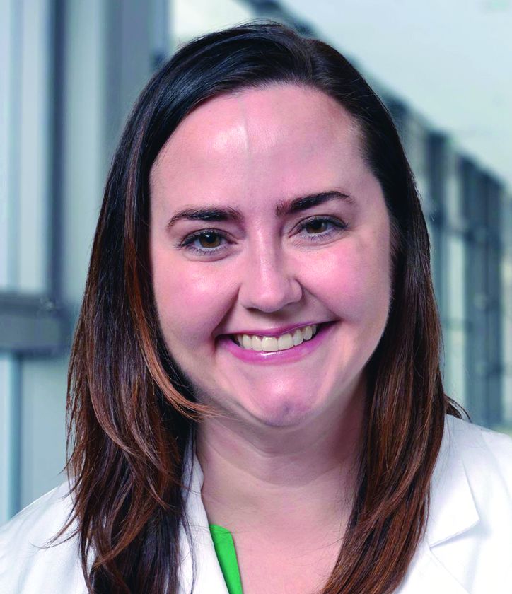
But the Food and Drug Administration’s new system of prescription drug labeling for pregnant women offers improved insight for dermatologists, and a variety of alternatives are available to commonly used dermatologic drugs that pose risks, said Dr. Tyler, who has a unique perspective on this issue: She used to practice as an ob.gyn.
In her presentation, she provided the following tips when caring for pregnant patients.
Check the prescription drug labeling for pregnant and lactating women
Prior to 2015, the FDA used a letter ratings to denote the risk of medications during pregnancy. The ratings – A, B, C, D, and X – go from “A” showing no fetal risk in controlled studies to “X” in which a drug is contraindicated because “there is no reason to risk use of drug in pregnancy.”
This rating system is problematic, Dr. Tyler said, since it’s imprecise. She noted that about two-thirds of medications have a risk rating of C, which means “Risk cannot be ruled out; human studies may or may not show risk; potential benefits may justify potential risk.” But, she asked, “How are you supposed to know what to do with that kind of information?”
By June 30, 2015, FDA had retired the letter classifications and replaced this system with the Pregnancy and Lactation Labeling final rule, which requires that labels include a pregnancy section with information on pregnancy exposure registries, in addition to risk summaries, clinical considerations, and available data. There are also sections on lactation, including nursing mothers, and females and males of reproductive potential. (This information appears in the prescription drug labeling sections 8.1-8.3.) Labels for medications approved prior to June 30, 2001 did not need to be updated. Labels for newer medications are required to comply within 3-5 years of the 2015 policy change.
Ask female patients about sexual activity
It’s important to ask all premenopausal female patients whether they’re sexually active and whether they are using birth control, trying to conceive, or are currently pregnant. If they’re pregnant, what trimester are they in?
Keep in mind, she said, that women can be pregnant for quite some time without knowing it. Home pregnancy tests may not show positive results for up to 5 weeks after conception, she said. But the embryonic/organogenesis stage – from 2 to 8 weeks – is the most important period for a pregnant mother avoid drugs that could damage the unborn child and is “when you want to avoid most medications,” she said.
Risks don’t end after a few months, however. Dr. Tyler urged dermatologists to keep in mind that the brain, teeth, and bones remain susceptible to damage from teratogenic medications after the eighth week.
Keep topical medications in mind
When it comes to pregnant women, “for most dermatologic therapies, topical medications will be your No. 1 choice” because of less absorption, Dr. Tyler said.
But there are exceptions, she noted. In acne and rosacea, for example, there’s controversy about the safety of topical retinoids. “I would just urge you to not use those medications in pregnancy because you don’t really need to use them,” Dr. Tyler said. “You have other options that are safer.”
Adapalene and tretinoin are listed as category C (risk cannot be ruled out in pregnancy) and topical tazarotene is category X (contraindicated in pregnancy) because of retinoid-like anomalies in animal studies, she said.
Avoid 3 major contraindicated systemic drugs
Isotretinoin, acitretin, and methotrexate are “absolutely contraindicated” for anyone who is pregnant or could become pregnant, Dr. Tyler said.
Use some systemic drugs with caution
Tetracyclines are category D (positive evidence of risk to human fetus, but benefits may outweigh risks of drug) with the highest risk in the second and third trimesters, Dr. Tyler said. “If for some reason you have a patient who’s on tetracycline, stop before she’s in her second trimester.”
She recommended avoiding the antibiotic erythromycin because of studies hinting at risks when used early in pregnancy. Spironolactone is theoretically risky after the week 8 of pregnancy, she said. “Because of the animal studies, we typically do not use this during pregnancy.”
Psoriasis often improves during pregnancy
Psoriasis improves during pregnancy in about half of women with psoriasis and worsens in about 20%, Dr. Tyler said. During pregnancy, topical treatments are the first-line treatment, said Dr. Tyler, who recommends that treatment should begin with topical steroids, then calcipotriene or tacrolimus if needed (J Am Acad Dermatol. 2013 Apr;68[4]:663-71). Cyclosporine is an option, as is phototherapy, she said. In an interview, she noted that phototherapy (narrowband UVB) would be considered next-line therapy in patients who are past the first 28 days of gestation and have failed topical therapy, given they are taking adequate folic acid supplementation, which is present in prenatal vitamins.
“As for biologics, you have to go with older ones that have more data. When we look at newer medications, we don’t know a lot about them,” she said during the presentation.
It’s okay to prescribe oral steroids and antihistamines
Oral steroids are safe during pregnancy, Dr. Tyler said, but “just be judicious” with moderate doses and short durations.
Antihistamines are also appropriate, she said, but be aware of the potential for neonatal sedation during lactation.
As for antibiotics for bacterial infections, azithromycin, penicillins, and cephalosporins are all category B (no risk to human fetus despite possible animal risk; or no risk in animal studies and human studies not done), as are all topical antibiotics except dapsone, which is category C because of a theoretical risk of neonatal hyperbilirubinemia if used near the time of delivery.
Hydroxychloroquine may be appropriate for connective tissue disease, she said, although steroids may be a better option in some cases.
And topical antifungals are considered safer for fungal diseases than systemic medications. She said she prefers clotrimazole and oxiconazole, both category B.
Finally, Dr. Tyler recommended permethrin (category B) for parasitic infections since it has been used extensively in pregnancy without a sign of risk and is the preferred treatment for scabies. It’s a better option than ivermectin, she said.
Postpone surgery until at least the second trimester
If it’s not possible to delay nonemergent dermatologic surgery until after pregnancy, she recommended performing procedures during the second trimester. Destruction of local lesions, however, is safe without anesthesia.
“In summary,” when treating pregnant patients, Dr. Tyler said, “a conservative approach is always best, topical medications are always first-line for any condition, and certain oral medications are safe.”
Dr. Tyler reported no relevant disclosures.
SAN DIEGO – “Some doctors don’t treat them at all, or they undertreat because they’re afraid of prescribing anything,” said dermatologist Kelly H. Tyler, MD.
But the Food and Drug Administration’s new system of prescription drug labeling for pregnant women offers improved insight for dermatologists, and a variety of alternatives are available to commonly used dermatologic drugs that pose risks, said Dr. Tyler, who has a unique perspective on this issue: She used to practice as an ob.gyn.
In her presentation, she provided the following tips when caring for pregnant patients.
Check the prescription drug labeling for pregnant and lactating women
Prior to 2015, the FDA used a letter ratings to denote the risk of medications during pregnancy. The ratings – A, B, C, D, and X – go from “A” showing no fetal risk in controlled studies to “X” in which a drug is contraindicated because “there is no reason to risk use of drug in pregnancy.”
This rating system is problematic, Dr. Tyler said, since it’s imprecise. She noted that about two-thirds of medications have a risk rating of C, which means “Risk cannot be ruled out; human studies may or may not show risk; potential benefits may justify potential risk.” But, she asked, “How are you supposed to know what to do with that kind of information?”
By June 30, 2015, FDA had retired the letter classifications and replaced this system with the Pregnancy and Lactation Labeling final rule, which requires that labels include a pregnancy section with information on pregnancy exposure registries, in addition to risk summaries, clinical considerations, and available data. There are also sections on lactation, including nursing mothers, and females and males of reproductive potential. (This information appears in the prescription drug labeling sections 8.1-8.3.) Labels for medications approved prior to June 30, 2001 did not need to be updated. Labels for newer medications are required to comply within 3-5 years of the 2015 policy change.
Ask female patients about sexual activity
It’s important to ask all premenopausal female patients whether they’re sexually active and whether they are using birth control, trying to conceive, or are currently pregnant. If they’re pregnant, what trimester are they in?
Keep in mind, she said, that women can be pregnant for quite some time without knowing it. Home pregnancy tests may not show positive results for up to 5 weeks after conception, she said. But the embryonic/organogenesis stage – from 2 to 8 weeks – is the most important period for a pregnant mother avoid drugs that could damage the unborn child and is “when you want to avoid most medications,” she said.
Risks don’t end after a few months, however. Dr. Tyler urged dermatologists to keep in mind that the brain, teeth, and bones remain susceptible to damage from teratogenic medications after the eighth week.
Keep topical medications in mind
When it comes to pregnant women, “for most dermatologic therapies, topical medications will be your No. 1 choice” because of less absorption, Dr. Tyler said.
But there are exceptions, she noted. In acne and rosacea, for example, there’s controversy about the safety of topical retinoids. “I would just urge you to not use those medications in pregnancy because you don’t really need to use them,” Dr. Tyler said. “You have other options that are safer.”
Adapalene and tretinoin are listed as category C (risk cannot be ruled out in pregnancy) and topical tazarotene is category X (contraindicated in pregnancy) because of retinoid-like anomalies in animal studies, she said.
Avoid 3 major contraindicated systemic drugs
Isotretinoin, acitretin, and methotrexate are “absolutely contraindicated” for anyone who is pregnant or could become pregnant, Dr. Tyler said.
Use some systemic drugs with caution
Tetracyclines are category D (positive evidence of risk to human fetus, but benefits may outweigh risks of drug) with the highest risk in the second and third trimesters, Dr. Tyler said. “If for some reason you have a patient who’s on tetracycline, stop before she’s in her second trimester.”
She recommended avoiding the antibiotic erythromycin because of studies hinting at risks when used early in pregnancy. Spironolactone is theoretically risky after the week 8 of pregnancy, she said. “Because of the animal studies, we typically do not use this during pregnancy.”
Psoriasis often improves during pregnancy
Psoriasis improves during pregnancy in about half of women with psoriasis and worsens in about 20%, Dr. Tyler said. During pregnancy, topical treatments are the first-line treatment, said Dr. Tyler, who recommends that treatment should begin with topical steroids, then calcipotriene or tacrolimus if needed (J Am Acad Dermatol. 2013 Apr;68[4]:663-71). Cyclosporine is an option, as is phototherapy, she said. In an interview, she noted that phototherapy (narrowband UVB) would be considered next-line therapy in patients who are past the first 28 days of gestation and have failed topical therapy, given they are taking adequate folic acid supplementation, which is present in prenatal vitamins.
“As for biologics, you have to go with older ones that have more data. When we look at newer medications, we don’t know a lot about them,” she said during the presentation.
It’s okay to prescribe oral steroids and antihistamines
Oral steroids are safe during pregnancy, Dr. Tyler said, but “just be judicious” with moderate doses and short durations.
Antihistamines are also appropriate, she said, but be aware of the potential for neonatal sedation during lactation.
As for antibiotics for bacterial infections, azithromycin, penicillins, and cephalosporins are all category B (no risk to human fetus despite possible animal risk; or no risk in animal studies and human studies not done), as are all topical antibiotics except dapsone, which is category C because of a theoretical risk of neonatal hyperbilirubinemia if used near the time of delivery.
Hydroxychloroquine may be appropriate for connective tissue disease, she said, although steroids may be a better option in some cases.
And topical antifungals are considered safer for fungal diseases than systemic medications. She said she prefers clotrimazole and oxiconazole, both category B.
Finally, Dr. Tyler recommended permethrin (category B) for parasitic infections since it has been used extensively in pregnancy without a sign of risk and is the preferred treatment for scabies. It’s a better option than ivermectin, she said.
Postpone surgery until at least the second trimester
If it’s not possible to delay nonemergent dermatologic surgery until after pregnancy, she recommended performing procedures during the second trimester. Destruction of local lesions, however, is safe without anesthesia.
“In summary,” when treating pregnant patients, Dr. Tyler said, “a conservative approach is always best, topical medications are always first-line for any condition, and certain oral medications are safe.”
Dr. Tyler reported no relevant disclosures.
SAN DIEGO – “Some doctors don’t treat them at all, or they undertreat because they’re afraid of prescribing anything,” said dermatologist Kelly H. Tyler, MD.
But the Food and Drug Administration’s new system of prescription drug labeling for pregnant women offers improved insight for dermatologists, and a variety of alternatives are available to commonly used dermatologic drugs that pose risks, said Dr. Tyler, who has a unique perspective on this issue: She used to practice as an ob.gyn.
In her presentation, she provided the following tips when caring for pregnant patients.
Check the prescription drug labeling for pregnant and lactating women
Prior to 2015, the FDA used a letter ratings to denote the risk of medications during pregnancy. The ratings – A, B, C, D, and X – go from “A” showing no fetal risk in controlled studies to “X” in which a drug is contraindicated because “there is no reason to risk use of drug in pregnancy.”
This rating system is problematic, Dr. Tyler said, since it’s imprecise. She noted that about two-thirds of medications have a risk rating of C, which means “Risk cannot be ruled out; human studies may or may not show risk; potential benefits may justify potential risk.” But, she asked, “How are you supposed to know what to do with that kind of information?”
By June 30, 2015, FDA had retired the letter classifications and replaced this system with the Pregnancy and Lactation Labeling final rule, which requires that labels include a pregnancy section with information on pregnancy exposure registries, in addition to risk summaries, clinical considerations, and available data. There are also sections on lactation, including nursing mothers, and females and males of reproductive potential. (This information appears in the prescription drug labeling sections 8.1-8.3.) Labels for medications approved prior to June 30, 2001 did not need to be updated. Labels for newer medications are required to comply within 3-5 years of the 2015 policy change.
Ask female patients about sexual activity
It’s important to ask all premenopausal female patients whether they’re sexually active and whether they are using birth control, trying to conceive, or are currently pregnant. If they’re pregnant, what trimester are they in?
Keep in mind, she said, that women can be pregnant for quite some time without knowing it. Home pregnancy tests may not show positive results for up to 5 weeks after conception, she said. But the embryonic/organogenesis stage – from 2 to 8 weeks – is the most important period for a pregnant mother avoid drugs that could damage the unborn child and is “when you want to avoid most medications,” she said.
Risks don’t end after a few months, however. Dr. Tyler urged dermatologists to keep in mind that the brain, teeth, and bones remain susceptible to damage from teratogenic medications after the eighth week.
Keep topical medications in mind
When it comes to pregnant women, “for most dermatologic therapies, topical medications will be your No. 1 choice” because of less absorption, Dr. Tyler said.
But there are exceptions, she noted. In acne and rosacea, for example, there’s controversy about the safety of topical retinoids. “I would just urge you to not use those medications in pregnancy because you don’t really need to use them,” Dr. Tyler said. “You have other options that are safer.”
Adapalene and tretinoin are listed as category C (risk cannot be ruled out in pregnancy) and topical tazarotene is category X (contraindicated in pregnancy) because of retinoid-like anomalies in animal studies, she said.
Avoid 3 major contraindicated systemic drugs
Isotretinoin, acitretin, and methotrexate are “absolutely contraindicated” for anyone who is pregnant or could become pregnant, Dr. Tyler said.
Use some systemic drugs with caution
Tetracyclines are category D (positive evidence of risk to human fetus, but benefits may outweigh risks of drug) with the highest risk in the second and third trimesters, Dr. Tyler said. “If for some reason you have a patient who’s on tetracycline, stop before she’s in her second trimester.”
She recommended avoiding the antibiotic erythromycin because of studies hinting at risks when used early in pregnancy. Spironolactone is theoretically risky after the week 8 of pregnancy, she said. “Because of the animal studies, we typically do not use this during pregnancy.”
Psoriasis often improves during pregnancy
Psoriasis improves during pregnancy in about half of women with psoriasis and worsens in about 20%, Dr. Tyler said. During pregnancy, topical treatments are the first-line treatment, said Dr. Tyler, who recommends that treatment should begin with topical steroids, then calcipotriene or tacrolimus if needed (J Am Acad Dermatol. 2013 Apr;68[4]:663-71). Cyclosporine is an option, as is phototherapy, she said. In an interview, she noted that phototherapy (narrowband UVB) would be considered next-line therapy in patients who are past the first 28 days of gestation and have failed topical therapy, given they are taking adequate folic acid supplementation, which is present in prenatal vitamins.
“As for biologics, you have to go with older ones that have more data. When we look at newer medications, we don’t know a lot about them,” she said during the presentation.
It’s okay to prescribe oral steroids and antihistamines
Oral steroids are safe during pregnancy, Dr. Tyler said, but “just be judicious” with moderate doses and short durations.
Antihistamines are also appropriate, she said, but be aware of the potential for neonatal sedation during lactation.
As for antibiotics for bacterial infections, azithromycin, penicillins, and cephalosporins are all category B (no risk to human fetus despite possible animal risk; or no risk in animal studies and human studies not done), as are all topical antibiotics except dapsone, which is category C because of a theoretical risk of neonatal hyperbilirubinemia if used near the time of delivery.
Hydroxychloroquine may be appropriate for connective tissue disease, she said, although steroids may be a better option in some cases.
And topical antifungals are considered safer for fungal diseases than systemic medications. She said she prefers clotrimazole and oxiconazole, both category B.
Finally, Dr. Tyler recommended permethrin (category B) for parasitic infections since it has been used extensively in pregnancy without a sign of risk and is the preferred treatment for scabies. It’s a better option than ivermectin, she said.
Postpone surgery until at least the second trimester
If it’s not possible to delay nonemergent dermatologic surgery until after pregnancy, she recommended performing procedures during the second trimester. Destruction of local lesions, however, is safe without anesthesia.
“In summary,” when treating pregnant patients, Dr. Tyler said, “a conservative approach is always best, topical medications are always first-line for any condition, and certain oral medications are safe.”
Dr. Tyler reported no relevant disclosures.
EXPERT ANALYSIS FROM AAD 18
Dramatic improvements reported after surgery for hidradenitis suppurativa
A retrospective German study found that the majority of , with many saying they no longer suffered from everyday impairment from the disease.
“We were able to show that surgical therapy resulted in convincing improvement of life quality and long-term results for HS that are at least as effective as biologicals,” the researchers wrote. The study was published online in the Journal of the European Academy of Dermatology and Venereology.
Lukas Kofler, MD, and associates from the department of dermatology at Eberhard Karls University’s University Medical Center, Tübingen, Germany, surveyed 910 of the facility’s patients who had undergone wide local excision for HS from 2006 to 2015. Surgery was “designed to reach into clinically disease-free subcutaneous fatty tissue,” followed by second intention healing, they wrote.
A total of 255 patients answered the survey, a response rate of 28%. There were 103 men and 152 women with a median age of 38 years (range, 14-66 years); 75% reported prior “nicotine abuse.” Almost half had been treated previously, most often with systemic antibiotics in 68%. The mean follow-up time was 57 months (range, 19-127 months);
All cases were Hurley grade III. Just over three-quarters of the patients described disease-related limitations in private life prior to surgery as “very strong” or “strong,” and 95% reported that their day-to-day life was impaired. Sixty percent said the disease impaired their work life (another 8% were not employed).
After surgery, 27% experienced postoperative complications, including minor bleeding, infection, and limited mobility; 65% experienced pain but 38% of the patients required analgesics postoperatively.
After surgery, 80% were satisfied or very satisfied with the results, and more than two-thirds were satisfied with the cosmetic results. Just over half said their private life was not impaired by the disease at all, compared with 3% who said so before surgery. After surgery, 20% reported being strongly or very strongly impaired, compared with 77% before surgery.
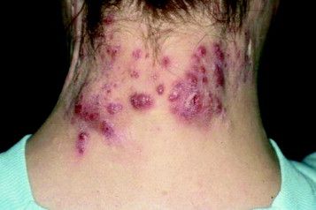
Nearly 70% reported that HS recurred after surgery, but 62% of those with recurrences said the disease was not as severe as before surgery.
“Surgery represents an important treatment option by itself but should also be part of combined therapeutic strategies, especially in severe disease stages. However, consistent approaches combining systemic and surgical treatments have not been established yet,” the authors noted.
The study had no funding source. The authors had no conflicts to disclose.
SOURCE: Kofler L et al. J Eur Acad Dermatol Venereol. 2018 Mar 23. doi: 10.1111/jdv.14892.
A retrospective German study found that the majority of , with many saying they no longer suffered from everyday impairment from the disease.
“We were able to show that surgical therapy resulted in convincing improvement of life quality and long-term results for HS that are at least as effective as biologicals,” the researchers wrote. The study was published online in the Journal of the European Academy of Dermatology and Venereology.
Lukas Kofler, MD, and associates from the department of dermatology at Eberhard Karls University’s University Medical Center, Tübingen, Germany, surveyed 910 of the facility’s patients who had undergone wide local excision for HS from 2006 to 2015. Surgery was “designed to reach into clinically disease-free subcutaneous fatty tissue,” followed by second intention healing, they wrote.
A total of 255 patients answered the survey, a response rate of 28%. There were 103 men and 152 women with a median age of 38 years (range, 14-66 years); 75% reported prior “nicotine abuse.” Almost half had been treated previously, most often with systemic antibiotics in 68%. The mean follow-up time was 57 months (range, 19-127 months);
All cases were Hurley grade III. Just over three-quarters of the patients described disease-related limitations in private life prior to surgery as “very strong” or “strong,” and 95% reported that their day-to-day life was impaired. Sixty percent said the disease impaired their work life (another 8% were not employed).
After surgery, 27% experienced postoperative complications, including minor bleeding, infection, and limited mobility; 65% experienced pain but 38% of the patients required analgesics postoperatively.
After surgery, 80% were satisfied or very satisfied with the results, and more than two-thirds were satisfied with the cosmetic results. Just over half said their private life was not impaired by the disease at all, compared with 3% who said so before surgery. After surgery, 20% reported being strongly or very strongly impaired, compared with 77% before surgery.
Nearly 70% reported that HS recurred after surgery, but 62% of those with recurrences said the disease was not as severe as before surgery.
“Surgery represents an important treatment option by itself but should also be part of combined therapeutic strategies, especially in severe disease stages. However, consistent approaches combining systemic and surgical treatments have not been established yet,” the authors noted.
The study had no funding source. The authors had no conflicts to disclose.
SOURCE: Kofler L et al. J Eur Acad Dermatol Venereol. 2018 Mar 23. doi: 10.1111/jdv.14892.
A retrospective German study found that the majority of , with many saying they no longer suffered from everyday impairment from the disease.
“We were able to show that surgical therapy resulted in convincing improvement of life quality and long-term results for HS that are at least as effective as biologicals,” the researchers wrote. The study was published online in the Journal of the European Academy of Dermatology and Venereology.
Lukas Kofler, MD, and associates from the department of dermatology at Eberhard Karls University’s University Medical Center, Tübingen, Germany, surveyed 910 of the facility’s patients who had undergone wide local excision for HS from 2006 to 2015. Surgery was “designed to reach into clinically disease-free subcutaneous fatty tissue,” followed by second intention healing, they wrote.
A total of 255 patients answered the survey, a response rate of 28%. There were 103 men and 152 women with a median age of 38 years (range, 14-66 years); 75% reported prior “nicotine abuse.” Almost half had been treated previously, most often with systemic antibiotics in 68%. The mean follow-up time was 57 months (range, 19-127 months);
All cases were Hurley grade III. Just over three-quarters of the patients described disease-related limitations in private life prior to surgery as “very strong” or “strong,” and 95% reported that their day-to-day life was impaired. Sixty percent said the disease impaired their work life (another 8% were not employed).
After surgery, 27% experienced postoperative complications, including minor bleeding, infection, and limited mobility; 65% experienced pain but 38% of the patients required analgesics postoperatively.
After surgery, 80% were satisfied or very satisfied with the results, and more than two-thirds were satisfied with the cosmetic results. Just over half said their private life was not impaired by the disease at all, compared with 3% who said so before surgery. After surgery, 20% reported being strongly or very strongly impaired, compared with 77% before surgery.
Nearly 70% reported that HS recurred after surgery, but 62% of those with recurrences said the disease was not as severe as before surgery.
“Surgery represents an important treatment option by itself but should also be part of combined therapeutic strategies, especially in severe disease stages. However, consistent approaches combining systemic and surgical treatments have not been established yet,” the authors noted.
The study had no funding source. The authors had no conflicts to disclose.
SOURCE: Kofler L et al. J Eur Acad Dermatol Venereol. 2018 Mar 23. doi: 10.1111/jdv.14892.
FROM THE JOURNAL OF THE EUROPEAN ACADEMY OF DERMATOLOGY AND VENEREOLOGY.
Key clinical point: Patients with hidradenitis suppurativa (HS) report dramatic improvement after radical surgery.
Major finding: The percentage reporting strong or very strong impairment of private life fell from 77% before surgery to 20% afterward.
Study details: A retrospective survey of 255 patients who had undergone surgery for HS (Hurley stage III).
Disclosures: The study had no funding source. The authors had no conflicts to disclose.
Source: Kofler L et al. J Eur Acad Dermatol Venereol. 2018 Mar 23. doi: 10.1111/jdv.14892.
From weekend warriors to pros, athletes are plagued by skin disorders
SAN DIEGO – Think beyond the foot: Fungal infections are just the beginning when it comes to skin disorders in athletes, which include ringworm in wrestlers, “jogger’s nipple” in runners, and more serious conditions – like skin cancer.
“from weekend warriors to professional athletes,” said Brian B. Adams, MD, MPH, professor and chair of the department of dermatology at the University of Cincinnati.
In an interview, he discussed specific risks for athletes, the scarcity of data on skin cancer in athletes, and the hazards posed by cotton clothing.
Skin cancer: Risk seems clear, but data are sparse
“Athletes in general appear to be at increased risk in the long term for skin cancer” since they often play and practice during the hours between 10 a.m. and 4 p.m., when the danger of sun exposure is at its highest, he said. In addition, “sweating removes the sunscreen that athletes put on, and there is evidence that sweating actually increases the chance of burning,” he said.
Skiers and snowboarders face unique sun exposure risks, he added. “Snow reflects up to 100% of UV, so even though they may be in shade, they experience UV. And mountain athletes experience greater amounts of UV as the altitude of the mountain increases: At higher altitudes, the atmosphere has less chance of filtering out the damaging rays.”
While it’s obvious that many athletes face extra sun exposure, Dr. Adams pointed out, “very little is definitively known about the actual degree of risk of athletic activities.”
Still, the research that does exist provides plenty of hints about risk. “Large epidemiological studies showed that recreational activities such as sun exposure on the beach or during water sports were associated with an increased risk of basal cell carcinoma, whereas skiing has been shown to be at increased risk for squamous cell carcinoma,” according to a 2008 report on outdoor sports and skin cancer (Clin Dermatol. 2008 Jan-Feb;26[1]:12-5).
“Risk factors of cutaneous melanoma, such as the number of melanocytic nevi and solar lentigines, have been found to be more frequent in subjects practicing endurance outdoor sports. An increased risk for cutaneous melanoma may be assumed for these athletes,” Dr. Adams commented.
Another study, this one published in 2006, found more atypical melanocytic nevi, solar lentigines, and lesions suggestive of nonmelanoma skin cancer in marathon runners, compared with a control group, and the risk was associated with the level of training intensity. The control subjects were more sensitive to the sun and had more common melanocytic nevi (Arch Dermatol. 2006 Nov;142[11]:1471-4).
Counseling about sunscreen may actually work
One strategy to reduce sun exposure is to advise athletes to avoid peak sun hours. However, “the key to caring for the athletes is not only recognizing that their sport may play a role in their disease but also realizing that your therapeutic approach must be tailored to minimize disruption to their practices and competitions,” Dr. Adams said.

However, there’s good news for dermatologists who are willing to push: The study also reported that athletes who were encouraged to use sunscreen were significantly more likely to use sunscreen (P less than .0001).
Watch for other conditions, from jogger’s nipple to ringworm
Dr. Adams offered advice about detection, treatment, and prevention of other skin disorders that affect athletes:
- “Jogger’s nipples” and other kinds of chafing. He has learned to recognize the “red eleven” – two vertical streaks of blood on a runner’s shirt – that represent a case of “jogger’s nipples” caused by chafing. Antibacterial ointment or petroleum jelly are useful treatments, he said, and an application of plenty of petroleum jelly on the nipples prior to a run can be helpful. Cotton shirts should be avoided, he said, in favor of synthetic, moisture-wicking shirts and bras. Chafing can also occur in the underarms and inner thighs, he said, and the same treatments and preventive techniques are useful.
- Callused and bleeding “jogger’s toes.” This can strike runners, especially on the second toe, which is often the longest and most likely to strike the toe box of a shoe. Specialty shoes can help prevent this condition, he said.
- Tinea corporis (ringworm) and herpes gladiatorum. In wrestlers, ringworm is known as tinea corporis gladiatorum because the intensity of skin-to-skin contact in wrestling makes the condition especially common in these athletes. Lesions don’t develop as rings at first; instead, they first appear as relatively nonspecific red round lesions and are most likely to be found in the head, neck, and upper extremities, Dr. Adams noted. Herpes gladiatorum is caused by herpes simplex virus 1; it is also seen in wrestlers and caused by skin-to-skin contact. Topical and oral antifungals clear ringworm, while oral antiviral agents are appropriate for herpes gladiatorum, Dr. Adams said. While herpes gladiatorum clears up and is no longer contagious after 4-5 days, he said, it’s not clear how long wrestlers with ringworm should be disqualified from playing.
Dr. Adams disclosed advising Mission, a company that focuses on sunscreen designed by and for athletes.
SAN DIEGO – Think beyond the foot: Fungal infections are just the beginning when it comes to skin disorders in athletes, which include ringworm in wrestlers, “jogger’s nipple” in runners, and more serious conditions – like skin cancer.
“from weekend warriors to professional athletes,” said Brian B. Adams, MD, MPH, professor and chair of the department of dermatology at the University of Cincinnati.
In an interview, he discussed specific risks for athletes, the scarcity of data on skin cancer in athletes, and the hazards posed by cotton clothing.
Skin cancer: Risk seems clear, but data are sparse
“Athletes in general appear to be at increased risk in the long term for skin cancer” since they often play and practice during the hours between 10 a.m. and 4 p.m., when the danger of sun exposure is at its highest, he said. In addition, “sweating removes the sunscreen that athletes put on, and there is evidence that sweating actually increases the chance of burning,” he said.
Skiers and snowboarders face unique sun exposure risks, he added. “Snow reflects up to 100% of UV, so even though they may be in shade, they experience UV. And mountain athletes experience greater amounts of UV as the altitude of the mountain increases: At higher altitudes, the atmosphere has less chance of filtering out the damaging rays.”
While it’s obvious that many athletes face extra sun exposure, Dr. Adams pointed out, “very little is definitively known about the actual degree of risk of athletic activities.”
Still, the research that does exist provides plenty of hints about risk. “Large epidemiological studies showed that recreational activities such as sun exposure on the beach or during water sports were associated with an increased risk of basal cell carcinoma, whereas skiing has been shown to be at increased risk for squamous cell carcinoma,” according to a 2008 report on outdoor sports and skin cancer (Clin Dermatol. 2008 Jan-Feb;26[1]:12-5).
“Risk factors of cutaneous melanoma, such as the number of melanocytic nevi and solar lentigines, have been found to be more frequent in subjects practicing endurance outdoor sports. An increased risk for cutaneous melanoma may be assumed for these athletes,” Dr. Adams commented.
Another study, this one published in 2006, found more atypical melanocytic nevi, solar lentigines, and lesions suggestive of nonmelanoma skin cancer in marathon runners, compared with a control group, and the risk was associated with the level of training intensity. The control subjects were more sensitive to the sun and had more common melanocytic nevi (Arch Dermatol. 2006 Nov;142[11]:1471-4).
Counseling about sunscreen may actually work
One strategy to reduce sun exposure is to advise athletes to avoid peak sun hours. However, “the key to caring for the athletes is not only recognizing that their sport may play a role in their disease but also realizing that your therapeutic approach must be tailored to minimize disruption to their practices and competitions,” Dr. Adams said.
However, there’s good news for dermatologists who are willing to push: The study also reported that athletes who were encouraged to use sunscreen were significantly more likely to use sunscreen (P less than .0001).
Watch for other conditions, from jogger’s nipple to ringworm
Dr. Adams offered advice about detection, treatment, and prevention of other skin disorders that affect athletes:
- “Jogger’s nipples” and other kinds of chafing. He has learned to recognize the “red eleven” – two vertical streaks of blood on a runner’s shirt – that represent a case of “jogger’s nipples” caused by chafing. Antibacterial ointment or petroleum jelly are useful treatments, he said, and an application of plenty of petroleum jelly on the nipples prior to a run can be helpful. Cotton shirts should be avoided, he said, in favor of synthetic, moisture-wicking shirts and bras. Chafing can also occur in the underarms and inner thighs, he said, and the same treatments and preventive techniques are useful.
- Callused and bleeding “jogger’s toes.” This can strike runners, especially on the second toe, which is often the longest and most likely to strike the toe box of a shoe. Specialty shoes can help prevent this condition, he said.
- Tinea corporis (ringworm) and herpes gladiatorum. In wrestlers, ringworm is known as tinea corporis gladiatorum because the intensity of skin-to-skin contact in wrestling makes the condition especially common in these athletes. Lesions don’t develop as rings at first; instead, they first appear as relatively nonspecific red round lesions and are most likely to be found in the head, neck, and upper extremities, Dr. Adams noted. Herpes gladiatorum is caused by herpes simplex virus 1; it is also seen in wrestlers and caused by skin-to-skin contact. Topical and oral antifungals clear ringworm, while oral antiviral agents are appropriate for herpes gladiatorum, Dr. Adams said. While herpes gladiatorum clears up and is no longer contagious after 4-5 days, he said, it’s not clear how long wrestlers with ringworm should be disqualified from playing.
Dr. Adams disclosed advising Mission, a company that focuses on sunscreen designed by and for athletes.
SAN DIEGO – Think beyond the foot: Fungal infections are just the beginning when it comes to skin disorders in athletes, which include ringworm in wrestlers, “jogger’s nipple” in runners, and more serious conditions – like skin cancer.
“from weekend warriors to professional athletes,” said Brian B. Adams, MD, MPH, professor and chair of the department of dermatology at the University of Cincinnati.
In an interview, he discussed specific risks for athletes, the scarcity of data on skin cancer in athletes, and the hazards posed by cotton clothing.
Skin cancer: Risk seems clear, but data are sparse
“Athletes in general appear to be at increased risk in the long term for skin cancer” since they often play and practice during the hours between 10 a.m. and 4 p.m., when the danger of sun exposure is at its highest, he said. In addition, “sweating removes the sunscreen that athletes put on, and there is evidence that sweating actually increases the chance of burning,” he said.
Skiers and snowboarders face unique sun exposure risks, he added. “Snow reflects up to 100% of UV, so even though they may be in shade, they experience UV. And mountain athletes experience greater amounts of UV as the altitude of the mountain increases: At higher altitudes, the atmosphere has less chance of filtering out the damaging rays.”
While it’s obvious that many athletes face extra sun exposure, Dr. Adams pointed out, “very little is definitively known about the actual degree of risk of athletic activities.”
Still, the research that does exist provides plenty of hints about risk. “Large epidemiological studies showed that recreational activities such as sun exposure on the beach or during water sports were associated with an increased risk of basal cell carcinoma, whereas skiing has been shown to be at increased risk for squamous cell carcinoma,” according to a 2008 report on outdoor sports and skin cancer (Clin Dermatol. 2008 Jan-Feb;26[1]:12-5).
“Risk factors of cutaneous melanoma, such as the number of melanocytic nevi and solar lentigines, have been found to be more frequent in subjects practicing endurance outdoor sports. An increased risk for cutaneous melanoma may be assumed for these athletes,” Dr. Adams commented.
Another study, this one published in 2006, found more atypical melanocytic nevi, solar lentigines, and lesions suggestive of nonmelanoma skin cancer in marathon runners, compared with a control group, and the risk was associated with the level of training intensity. The control subjects were more sensitive to the sun and had more common melanocytic nevi (Arch Dermatol. 2006 Nov;142[11]:1471-4).
Counseling about sunscreen may actually work
One strategy to reduce sun exposure is to advise athletes to avoid peak sun hours. However, “the key to caring for the athletes is not only recognizing that their sport may play a role in their disease but also realizing that your therapeutic approach must be tailored to minimize disruption to their practices and competitions,” Dr. Adams said.
However, there’s good news for dermatologists who are willing to push: The study also reported that athletes who were encouraged to use sunscreen were significantly more likely to use sunscreen (P less than .0001).
Watch for other conditions, from jogger’s nipple to ringworm
Dr. Adams offered advice about detection, treatment, and prevention of other skin disorders that affect athletes:
- “Jogger’s nipples” and other kinds of chafing. He has learned to recognize the “red eleven” – two vertical streaks of blood on a runner’s shirt – that represent a case of “jogger’s nipples” caused by chafing. Antibacterial ointment or petroleum jelly are useful treatments, he said, and an application of plenty of petroleum jelly on the nipples prior to a run can be helpful. Cotton shirts should be avoided, he said, in favor of synthetic, moisture-wicking shirts and bras. Chafing can also occur in the underarms and inner thighs, he said, and the same treatments and preventive techniques are useful.
- Callused and bleeding “jogger’s toes.” This can strike runners, especially on the second toe, which is often the longest and most likely to strike the toe box of a shoe. Specialty shoes can help prevent this condition, he said.
- Tinea corporis (ringworm) and herpes gladiatorum. In wrestlers, ringworm is known as tinea corporis gladiatorum because the intensity of skin-to-skin contact in wrestling makes the condition especially common in these athletes. Lesions don’t develop as rings at first; instead, they first appear as relatively nonspecific red round lesions and are most likely to be found in the head, neck, and upper extremities, Dr. Adams noted. Herpes gladiatorum is caused by herpes simplex virus 1; it is also seen in wrestlers and caused by skin-to-skin contact. Topical and oral antifungals clear ringworm, while oral antiviral agents are appropriate for herpes gladiatorum, Dr. Adams said. While herpes gladiatorum clears up and is no longer contagious after 4-5 days, he said, it’s not clear how long wrestlers with ringworm should be disqualified from playing.
Dr. Adams disclosed advising Mission, a company that focuses on sunscreen designed by and for athletes.
EXPERT ANALYSIS FROM AAD 18
Sensitivity of vibration-based neuropathy detectors varies widely
Three vibration-based detection tools vary widely in their sensitivity for detection of diabetic peripheral neuropathy. Researchers are suggesting that physicians use more than a single vibration-based device when they check the sensitivity of their diabetic patients’ feet.
“This would significantly reduce the proportion of patients with diabetes who would be falsely identified as having no peripheral neuropathy and subsequently denied the benefit of beneficial and effective secondary risk factor control,” said study coauthor Cynthia Formosa, PhD, of the University of Malta, Msida, and Staffordshire University, Stoke-on-Trent, England, in an interview.
Researchers have estimated that 50% of patients with diabetes develop diabetic polyneuropathy over their lives, and that the condition is responsible for more than a quarter of all diabetes spending. “Despite a long history of research in this area, we are only starting to understand the pathophysiology of the disease,” reports a 2016 review of recent findings.
In a previous report, several of the authors of the new study analyzed 10 sets of guidelines regarding diabetic foot and found that some lacked information to back up the recommendations. Guideline limitations “are responsible for the current gaps between guidelines, standard clinical practice, and development of complications,” the researchers wrote (Rev Diabet Stud. 2016, 13[2-3]:158-86).
Most of the guidelines recommended the use of a 10-g monofilament test, in which a monofilament is placed against the skin, plus a vibration-based test, the researchers reported.
For the new study in Primary Care Diabetes, the researchers examined the efficacy of three noninvasive tools that are used to detect neuropathy in the foot: the 128-Hz tuning fork; the VibraTip, a pocket-sized, motorized device; and the neurothesiometer, a 7-pound electromechanical device that comes in a carrying case and includes an attachment that delivers vibrations to the body.
According to Dr. Formosa, the tuning fork is the most commonly used testing device in normal clinical practice.
Physicians such as endocrinologists and podiatrists use the vibrations produced by the devices to detect whether patients have normal levels of sensation in various parts of the foot and ankle. “Vibration testing is extremely important,” the study investigators wrote, “since in the initial stages of neuropathy, the vibratory sensory system is amongst the first component of the nervous system to be affected.”
However, research into the accuracy of the devices has been inconsistent, according to the investigators.
The researchers prospectively recruited 100 patients at primary health centers diagnosed with type 2 diabetes for at least 10 years. The subjects included 57 males and 43 females, with a mean age of 73 years (± 7.8 years). Practitioners tested the neuropathy detection tools on the patients and recorded whether they felt vibrations.
The study authors reported that 29% of patients didn’t perceive vibrations produced by the VibraTip, compared with 21% for the neurothesiometer and 12% for the 128-Hz tuning fork (P less than .001).
The VibraTip device is available for about $90 on Amazon. The cost of a neurothesiometer is listed by one medical supply company as $2,850. Prices for 128-Hz tuning forks vary on Amazon, with prices listed from $6 to $40.
The study researchers did not confirm neuropathy via more sophisticated neurologic testing, so it remains unclear which device is actually the most accurate at detecting cases of neuropathy.
Still, “the findings of this study suggest that use of only one screening tool to assess vibration perception is likely to yield high false negative results,” the researchers wrote. “We recommend that peripheral neuropathy in patients with diabetes should be assessed utilizing two or more modalities.”

Moving forward, Dr. Chockalingam said, the research team plans to launch “a structured clinical trial using a gold-standard tool such as nerve conduction testing to confirm which simple screening method best detects neuropathy.”
No study funding was reported. The authors reported no relevant disclosures.
SOURCE: Azzopardia K et al. Prim Care Diabetes. 2018 Apr;12(2):111-5.
Three vibration-based detection tools vary widely in their sensitivity for detection of diabetic peripheral neuropathy. Researchers are suggesting that physicians use more than a single vibration-based device when they check the sensitivity of their diabetic patients’ feet.
“This would significantly reduce the proportion of patients with diabetes who would be falsely identified as having no peripheral neuropathy and subsequently denied the benefit of beneficial and effective secondary risk factor control,” said study coauthor Cynthia Formosa, PhD, of the University of Malta, Msida, and Staffordshire University, Stoke-on-Trent, England, in an interview.
Researchers have estimated that 50% of patients with diabetes develop diabetic polyneuropathy over their lives, and that the condition is responsible for more than a quarter of all diabetes spending. “Despite a long history of research in this area, we are only starting to understand the pathophysiology of the disease,” reports a 2016 review of recent findings.
In a previous report, several of the authors of the new study analyzed 10 sets of guidelines regarding diabetic foot and found that some lacked information to back up the recommendations. Guideline limitations “are responsible for the current gaps between guidelines, standard clinical practice, and development of complications,” the researchers wrote (Rev Diabet Stud. 2016, 13[2-3]:158-86).
Most of the guidelines recommended the use of a 10-g monofilament test, in which a monofilament is placed against the skin, plus a vibration-based test, the researchers reported.
For the new study in Primary Care Diabetes, the researchers examined the efficacy of three noninvasive tools that are used to detect neuropathy in the foot: the 128-Hz tuning fork; the VibraTip, a pocket-sized, motorized device; and the neurothesiometer, a 7-pound electromechanical device that comes in a carrying case and includes an attachment that delivers vibrations to the body.
According to Dr. Formosa, the tuning fork is the most commonly used testing device in normal clinical practice.
Physicians such as endocrinologists and podiatrists use the vibrations produced by the devices to detect whether patients have normal levels of sensation in various parts of the foot and ankle. “Vibration testing is extremely important,” the study investigators wrote, “since in the initial stages of neuropathy, the vibratory sensory system is amongst the first component of the nervous system to be affected.”
However, research into the accuracy of the devices has been inconsistent, according to the investigators.
The researchers prospectively recruited 100 patients at primary health centers diagnosed with type 2 diabetes for at least 10 years. The subjects included 57 males and 43 females, with a mean age of 73 years (± 7.8 years). Practitioners tested the neuropathy detection tools on the patients and recorded whether they felt vibrations.
The study authors reported that 29% of patients didn’t perceive vibrations produced by the VibraTip, compared with 21% for the neurothesiometer and 12% for the 128-Hz tuning fork (P less than .001).
The VibraTip device is available for about $90 on Amazon. The cost of a neurothesiometer is listed by one medical supply company as $2,850. Prices for 128-Hz tuning forks vary on Amazon, with prices listed from $6 to $40.
The study researchers did not confirm neuropathy via more sophisticated neurologic testing, so it remains unclear which device is actually the most accurate at detecting cases of neuropathy.
Still, “the findings of this study suggest that use of only one screening tool to assess vibration perception is likely to yield high false negative results,” the researchers wrote. “We recommend that peripheral neuropathy in patients with diabetes should be assessed utilizing two or more modalities.”
Moving forward, Dr. Chockalingam said, the research team plans to launch “a structured clinical trial using a gold-standard tool such as nerve conduction testing to confirm which simple screening method best detects neuropathy.”
No study funding was reported. The authors reported no relevant disclosures.
SOURCE: Azzopardia K et al. Prim Care Diabetes. 2018 Apr;12(2):111-5.
Three vibration-based detection tools vary widely in their sensitivity for detection of diabetic peripheral neuropathy. Researchers are suggesting that physicians use more than a single vibration-based device when they check the sensitivity of their diabetic patients’ feet.
“This would significantly reduce the proportion of patients with diabetes who would be falsely identified as having no peripheral neuropathy and subsequently denied the benefit of beneficial and effective secondary risk factor control,” said study coauthor Cynthia Formosa, PhD, of the University of Malta, Msida, and Staffordshire University, Stoke-on-Trent, England, in an interview.
Researchers have estimated that 50% of patients with diabetes develop diabetic polyneuropathy over their lives, and that the condition is responsible for more than a quarter of all diabetes spending. “Despite a long history of research in this area, we are only starting to understand the pathophysiology of the disease,” reports a 2016 review of recent findings.
In a previous report, several of the authors of the new study analyzed 10 sets of guidelines regarding diabetic foot and found that some lacked information to back up the recommendations. Guideline limitations “are responsible for the current gaps between guidelines, standard clinical practice, and development of complications,” the researchers wrote (Rev Diabet Stud. 2016, 13[2-3]:158-86).
Most of the guidelines recommended the use of a 10-g monofilament test, in which a monofilament is placed against the skin, plus a vibration-based test, the researchers reported.
For the new study in Primary Care Diabetes, the researchers examined the efficacy of three noninvasive tools that are used to detect neuropathy in the foot: the 128-Hz tuning fork; the VibraTip, a pocket-sized, motorized device; and the neurothesiometer, a 7-pound electromechanical device that comes in a carrying case and includes an attachment that delivers vibrations to the body.
According to Dr. Formosa, the tuning fork is the most commonly used testing device in normal clinical practice.
Physicians such as endocrinologists and podiatrists use the vibrations produced by the devices to detect whether patients have normal levels of sensation in various parts of the foot and ankle. “Vibration testing is extremely important,” the study investigators wrote, “since in the initial stages of neuropathy, the vibratory sensory system is amongst the first component of the nervous system to be affected.”
However, research into the accuracy of the devices has been inconsistent, according to the investigators.
The researchers prospectively recruited 100 patients at primary health centers diagnosed with type 2 diabetes for at least 10 years. The subjects included 57 males and 43 females, with a mean age of 73 years (± 7.8 years). Practitioners tested the neuropathy detection tools on the patients and recorded whether they felt vibrations.
The study authors reported that 29% of patients didn’t perceive vibrations produced by the VibraTip, compared with 21% for the neurothesiometer and 12% for the 128-Hz tuning fork (P less than .001).
The VibraTip device is available for about $90 on Amazon. The cost of a neurothesiometer is listed by one medical supply company as $2,850. Prices for 128-Hz tuning forks vary on Amazon, with prices listed from $6 to $40.
The study researchers did not confirm neuropathy via more sophisticated neurologic testing, so it remains unclear which device is actually the most accurate at detecting cases of neuropathy.
Still, “the findings of this study suggest that use of only one screening tool to assess vibration perception is likely to yield high false negative results,” the researchers wrote. “We recommend that peripheral neuropathy in patients with diabetes should be assessed utilizing two or more modalities.”
Moving forward, Dr. Chockalingam said, the research team plans to launch “a structured clinical trial using a gold-standard tool such as nerve conduction testing to confirm which simple screening method best detects neuropathy.”
No study funding was reported. The authors reported no relevant disclosures.
SOURCE: Azzopardia K et al. Prim Care Diabetes. 2018 Apr;12(2):111-5.
FROM PRIMARY CARE DIABETES
Key clinical point: Results vary widely among three vibration-based devices used to detect peripheral neuropathy.
Major finding: 29% of patients didn’t perceive vibrations produced by the VibraTip to 21% for the neurothesiometer and 12% for the 128 Hz tuning fork (P less than .001).
Study details: A prospective, multicenter, cross-sectional study of 100 patients diagnosed with type 2 diabetes for at least 10 years.
Disclosures: No study funding was reported. The authors reported no relevant disclosures.
Source: Azzopardia K et al. Prim Care Diabetes. 2018 Apr;12(2):111-5.
Posttransplant skin conditions vary widely by ethnicity
SAN DIEGO – A new study finds that the risk of skin cancers in organ transplant recipients may vary widely by ethnicity.
“The most important findings from our study are the high rates of keratinocyte neoplasms observed in our white Northern European patients, but also in those of Far East Asian descent. Dermatologists should also appreciate the high risk of Kaposi’s sarcoma (KS) in patients originating from Sub-Saharan Africa,” Jonathan Kentley, MBBS, of Royal London Hospital, said in an interview. He presented the study findings at the annual meeting of the American Academy of Dermatology.

For the study, Dr. Kentley and colleagues sought to better understand .
They analyzed an organ transplant center database for the years 1989-2016, and tracked 1,304 consecutive patients – which included 1,125 with skin problems. The overall population was 64% male with a median age in the early 40s, and almost all (1,276) had undergone renal transplants. A relative handful underwent liver, lung, heart, and pancreas transplants.
The majority of patients (885) were white Northern Europeans, but there were also significant numbers of people with South Asian (202), black African/Caribbean (131) and white/Mediterranean (52) heritage. A small number were Far East Asian (26) and Middle Eastern (8). The median follow-up time for the ethnic groups varied from about 5 years to about 12 years.
The researchers found that basal cell carcinoma was most common in white Northern European patients, at nearly 25%, with other groups under 10%. SCC was common in white Northern European patients and Far East Asians, both at nearly 25%.
By far, KS was the most common in black African/Caribbean patients, at nearly 11%. According to Dr. Kentley, researchers found the number of KS cases to be surprisingly high in this group, “compounded by the fact that we have had a number of additional cases in the past year after we had collected the data for this study.” He attributes the higher number of KS cases in these patients to an increased seroprevalence of its causative agent, human herpesvirus-8, in Sub-Saharan Africa. The rate of KS in the second most commonly affected group – white Mediterranean patients – was almost 2%.
Viral warts were common in most groups, with the rate in both white groups (white Northern European and white Mediterranean) at nearly 60%, and Far East Asians at about 65%. Porokeratosis was by far the most common in white Norther Europeans, at nearly 8%, and sebaceous hyperplasia was common in all groups (more than 20% to about 27%) except in the black African/Caribbean and South Asian groups.
All these results were statistically significant with P values less than .05.
“Our study has confirmed the increased risk of keratinocyte cancers in patients of white Northern European descent, as well as providing more information on the increased risk in patients of Far East Asian descent,” Dr. Kentley said. “We have also confirmed the propensity of black African/Caribbean patients to develop Kaposi’s sarcoma in the first 5 years post transplant and highlighted that white Mediterranean patients are also at high risk. Beyond this, we have been able to review the prevalence of rare malignancies, such as Merkel cell carcinoma and appendageal tumors, and highlight that white Northern European patients remain at high risk of developing these conditions.”
As for the impact on clinical practice, “the patterns of skin disease susceptibility we have identified have important implications for rational design of transplant skin surveillance programs, targeted patient (and provider) education, and optimized clinical management,” Dr. Kentley said. “Ultimately, this is likely to have a significant impact on strategic deployment of limited dermatology health care resources.”
Specifically, the study suggests that all organ transplant patients receive a baseline skin assessment visit and nurse-led targeted education. Black African/Caribbean patients should be followed up for at least 5 years after transplant.
In the United States, at least 724,000 people have undergone organ transplants since 1988, with most getting kidney transplants, according to the United Network for Organ Sharing (UNOS).
No study funding was reported. The authors had no disclosures.
SOURCE: Kentley J et al. AAD 2018, Session F055.
SAN DIEGO – A new study finds that the risk of skin cancers in organ transplant recipients may vary widely by ethnicity.
“The most important findings from our study are the high rates of keratinocyte neoplasms observed in our white Northern European patients, but also in those of Far East Asian descent. Dermatologists should also appreciate the high risk of Kaposi’s sarcoma (KS) in patients originating from Sub-Saharan Africa,” Jonathan Kentley, MBBS, of Royal London Hospital, said in an interview. He presented the study findings at the annual meeting of the American Academy of Dermatology.
For the study, Dr. Kentley and colleagues sought to better understand .
They analyzed an organ transplant center database for the years 1989-2016, and tracked 1,304 consecutive patients – which included 1,125 with skin problems. The overall population was 64% male with a median age in the early 40s, and almost all (1,276) had undergone renal transplants. A relative handful underwent liver, lung, heart, and pancreas transplants.
The majority of patients (885) were white Northern Europeans, but there were also significant numbers of people with South Asian (202), black African/Caribbean (131) and white/Mediterranean (52) heritage. A small number were Far East Asian (26) and Middle Eastern (8). The median follow-up time for the ethnic groups varied from about 5 years to about 12 years.
The researchers found that basal cell carcinoma was most common in white Northern European patients, at nearly 25%, with other groups under 10%. SCC was common in white Northern European patients and Far East Asians, both at nearly 25%.
By far, KS was the most common in black African/Caribbean patients, at nearly 11%. According to Dr. Kentley, researchers found the number of KS cases to be surprisingly high in this group, “compounded by the fact that we have had a number of additional cases in the past year after we had collected the data for this study.” He attributes the higher number of KS cases in these patients to an increased seroprevalence of its causative agent, human herpesvirus-8, in Sub-Saharan Africa. The rate of KS in the second most commonly affected group – white Mediterranean patients – was almost 2%.
Viral warts were common in most groups, with the rate in both white groups (white Northern European and white Mediterranean) at nearly 60%, and Far East Asians at about 65%. Porokeratosis was by far the most common in white Norther Europeans, at nearly 8%, and sebaceous hyperplasia was common in all groups (more than 20% to about 27%) except in the black African/Caribbean and South Asian groups.
All these results were statistically significant with P values less than .05.
“Our study has confirmed the increased risk of keratinocyte cancers in patients of white Northern European descent, as well as providing more information on the increased risk in patients of Far East Asian descent,” Dr. Kentley said. “We have also confirmed the propensity of black African/Caribbean patients to develop Kaposi’s sarcoma in the first 5 years post transplant and highlighted that white Mediterranean patients are also at high risk. Beyond this, we have been able to review the prevalence of rare malignancies, such as Merkel cell carcinoma and appendageal tumors, and highlight that white Northern European patients remain at high risk of developing these conditions.”
As for the impact on clinical practice, “the patterns of skin disease susceptibility we have identified have important implications for rational design of transplant skin surveillance programs, targeted patient (and provider) education, and optimized clinical management,” Dr. Kentley said. “Ultimately, this is likely to have a significant impact on strategic deployment of limited dermatology health care resources.”
Specifically, the study suggests that all organ transplant patients receive a baseline skin assessment visit and nurse-led targeted education. Black African/Caribbean patients should be followed up for at least 5 years after transplant.
In the United States, at least 724,000 people have undergone organ transplants since 1988, with most getting kidney transplants, according to the United Network for Organ Sharing (UNOS).
No study funding was reported. The authors had no disclosures.
SOURCE: Kentley J et al. AAD 2018, Session F055.
SAN DIEGO – A new study finds that the risk of skin cancers in organ transplant recipients may vary widely by ethnicity.
“The most important findings from our study are the high rates of keratinocyte neoplasms observed in our white Northern European patients, but also in those of Far East Asian descent. Dermatologists should also appreciate the high risk of Kaposi’s sarcoma (KS) in patients originating from Sub-Saharan Africa,” Jonathan Kentley, MBBS, of Royal London Hospital, said in an interview. He presented the study findings at the annual meeting of the American Academy of Dermatology.
For the study, Dr. Kentley and colleagues sought to better understand .
They analyzed an organ transplant center database for the years 1989-2016, and tracked 1,304 consecutive patients – which included 1,125 with skin problems. The overall population was 64% male with a median age in the early 40s, and almost all (1,276) had undergone renal transplants. A relative handful underwent liver, lung, heart, and pancreas transplants.
The majority of patients (885) were white Northern Europeans, but there were also significant numbers of people with South Asian (202), black African/Caribbean (131) and white/Mediterranean (52) heritage. A small number were Far East Asian (26) and Middle Eastern (8). The median follow-up time for the ethnic groups varied from about 5 years to about 12 years.
The researchers found that basal cell carcinoma was most common in white Northern European patients, at nearly 25%, with other groups under 10%. SCC was common in white Northern European patients and Far East Asians, both at nearly 25%.
By far, KS was the most common in black African/Caribbean patients, at nearly 11%. According to Dr. Kentley, researchers found the number of KS cases to be surprisingly high in this group, “compounded by the fact that we have had a number of additional cases in the past year after we had collected the data for this study.” He attributes the higher number of KS cases in these patients to an increased seroprevalence of its causative agent, human herpesvirus-8, in Sub-Saharan Africa. The rate of KS in the second most commonly affected group – white Mediterranean patients – was almost 2%.
Viral warts were common in most groups, with the rate in both white groups (white Northern European and white Mediterranean) at nearly 60%, and Far East Asians at about 65%. Porokeratosis was by far the most common in white Norther Europeans, at nearly 8%, and sebaceous hyperplasia was common in all groups (more than 20% to about 27%) except in the black African/Caribbean and South Asian groups.
All these results were statistically significant with P values less than .05.
“Our study has confirmed the increased risk of keratinocyte cancers in patients of white Northern European descent, as well as providing more information on the increased risk in patients of Far East Asian descent,” Dr. Kentley said. “We have also confirmed the propensity of black African/Caribbean patients to develop Kaposi’s sarcoma in the first 5 years post transplant and highlighted that white Mediterranean patients are also at high risk. Beyond this, we have been able to review the prevalence of rare malignancies, such as Merkel cell carcinoma and appendageal tumors, and highlight that white Northern European patients remain at high risk of developing these conditions.”
As for the impact on clinical practice, “the patterns of skin disease susceptibility we have identified have important implications for rational design of transplant skin surveillance programs, targeted patient (and provider) education, and optimized clinical management,” Dr. Kentley said. “Ultimately, this is likely to have a significant impact on strategic deployment of limited dermatology health care resources.”
Specifically, the study suggests that all organ transplant patients receive a baseline skin assessment visit and nurse-led targeted education. Black African/Caribbean patients should be followed up for at least 5 years after transplant.
In the United States, at least 724,000 people have undergone organ transplants since 1988, with most getting kidney transplants, according to the United Network for Organ Sharing (UNOS).
No study funding was reported. The authors had no disclosures.
SOURCE: Kentley J et al. AAD 2018, Session F055.
REPORTING FROM AAD 18
Key clinical point: Skin disorders after organ transplant differ widely by ethnicity.
Major finding: Posttransplant basal cell and squamous cell carcinomas are most common in white Northern Europeans (at nearly 25%), while Kaposi’s sarcoma was higher than expected (nearly 10%) in black African/Caribbean patients.
Study details: Analysis of 1,125 patients from a single transplant center who received organ transplants and developed skin problems over a median follow-up time of 5 to more than 12 years, depending on ethnicity.
Disclosures: No study funding was reported. The authors had no disclosures.
Source: Kentley J et al. AAD 2018, Session F055.
Microneedling improved acne scars in small study of patients with darker skin
, and did not contribute to more pigmentation, the study authors reported.
Most patients were pleased with the results. “Microneedling is an effective and safe treatment for acne scars associated with pigmentation in dark-skinned patients, without adding any risk of causing worsening of pigmentation,” the study’s lead author, Firas Al Qarqaz, MD said in an interview.
The study was published online in the Journal of Cosmetic Dermatology.
Dr. Al Qarqaz, of the department of dermatology, Jordan University of Science and Technology, Irbid, Jordan, pointed out that patients with darker skin and acne scars pose a unique challenge because some current treatments “can improve the scars but carry a risk of worsening the pigmentation and making skin/scars darker, which can be as troublesome to patients as their original scars.” Indeed, a review of microneedling as a treatment for dermatologic conditions in patients with darker skin noted that conventional resurfacing procedures can be limited in this patient population, because of concerns of adverse effects, including dyspigmentation (J Am Acad Dermatol. 2016 Feb;74[2]:348-55).
The situation is especially complex because “the current assessment methods for evaluating acne scars are not addressing clearly the important aspect of pigmentation that is associated with such scars, especially in darker skin, which can make objective assessment for improvement lacking,” Dr. Al Qarqaz noted.
He and a coauthor conducted the new study to determine whether microneedling can safely and effectively improve both acne scars and related hyperpigmentation in patients with darker skin. The study of 39 patients with postacne scarring comprised 31 women and 8 men aged 18-43 years (mean age, 27); their skin colors ranged from Fitzpatrick skin types type III to V. Most (27) were type IV.
The patients were treated with an electronic microneedling device and were evaluated at 2 weeks, and at least 4 weeks after their initial assessment (range, 4-14 weeks) for the final evaluation. The researchers found statistically significant improvement in two measures: The Postacne Hyperpigmentation Index improved from a mean score of 13 at baseline to a mean of 10 post procedure (P = .0035), and the Goodman-Baron acne scarring scale improved from a mean of 18 at baseline to a mean of 12 post procedure (P = .008).
Side effects were mild and temporary. “This treatment seems to be safe apart from short-lived erythema and occasional small hematoma following procedure,” the researchers concluded.
Nearly 80% of patients said they were satisfied with the procedure; the rest were not satisfied with the results (no reasons were cited). “Additional studies focusing on postacne scarring with hyperpigmentation are needed,” in addition to “assessment tools designed for this particular patient group,” the authors noted. They added that more treatments may be needed in some patients for hyperpigmentation.
Jordan University of Science and Technology funded the study. The study authors reported no relevant disclosures.
SOURCE: Al-Qarqaz F et al. J Cosmet Dermatol. 2018 Mar 15. (doi: 10.1111/jocd.12520.)
, and did not contribute to more pigmentation, the study authors reported.
Most patients were pleased with the results. “Microneedling is an effective and safe treatment for acne scars associated with pigmentation in dark-skinned patients, without adding any risk of causing worsening of pigmentation,” the study’s lead author, Firas Al Qarqaz, MD said in an interview.
The study was published online in the Journal of Cosmetic Dermatology.
Dr. Al Qarqaz, of the department of dermatology, Jordan University of Science and Technology, Irbid, Jordan, pointed out that patients with darker skin and acne scars pose a unique challenge because some current treatments “can improve the scars but carry a risk of worsening the pigmentation and making skin/scars darker, which can be as troublesome to patients as their original scars.” Indeed, a review of microneedling as a treatment for dermatologic conditions in patients with darker skin noted that conventional resurfacing procedures can be limited in this patient population, because of concerns of adverse effects, including dyspigmentation (J Am Acad Dermatol. 2016 Feb;74[2]:348-55).
The situation is especially complex because “the current assessment methods for evaluating acne scars are not addressing clearly the important aspect of pigmentation that is associated with such scars, especially in darker skin, which can make objective assessment for improvement lacking,” Dr. Al Qarqaz noted.
He and a coauthor conducted the new study to determine whether microneedling can safely and effectively improve both acne scars and related hyperpigmentation in patients with darker skin. The study of 39 patients with postacne scarring comprised 31 women and 8 men aged 18-43 years (mean age, 27); their skin colors ranged from Fitzpatrick skin types type III to V. Most (27) were type IV.
The patients were treated with an electronic microneedling device and were evaluated at 2 weeks, and at least 4 weeks after their initial assessment (range, 4-14 weeks) for the final evaluation. The researchers found statistically significant improvement in two measures: The Postacne Hyperpigmentation Index improved from a mean score of 13 at baseline to a mean of 10 post procedure (P = .0035), and the Goodman-Baron acne scarring scale improved from a mean of 18 at baseline to a mean of 12 post procedure (P = .008).
Side effects were mild and temporary. “This treatment seems to be safe apart from short-lived erythema and occasional small hematoma following procedure,” the researchers concluded.
Nearly 80% of patients said they were satisfied with the procedure; the rest were not satisfied with the results (no reasons were cited). “Additional studies focusing on postacne scarring with hyperpigmentation are needed,” in addition to “assessment tools designed for this particular patient group,” the authors noted. They added that more treatments may be needed in some patients for hyperpigmentation.
Jordan University of Science and Technology funded the study. The study authors reported no relevant disclosures.
SOURCE: Al-Qarqaz F et al. J Cosmet Dermatol. 2018 Mar 15. (doi: 10.1111/jocd.12520.)
, and did not contribute to more pigmentation, the study authors reported.
Most patients were pleased with the results. “Microneedling is an effective and safe treatment for acne scars associated with pigmentation in dark-skinned patients, without adding any risk of causing worsening of pigmentation,” the study’s lead author, Firas Al Qarqaz, MD said in an interview.
The study was published online in the Journal of Cosmetic Dermatology.
Dr. Al Qarqaz, of the department of dermatology, Jordan University of Science and Technology, Irbid, Jordan, pointed out that patients with darker skin and acne scars pose a unique challenge because some current treatments “can improve the scars but carry a risk of worsening the pigmentation and making skin/scars darker, which can be as troublesome to patients as their original scars.” Indeed, a review of microneedling as a treatment for dermatologic conditions in patients with darker skin noted that conventional resurfacing procedures can be limited in this patient population, because of concerns of adverse effects, including dyspigmentation (J Am Acad Dermatol. 2016 Feb;74[2]:348-55).
The situation is especially complex because “the current assessment methods for evaluating acne scars are not addressing clearly the important aspect of pigmentation that is associated with such scars, especially in darker skin, which can make objective assessment for improvement lacking,” Dr. Al Qarqaz noted.
He and a coauthor conducted the new study to determine whether microneedling can safely and effectively improve both acne scars and related hyperpigmentation in patients with darker skin. The study of 39 patients with postacne scarring comprised 31 women and 8 men aged 18-43 years (mean age, 27); their skin colors ranged from Fitzpatrick skin types type III to V. Most (27) were type IV.
The patients were treated with an electronic microneedling device and were evaluated at 2 weeks, and at least 4 weeks after their initial assessment (range, 4-14 weeks) for the final evaluation. The researchers found statistically significant improvement in two measures: The Postacne Hyperpigmentation Index improved from a mean score of 13 at baseline to a mean of 10 post procedure (P = .0035), and the Goodman-Baron acne scarring scale improved from a mean of 18 at baseline to a mean of 12 post procedure (P = .008).
Side effects were mild and temporary. “This treatment seems to be safe apart from short-lived erythema and occasional small hematoma following procedure,” the researchers concluded.
Nearly 80% of patients said they were satisfied with the procedure; the rest were not satisfied with the results (no reasons were cited). “Additional studies focusing on postacne scarring with hyperpigmentation are needed,” in addition to “assessment tools designed for this particular patient group,” the authors noted. They added that more treatments may be needed in some patients for hyperpigmentation.
Jordan University of Science and Technology funded the study. The study authors reported no relevant disclosures.
SOURCE: Al-Qarqaz F et al. J Cosmet Dermatol. 2018 Mar 15. (doi: 10.1111/jocd.12520.)
FROM THE JOURNAL OF COSMETIC DERMATOLOGY
Key clinical point: Microneedling produced statistically significant improvements without worsening pigmentation in darker-skinned patients with postacne scarring.
Major finding: The postacne hyperpigmentation index score improved from a mean of 13 to 10 (P = .0035) and the acne scarring scale improved from 18 to 12 (P = .008).
Study details: Microneedling was used to treat postacne scarring in 31 women and 8 men with Fitzpatrick skin types III-V.
Disclosures: Jordan University of Science and Technology funded the study. The study authors reported no relevant disclosures.
Source: Al Qarqaz F et al. J Cosmet Dermatol. 2018 Mar 15. doi: 10.1111/jocd.12520.
Hope and hype: Inside the push for wearable diabetes technology
Want to make a billion dollars? Here’s a hot tip: Invent wearable technology that detects diabetes, measures glucose levels, and determines how much insulin is needed – all without the need for a single drop of blood.
If you accept this mission, there’s a catch: You’ll have a whole bunch of company. When it comes to using technology to free patients with diabetes from the dreaded finger stick, “hope springs eternal in the hearts of scientists, entrepreneurs, opportunists, and charlatans alike,” writes electrochemical specialist and consultant John L. Smith, PhD, in his book “The Pursuit of Noninvasive Glucose.”
Google and Apple have been in the hunt, along with countless makers of devices and software. A noninvasive glucose monitoring system is the prime target, but there’s also plenty of interest in software that puts data from such devices as heartbeat sensors to work.

For the moment, however, results are elusive, and the name of the game is hype.
Early failure has a lasting impact
In the beginning, there was Glucowatch. And it was not good.
The GlucoWatch G2 Biographer product received approval from the Food and Drug Administration back in 2001 and touted as a high-tech tool to monitor glucose levels via interstitial concentrations every 10 minutes. The device promised to draw glucose to the skin surface for measurement via electric shocks, and alarms were to go off when hypoglycemia or hyperglycemia was detected.
But numerous problems cropped up. There was a time lag, with the device estimating glucose levels that actually occurred 15-20 minutes earlier. Some patients couldn’t tolerate wearing the watch, and some were burned by the electric current.
And perhaps worst of all, the measurements often weren’t accurate, with one study finding that more than half of 240 nighttime alarms incorrectly warned children with diabetes of dangerously high or low glucose. (Diabetes Technol Ther. 2005 June; 7[3]:440-7).
As a result of the Glucowatch debacle, the FDA “become a little gun shy about approving anything. It made it even harder to approve something,” said Mark J. Rice, MD, of Vanderbilt University, Nashville, Tenn., who has tried to develop glucose-measuring technology.

“If a device were to be commercialized, it would be hugely disruptive to the industry,” according to Dr. Baers, who said that she expects a device eventually will lead to higher levels of diabetes control and fewer side effects. “This would result in billion-dollar savings for the health care industry and reduced complications,” she said.
On tech front, promises and more promises
So far, there have been more promises than actual products.
If you don’t look too closely at the website of a device called GlucoWise, you might assume a noninvasive glucose monitor already exists. Under a photo of a smiling woman, the site promises a “100% pain-free device that makes traditional blood sampling a thing of the past.”
The “simple yet highly reliable” device, which looks a bit like a large clip for a potato chip bag, promises to measure glucose through high-frequency radio waves that penetrate thin body tissue in the earlobe or the area between the thumb and forefinger.
But the GlucoWise device is neither approved nor available, and the company’s predictions that it would take preorders by late 2016 didn’t come true.
Another product called SugarBEAT missed its planned 2016 release and now hopes to be available in the Britain later this year. It promises to measure glucose levels every 5 minutes via a small disposable patch that draws interstitial fluid from the skin.
Meanwhile, Apple has enlisted biomedical engineers to work on a secret project to measure glucose continuously and noninvasively, CNBC reported last year. And Google announced in 2014 that it was working on a glucose-detecting contact lens that could alert patients via tiny LED lights – yes, apparently in the lenses themselves – if levels go too high or low. But neither of these technologies is ready for prime time.
Sneaky glucose molecules elude scientists
According to Dr. Rice, no truly noninvasive glucose-measuring technique has worked so far.
The challenge, he said, is that it’s difficult to measure tiny glucose molecules, which have no color and share many characteristics with H2O.
“The real problem is trying to measure a colorless molecule in a sea of water,” Dr. Rice said.
Glucose lab tests rely on indicators from reactions with other substances, he said, “but you can’t do that in the body.” Measuring glucose in tears or urine is one possibility, he said, but the scarcity in those liquids poses a challenge: “Your body doesn’t want to spill glucose and lose energy.”
Dr. Rice himself explored a glucose-measuring technique that aimed to correlate glucose levels to the speed of the retina’s reaction to light. The idea was that patients would wear special glasses that would shine a light in the eye at the press of a button. The project ultimately failed.
There are other challenges, said Dr. Baers, the technology adviser. “Glucose concentrations in sweat or tears are not reflective of blood glucose concentrations. To make things even more challenging, glucose levels in these fluids are orders of magnitude smaller than that found in blood.”
And, she said, there’s a time lag between glucose levels in blood and in other body fluids. “This means that a sweat glucose level is really giving information from an hour previous, which can be dangerous if you’re operating machinery or driving.”
Wearable diabetes tech targets more than glucose
There’s more to wearable, noninvasive diabetes technology than glucose-monitoring. One of the new frontiers is diagnostics.
Earlier this year, researchers from the University of California at San Francisco and the digital startup Cardiogram reported that they were able to use data from digital heart rate sensors (like those found in Apple Watches, Fitbits and other devices) to correctly detect diabetes in patients.
In a study presented at the 2018 meeting of the Association for the Advancement of Artificial Intelligence, the researchers said they detected diabetes in 85% of 462 participants (out of a pool of 14,011) who’d previously been diagnosed with the condition (AAAAI abstract arXiv:1802.02511v1 [cs.LG]).

The next step is to test whether the data analysis can detect undiagnosed diabetes, Mr. Ballinger said.
As tech advances, questions remain
San Diego’s Scripps Whittier Diabetes Institute is another player in the diabetes/digital health world. It’s currently working on several clinical trials of diabetes technology, including a study into whether older adults with type 1 diabetes will benefit from a continuous glucose monitoring device with a wireless connection.

Devices that measure glucose can also suffer from errors in transmission, she said. And the existing continuous glucose monitors have trouble with accuracy at the very highest and lowest glucose levels, she said, although they are improving.
There are other questions about future wearable technology for diabetes: Will the devices cost more than continuous glucose monitoring systems (CGM), which are already pricey? How will private health information be protected? (As Mr. Ballinger noted, “wearable data itself is out of the scope of HIPAA.”) And will patients actually take action when their devices diagnose diabetes or warn them that their glucose levels are out of whack?
CGM systems provide insight into the latter issue. Repeated alarms about highs and lows can drive patients crazy, Dr. Philis-Tsimikas said. “You might end up with alarm fatigue and annoyance. They might hit a 250, but they won’t want the alarm to go off, and they don’t want to be reminded of it,” she said. “And they might go down to 60-80 at night, but they don’t want to be woken up because they’re used to that range.”
Even if patients do pay attention to their diabetes devices, they may not take the proper action. Dr. Philis-Tsimikas pointed to a 2016 study that found adding an exercise-tracking device to traditional weight-loss intervention didn’t lead to more weight loss. In fact, those who used the device actually loss less weight. (JAMA. 2016;316[11]:1161-71)
The lesson? “There has to be a combination of some education together with the physiologic information,” she said. For now, the good news is that “we still have other options,” Dr. Philis-Tsimikas said. The newly released CGM system known as the Freestyle Libre, she said, is one alternative.
And she mentioned another technique that’s still around. You could call it Old Faithful: the low-tech, high-hassle but highly accurate finger stick.
Dr. Baers and Dr. Rice report no disclosures. Dr. Philis-Tsimikas has no disclosures but notes that Scripps Whittier Diabetes Institute receives grants and funding in the diabetes field and works with a number of drug makers and device makers. Mr. Ballinger discloses salary and equity from Cardiogram.
cenews@mdedge.com
Want to make a billion dollars? Here’s a hot tip: Invent wearable technology that detects diabetes, measures glucose levels, and determines how much insulin is needed – all without the need for a single drop of blood.
If you accept this mission, there’s a catch: You’ll have a whole bunch of company. When it comes to using technology to free patients with diabetes from the dreaded finger stick, “hope springs eternal in the hearts of scientists, entrepreneurs, opportunists, and charlatans alike,” writes electrochemical specialist and consultant John L. Smith, PhD, in his book “The Pursuit of Noninvasive Glucose.”
Google and Apple have been in the hunt, along with countless makers of devices and software. A noninvasive glucose monitoring system is the prime target, but there’s also plenty of interest in software that puts data from such devices as heartbeat sensors to work.
For the moment, however, results are elusive, and the name of the game is hype.
Early failure has a lasting impact
In the beginning, there was Glucowatch. And it was not good.
The GlucoWatch G2 Biographer product received approval from the Food and Drug Administration back in 2001 and touted as a high-tech tool to monitor glucose levels via interstitial concentrations every 10 minutes. The device promised to draw glucose to the skin surface for measurement via electric shocks, and alarms were to go off when hypoglycemia or hyperglycemia was detected.
But numerous problems cropped up. There was a time lag, with the device estimating glucose levels that actually occurred 15-20 minutes earlier. Some patients couldn’t tolerate wearing the watch, and some were burned by the electric current.
And perhaps worst of all, the measurements often weren’t accurate, with one study finding that more than half of 240 nighttime alarms incorrectly warned children with diabetes of dangerously high or low glucose. (Diabetes Technol Ther. 2005 June; 7[3]:440-7).
As a result of the Glucowatch debacle, the FDA “become a little gun shy about approving anything. It made it even harder to approve something,” said Mark J. Rice, MD, of Vanderbilt University, Nashville, Tenn., who has tried to develop glucose-measuring technology.
“If a device were to be commercialized, it would be hugely disruptive to the industry,” according to Dr. Baers, who said that she expects a device eventually will lead to higher levels of diabetes control and fewer side effects. “This would result in billion-dollar savings for the health care industry and reduced complications,” she said.
On tech front, promises and more promises
So far, there have been more promises than actual products.
If you don’t look too closely at the website of a device called GlucoWise, you might assume a noninvasive glucose monitor already exists. Under a photo of a smiling woman, the site promises a “100% pain-free device that makes traditional blood sampling a thing of the past.”
The “simple yet highly reliable” device, which looks a bit like a large clip for a potato chip bag, promises to measure glucose through high-frequency radio waves that penetrate thin body tissue in the earlobe or the area between the thumb and forefinger.
But the GlucoWise device is neither approved nor available, and the company’s predictions that it would take preorders by late 2016 didn’t come true.
Another product called SugarBEAT missed its planned 2016 release and now hopes to be available in the Britain later this year. It promises to measure glucose levels every 5 minutes via a small disposable patch that draws interstitial fluid from the skin.
Meanwhile, Apple has enlisted biomedical engineers to work on a secret project to measure glucose continuously and noninvasively, CNBC reported last year. And Google announced in 2014 that it was working on a glucose-detecting contact lens that could alert patients via tiny LED lights – yes, apparently in the lenses themselves – if levels go too high or low. But neither of these technologies is ready for prime time.
Sneaky glucose molecules elude scientists
According to Dr. Rice, no truly noninvasive glucose-measuring technique has worked so far.
The challenge, he said, is that it’s difficult to measure tiny glucose molecules, which have no color and share many characteristics with H2O.
“The real problem is trying to measure a colorless molecule in a sea of water,” Dr. Rice said.
Glucose lab tests rely on indicators from reactions with other substances, he said, “but you can’t do that in the body.” Measuring glucose in tears or urine is one possibility, he said, but the scarcity in those liquids poses a challenge: “Your body doesn’t want to spill glucose and lose energy.”
Dr. Rice himself explored a glucose-measuring technique that aimed to correlate glucose levels to the speed of the retina’s reaction to light. The idea was that patients would wear special glasses that would shine a light in the eye at the press of a button. The project ultimately failed.
There are other challenges, said Dr. Baers, the technology adviser. “Glucose concentrations in sweat or tears are not reflective of blood glucose concentrations. To make things even more challenging, glucose levels in these fluids are orders of magnitude smaller than that found in blood.”
And, she said, there’s a time lag between glucose levels in blood and in other body fluids. “This means that a sweat glucose level is really giving information from an hour previous, which can be dangerous if you’re operating machinery or driving.”
Wearable diabetes tech targets more than glucose
There’s more to wearable, noninvasive diabetes technology than glucose-monitoring. One of the new frontiers is diagnostics.
Earlier this year, researchers from the University of California at San Francisco and the digital startup Cardiogram reported that they were able to use data from digital heart rate sensors (like those found in Apple Watches, Fitbits and other devices) to correctly detect diabetes in patients.
In a study presented at the 2018 meeting of the Association for the Advancement of Artificial Intelligence, the researchers said they detected diabetes in 85% of 462 participants (out of a pool of 14,011) who’d previously been diagnosed with the condition (AAAAI abstract arXiv:1802.02511v1 [cs.LG]).
The next step is to test whether the data analysis can detect undiagnosed diabetes, Mr. Ballinger said.
As tech advances, questions remain
San Diego’s Scripps Whittier Diabetes Institute is another player in the diabetes/digital health world. It’s currently working on several clinical trials of diabetes technology, including a study into whether older adults with type 1 diabetes will benefit from a continuous glucose monitoring device with a wireless connection.
Devices that measure glucose can also suffer from errors in transmission, she said. And the existing continuous glucose monitors have trouble with accuracy at the very highest and lowest glucose levels, she said, although they are improving.
There are other questions about future wearable technology for diabetes: Will the devices cost more than continuous glucose monitoring systems (CGM), which are already pricey? How will private health information be protected? (As Mr. Ballinger noted, “wearable data itself is out of the scope of HIPAA.”) And will patients actually take action when their devices diagnose diabetes or warn them that their glucose levels are out of whack?
CGM systems provide insight into the latter issue. Repeated alarms about highs and lows can drive patients crazy, Dr. Philis-Tsimikas said. “You might end up with alarm fatigue and annoyance. They might hit a 250, but they won’t want the alarm to go off, and they don’t want to be reminded of it,” she said. “And they might go down to 60-80 at night, but they don’t want to be woken up because they’re used to that range.”
Even if patients do pay attention to their diabetes devices, they may not take the proper action. Dr. Philis-Tsimikas pointed to a 2016 study that found adding an exercise-tracking device to traditional weight-loss intervention didn’t lead to more weight loss. In fact, those who used the device actually loss less weight. (JAMA. 2016;316[11]:1161-71)
The lesson? “There has to be a combination of some education together with the physiologic information,” she said. For now, the good news is that “we still have other options,” Dr. Philis-Tsimikas said. The newly released CGM system known as the Freestyle Libre, she said, is one alternative.
And she mentioned another technique that’s still around. You could call it Old Faithful: the low-tech, high-hassle but highly accurate finger stick.
Dr. Baers and Dr. Rice report no disclosures. Dr. Philis-Tsimikas has no disclosures but notes that Scripps Whittier Diabetes Institute receives grants and funding in the diabetes field and works with a number of drug makers and device makers. Mr. Ballinger discloses salary and equity from Cardiogram.
cenews@mdedge.com
Want to make a billion dollars? Here’s a hot tip: Invent wearable technology that detects diabetes, measures glucose levels, and determines how much insulin is needed – all without the need for a single drop of blood.
If you accept this mission, there’s a catch: You’ll have a whole bunch of company. When it comes to using technology to free patients with diabetes from the dreaded finger stick, “hope springs eternal in the hearts of scientists, entrepreneurs, opportunists, and charlatans alike,” writes electrochemical specialist and consultant John L. Smith, PhD, in his book “The Pursuit of Noninvasive Glucose.”
Google and Apple have been in the hunt, along with countless makers of devices and software. A noninvasive glucose monitoring system is the prime target, but there’s also plenty of interest in software that puts data from such devices as heartbeat sensors to work.
For the moment, however, results are elusive, and the name of the game is hype.
Early failure has a lasting impact
In the beginning, there was Glucowatch. And it was not good.
The GlucoWatch G2 Biographer product received approval from the Food and Drug Administration back in 2001 and touted as a high-tech tool to monitor glucose levels via interstitial concentrations every 10 minutes. The device promised to draw glucose to the skin surface for measurement via electric shocks, and alarms were to go off when hypoglycemia or hyperglycemia was detected.
But numerous problems cropped up. There was a time lag, with the device estimating glucose levels that actually occurred 15-20 minutes earlier. Some patients couldn’t tolerate wearing the watch, and some were burned by the electric current.
And perhaps worst of all, the measurements often weren’t accurate, with one study finding that more than half of 240 nighttime alarms incorrectly warned children with diabetes of dangerously high or low glucose. (Diabetes Technol Ther. 2005 June; 7[3]:440-7).
As a result of the Glucowatch debacle, the FDA “become a little gun shy about approving anything. It made it even harder to approve something,” said Mark J. Rice, MD, of Vanderbilt University, Nashville, Tenn., who has tried to develop glucose-measuring technology.
“If a device were to be commercialized, it would be hugely disruptive to the industry,” according to Dr. Baers, who said that she expects a device eventually will lead to higher levels of diabetes control and fewer side effects. “This would result in billion-dollar savings for the health care industry and reduced complications,” she said.
On tech front, promises and more promises
So far, there have been more promises than actual products.
If you don’t look too closely at the website of a device called GlucoWise, you might assume a noninvasive glucose monitor already exists. Under a photo of a smiling woman, the site promises a “100% pain-free device that makes traditional blood sampling a thing of the past.”
The “simple yet highly reliable” device, which looks a bit like a large clip for a potato chip bag, promises to measure glucose through high-frequency radio waves that penetrate thin body tissue in the earlobe or the area between the thumb and forefinger.
But the GlucoWise device is neither approved nor available, and the company’s predictions that it would take preorders by late 2016 didn’t come true.
Another product called SugarBEAT missed its planned 2016 release and now hopes to be available in the Britain later this year. It promises to measure glucose levels every 5 minutes via a small disposable patch that draws interstitial fluid from the skin.
Meanwhile, Apple has enlisted biomedical engineers to work on a secret project to measure glucose continuously and noninvasively, CNBC reported last year. And Google announced in 2014 that it was working on a glucose-detecting contact lens that could alert patients via tiny LED lights – yes, apparently in the lenses themselves – if levels go too high or low. But neither of these technologies is ready for prime time.
Sneaky glucose molecules elude scientists
According to Dr. Rice, no truly noninvasive glucose-measuring technique has worked so far.
The challenge, he said, is that it’s difficult to measure tiny glucose molecules, which have no color and share many characteristics with H2O.
“The real problem is trying to measure a colorless molecule in a sea of water,” Dr. Rice said.
Glucose lab tests rely on indicators from reactions with other substances, he said, “but you can’t do that in the body.” Measuring glucose in tears or urine is one possibility, he said, but the scarcity in those liquids poses a challenge: “Your body doesn’t want to spill glucose and lose energy.”
Dr. Rice himself explored a glucose-measuring technique that aimed to correlate glucose levels to the speed of the retina’s reaction to light. The idea was that patients would wear special glasses that would shine a light in the eye at the press of a button. The project ultimately failed.
There are other challenges, said Dr. Baers, the technology adviser. “Glucose concentrations in sweat or tears are not reflective of blood glucose concentrations. To make things even more challenging, glucose levels in these fluids are orders of magnitude smaller than that found in blood.”
And, she said, there’s a time lag between glucose levels in blood and in other body fluids. “This means that a sweat glucose level is really giving information from an hour previous, which can be dangerous if you’re operating machinery or driving.”
Wearable diabetes tech targets more than glucose
There’s more to wearable, noninvasive diabetes technology than glucose-monitoring. One of the new frontiers is diagnostics.
Earlier this year, researchers from the University of California at San Francisco and the digital startup Cardiogram reported that they were able to use data from digital heart rate sensors (like those found in Apple Watches, Fitbits and other devices) to correctly detect diabetes in patients.
In a study presented at the 2018 meeting of the Association for the Advancement of Artificial Intelligence, the researchers said they detected diabetes in 85% of 462 participants (out of a pool of 14,011) who’d previously been diagnosed with the condition (AAAAI abstract arXiv:1802.02511v1 [cs.LG]).
The next step is to test whether the data analysis can detect undiagnosed diabetes, Mr. Ballinger said.
As tech advances, questions remain
San Diego’s Scripps Whittier Diabetes Institute is another player in the diabetes/digital health world. It’s currently working on several clinical trials of diabetes technology, including a study into whether older adults with type 1 diabetes will benefit from a continuous glucose monitoring device with a wireless connection.
Devices that measure glucose can also suffer from errors in transmission, she said. And the existing continuous glucose monitors have trouble with accuracy at the very highest and lowest glucose levels, she said, although they are improving.
There are other questions about future wearable technology for diabetes: Will the devices cost more than continuous glucose monitoring systems (CGM), which are already pricey? How will private health information be protected? (As Mr. Ballinger noted, “wearable data itself is out of the scope of HIPAA.”) And will patients actually take action when their devices diagnose diabetes or warn them that their glucose levels are out of whack?
CGM systems provide insight into the latter issue. Repeated alarms about highs and lows can drive patients crazy, Dr. Philis-Tsimikas said. “You might end up with alarm fatigue and annoyance. They might hit a 250, but they won’t want the alarm to go off, and they don’t want to be reminded of it,” she said. “And they might go down to 60-80 at night, but they don’t want to be woken up because they’re used to that range.”
Even if patients do pay attention to their diabetes devices, they may not take the proper action. Dr. Philis-Tsimikas pointed to a 2016 study that found adding an exercise-tracking device to traditional weight-loss intervention didn’t lead to more weight loss. In fact, those who used the device actually loss less weight. (JAMA. 2016;316[11]:1161-71)
The lesson? “There has to be a combination of some education together with the physiologic information,” she said. For now, the good news is that “we still have other options,” Dr. Philis-Tsimikas said. The newly released CGM system known as the Freestyle Libre, she said, is one alternative.
And she mentioned another technique that’s still around. You could call it Old Faithful: the low-tech, high-hassle but highly accurate finger stick.
Dr. Baers and Dr. Rice report no disclosures. Dr. Philis-Tsimikas has no disclosures but notes that Scripps Whittier Diabetes Institute receives grants and funding in the diabetes field and works with a number of drug makers and device makers. Mr. Ballinger discloses salary and equity from Cardiogram.
cenews@mdedge.com
Smokers face higher infection risk after hernia operations
Jonah Stulberg, MD, FACS, is stickler about requiring patients to stop smoking at least 3 months before hernia surgery. He even uses urine tests to confirm whether they actually quit. A study by Dr. Stulberg and his colleagues supports this approach: .
The finding held up even after the researchers controlled for various factors. “Our findings are in agreement with other findings in higher risk surgeries, and they provide evidence that low-risk surgeries are not exempt from the risks associated with smoking,” said Dr. Stulberg in an interview. “Our data would suggest that there is significant clinical benefit to encouraging smoking cessation before elective hernia repair.”

The researchers launched the study to better understand how smoking affects complication rates in light of the fact that “surgeons in the U.S. tend to offer low-risk elective surgical procedures to patients who are actively smoking despite overwhelming evidence that smoking increases surgical risks,” Dr. Stulberg said.
The researchers tracked 220,629 patients in the American College of Surgeons National Surgical Quality Improvement Project (NSQIP) database who underwent several types of elective hernia repair from 2011 to 2014.
Just over 18% of the patients said they’d smoked over the past year; they were more likely to be younger (median age, 50 for smokers vs. 57 for nonsmokers). Smokers also were more likely to be black, to be underweight, and to consume two or more alcoholic beverages per day (P less than .05).
The researchers tracked serious complications in the 30 days after surgery such as death, sepsis, and readmission.
Complications developed in 6.34% of smokers and 4.72% of nonsmokers (P less than .001). Numerous kinds of complications were more common in the smokers prior to adjustment: death, return to the operating room, readmission, and transfusion plus wound, pulmonary, thromboembolic and cardiac complications.
The researchers adjusted their statistics to account for factors such as ethnicity, sex, body mass index, preexisting comorbidities, and type of hernia operation. They found that risk of all complications was higher in smokers, compared with nonsmokers (odds ratio, 1.30) as were several other complications: death (OR, 1.53), return to operating room (OR, 1.23), readmission (OR, 1.24), wound complication (OR, 1.36), sepsis/septic shock (OR, 1.31), pulmonary complication (OR 1.77-2.30) and cardiac complication (OR, 1.27-1.43).
Only transfusion (OR, 0.90) and thromboembolic (OR, 0.87) complications were less likely in smokers.
The researchers noted that the statistics don’t allow them to analyze whether it makes any difference if smokers quit shortly before their procedures. Still, Dr. Stulberg stands by his you-must-quit-smoking-before-surgery edict. “I believe that their active smoking habit is a bigger health threat than their asymptomatic hernia, and therefore feel the right thing to do as their physician is support them through their smoking cessation,” he said. “I offer counseling and nicotine replacement if needed. I have very good quit rates and would encourage other surgeons to do the same.”
Dr. Stulberg noted that he can’t point to evidence supporting his requirement that patients quit at least 3 months before surgery. “Most data out there says the farther away from your last cigarette, the better,” he said. “But there isn’t any magic to 3 months, other than I believe it will lead to a higher likelihood of permanent cessation.”
Northwestern Memorial Hospital and Northwestern University funded the study. Four of the nine authors, including Dr. Stulberg, report various disclosures that are not directly related to the study, including funding from government agencies, physician organizations, Health Care Services Corporation and Blue Cross Blue Shield of Illinois, Mallinckrodt, and Northwestern University. The other authors report no disclosures.
SOURCE: DeLancey JO et al. Am J Surg. 2018 Mar 6. doi: 10.1016/j.amjsurg.2018.03.004.
Jonah Stulberg, MD, FACS, is stickler about requiring patients to stop smoking at least 3 months before hernia surgery. He even uses urine tests to confirm whether they actually quit. A study by Dr. Stulberg and his colleagues supports this approach: .
The finding held up even after the researchers controlled for various factors. “Our findings are in agreement with other findings in higher risk surgeries, and they provide evidence that low-risk surgeries are not exempt from the risks associated with smoking,” said Dr. Stulberg in an interview. “Our data would suggest that there is significant clinical benefit to encouraging smoking cessation before elective hernia repair.”
The researchers launched the study to better understand how smoking affects complication rates in light of the fact that “surgeons in the U.S. tend to offer low-risk elective surgical procedures to patients who are actively smoking despite overwhelming evidence that smoking increases surgical risks,” Dr. Stulberg said.
The researchers tracked 220,629 patients in the American College of Surgeons National Surgical Quality Improvement Project (NSQIP) database who underwent several types of elective hernia repair from 2011 to 2014.
Just over 18% of the patients said they’d smoked over the past year; they were more likely to be younger (median age, 50 for smokers vs. 57 for nonsmokers). Smokers also were more likely to be black, to be underweight, and to consume two or more alcoholic beverages per day (P less than .05).
The researchers tracked serious complications in the 30 days after surgery such as death, sepsis, and readmission.
Complications developed in 6.34% of smokers and 4.72% of nonsmokers (P less than .001). Numerous kinds of complications were more common in the smokers prior to adjustment: death, return to the operating room, readmission, and transfusion plus wound, pulmonary, thromboembolic and cardiac complications.
The researchers adjusted their statistics to account for factors such as ethnicity, sex, body mass index, preexisting comorbidities, and type of hernia operation. They found that risk of all complications was higher in smokers, compared with nonsmokers (odds ratio, 1.30) as were several other complications: death (OR, 1.53), return to operating room (OR, 1.23), readmission (OR, 1.24), wound complication (OR, 1.36), sepsis/septic shock (OR, 1.31), pulmonary complication (OR 1.77-2.30) and cardiac complication (OR, 1.27-1.43).
Only transfusion (OR, 0.90) and thromboembolic (OR, 0.87) complications were less likely in smokers.
The researchers noted that the statistics don’t allow them to analyze whether it makes any difference if smokers quit shortly before their procedures. Still, Dr. Stulberg stands by his you-must-quit-smoking-before-surgery edict. “I believe that their active smoking habit is a bigger health threat than their asymptomatic hernia, and therefore feel the right thing to do as their physician is support them through their smoking cessation,” he said. “I offer counseling and nicotine replacement if needed. I have very good quit rates and would encourage other surgeons to do the same.”
Dr. Stulberg noted that he can’t point to evidence supporting his requirement that patients quit at least 3 months before surgery. “Most data out there says the farther away from your last cigarette, the better,” he said. “But there isn’t any magic to 3 months, other than I believe it will lead to a higher likelihood of permanent cessation.”
Northwestern Memorial Hospital and Northwestern University funded the study. Four of the nine authors, including Dr. Stulberg, report various disclosures that are not directly related to the study, including funding from government agencies, physician organizations, Health Care Services Corporation and Blue Cross Blue Shield of Illinois, Mallinckrodt, and Northwestern University. The other authors report no disclosures.
SOURCE: DeLancey JO et al. Am J Surg. 2018 Mar 6. doi: 10.1016/j.amjsurg.2018.03.004.
Jonah Stulberg, MD, FACS, is stickler about requiring patients to stop smoking at least 3 months before hernia surgery. He even uses urine tests to confirm whether they actually quit. A study by Dr. Stulberg and his colleagues supports this approach: .
The finding held up even after the researchers controlled for various factors. “Our findings are in agreement with other findings in higher risk surgeries, and they provide evidence that low-risk surgeries are not exempt from the risks associated with smoking,” said Dr. Stulberg in an interview. “Our data would suggest that there is significant clinical benefit to encouraging smoking cessation before elective hernia repair.”
The researchers launched the study to better understand how smoking affects complication rates in light of the fact that “surgeons in the U.S. tend to offer low-risk elective surgical procedures to patients who are actively smoking despite overwhelming evidence that smoking increases surgical risks,” Dr. Stulberg said.
The researchers tracked 220,629 patients in the American College of Surgeons National Surgical Quality Improvement Project (NSQIP) database who underwent several types of elective hernia repair from 2011 to 2014.
Just over 18% of the patients said they’d smoked over the past year; they were more likely to be younger (median age, 50 for smokers vs. 57 for nonsmokers). Smokers also were more likely to be black, to be underweight, and to consume two or more alcoholic beverages per day (P less than .05).
The researchers tracked serious complications in the 30 days after surgery such as death, sepsis, and readmission.
Complications developed in 6.34% of smokers and 4.72% of nonsmokers (P less than .001). Numerous kinds of complications were more common in the smokers prior to adjustment: death, return to the operating room, readmission, and transfusion plus wound, pulmonary, thromboembolic and cardiac complications.
The researchers adjusted their statistics to account for factors such as ethnicity, sex, body mass index, preexisting comorbidities, and type of hernia operation. They found that risk of all complications was higher in smokers, compared with nonsmokers (odds ratio, 1.30) as were several other complications: death (OR, 1.53), return to operating room (OR, 1.23), readmission (OR, 1.24), wound complication (OR, 1.36), sepsis/septic shock (OR, 1.31), pulmonary complication (OR 1.77-2.30) and cardiac complication (OR, 1.27-1.43).
Only transfusion (OR, 0.90) and thromboembolic (OR, 0.87) complications were less likely in smokers.
The researchers noted that the statistics don’t allow them to analyze whether it makes any difference if smokers quit shortly before their procedures. Still, Dr. Stulberg stands by his you-must-quit-smoking-before-surgery edict. “I believe that their active smoking habit is a bigger health threat than their asymptomatic hernia, and therefore feel the right thing to do as their physician is support them through their smoking cessation,” he said. “I offer counseling and nicotine replacement if needed. I have very good quit rates and would encourage other surgeons to do the same.”
Dr. Stulberg noted that he can’t point to evidence supporting his requirement that patients quit at least 3 months before surgery. “Most data out there says the farther away from your last cigarette, the better,” he said. “But there isn’t any magic to 3 months, other than I believe it will lead to a higher likelihood of permanent cessation.”
Northwestern Memorial Hospital and Northwestern University funded the study. Four of the nine authors, including Dr. Stulberg, report various disclosures that are not directly related to the study, including funding from government agencies, physician organizations, Health Care Services Corporation and Blue Cross Blue Shield of Illinois, Mallinckrodt, and Northwestern University. The other authors report no disclosures.
SOURCE: DeLancey JO et al. Am J Surg. 2018 Mar 6. doi: 10.1016/j.amjsurg.2018.03.004.
FROM AMERICAN JOURNAL OF SURGERY
Key clinical point: Smokers are more likely than are nonsmokers to develop serious complications after elective hernia surgery.
Major finding: The adjusted risk of serious complications after elective hernia surgery is higher (odds ratio, 1.30) in smokers than nonsmokers.
Study details: Retrospective study of ACS NSQIP data on 220,629 patients in the United States (18% smokers) who underwent elective hernia operations during 2011-2014.
Disclosures: Northwestern Memorial Hospital and Northwestern University funded the study. Four of the nine authors reported various disclosures. The other authors report no disclosures.
Source: DeLancey JO et al. Am J Surg. 2018 Mar 6. doi: 10.1016/j.amjsurg.2018.03.004.
Single screening for Lynch syndrome beats sequential tests in CRC
Physicians could more accurately test patients with colon cancer for Lynch syndrome by using a single tumor sequencing test instead of the current protocol of up to six sequential tests, a new study suggests. The process may also be faster in some cases.
“We found that up-front tumor testing is actually more sensitive and more specific for detecting Lynch syndrome than the old, multiple-test model,” study coauthor Rachel Pearlman, MS, a genetic counselor at Ohio State University Wexner Medical Center, said in an interview. “Tumor sequencing resulted in a 10% improvement in Lynch syndrome detection rates while also providing important information about treatment options for the patients.”
According to Ms. Pearlman, screening for Lynch syndrome is recommended for all patients with colon cancer and can require multiple sequential tests. It affects an estimated 3% of these patients, putting them at higher risk of several kinds of cancers including endometrial, ovarian, and gastric.
“Identifying the condition at the time of diagnosis can potentially impact treatment options and also help to facilitate intensive surveillance for other types of cancer,” Ms. Pearlman said. “In addition, we’ll know that the patients’ family members are at risk and will benefit from genetic counseling and testing.”
However, “traditional sequential testing is complex and confusing to patients and clinicians and occurs over a prolonged period, incurring risk for loss to follow-up,” the investigators wrote in JAMA Oncology.
For the new study, the researchers sought to confirm whether tumor sequencing, a form of genetic testing, would be faster and more accurate than the current sequential testing approach.
In a multicenter study, they prospectively tested tumor DNA in 2015 and 2016. They also tested another 46 patients who had been previously confirmed to have Lynch syndrome.
The average age of the patients was 60 years, 52% were women, 89% were white. Hispanics and Asians made up just 1% each of the total. Most of the cancers were stage II (26%) or stage III (40%).
Tumor sequencing identified all of the 46 confirmed cases of Lynch syndrome and turned up 12 more in the larger group, the researchers found.
Sensitivity of tumor sequencing was better (100%; 95% confidence interval, 93.8%-100%) than immunohistochemical testing plus BRAF (89.7%; 95% CI, 78.8%-96.1%; P = .04) and microsatellite instability testing plus BRAF (91.4%; 95% CI, 81.0%-97.1%; P = .07), and its specificity was equal to the other approaches, Ms. Pearlman and her associates reported.
Researchers also reported that tumor sequencing identified nearly 300 cases of tumors with genetic mutations that could impact therapy.
Eesults from tumor sequencing are available in a median of 2 weeks, which may be longer than some other tests, but “it requires less time overall by eliminating multiple follow-up tests in a subset of cases,” the study authors wrote.
“While this new test is currently more expensive than traditional step-wise testing, it will eliminate many other tests for a subset of patients so that it may be more cost-effective overall. If it is not now, it will certainly be in the future as the costs of tumor sequencing continue to decline,” Ms. Pearlman said. “However, formal cost-analysis studies will be necessary to determine if this is a cost-effective approach.”
The study was funded by a grant from Pelotonia, an annual cycling event that supports cancer research, and the National Cancer Institute. Myriad Genetics donated the sequence testing used for some patients.
SOURCE: Hampel H et al. JAMA Oncol. 2018 Mar 29. doi: 10.1001/jamaoncol.2018.0104.
Physicians could more accurately test patients with colon cancer for Lynch syndrome by using a single tumor sequencing test instead of the current protocol of up to six sequential tests, a new study suggests. The process may also be faster in some cases.
“We found that up-front tumor testing is actually more sensitive and more specific for detecting Lynch syndrome than the old, multiple-test model,” study coauthor Rachel Pearlman, MS, a genetic counselor at Ohio State University Wexner Medical Center, said in an interview. “Tumor sequencing resulted in a 10% improvement in Lynch syndrome detection rates while also providing important information about treatment options for the patients.”
According to Ms. Pearlman, screening for Lynch syndrome is recommended for all patients with colon cancer and can require multiple sequential tests. It affects an estimated 3% of these patients, putting them at higher risk of several kinds of cancers including endometrial, ovarian, and gastric.
“Identifying the condition at the time of diagnosis can potentially impact treatment options and also help to facilitate intensive surveillance for other types of cancer,” Ms. Pearlman said. “In addition, we’ll know that the patients’ family members are at risk and will benefit from genetic counseling and testing.”
However, “traditional sequential testing is complex and confusing to patients and clinicians and occurs over a prolonged period, incurring risk for loss to follow-up,” the investigators wrote in JAMA Oncology.
For the new study, the researchers sought to confirm whether tumor sequencing, a form of genetic testing, would be faster and more accurate than the current sequential testing approach.
In a multicenter study, they prospectively tested tumor DNA in 2015 and 2016. They also tested another 46 patients who had been previously confirmed to have Lynch syndrome.
The average age of the patients was 60 years, 52% were women, 89% were white. Hispanics and Asians made up just 1% each of the total. Most of the cancers were stage II (26%) or stage III (40%).
Tumor sequencing identified all of the 46 confirmed cases of Lynch syndrome and turned up 12 more in the larger group, the researchers found.
Sensitivity of tumor sequencing was better (100%; 95% confidence interval, 93.8%-100%) than immunohistochemical testing plus BRAF (89.7%; 95% CI, 78.8%-96.1%; P = .04) and microsatellite instability testing plus BRAF (91.4%; 95% CI, 81.0%-97.1%; P = .07), and its specificity was equal to the other approaches, Ms. Pearlman and her associates reported.
Researchers also reported that tumor sequencing identified nearly 300 cases of tumors with genetic mutations that could impact therapy.
Eesults from tumor sequencing are available in a median of 2 weeks, which may be longer than some other tests, but “it requires less time overall by eliminating multiple follow-up tests in a subset of cases,” the study authors wrote.
“While this new test is currently more expensive than traditional step-wise testing, it will eliminate many other tests for a subset of patients so that it may be more cost-effective overall. If it is not now, it will certainly be in the future as the costs of tumor sequencing continue to decline,” Ms. Pearlman said. “However, formal cost-analysis studies will be necessary to determine if this is a cost-effective approach.”
The study was funded by a grant from Pelotonia, an annual cycling event that supports cancer research, and the National Cancer Institute. Myriad Genetics donated the sequence testing used for some patients.
SOURCE: Hampel H et al. JAMA Oncol. 2018 Mar 29. doi: 10.1001/jamaoncol.2018.0104.
Physicians could more accurately test patients with colon cancer for Lynch syndrome by using a single tumor sequencing test instead of the current protocol of up to six sequential tests, a new study suggests. The process may also be faster in some cases.
“We found that up-front tumor testing is actually more sensitive and more specific for detecting Lynch syndrome than the old, multiple-test model,” study coauthor Rachel Pearlman, MS, a genetic counselor at Ohio State University Wexner Medical Center, said in an interview. “Tumor sequencing resulted in a 10% improvement in Lynch syndrome detection rates while also providing important information about treatment options for the patients.”
According to Ms. Pearlman, screening for Lynch syndrome is recommended for all patients with colon cancer and can require multiple sequential tests. It affects an estimated 3% of these patients, putting them at higher risk of several kinds of cancers including endometrial, ovarian, and gastric.
“Identifying the condition at the time of diagnosis can potentially impact treatment options and also help to facilitate intensive surveillance for other types of cancer,” Ms. Pearlman said. “In addition, we’ll know that the patients’ family members are at risk and will benefit from genetic counseling and testing.”
However, “traditional sequential testing is complex and confusing to patients and clinicians and occurs over a prolonged period, incurring risk for loss to follow-up,” the investigators wrote in JAMA Oncology.
For the new study, the researchers sought to confirm whether tumor sequencing, a form of genetic testing, would be faster and more accurate than the current sequential testing approach.
In a multicenter study, they prospectively tested tumor DNA in 2015 and 2016. They also tested another 46 patients who had been previously confirmed to have Lynch syndrome.
The average age of the patients was 60 years, 52% were women, 89% were white. Hispanics and Asians made up just 1% each of the total. Most of the cancers were stage II (26%) or stage III (40%).
Tumor sequencing identified all of the 46 confirmed cases of Lynch syndrome and turned up 12 more in the larger group, the researchers found.
Sensitivity of tumor sequencing was better (100%; 95% confidence interval, 93.8%-100%) than immunohistochemical testing plus BRAF (89.7%; 95% CI, 78.8%-96.1%; P = .04) and microsatellite instability testing plus BRAF (91.4%; 95% CI, 81.0%-97.1%; P = .07), and its specificity was equal to the other approaches, Ms. Pearlman and her associates reported.
Researchers also reported that tumor sequencing identified nearly 300 cases of tumors with genetic mutations that could impact therapy.
Eesults from tumor sequencing are available in a median of 2 weeks, which may be longer than some other tests, but “it requires less time overall by eliminating multiple follow-up tests in a subset of cases,” the study authors wrote.
“While this new test is currently more expensive than traditional step-wise testing, it will eliminate many other tests for a subset of patients so that it may be more cost-effective overall. If it is not now, it will certainly be in the future as the costs of tumor sequencing continue to decline,” Ms. Pearlman said. “However, formal cost-analysis studies will be necessary to determine if this is a cost-effective approach.”
The study was funded by a grant from Pelotonia, an annual cycling event that supports cancer research, and the National Cancer Institute. Myriad Genetics donated the sequence testing used for some patients.
SOURCE: Hampel H et al. JAMA Oncol. 2018 Mar 29. doi: 10.1001/jamaoncol.2018.0104.
FROM JAMA ONCOLOGY
Key clinical point: Tumor sequencing provides more accurate Lynch syndrome testing in colon cancer.
Major finding: Sensitivity of tumor sequencing was better (100%) than immunohistochemical testing plus BRAF (89.7%) and microsatellite instability testing plus BRAF (91.4%). Specificity was the same.
Study details: Prospective testing of 419 consecutive patients with colon cancer plus analysis of samples from 46 patients with confirmed Lynch syndrome.
Disclosures: The study was funded by a grant from Pelotonia, an annual cycling event that supports cancer research, and the National Cancer Institute. Myriad Genetics provided the sequence testing used for some of the patients.
Source: Hampel H et al. JAMA Oncol. 2018 Mar 29. doi: 10.1001/jamaoncol.2018.0104.