User login
Multiple career options for hospitalists
A Tuesday morning session at 10:35–11:15 a.m. called “Hospitalist Careers: So Many Options” will highlight the wide range of job possibilities for young hospitalists and provide a framework for advancing your career.
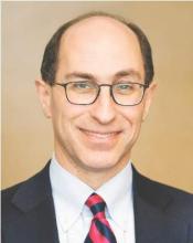
“Especially early in their careers, hospitalists can sometimes be overwhelmed by the possibilities of where their career can go and how to get to the position that they want, whether it’s leadership or another ultimate goal,” said copresenter Brian Markoff, MD, FACP, SFHM, chief of the division of hospital medicine at Mount Sinai St. Luke’s Hospital in New York. “This session is designed to help young physicians figure out where they are, where they want to be, and how they might be able to get there.”
The session – aimed at medical students, residents, fellows, and new hospitalists – will review different careers in hospital medicine, including academics and clinical work; share some individual success stories of currently practicing hospitalists; discuss the importance of mentorship; and note how you can use the Society of Hospital Medicine to advance your career status. “This is to help you take a step back and think about the big picture,” said copresenter Alfred Burger, MD, FACP, SFHM, associate program director of the internal medicine residency at Mount Sinai Beth Israel Hospital in New York.
Hospitalist careers today are quite varied, Dr. Burger said. Some physicians have a career that is primarily clinical while others have moved into a wide variety of leadership roles. “Hospitalists have ascended to some of the highest levels of leadership, whether it’s surgeon general or a chief medical officer or chief operating officer of a government agency or hospital group,” he said.
Some hospitalists have had success moving into the business and consulting range after practicing in a hospital setting, advising organizations how to do things more efficiently, while others have taken on health services research or patient safety and quality improvement work, he added.
Young hospitalists lately have been concerned with how much job advancement depends on gaining an extra degree like an MBA, versus being a really good hospitalist and working your way up, Dr. Markoff said. “We tell them, ‘You can do both,’ ” he said. “Depending on your career choice, you may or may not need an advanced degree.” In the past, Dr. Burger added, most hospitalists started a career and then gained additional degrees as needed. Today more hospitalists are starting their careers as a dual or sometimes triple degree holder.
“Hopefully, the session will help young hospitalists reflect on their own practice and help them sort out what they are doing that will get them to their ultimate career goal,” Dr. Markoff said. “The main take-home message is ‘There’s no one right way to get the career you want, but it’s going to take some active management on your part to get on the right path.’ ”
Dr. Burger thinks that the career pathways in hospital medicine are limited only by your own imagination. “Our talk is meant to inspire you to see the wide possibilities,” he said.
Attendees of the session, now in its third year, can serve as resources to peers and others who are more junior, Dr. Burger noted. “You may be able to inspire somebody based on something you heard” or share advice learned with a friend or colleague, he said, noting that some hospitalists have attended more than once.
“Hospitalist Careers: So Many Options”Tuesday, 10:35 a.m.–11:15 a.m.
Learn how to lead the battle against unnecessary testing
The steps that hospitalists can take to effect change in their home institutions will be the focus of Tuesday morning’s session at 10:35–11:15 a.m., “Overcoming a Culture Overrun with Overuse.”
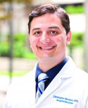
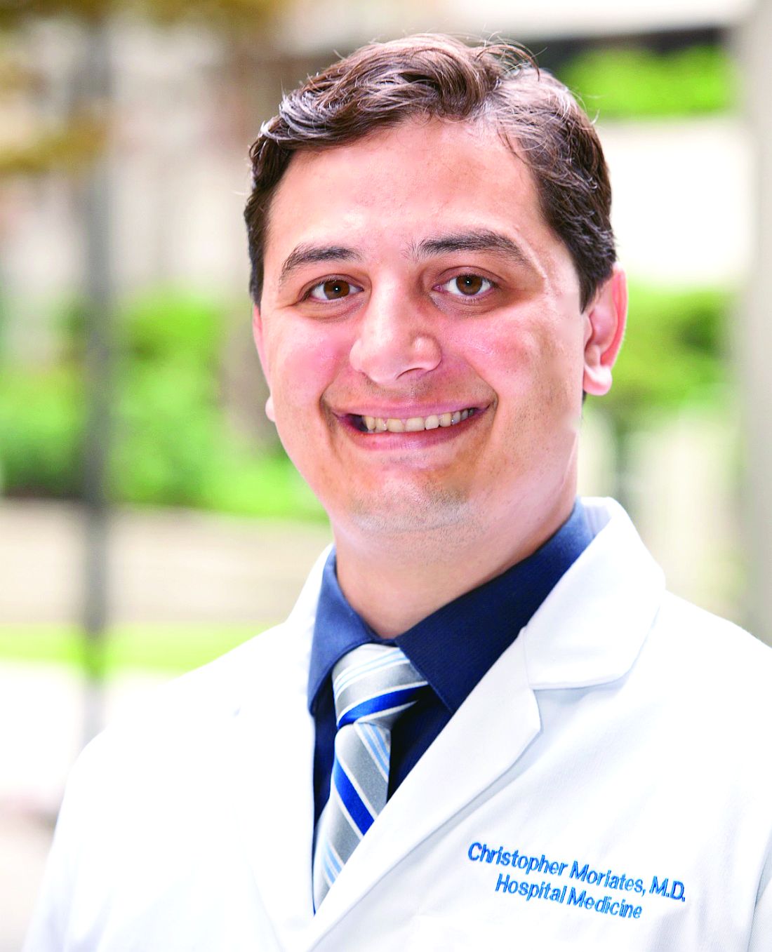
“Overuse is one of the [biggest] issues that we’re facing right now in health care,” said presenter Christopher Moriates, MD, assistant dean for health care value at the University of Texas at Austin. “The estimates are that about a third of what we do in health care is unnecessary. If you look at areas like lab testing in hospitals, it might be even more than that.”
“We must also address our medical culture that, through many different mechanisms, reinforces overuse,” he added. “The way we’re trained, the way we think about addressing problems in the hospital, the way that hospitals are set up and organized – all of these things contribute to us ordering more tests or doing more things.”
The session will discuss culture change as a general concept and introduce a framework for understanding and targeting culture change. The presentation also will demonstrate how culture contributes to overuse and low-value care, describe specific interventions that support culture change, and define opportunities to ensure the delivery of high-value care to patients.
One initiative to be discussed is Caring Wisely, which Dr. Moriates helped implement in 2012 at the University of California, San Francisco. The program takes the ideals of Choosing Wisely and, by crowdsourcing among front-line hospital staff, looks for ways to cut health care waste.
“We will highlight specific examples for how hospitalists can lead programs and work with others to decrease overuse, even within other departments,” Dr. Moriates said. “For example, we will discuss a successful resident-led program to decrease phlebotomy rates in the hospital, which can be replicated by other medical center or resident groups.”
Hospitalists at any level, in any setting, can help contribute to culture change, either by mimicking programs like Caring Wisely or by implementing simple changes in the way we speak to patients and each other.
“Every time we make decisions and we have conversations with patients, trainees, or consultants, we have the ability to either contribute to or take away from that culture of overuse,” Dr. Moriates said. “We’ll also discuss how hospitalists can lead efforts to organize multidisciplinary teams around decreasing common areas of overuse.”
On all levels, hospitalists have an important role to play. “We should all recognize that we lead from where we stand, no matter what our position or title may be,” Dr. Moriates said.
Overcoming a Culture Overrun with Overuse
Tuesday, 10:35 a.m.-11:15 a.m.
What hospitalists must know about co-management
With patient co-management arrangements between hospitalists and other surgical and medical subspecialists becoming more common, HM17 attendees won’t want to miss Tuesday afternoon’s session at 3:15–4:20 p.m., “Redefining Co-management in Hospital Medicine.”
“We’ll provide hospitalists effective co-management programs at their respective hospitals,” said copresenter William Atchley Jr., MD, FACP, SFHM, a hospitalist with Sentara Heart Hospital in Norfolk, Va.
More hospital medicine groups are getting involved in co-management, the presenters said. There are two primary models: one in which the hospitalist is the attending of record and the subspecialist is the co-manager and another in which the subspecialist is the attending of record and the hospitalist serves as the co-manager. “Either model can work with the right agreements put in place,” Dr. Atchley said.
“We’ll go through some of what we feel are the undiscovered benefits of having a co-management service,” said copresenter Mark Goldin, MD, FACP, a hospitalist at Long Island Jewish Medical Center. “I think a lot of people will be interested to hear that because research on co-management has been mixed, up to this point.” For example, he said, SHM engagement surveys have indicated that hospitalists who do co-management may be at reduced risk of burnout.
Also important is creating a metrics dashboard and monitoring and updating it regularly, Dr. Karlin-Zysman added. “Not only does it keep both sides honest, but it’s how you garner support from the C-suite.”
The Society of Hospital Medicine has resources available to help, Dr. Atchley said. The SHM website includes a white paper on co-management. There also is a listserv called HMS Exchange, in which hospitalists can discuss comanagement topics.
“Co-management is not going away. It’s something that hospitalists are going to be involved with,” Dr. Atchley said. “It’s important to come up with the right agreement and, at the same time, work with everybody in collaboration to improve patient care.”
Redefining Co-management in Hospital Medicine
Tuesday, 3:15–4:20 p.m.
Simplify Cardiac Risk Assessment for Rheumatologic Conditions
LONDON – Cardiovascular disease (CVD) risk assessment for patients with rheumatic diseases can be simple and integrated into general practice or rheumatology clinics, experts said during an Outcomes Science Session at the European Congress of Rheumatology
Patients with rheumatoid arthritis have a 50% higher risk of heart disease than do their counterparts without the disease, but “just having RA on its own isn’t sufficient to render that individual at high risk,” Dr. Naveed Sattar, professor of metabolic medicine at the University of Glasgow’s Institute of Cardiovascular and Medical Sciences in Scotland, said in an interview.
It’s simple enough to use traditional CVD risk factors in an RA population by including a patient’s age, gender, smoking status, and family history of heart disease, in addition to measuring blood pressure and blood lipid levels. Most risk scores will compile those features into a 10-year risk of a fatal CVD event. To account for the contribution of RA, Dr. Sattar said, simply multiply that score by 1.5.
While “there’s a fixation in some parts of Europe for [measuring] fasting lipids,” it is not necessary, Dr. Sattar said. The two lipid parameters that go in risk scores tend to be cholesterol and HDL cholesterol, he said, which change only minimally in fasting versus nonfasting states.
“The evidence overwhelmingly shows that nonfasting lipids, which can be done easily on the same sample as other clinic tests, are just as predictive of CVD risk as fasting lipids,” he said. “That really matters because many of our patients with RA or other conditions come to the hospital when they’re not fasting, and we shouldn’t be sending them away to come back fasting to do risk scores for CVD. That just doesn’t make sense.”
Updated guidelines from the European Society of Cardiology and guidelines soon to be released from the European League Against Rheumatism suggest that risk scores can be calculated every 5 years for most patients, a change from previous recommendations to calculate risk annually. Risk scoring is not perfect, however, and there is some debate about whether additional blood tests or ultrasound scanning of the carotid artery could augment the ability to predict heart disease risk. “We’re not quite there yet,” Dr. Sattar said. “I think we should do the simple things first and do them well.”
CV risk raised in all inflammatory arthritic diseases
During the same session, Dr. Paola de Pablo of the University of Birmingham, England, focused on how immune-mediated diseases predispose to premature, accelerated atherosclerosis and subsequent increased cardiovascular morbidity and mortality.
Cardiovascular risk is not only elevated in those with RA, she observed, but also in those with systemic lupus erythematosus, ankylosing spondylitis, psoriatic arthritis, vasculitides, and inflammatory myopathies. The risk varies but as a rule is more than 50% higher than the rate seen in the general population.
The underlying mechanisms are not clear, but chronic inflammation is closely linked with atherosclerosis, which in turn ups the risk for myocardial infarction and cerebrovascular accident.
Despite treatment, the risk often remains, Dr. de Pablo said. She highlighted how treatment with methotrexate and anti–tumor necrosis factor (TNF)–alpha drugs in RA had been associated with a reduction in the risk for heart attack of 20% and 40%, respectively (Ann Rheum Dis. 2014;74:480–89) so targeting inflammation with these drugs may have positive effects, at least in RA.
Managing traditional cardiovascular risk factors remains important, Dr. de Pablo said. That was a sentiment echoed by Dr. Sattar and by rheumatologist Dr. Michael Nurmohamed of the VU Medical Center in Amsterdam. This includes controlling blood pressure with antihypertensive medications and blood lipids with statins, and advocating smoking cessation and perhaps other appropriate lifestyle changes such as increasing physical exercise and controlling weight.
Dr. Nurmohamed, who was involved in the 2015 update of the EULAR recommendations on cardiovascular risk management, noted that traditional risk factor management in patients with arthritis in current clinical practice is often poor and that strategies to address this were urgently needed.
Although treating to-target and preventing disease flares in the rheumatic diseases is important, it lowers but does not normalize cardiovascular risk. “This appears to be irrespective of the drug used,” Dr. Nurmohamed said. Rheumatologists need to be careful when tapering medication, particularly the biologics, as these are perhaps helping to temper cardiovascular inflammation, which could worsen when doses are reduced. “Antirheumatic treatment only is not good enough to decrease or normalize the cardiovascular risk of our patients”, he emphasized.
Norwegian project shows how to integrate CVD assessment into routine practice
In a separate presentation, Dr. Eirik Ikdahl, a PhD student at Diakonhjemmet Hospital in Oslo, discussed how some rheumatology clinics in Norway are successfully incorporating CVD risk screening.
Through the Norwegian Collaboration on Atherosclerotic disease in patients with Rheumatic joint diseases (NOCAR), which started in April 2014, annual cardiovascular disease risk evaluations of patients with inflammatory joint diseases are being implemented into the practices of 11 rheumatology outpatient clinics. While waiting for clinic appointments, patients are given electronic devices through which they can report CVD risk factors via an electronic patient journal program called GoTreatIt Rheuma. From there, the clinic can order nonfasting lipid measurements and nurses can record patients’ blood pressure.
Then, using the ESC Systematic Coronary Risk Evaluation (SCORE) algorithm, the program automatically calculates a patient’s 10-year risk of a fatal CVD event. If the SCORE estimate is 5% or greater, the rheumatologist forwards a note to the patient’s primary care physician or cardiologist saying there is an indication for initiation of CVD-preventive measures such as medication or lifestyle changes. Rheumatologists and rheumatology nurses also deliver brief advice regarding smoking cessation and healthy diet.
“The main aim of the project is to raise awareness of the cardiovascular burden that these patients experience, and to ensure that patients with inflammatory joint diseases receive guideline-recommended cardiovascular preventive treatment,” Dr. Ikdahl said.
Of 6,150 patients defined as eligible for the NOCAR project in three of the centers, 41% (n = 2,519) received a CVD risk assessment during the first year and a half of the program, officers found in a recent review. Of those, 1,569 had RA, 418 had ankylosing spondylitis, 350 had psoriatic arthritis, and 122 had other spondyloarthritides.
Through the program, “a large number of high-risk patients have received screening that they would not otherwise have been offered,” Dr. Ikdahl said.
The major obstacles to successful implementation were time scarcity, defining a date for annual CVD risk assessment among patients who visit the clinics multiple times per year, and making sure lipids were measured before seeing the rheumatologist, he said. “We acknowledge there is room for improvement. It is challenging to implement new work tasks in an already busy rheumatology outpatient clinic, and since the project does not offer financial incentives to the participating centers, we rely on a collective effort and voluntary work based on resources already available.”
Remember CVD, but don’t forget other comorbidities
Other research presented by Dr. Laure Gossec, professor of rheumatology at Pitie-Salpétriere Hospital and Pierre & Marie Curie University in Paris highlighted the importance of identifying all comorbidities and their risk factors in patients with rheumatic diseases, and not just cardiovascular disease.
Dr. Gossec presented the results of an initiative aiming to make the collection and management of comorbidities easier in routine rheumatologic practice. The aim was to develop a simple, more pragmatic form that could be used to help rheumatologists manage selected comorbidities, and know when to refer for other specialist assessment. The focus was on ischemic cardiovascular disease, malignancies, infections such as chronic bronchitis, gastrointestinal disease such as diverticulitis, osteoporosis, and depression.
A committee of 18 experts, both physicians and nurses, was convened to examine the results of a systematic literature review of recommendations on comorbidity management and come up with concise recommendations for rheumatologists. Each of their recommendations covered whether or not the comorbidity was present (yes/no/don’t know) and if screening had been undertaken, such as measurement of blood lipids, and when this had occurred if known. There was then guidance on how to interpret these findings, calculate risk, and what to do if findings were abnormal.
The project is ongoing, and so far the expert panel has developed a pragmatic document with forms to help collect, report, and manage each specific comorbidity and its known risk factors. But it is still perhaps too long to be feasibly used in everyday practice, Dr. Gossec conceded. So the aim is to create a short, 2-page form that could summarize the recommendations briefly, and also develop a questionnaire for the patient to fill out and understand how to self-manage some comorbidities.
“We feel that this is a way to disseminate and adapt to the national context for France the EULAR comorbidities initiative,” Dr. Gossec said. “It also defines exactly what rheumatologists should be doing and when they should refer, hopefully to the benefit of our patients.”
Dr. Sattar has participated in advisory boards for Amgen, AstraZeneca, Boehringer Ingelheim, Eli Lily and UCB. He has also consulted for Merck and is a member of Roche’s speakers’ bureau. Dr. de Paolo and Dr. Nurmohamed reported no relevant financial disclosures. Dr. Ikdahl has received speaker’s honoraria from Pfizer. Dr. Gossec and coauthors have received honoraria from Abbvie France.
The video associated with this article is no longer available on this site. Please view all of our videos on the MDedge YouTube channel
The video associated with this article is no longer available on this site. Please view all of our videos on the MDedge YouTube channel
AT THE EULAR 2016 CONGRESS
Simplify cardiac risk assessment for rheumatologic conditions
LONDON – Cardiovascular disease (CVD) risk assessment for patients with rheumatic diseases can be simple and integrated into general practice or rheumatology clinics, experts said during an Outcomes Science Session at the European Congress of Rheumatology
Patients with rheumatoid arthritis have a 50% higher risk of heart disease than do their counterparts without the disease, but “just having RA on its own isn’t sufficient to render that individual at high risk,” Dr. Naveed Sattar, professor of metabolic medicine at the University of Glasgow’s Institute of Cardiovascular and Medical Sciences in Scotland, said in an interview.
It’s simple enough to use traditional CVD risk factors in an RA population by including a patient’s age, gender, smoking status, and family history of heart disease, in addition to measuring blood pressure and blood lipid levels. Most risk scores will compile those features into a 10-year risk of a fatal CVD event. To account for the contribution of RA, Dr. Sattar said, simply multiply that score by 1.5.
While “there’s a fixation in some parts of Europe for [measuring] fasting lipids,” it is not necessary, Dr. Sattar said. The two lipid parameters that go in risk scores tend to be cholesterol and HDL cholesterol, he said, which change only minimally in fasting versus nonfasting states.
“The evidence overwhelmingly shows that nonfasting lipids, which can be done easily on the same sample as other clinic tests, are just as predictive of CVD risk as fasting lipids,” he said. “That really matters because many of our patients with RA or other conditions come to the hospital when they’re not fasting, and we shouldn’t be sending them away to come back fasting to do risk scores for CVD. That just doesn’t make sense.”
Updated guidelines from the European Society of Cardiology and guidelines soon to be released from the European League Against Rheumatism suggest that risk scores can be calculated every 5 years for most patients, a change from previous recommendations to calculate risk annually. Risk scoring is not perfect, however, and there is some debate about whether additional blood tests or ultrasound scanning of the carotid artery could augment the ability to predict heart disease risk. “We’re not quite there yet,” Dr. Sattar said. “I think we should do the simple things first and do them well.”
CV risk raised in all inflammatory arthritic diseases
During the same session, Dr. Paola de Pablo of the University of Birmingham, England, focused on how immune-mediated diseases predispose to premature, accelerated atherosclerosis and subsequent increased cardiovascular morbidity and mortality.
Cardiovascular risk is not only elevated in those with RA, she observed, but also in those with systemic lupus erythematosus, ankylosing spondylitis, psoriatic arthritis, vasculitides, and inflammatory myopathies. The risk varies but as a rule is more than 50% higher than the rate seen in the general population.
The underlying mechanisms are not clear, but chronic inflammation is closely linked with atherosclerosis, which in turn ups the risk for myocardial infarction and cerebrovascular accident.
Despite treatment, the risk often remains, Dr. de Pablo said. She highlighted how treatment with methotrexate and anti–tumor necrosis factor (TNF)–alpha drugs in RA had been associated with a reduction in the risk for heart attack of 20% and 40%, respectively (Ann Rheum Dis. 2014;74:480–89) so targeting inflammation with these drugs may have positive effects, at least in RA.
Managing traditional cardiovascular risk factors remains important, Dr. de Pablo said. That was a sentiment echoed by Dr. Sattar and by rheumatologist Dr. Michael Nurmohamed of the VU Medical Center in Amsterdam. This includes controlling blood pressure with antihypertensive medications and blood lipids with statins, and advocating smoking cessation and perhaps other appropriate lifestyle changes such as increasing physical exercise and controlling weight.
Dr. Nurmohamed, who was involved in the 2015 update of the EULAR recommendations on cardiovascular risk management, noted that traditional risk factor management in patients with arthritis in current clinical practice is often poor and that strategies to address this were urgently needed.
Although treating to-target and preventing disease flares in the rheumatic diseases is important, it lowers but does not normalize cardiovascular risk. “This appears to be irrespective of the drug used,” Dr. Nurmohamed said. Rheumatologists need to be careful when tapering medication, particularly the biologics, as these are perhaps helping to temper cardiovascular inflammation, which could worsen when doses are reduced. “Antirheumatic treatment only is not good enough to decrease or normalize the cardiovascular risk of our patients”, he emphasized.
Norwegian project shows how to integrate CVD assessment into routine practice
In a separate presentation, Dr. Eirik Ikdahl, a PhD student at Diakonhjemmet Hospital in Oslo, discussed how some rheumatology clinics in Norway are successfully incorporating CVD risk screening.
Through the Norwegian Collaboration on Atherosclerotic disease in patients with Rheumatic joint diseases (NOCAR), which started in April 2014, annual cardiovascular disease risk evaluations of patients with inflammatory joint diseases are being implemented into the practices of 11 rheumatology outpatient clinics. While waiting for clinic appointments, patients are given electronic devices through which they can report CVD risk factors via an electronic patient journal program called GoTreatIt Rheuma. From there, the clinic can order nonfasting lipid measurements and nurses can record patients’ blood pressure.
Then, using the ESC Systematic Coronary Risk Evaluation (SCORE) algorithm, the program automatically calculates a patient’s 10-year risk of a fatal CVD event. If the SCORE estimate is 5% or greater, the rheumatologist forwards a note to the patient’s primary care physician or cardiologist saying there is an indication for initiation of CVD-preventive measures such as medication or lifestyle changes. Rheumatologists and rheumatology nurses also deliver brief advice regarding smoking cessation and healthy diet.
“The main aim of the project is to raise awareness of the cardiovascular burden that these patients experience, and to ensure that patients with inflammatory joint diseases receive guideline-recommended cardiovascular preventive treatment,” Dr. Ikdahl said.
Of 6,150 patients defined as eligible for the NOCAR project in three of the centers, 41% (n = 2,519) received a CVD risk assessment during the first year and a half of the program, officers found in a recent review. Of those, 1,569 had RA, 418 had ankylosing spondylitis, 350 had psoriatic arthritis, and 122 had other spondyloarthritides.
Through the program, “a large number of high-risk patients have received screening that they would not otherwise have been offered,” Dr. Ikdahl said.
The major obstacles to successful implementation were time scarcity, defining a date for annual CVD risk assessment among patients who visit the clinics multiple times per year, and making sure lipids were measured before seeing the rheumatologist, he said. “We acknowledge there is room for improvement. It is challenging to implement new work tasks in an already busy rheumatology outpatient clinic, and since the project does not offer financial incentives to the participating centers, we rely on a collective effort and voluntary work based on resources already available.”
Remember CVD, but don’t forget other comorbidities
Other research presented by Dr. Laure Gossec, professor of rheumatology at Pitie-Salpétriere Hospital and Pierre & Marie Curie University in Paris highlighted the importance of identifying all comorbidities and their risk factors in patients with rheumatic diseases, and not just cardiovascular disease.
Dr. Gossec presented the results of an initiative aiming to make the collection and management of comorbidities easier in routine rheumatologic practice. The aim was to develop a simple, more pragmatic form that could be used to help rheumatologists manage selected comorbidities, and know when to refer for other specialist assessment. The focus was on ischemic cardiovascular disease, malignancies, infections such as chronic bronchitis, gastrointestinal disease such as diverticulitis, osteoporosis, and depression.
A committee of 18 experts, both physicians and nurses, was convened to examine the results of a systematic literature review of recommendations on comorbidity management and come up with concise recommendations for rheumatologists. Each of their recommendations covered whether or not the comorbidity was present (yes/no/don’t know) and if screening had been undertaken, such as measurement of blood lipids, and when this had occurred if known. There was then guidance on how to interpret these findings, calculate risk, and what to do if findings were abnormal.
The project is ongoing, and so far the expert panel has developed a pragmatic document with forms to help collect, report, and manage each specific comorbidity and its known risk factors. But it is still perhaps too long to be feasibly used in everyday practice, Dr. Gossec conceded. So the aim is to create a short, 2-page form that could summarize the recommendations briefly, and also develop a questionnaire for the patient to fill out and understand how to self-manage some comorbidities.
“We feel that this is a way to disseminate and adapt to the national context for France the EULAR comorbidities initiative,” Dr. Gossec said. “It also defines exactly what rheumatologists should be doing and when they should refer, hopefully to the benefit of our patients.”
Dr. Sattar has participated in advisory boards for Amgen, AstraZeneca, Boehringer Ingelheim, Eli Lily and UCB. He has also consulted for Merck and is a member of Roche’s speakers’ bureau. Dr. de Paolo and Dr. Nurmohamed reported no relevant financial disclosures. Dr. Ikdahl has received speaker’s honoraria from Pfizer. Dr. Gossec and coauthors have received honoraria from Abbvie France.
The video associated with this article is no longer available on this site. Please view all of our videos on the MDedge YouTube channel
The video associated with this article is no longer available on this site. Please view all of our videos on the MDedge YouTube channel
AT THE EULAR 2016 CONGRESS
Diagnosis, treatment of gout lag behind prevalence
LONDON – The prevalence of gout is increasing and to prevent an epidemic, rheumatologists and other physicians need to diagnose and treat cases promptly and better explain the treatment process to their patients, according to a Swedish expert on the disease.
“It’s a disease for which we understand the mechanisms, we know how to diagnose it, and we have had good treatments for the last 50 years,” said Dr. Lennart Jacobsson, professor of rheumatology at the University of Gothenburg in Sweden. “Despite that, we don’t treat it properly. It’s difficult to understand why that is the case, why there is such a lack of knowledge and such a lack of willingness to pursue treatment.”
Gout is the most common nondegenerative inflammatory joint disease, exemplified by a prevalence of 0.9%-2.5% in Europe and 3.9% in the United States. The incidence is increasing along with factors such as the aging population, as well as lifestyle changes such as rising body mass index and physical inactivity, Dr. Jacobsson said at the European Congress of Rheumatology.
“It’s the same story everywhere,” he said. “Gout is underdiagnosed, it’s diagnosed late, and once it’s diagnosed people don’t get treated with urate-lowering therapy, which aims at the heart of the disease. If they are treated, treatment often is discontinued, which we think is largely due to lack of education and information to patients.”
Urate crystals can build up over as much as a decade before a person experiences a first gout attack, Dr. Jacobsson said. “It can take 3-5 years of effective treatment to get rid of those masses within the body. Over that time, especially at the beginning, patients may still have gout attacks, which they often interpret as side effects of the medication or a misinterpretation that it doesn’t work because they’re not properly informed that medication needs to be a long-term treatment.”
Close to 10% of men aged 70-80 years have gout, he said, but it’s still unclear how many of those have mild, moderate, or severe disease. It’s also not well studied how gout itself can affect health-related quality of life and costs to society. “You can easily imagine that the costs are pretty large, however, and they will increase,” he said.
Gout is interrelated with several metabolic syndrome disorders such as obesity, hypertension, and diabetes, Dr. Jacobsson said: “If you have renal disease, you have higher uric acid levels and can more easily get gout, but from having high uric acid levels you may also get decreased renal function, so it’s sort of circular. The same is true of gout and hypertension.”
Once considered a disease of the wealthy, Dr. Jacobsson said, gout has recently been shown to be associated with lower income and socioeconomic class, as is the case for many other chronic diseases. There are still large opportunities for improvements regarding early detection and the initiation of urate-lowering therapy, he said, as well as counseling patients on lifestyle improvements.
“We know what to do, we have the tools, and we should start using them more effectively,” he said. “It’s really very simple, but that message has to get through. It’s an increasing problem, and it’s bad not just for the joints but also for quality of life and comorbidities.”
Dr. Jacobsson reported no relevant financial disclosures.
EXPERT ANALYSIS FROM THE EULAR 2016 CONGRESS
Anti-TNF agents may slow erosive hand osteoarthritis
LONDON – Tumor necrosis factor may play a role in erosive hand osteoarthritis, and treatments such as etanercept that target this cytokine may help prevent progression of the condition, according to the results of two studies presented at the European Congress of Rheumatology.
In one of the studies, immunoscintigraphic detection of radiolabeled certolizumab pegol was used to show that tumor necrosis factor (TNF) was present in swollen finger joints. In another study, researchers looked to see if treatment with etanercept would have any specific effects on the joints of patients with erosive hand osteoarthritis (OA) and performed a separate analysis of the potential effect on synovitis and the effect on bone marrow lesions.
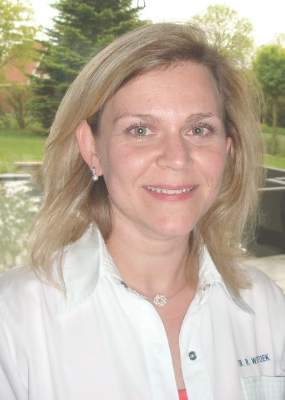
“We previously had the idea that TNF is an important cytokine in the pathogenesis of erosive [hand] osteoarthritis; but there have been no animal studies, and it’s very difficult to take biopsies or fluid aspiration from these small finger joints,” Dr. Ruth Wittoek, a staff rheumatologist at Ghent University Hospital in Belgium and a coauthor of all three studies, explained in a precongress interview. “We needed to look for other possibilities to really identify the presence of TNF in those affected joints.”
Dr. Wittoek and her associates used immunoscintigraphy to take static images of both hands of five patients with erosive OA immediately (less than 15 minutes) after administration of radiolabeled certolizumab pegol (early phase) and 4-6 hours following the injection (late phase).
The patients studied had erosive OA for a median of 8.4 years, and their median age was 55.6 years. All patients underwent clinical examination for presence of tenderness and palpable swelling of the joints and ultrasound 1 day prior to undergoing immunoscintigraphy.
All 18 interphalangeal (IP) finger joints were scored according to the anatomical phase scoring system on x-ray, and 90 IP finger joints were studied in total. The uptake of radiolabeled certolizumab pegol was semiquantitatively described as being absent, weak, or strong.
During the early phase following administration, uptake of the radiolabeled TNF inhibitor was seen in seven (7.8%) joints, although the uptake was described as weak in all cases. The radiolabeled TNF inhibitor was seen in 24 (26.7%) joints during the late phase following administration, with five instances described as strong uptake and the remaining 19 instances being weak uptake. No uptake of the radiolabeled TNF inhibitor was seen in metacarpophalangeal joints.
Uptake of the radiolabeled TNF inhibitor was linked to signs of disease activity, including tender, swollen, and radiographically active joints.
Late uptake was present in 12 (36.4%) of 33 tender joints and in 12 (21.1%) of 57 nontender joints (odds ratio, 2.1; 95% confidence interval, 0.8-5.6; P was nonsignificant).
The relationship was most pronounced with palpable joint swelling: Late uptake was present in 14 (61%) of 23 swollen joints and 10 (14.9%) of 67 nonswollen joints (OR, 8.9; 95% CI, 3.3-26.0; P less than .001).
Late uptake was present in 18 (29%) of 62 sonographically active joints (defined as any presence of effusion or synovial proliferation) but just 6 (21.4%) of 28 noninflamed joints (OR, 1.5; 95% CI, 0.5-4.3; P was nonsignificant).
Uptake of the radiolabeled TNF inhibitor was observed in all anatomical phases of erosive hand OA, Dr. Wittoek noted, but the strongest association was found during the final remodeling (R) phase.
“Soft-tissue swelling strongly correlated with uptake of certolizumab, meaning in these joints a lot of TNF was present,” Dr. Wittoek said. “These data further solidify the rationale for cytokine-directed therapies in erosive OA.”
Although the data provide proof of concept that TNF may be involved in erosive hand OA, the lack of a control tracer was noted as a limitation of the study after the investigator’s presentation. Dr. Wittoek conceded that it would be interesting to examine that in a future study.
So, if TNF is present, what effect does anti-TNF therapy have on the joints in erosive hand OA?
That question was addressed in a multicenter, double-blind, randomized, placebo-controlled trial involving 90 patients who were randomized to receive either 50 mg of subcutaneous etanercept weekly for 24 weeks, then 25 mg weekly for the remainder of 1 year (45 patients), or placebo (45 patients). Participants were a mean age of 60 years, 81% were women, and 96% fulfilled the American College of Rheumatology hand OA criteria.
“Synovial inflammation is often present in erosive hand OA; moreover, synovitis is associated with pain and with structural damage after around 2 and a half years,” the lead study author, Dr. Margreet Kloppenburg, a professor of rheumatology at Leiden University Medical Centre in the Netherlands, said in an interview.
“Therefore, we wanted to know whether blocking of synovial inflammation by a well-known drug such as etanercept would also have a positive effect on outcomes in erosive hand OA,” Dr. Kloppenburg explained.
The primary outcome measure was the level of OA pain assessed on a visual analog scale (VAS) at 24 weeks.
Secondary endpoints included assessment of hand function, quality of life, the number of tender joints, and grip strength after 4, 8, 12, 24, and 36 weeks, and after 1 year. Radiographic progression of IP joints was scored blindly at baseline, 24 weeks, and 1 year following the quantitative Ghent University Scoring System (GUSS). VAS pain was compared between treatment groups at 24 weeks and 1 year in intention-to-treat analyses.
Although etanercept was not superior to placebo on VAS pain at 24 weeks, it was superior to placebo both on pain and structural damage assessed by GUSS in the symptomatic and inflammatory patients who completed the study. The drug was especially effective in joints with signs of inflammation.
Overall, VAS pain in all patients decreased by 24.8 mm (95% CI, –29.2 to –20.5; P less than .001) at 24 weeks. In intention-to-treat analysis, differences in pain between the groups were in favor of etanercept but did not reach statistical significance. The per-protocol analysis of GUSS showed a mean difference in favor of etanercept, indicating more remodeling in the etanercept group.
Additional analyses showed an interaction between soft swelling/erythema and etanercept treatment on GUSS, resulting in a statistically significant (P less than .05) mean difference between the two treatment groups. More patients dropped out on placebo than on etanercept (six vs. three) because of inefficacy, whereas more dropped out on etanercept than on placebo (six vs. one) because of adverse effects.
“Synovial inflammation is an interesting target for treatment in OA patients with an inflammatory hand osteoarthritis phenotype,” Dr. Kloppenburg said.
A separate analysis of the same multicenter study suggests that etanercept is effective in inhibiting bone marrow lesions (BMLs) in patients with erosive hand OA.
The researchers studied 20 participants with symptomatic erosive OA with clinical and ultrasonographic signs of inflammation in at least one IP joint. The patients underwent contrast-enhanced MRI of the eight distal and proximal IP joints of one hand at baseline and 1 year. Images were scored for synovitis and BMLs (0-3 per joint, total score 0-24), blinded for patient characteristics.
Radiographs of the same hand were scored according to the Verbruggen-Veys system. Logistic regression was used to associate the presence of an MRI feature in a joint with being in an erosive versus nonerosive anatomical phase.
“Although erosive hand OA is a condition with a high disease burden, no disease-modifying treatments are available yet,” explained Féline Kroon, a PhD student in the department of rheumatology at Leiden University Medical Center in the Netherlands, in an interview. “Research to find a new form of therapy is partly hindered by our limited knowledge on the pathophysiology of the disease.”
However, “new imaging modalities like MRI enable us to study the pathophysiology of erosive OA more closely,” she said. “This study also gave us the unique opportunity to investigate whether anti-TNF, which is known to lead to clinical improvement and improvement of inflammatory lesions on MRI in other rheumatic diseases like rheumatoid arthritis, might also be effective in erosive OA.”
The presence of BMLs, but not synovitis, was associated with the presence of an erosive anatomical phase in a joint, and treatment with etanercept appeared to be effective in inhibiting these lesions. That suggests a role for TNF in the pathophysiology of erosive OA.
The inhibitory effect of etanercept on BMLs was more pronounced in IP joints with severe synovitis at baseline, suggesting that inflamed synovial tissue could be a source of TNF production in erosive OA.
The total synovitis score was similar at baseline and 1 year in both study groups. For BMLs, the median total score at baseline was 5.4 (range 2-9) and 7.0 (0-9) at 1 year in the placebo group, versus 4.5 (3-9) and 3.7 (0-8), respectively, in the etanercept group.
The presence of BMLs was associated with being in the erosive and remodeling anatomical phases of erosive hand OA. Synovitis was not associated with those phases.
“We think that TNF-alpha plays a role in the pathophysiology of erosive OA via an effect on the subchondral bone,” Ms. Kroon said. “Because we saw that the beneficial effect of etanercept on BMLs was more pronounced in joints with synovitis at baseline, we think that, in an inflamed synovial hand joint, an interaction takes place between synovium and subchondral bone, which could be influenced by blocking TNF.”
Pfizer and UCB supported the investigator-initiated studies and provided the study drugs. Dr. Kloppenburg has received lecturing, consultancy, and investigator fees or grants from AbbVie, APPROACH, GlaxoSmithKline, Levicept, Pfizer, Servier, and UCB, all paid to her institution. All other authors declared no conflicts of interests.
AT THE EULAR 2016 CONGRESS
Key clinical point: Early data suggest targeting tumor necrosis factor could be of benefit in patients with erosive hand disease.
Major finding: TNF was found in the hand joints, and anti-TNF therapy reduced synovial inflammation and bone marrow lesions.
Data source: Two studies looking at the effects of anti-TNF treatments in patients with erosive hand osteoarthritis.
Disclosures: Pfizer and UCB supported the investigator-initiated studies and provided the study drugs. Dr. Kloppenburg has received lecturing, consultancy, and investigator fees or grants from AbbVie, APPROACH, GlaxoSmithKline, Levicept, Pfizer, Servier, and UCB, all paid to her institution. All other authors declared no conflicts of interests.
Actionable traits will guide gastroenterologists in treatment of obesity
BOSTON – Gastroenterologists need to recognize that obesity is not one but multiple diseases and band together with other specialists in an interdisciplinary fashion to develop workable plans for long-term weight control, according to a panel of experts at the 2016 AGA Tech Summit, which is sponsored by the AGA Center for GI Innovation and Technology.
More than one-third of U.S. adults have obesity, at a medical cost of between $147 and $300 billion annually, according to the Centers for Disease Control and Prevention. There are many reasons people suffer from obesity, said Dr. Robert Fanelli, chief of minimally invasive surgery and surgical endoscopy at the Guthrie Clinic in Sayre, Pa. These range from poor diet to genetics, and, accordingly, physicians are going to have to turn to personalized care.
“The time has come for gastroenterologists to play a role in addressing this crisis,” said Dr. Sarah Streett, clinical associate professor of medicine at Stanford (Calif.) University. “Gastroenterologists are uniquely positioned to help in this area because we are, by training, internists, digestive disease specialists, and endoscopists, and endoscopic bariatric therapies are entering the armamentarium of tools to treat obesity,” she added.
Over the last few decades, GI experts’ attention has focused on colon cancer screening and significant innovations in the areas of endoscopic therapeutics and the treatment of hepatitis and inflammatory bowel disease, Dr. Streett said, “but many in GI appreciate that obesity is a root cause for so many of the conditions that we’re already treating: esophageal reflux and its complications, fatty liver disease, gallbladder disease, and gastrointestinal cancers.”
Because the disease is complex, said Dr. Streett, who is also the chair of the AGA Practice Management and Economics Committee, it’s imperative to work collaboratively with others, including obesity medicine specialists, dietitians, counselors, psychologists, and bariatric surgeons, to create a continuum of care tailored to patients’ individual needs, with good nutrition, physical activity, and stress reduction as central tenets.
AGA – in conjunction with the Society of American Gastrointestinal and Endoscopic Surgeons, the Obesity Society, and the North American Society for Pediatric Gastroenterology, Hepatology and Nutrition – is developing a program to guide gastroenterologists who want to develop comprehensive care management programs to treat obesity with a multimodal approach, she said. The initiative, called “POWER: Practice Guide on Obesity and Weight Management, Education and Resources,” will be made available on the AGA website later this year.
For patients in need, there are safe, robust surgical options to manage obesity, including gastric bypass and sleeve gastrectomy, said Dr. Kevin Reavis, a bariatric surgeon at the Oregon Clinic in Portland. The safety record for bariatric surgery is now on par with elective gallbladder surgery and matches or betters heart surgery, he said. A less invasive option is the endoscopic placement of devices like gastric balloons that generate an early sense of satiety by applying pressure to the upper stomach.
Moving toward the future, there is a lot of “out of the box thinking” that has led to research into ways to alter consumption and digestion of food with such approaches as blocking the vagus nerve or employing deep brain stimulation. Although some devices or interventions are designed to block calories from being absorbed from the GI tract, others may be able to reset patients’ metabolic balance, enabling easier weight loss and maintenance.
Physicians’ understanding of obesity is changing, said Dr. Lee Kaplan, director of the Obesity, Metabolism and Nutrition Institute at Massachusetts General Hospital, Boston. “We now understand that the body has an elaborate and powerful regulatory system that seeks to maintain a stable amount of fat,” a phenomenon he characterized as “the fat mass set point.”
Changes in modern society, including the type of foods we eat, lack of exercise, disturbed sleep, increased stress, and medications that stimulate appetite, all send aberrant signals to the brain, causing it to seek too high a fat mass, he said. Any therapy that is to be successful must lower that set point.
Different from other heterogeneous disorders like cancer, it’s not yet clear which treatments will work best for which types of obesity, Dr. Kaplan noted. Meanwhile, “we’re recommending more and more that we approach obesity in the same way that we approach any other disease, which is to start with lifestyle changes,” he said, “and if that doesn’t work, to move on to more classically medical approaches, including medications and surgery.”
Once an effective treatment is implemented, continuing care is essential. Obesity is not cured with current treatments, which means that patients require ongoing support.
After weight is reduced, “we abandon patients as a matter of routine,” said Dr. Kaplan, noting that this is not the case in other serious chronic diseases. This is one aspect of obesity care that “we must change.”
Dr. Kaplan receives consulting fees from Eisai, Ethicon, GI Dynamics, Novo Nordisk, and Vivus, and has an ownership interest in GI Dynamics. Dr. Fanelli is a part owner of Allurion Technologies, a company that manufacturers an intragastric balloon for weight loss, and an advisor to EndoGastric Solutions. Dr. Streett and Dr. Reavis reported no relevant financial disclosures.
AT THE AGA 2016 TECH SUMMIT
Gastroenterology is fertile ground for personalized medicine
BOSTON – The time is ripe for gastroenterologists to embrace personalized medicine, a leader in the genomics industry said at the 2016 AGA Tech Summit, which is sponsored by the AGA Center for GI Innovation and Technology.
Personalized medicine has made the most inroads in oncology, but there are compelling reasons for GI experts to get involved as well, said Mara Aspinall, executive chair of the computational genomics company GenePeeks, and president and CEO of Health Catalysts, a firm dedicated to the growth of new healthcare companies.
Virtually all aspects of gastroenterology today are emerging as fertile ground for genomic stratification of patients, Ms. Aspinall said. Inflammatory bowel disease is the first area where there already has been much work done on genomic polymorphism and how differences between patients’ disease progression can be explained, she said, and drug dosing is the first “low hanging fruit” in the area.
In addition, new areas of research can be viewed as opportunities for GI, Ms. Aspinall said. One example is the microbiome: “That’s a cutting-edge area where GI specialists can look to understand the current learning and translate them into the unique impact on their patients.” Another area is the increasingly important role of nutrition in managing patients with everything from metabolic disease to cancer. Nutritional analysis and advice has traditionally been delegated to dietitians more than gastroenterologists, but Ms. Aspinall said she believes personalized medicine in its broadest form will take the shape of using food as drugs, which puts it in GI’s wheelhouse.
Furthermore, there is much more work to be done on the gastrointestinal causes and consequences of immunological diseases, which have been understudied and underdeveloped, she said, looking in particular at patient populations at risk: children, whose immune systems and GI tracts are closely related; and women, who account for the vast majority of immune disorders such as allergies, multiple sclerosis, or lupus. “What role can the GI doc play in better diagnosis and treatment?” Ms. Aspinall said.
Over the past 50 years, the history of medicine has really been a story of diagnostics emergence, Ms. Aspinall said, including the growth of imaging, MRI, PET scans, molecular technologies, and sequencing. “We look at diagnostics as the bridge between theory and therapy,” she said.
“The headlines have been all about the new drugs and new procedures,” she said. “While that’s important, I would argue that it’s not as important as the change in diagnostics. A patient has nothing without an accurate and timely diagnosis.”
Ms. Aspinall said she sees a wider role for the field: “We need to define gastroenterology in a broader context in order to capture personalized medicine.” GI experts should start collecting data on patients’ outcomes, family histories, and complication rates as a start, she said, to look for patterns that could inform improved treatment regimens – particularly in light of genomic differences in those patients. “The reality is you’re probably not helping today’s patients but you will help patients 5 years from now,” she said.
“Cancer treatment today is organ-based,” said Ms. Aspinall. She predicted that in the not-too-distant future, cancer treatment will move more toward pathway-based treatments, as treatment is guided more and more by genetic polymorphisms.
The plummeting cost of genome sequencing is a key development that holds great promise for gastrointestinal cancers, said Raju Kucherlapati, Ph.D., who also spoke about personalized medicine in gastrointestinal cancers. Dr. Kucherlapati, a professor of genetics at Harvard Medical School and the Brigham and Women’s Hospital, Boston, said that sequencing tumor DNA is leading to risk stratification and targeted therapy in ever-increasing numbers of cancer types.
It turns out that many gastrointestinal cancers have mutations that are shared by other cancers, meaning that already-approved therapies may work for some patients with GI cancers, said Dr. Kucherlapati. For example, about 4% of colorectal cancers have amplifications of the ERBB2 gene, meaning that these patients could benefit from trastuzumab (Herceptin). “One of the aspects that’s remarkable is that drugs are already available for many of these targets, reducing the costs and time tremendously,” said Dr. Kucherlapati.
Where will this lead? The nomenclature of cancer is changing to the point where patients and their physicians in the future won’t say they have colon cancer or lung cancer but instead talk of EGFR cancer or c-KIT mutation cancer, Ms. Aspinall said: “If that happens in cancer, I am confident that it will be relevant to other specialties as well.”
In the panel discussion following the morning’s presentations, Dr. Michael Camilleri, AGA president and professor of medicine at Mayo Medical School, Rochester, Minn., agreed that a focus on personalized medicine makes sense in the GI space. “I love the idea that we need to focus on one or two big ideas,” and linking genotype to patient phenotype to target therapy should be one of them.
Ms. Aspinall and Dr. Kucherlapati reported no relevant financial disclosures.
AT THE 2016 AGA TECH SUMMIT
Top medtech successes in gastroenterology and metabolic diseases this year
BOSTON – Gastroenterology as a specialty is in transition when it comes to technology, and gastroenterologists will have to decide whether to stay the course, performing procedures like endoscopies and colonoscopies, or also embrace additional technologies, a GI tech expert said at the 2016 AGA Tech Summit, which is sponsored by the AGA Center for GI Innovation and Technology.
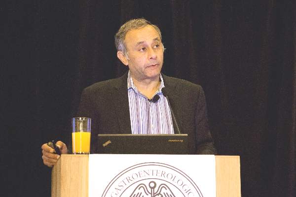
“We have the ability to seize the moment and transform the specialty into a major player in therapeutics that go beyond the gut to include conditions like diabetes, obesity, or other areas,” said Dr. Pankaj Jay Pasricha, past chair of the AGA Center for GI Innovation and Technology and director of the Johns Hopkins Center for Neurogastroenterology and Johns Hopkins division of digestive diseases in Baltimore. “It’s up to us.”
Last year saw the approval of an array of devices in areas involving new endoscopic platforms, new tools for mucosal resection, and new strategies for improving the utility of endoscopic ultrasound, Dr. Pasricha said. To be employed appropriately, many of these devices will require GI professionals to seek additional training. One example includes the use of new minimally invasive balloon technologies to control obesity. Two, the Orbera Intragastric Balloon System (Apollo Endosurgery) and the ReShape Integrated Dual Balloon System (ReShape Medical) were singled out as examples.
“It’s not just simply a matter of dropping the balloon in and hoping for the best,” he said. “This requires careful training. The balloon is not an end to itself; it’s a tool for helping patients get to their goals. GIs either need to jump into this and say, ‘Yes, we want to expand’ or potentially lose business to bariatric surgeons. We will be pushed as a specialty to figure out how we want to deal with this.”
The American Gastroenterological Association has been working on this issue and is developing, in conjunction with the Society of American Gastrointestinal and Endoscopic Surgeons, The Obesity Society, and the North American Society for Pediatric Gastroenterology, Hepatology and Nutrition, a program to guide gastroenterologists who want to develop comprehensive care management programs to treat obesity with a multimodal approach. The initiative, called “POWER: Practice Guide on Obesity and Weight Management, Education and Resources,” will be available later this year, along with a white paper and bundled payment model.
Overall, 2015 was a good year for GI technologies, although not as exciting as 2014, Dr. Pasricha said. Devices approved by the Food and Drug Administration last year included the Gastric-Emptying Breath Test (Advanced Breath Diagnostics), a breath test to aid in the diagnosis of delayed gastric emptying; the Splash M-Knife (Pentax Medical), a tool to achieve complete endoscopic submucosal dissection; the Eclipse System (Pelvalon), an appliance inserted into the vagina to treat fecal incontinence in women; and Ab Stats (GI Logic), a connected wearable stethoscope to monitor the digestive system after patients have surgery. Neuromodulation for inflammatory bowel disease and irritable bowel syndrome also is an emerging area for technology, he said.
In the field of endoscopy, Dr. Pasricha noted the ever-greater emphasis being placed by the manufacturers of endoscopes on tools for therapeutics.
“It has taken a long time for endoscope companies to recognize that they need to be players not just in providing the eyes but also the hands and the tools to address the pathology that we see in procedures,” Dr. Pasricha said.
In other news from 2015, outbreaks of carbapenem-resistant Enterobacteriaceae linked to patients undergoing procedures with duodenoscopes highlighted the need to move from risk reduction to risk elimination, Dr. Pasricha said. The AGA Center for GI Innovation and Technology worked closely with the FDA to ensure GIs had the guidance and resources they needed to eliminate the risk of patient infection from duodenoscopes. Following discussions with the AGA center leadership and other invested stakeholders, the FDA in October issued new guidelines to health care facilities for cleaning the devices and ordered the devices’ three manufacturers to conduct postmarket surveillance studies to better understand how the devices are reprocessed. Additional communications from the agency regarding this postmarket surveillance are likely forthcoming.
In a similar risk-elimination case, the FDA in March halted enrollment in a clinical trial of GI Dynamics’ EndoBarrier weight loss device after a few patients developed liver infections.
The AGA and its Center for GI Innovation and Technology recognizes that training is critical to the adoption of new technologies. The center is working to develop competency thresholds for new technologies and is developing an action plan to educate the next generation of interventionalists, Dr. Pasricha said: “We need to teach gastroenterologists how to do more skilled procedures than just diagnostic endoscopy or point and shoot procedures. We also need to teach surgeons how to be facile with flexible endoscopy.”
On the reimbursement front, the AGA center – working with the other GI societies – works to put together rational reimbursement codes to reflect the new reality of the complexities of interventional procedures, he said.
“We have learned that you should not even be getting involved in commercial development of new devices without first running it through the filter of economics,” Dr. Pasricha said. A good idea for a new technology by itself is not sufficient, “especially now with all the cost constraints.”
Dr. Pasricha is a consultant, advisor, or cofounder for Apollo Endosurgery, Pentax, EndoGastric Solutions, Crospon, GI Cryo, Glyscend, Enterastim, and GI Scientific.
Ted Bosworth contributed to this report.
AT THE AGA 2016 TECH SUMMIT
