User login
Patients with psoriatic arthritis perform accurate joint counts
NEW YORK – After instruction by a nurse, patients with psoriatic arthritis do at least as well as nurses but better than physicians in counting tender and swollen joints, according to a cross-sectional study that employed ultrasound as a gold standard.
"Patient counts of swollen joints correlated with both effusion and synovitis as detected by ultrasound, suggesting that patients can learn to monitor their own disease," reported Dr. Agnes Szentpetery of the department of rheumatology at St. Vincent’s Hospital, Dublin.
In this study, described as the first ever conducted in psoriatic arthritis to compare joint counts by patients, nurses, physicians, and ultrasound (US), 50 patients with a mean age of 50 years were enrolled. All received instruction from a nurse on how to assess and count tender and swollen joints. The major focus of the correlations was in 34 joints assessed with US, although further correlations were made between patients, nurses, and physicians for 28 (JC28) and 66 swollen and 68 tender (JC 66/68) counts.
"Joint counts [confirming active disease] were higher for patients and nurses than for physicians," reported Dr. Szentpetery, who presented these data at the joint meetings of the Group for Research and Assessment of Psoriasis and Psoriatic Arthritis and the Spondyloarthritis Research & Treatment Network (SPARTAN). Doctor and nurse counts for swollen joints, unlike patient counts, did not correlate with US evidence of effusion, although they did for synovitis.
For tender joints, there was moderate or good agreement between patient and doctor for 11 joints, doctor and nurse for 12 joints, and patient and nurse for 19 joints. (This nurse was different from the one who provided instruction on joint counts.) However, correlation between any of these observers and US evidence of tender joints was uniformly poor.
Small joints in the hand, particularly the proximal interphalangeal joint, offered the best interobserver agreement for correlations, according to Dr. Szentpetery. Although no correlations rose above good and most were moderate between any two observers, Dr. Szentpetery stressed that patients performed at levels comparable with doctors and nurses, compared with US.
"Although this was a small study, the results support patient joint counts, and we have already begun routinely instructing patients in our clinic," Dr. Szentpetery reported.
Asked for a comment, Dr. John D. Reveille, professor of rheumatology and clinical immunogenetics at the University of Texas Health Sciences Center at Houston, said that he is remaining cautious until a larger study generates more data. He said that similar efforts to instruct patients to perform joint counts in rheumatoid arthritis failed to produce acceptable rates of accuracy.
"It may be possible to enlist patients to perform accurate joint counts if they are motivated and receive appropriate education, but you may need a special population and the right education," said Dr. Reveille, who is chair of SPARTAN’s executive committee.
However, joint counts are perceived to be so time consuming that many clinics do not perform them at all, according to Dr. Szentpetery. Moreover, she suggested that the absolute number of tender or swollen joints at any moment in time may be less important than the change in joint involvement after treatment is initiated, an outcome that may be best measured by patients.
"I agree that this is a small study, and more data are needed, but our data suggest patients may have a role in joint counts, which is an attractive strategy for reducing work for the clinicians," Dr. Szentpetery said.
Dr. Szentpetery and Dr. Reveille reported no relevant financial conflicts.
AT THE 2014 GRAPPA AND SPARTAN ANNUAL MEETINGS
Key clinical point: Properly educated patients with psoriatic arthritis can count tender and swollen joints accurately, thereby saving time and effort by physicians or nurses.
Major finding: For tender joints, there was moderate or good agreement between the patient and the doctor for 11 joints, the doctor and the nurse for 12 joints, and the patient and the nurse for 19 joints, but correlations between any of these observers and ultrasound evidence of tender joints were uniformly poor.
Data source: A cross-sectional study of 50 patients with PsA.
Disclosures: Dr. Szentpetery and Dr. Reveille reported no potential conflicts of interest relevant to this study.
First comorbidity guidelines drafted for psoriatic arthritis
NEW YORK – For the first time, the Group for Research and Assessment of Psoriasis and Psoriatic Arthritis is preparing evidence-based treatment recommendations for the diagnosis and management of the comorbidities associated with psoriatic arthritis.
"I am really excited that we are taking the initiative to treat and manage the psoriatic arthritis [PsA] patient as a whole," reported Dr. Elaine Husni, director of the Arthritis and Musculoskeletal Center at the Cleveland Clinic. The guidelines are meant to be a "platform on which to raise awareness" and foster education.

An initial set of guidelines on comorbidities was drafted at the joint meetings of the Group for Research and Assessment of Psoriasis and Psoriatic Arthritis (GRAPPA) and the Spondyloarthritis Research & Treatment Network (SPARTAN). Dr. Husni led the consensus group and fielded questions about key recommendations.
The first of these recommendations, which will undergo a process of discussion and review prior to formal adoption, states that all patients with PsA should be evaluated for cardiovascular (CV) disease. The consensus group labeled this recommendation "strong" even while conferring it with grade D evidence.
"The grade D was based on the fact that there are no outcomes data specifically in patients with psoriatic arthritis," observed Dr. Alexis R. Ogdie-Beatty, director of the Penn Psoriatic Arthritis Clinic at the University of Pennsylvania, Philadelphia. A member of the consensus group, Dr. Ogdie-Beatty suggested that benefit from CV screening is still a reasonable expectation "given the growing evidence that patients with PsA are at increased risk."
Similar "strong" recommendations but grade D evidence were given for screening for ophthalmic complications and inflammatory bowel disease. In these cases, there is good evidence for an association with PsA but limited evidence that screening will lead to improved outcome. The exception was obesity for which the group gave a B rating to the evidence for benefit from diagnosis and treatment.
Screening for diabetes was also included among recommendations, but it was given a "weak" rating and a grade D for supportive evidence.
Most of the other recommendations involved screening for various infections, such as hepatitis C virus (HCV), HIV, and tuberculosis, prior to initiating immunosuppressant therapies, particularly biologics. The level of evidence for these recommendations typically ranged between B and C even though all were given strong recommendations.

The consensus recommendations are not expected to include much detail about the specific management of comorbidities. The reason is concern about their applicability across various settings of care. It was thought that GRAPPA, as an international organization, should accommodate different types of practice. For example, the group cautioned against outlining steps of CV risk management, which may be managed by rheumatologists in some areas of the world but by specialists in others.
"We want to stay away from being minicardiologists," Dr. Husni explained. She indicated that the goal of the recommendations is to simply identify the specific types of comorbidity screening that should be considered "core recommendations" in the approach to PsA.
However, there is interest in creating a table regarding the use of specific medications for PsA treatment in the context of comorbidities. In a color-coded draft presented at the GRAPPA and SPARTAN meeting, some examples included caution in the use of NSAIDs in patients with CV disease, a need for monitoring when using cyclosporine in patients with chronic kidney disease, and a preference for etanercept over other tumor necrosis factor inhibitors in patients with HCV infection.
Overall, recommendations for comorbidities were identified as an important step in defining optimal care for PsA. According to Dr. Ogdie-Beatty, "the most important point may be to raise awareness." As these recommendations wend their way through an approval process at the organizational level, Dr. Ogdie-Beatty expressed hope that the final wording is specific enough to encourage attention to comorbidities without restricting a variety of valid approaches.
Dr. Husni reported a financial relationship with Celgene. Dr. Ogdie-Beatty had no potential conflicts of interest to report.
NEW YORK – For the first time, the Group for Research and Assessment of Psoriasis and Psoriatic Arthritis is preparing evidence-based treatment recommendations for the diagnosis and management of the comorbidities associated with psoriatic arthritis.
"I am really excited that we are taking the initiative to treat and manage the psoriatic arthritis [PsA] patient as a whole," reported Dr. Elaine Husni, director of the Arthritis and Musculoskeletal Center at the Cleveland Clinic. The guidelines are meant to be a "platform on which to raise awareness" and foster education.
An initial set of guidelines on comorbidities was drafted at the joint meetings of the Group for Research and Assessment of Psoriasis and Psoriatic Arthritis (GRAPPA) and the Spondyloarthritis Research & Treatment Network (SPARTAN). Dr. Husni led the consensus group and fielded questions about key recommendations.
The first of these recommendations, which will undergo a process of discussion and review prior to formal adoption, states that all patients with PsA should be evaluated for cardiovascular (CV) disease. The consensus group labeled this recommendation "strong" even while conferring it with grade D evidence.
"The grade D was based on the fact that there are no outcomes data specifically in patients with psoriatic arthritis," observed Dr. Alexis R. Ogdie-Beatty, director of the Penn Psoriatic Arthritis Clinic at the University of Pennsylvania, Philadelphia. A member of the consensus group, Dr. Ogdie-Beatty suggested that benefit from CV screening is still a reasonable expectation "given the growing evidence that patients with PsA are at increased risk."
Similar "strong" recommendations but grade D evidence were given for screening for ophthalmic complications and inflammatory bowel disease. In these cases, there is good evidence for an association with PsA but limited evidence that screening will lead to improved outcome. The exception was obesity for which the group gave a B rating to the evidence for benefit from diagnosis and treatment.
Screening for diabetes was also included among recommendations, but it was given a "weak" rating and a grade D for supportive evidence.
Most of the other recommendations involved screening for various infections, such as hepatitis C virus (HCV), HIV, and tuberculosis, prior to initiating immunosuppressant therapies, particularly biologics. The level of evidence for these recommendations typically ranged between B and C even though all were given strong recommendations.
The consensus recommendations are not expected to include much detail about the specific management of comorbidities. The reason is concern about their applicability across various settings of care. It was thought that GRAPPA, as an international organization, should accommodate different types of practice. For example, the group cautioned against outlining steps of CV risk management, which may be managed by rheumatologists in some areas of the world but by specialists in others.
"We want to stay away from being minicardiologists," Dr. Husni explained. She indicated that the goal of the recommendations is to simply identify the specific types of comorbidity screening that should be considered "core recommendations" in the approach to PsA.
However, there is interest in creating a table regarding the use of specific medications for PsA treatment in the context of comorbidities. In a color-coded draft presented at the GRAPPA and SPARTAN meeting, some examples included caution in the use of NSAIDs in patients with CV disease, a need for monitoring when using cyclosporine in patients with chronic kidney disease, and a preference for etanercept over other tumor necrosis factor inhibitors in patients with HCV infection.
Overall, recommendations for comorbidities were identified as an important step in defining optimal care for PsA. According to Dr. Ogdie-Beatty, "the most important point may be to raise awareness." As these recommendations wend their way through an approval process at the organizational level, Dr. Ogdie-Beatty expressed hope that the final wording is specific enough to encourage attention to comorbidities without restricting a variety of valid approaches.
Dr. Husni reported a financial relationship with Celgene. Dr. Ogdie-Beatty had no potential conflicts of interest to report.
AT THE 2014 GRAPPA AND SPARTAN ANNUAL MEETINGS

First comorbidity guidelines drafted for psoriatic arthritis
NEW YORK – For the first time, the Group for Research and Assessment of Psoriasis and Psoriatic Arthritis is preparing evidence-based treatment recommendations for the diagnosis and management of the comorbidities associated with psoriatic arthritis.
"I am really excited that we are taking the initiative to treat and manage the psoriatic arthritis [PsA] patient as a whole," reported Dr. Elaine Husni, director of the Arthritis and Musculoskeletal Center at the Cleveland Clinic. The guidelines are meant to be a "platform on which to raise awareness" and foster education.
An initial set of guidelines on comorbidities was drafted at the joint meetings of the Group for Research and Assessment of Psoriasis and Psoriatic Arthritis (GRAPPA) and the Spondyloarthritis Research & Treatment Network (SPARTAN). Dr. Husni led the consensus group and fielded questions about key recommendations.
The first of these recommendations, which will undergo a process of discussion and review prior to formal adoption, states that all patients with PsA should be evaluated for cardiovascular (CV) disease. The consensus group labeled this recommendation "strong" even while conferring it with grade D evidence.
"The grade D was based on the fact that there are no outcomes data specifically in patients with psoriatic arthritis," observed Dr. Alexis R. Ogdie-Beatty, director of the Penn Psoriatic Arthritis Clinic at the University of Pennsylvania, Philadelphia. A member of the consensus group, Dr. Ogdie-Beatty suggested that benefit from CV screening is still a reasonable expectation "given the growing evidence that patients with PsA are at increased risk."
Similar "strong" recommendations but grade D evidence were given for screening for ophthalmic complications and inflammatory bowel disease. In these cases, there is good evidence for an association with PsA but limited evidence that screening will lead to improved outcome. The exception was obesity for which the group gave a B rating to the evidence for benefit from diagnosis and treatment.
Screening for diabetes was also included among recommendations, but it was given a "weak" rating and a grade D for supportive evidence.
Most of the other recommendations involved screening for various infections, such as hepatitis C virus (HCV), HIV, and tuberculosis, prior to initiating immunosuppressant therapies, particularly biologics. The level of evidence for these recommendations typically ranged between B and C even though all were given strong recommendations.
The consensus recommendations are not expected to include much detail about the specific management of comorbidities. The reason is concern about their applicability across various settings of care. It was thought that GRAPPA, as an international organization, should accommodate different types of practice. For example, the group cautioned against outlining steps of CV risk management, which may be managed by rheumatologists in some areas of the world but by specialists in others.
"We want to stay away from being minicardiologists," Dr. Husni explained. She indicated that the goal of the recommendations is to simply identify the specific types of comorbidity screening that should be considered "core recommendations" in the approach to PsA.
However, there is interest in creating a table regarding the use of specific medications for PsA treatment in the context of comorbidities. In a color-coded draft presented at the GRAPPA and SPARTAN meeting, some examples included caution in the use of NSAIDs in patients with CV disease, a need for monitoring when using cyclosporine in patients with chronic kidney disease, and a preference for etanercept over other tumor necrosis factor inhibitors in patients with HCV infection.
Overall, recommendations for comorbidities were identified as an important step in defining optimal care for PsA. According to Dr. Ogdie-Beatty, "the most important point may be to raise awareness." As these recommendations wend their way through an approval process at the organizational level, Dr. Ogdie-Beatty expressed hope that the final wording is specific enough to encourage attention to comorbidities without restricting a variety of valid approaches.
Dr. Husni reported a financial relationship with Celgene. Dr. Ogdie-Beatty had no potential conflicts of interest to report.
AT THE 2014 GRAPPA AND SPARTAN ANNUAL MEETINGS

Putting Psoriatic Arthritis First
We are all aware of the burden of psoriatic arthritis (PsA). Although psoriasis is more common, the arthritic component is a major factor in the morbidity of psoriatic disease. Accordingly, the National Psoriasis Foundation (NPF) is taking steps to expand the focus on PsA.
Last month, the NPF launched the largest realignment and expansion of its PsA program since the organization began its services almost 20 years ago. The NPF’s PsA Project will focus on 4 major areas: (1) Decrease the time to diagnosis. (2) Help those with PsA better manage their disease. (3) Reduce barriers to health care and treatments. (4) Improve understanding of PsA symptoms, disease management, and impact on patient quality of life among health care providers.
Through the PsA Project, the NPF has the following specific goals: (1) Reduce the average time of diagnosis of PsA from 4 years to 1 year. (2) Increase by 50% the number of people with PsA who are receiving the right treatment to 62% total. (3) Reduce from 50% to 30% the number of people who report PsA is a problem in their everyday lives. (4) Double the number of health resources available to people diagnosed with PsA. (5) Increase by 50% the number of National Institutes of Health–funded scientists studying psoriatic disease to 42 scientists to boost care, improve treatment, and find a cure for PsA.
What’s the issue?
Our patients with PsA deserve more resources. The earlier we diagnose this condition the less likely an individual is to suffer pain and possible joint destruction. A project such as the one created by the NPF can only help to raise awareness among both patients and physicians. How will you contribute to expanding awareness of psoriatic arthritis in your community?

Axial involvement found common in psoriatic arthritis mutilans
NEW YORK – Unexpectedly and conflicting with previous reports, a careful analysis of a relatively large series of patients with psoriatic arthritis mutilans found that most had axial involvement.
In a series of 56 patients with psoriatic arthritis (PsA) mutilans drawn from 610 consecutive patients with PsA, 65% had evidence of axial disease on the most recent radiograph. Of these, 57% had radiographic sacroiliitis, which was bilateral in 80%.
"This proportion far exceeds the typical 25% incidence of axial disease reported in the literature," according to Dr. Deepak Jadon, a specialist registrar in rheumatology at the Royal National Hospital for Rheumatic Diseases, Bath, England.

The proportion of confirmed PsA mutilans cases, at 9%, was also higher than the 5% typically reported, according to Dr. Jadon, who presented these findings at the joint meetings of the Group for Research and Assessment of Psoriasis and Psoriatic Arthritis (GRAPPA) and the Spondyloarthritis Research & Treatment Network (SPARTAN). Referral to a specialty clinic may explain the higher rate, but PsA mutilans could only be ruled out, because of inadequate radiographs, in 483 of the 610 patients, so the actual proportion could be even higher.
In 35 of the patients with PsA mutilans, defined as osteolysis involving at least 50% of the visualized articular surface on both sides of the joints, serial radiographs confirmed that this disease is progressive. In this series, 37% of the patients had PsA mutilans at the time of the first radiograph, but the rest developed PsA mutilans over the course of follow-up.
Of this second group, 84% had received a disease-modifying antirheumatic drug (DMARD) prior to developing PsA mutilans, an observation that "suggests DMARDs may not prevent the onset of this condition," Dr. Jadon reported, cautioning that there are other potential explanations.
Using a random-effects model, "different patterns of progression were observed in the feet and hands," Dr. Jadon reported. In both groups, the most common pattern was an initial surge of activity followed by a tapering of rate of progression. However, a second surge of activity in the hands was not observed in the feet.
When compared to unaffected PsA patients, those with PsA mutilans were on average younger (33 vs. 40 years; P = .039) and more likely to have nail dystrophy (83% vs. 45%; P = .0002). The severity of nail dystrophy appeared to correlate with the severity of joint involvement.
The most commonly affected joints in this series were the metatarsophalangeal joint of the big toe, the proximal interphalangeal joint of the thumb, and the metacarpophalangeal joint of the index finger. While many patients had monoarticular involvement at the time of diagnosis, 80% of patients in this series had polyarticular disease on the most recent radiograph.
Not surprisingly, functional limitations were significantly greater for patients with PsA mutilans on standardized health assessment questionnaire (HAQ) when compared with patients with PsA (P = .048). The differences involved functions affecting both the hands, such as those relevant to grip strength, and the feet.
Data from this series did not provide any insight on treatment efficacy. Biologic use in those with serial radiographs, which included a sizeable number of patients managed before the biologic era, was too infrequent to infer effect on disease progression. However, the frequent exposure to DMARDs in patients with documented progression suggests benefits may be limited.
Dr. Jadon suggested that prospective data for PsA mutilans, which is widely regarded as the least common but most severe form of PsA, are now needed to determine which treatments have the greatest potential to improve long-term outcome.
Dr. Jadon reported no potential conflicts of interest.
NEW YORK – Unexpectedly and conflicting with previous reports, a careful analysis of a relatively large series of patients with psoriatic arthritis mutilans found that most had axial involvement.
In a series of 56 patients with psoriatic arthritis (PsA) mutilans drawn from 610 consecutive patients with PsA, 65% had evidence of axial disease on the most recent radiograph. Of these, 57% had radiographic sacroiliitis, which was bilateral in 80%.
"This proportion far exceeds the typical 25% incidence of axial disease reported in the literature," according to Dr. Deepak Jadon, a specialist registrar in rheumatology at the Royal National Hospital for Rheumatic Diseases, Bath, England.
The proportion of confirmed PsA mutilans cases, at 9%, was also higher than the 5% typically reported, according to Dr. Jadon, who presented these findings at the joint meetings of the Group for Research and Assessment of Psoriasis and Psoriatic Arthritis (GRAPPA) and the Spondyloarthritis Research & Treatment Network (SPARTAN). Referral to a specialty clinic may explain the higher rate, but PsA mutilans could only be ruled out, because of inadequate radiographs, in 483 of the 610 patients, so the actual proportion could be even higher.
In 35 of the patients with PsA mutilans, defined as osteolysis involving at least 50% of the visualized articular surface on both sides of the joints, serial radiographs confirmed that this disease is progressive. In this series, 37% of the patients had PsA mutilans at the time of the first radiograph, but the rest developed PsA mutilans over the course of follow-up.
Of this second group, 84% had received a disease-modifying antirheumatic drug (DMARD) prior to developing PsA mutilans, an observation that "suggests DMARDs may not prevent the onset of this condition," Dr. Jadon reported, cautioning that there are other potential explanations.
Using a random-effects model, "different patterns of progression were observed in the feet and hands," Dr. Jadon reported. In both groups, the most common pattern was an initial surge of activity followed by a tapering of rate of progression. However, a second surge of activity in the hands was not observed in the feet.
When compared to unaffected PsA patients, those with PsA mutilans were on average younger (33 vs. 40 years; P = .039) and more likely to have nail dystrophy (83% vs. 45%; P = .0002). The severity of nail dystrophy appeared to correlate with the severity of joint involvement.
The most commonly affected joints in this series were the metatarsophalangeal joint of the big toe, the proximal interphalangeal joint of the thumb, and the metacarpophalangeal joint of the index finger. While many patients had monoarticular involvement at the time of diagnosis, 80% of patients in this series had polyarticular disease on the most recent radiograph.
Not surprisingly, functional limitations were significantly greater for patients with PsA mutilans on standardized health assessment questionnaire (HAQ) when compared with patients with PsA (P = .048). The differences involved functions affecting both the hands, such as those relevant to grip strength, and the feet.
Data from this series did not provide any insight on treatment efficacy. Biologic use in those with serial radiographs, which included a sizeable number of patients managed before the biologic era, was too infrequent to infer effect on disease progression. However, the frequent exposure to DMARDs in patients with documented progression suggests benefits may be limited.
Dr. Jadon suggested that prospective data for PsA mutilans, which is widely regarded as the least common but most severe form of PsA, are now needed to determine which treatments have the greatest potential to improve long-term outcome.
Dr. Jadon reported no potential conflicts of interest.
AT THE 2014 GRAPPA AND SPARTAN ANNUAL MEETINGS
Key clinical point: In a review of a relatively large series of patients with PsA mutilans, radiographic evidence of axial disease was found in the majority, which conflicts with previous reports.
Major finding: Sixty-five percent of patients with PsA mutilans had axial disease and 57% of those had sacroiliitis.
Data source: A case series of 56 patients with PsA mutilans.
Disclosures: Dr. Jadon reported no potential conflicts of interest.

Patients become full partners in inflammatory arthritis and psoriasis research
NEW YORK – Improvements in clinical trial design for psoriasis, psoriatic arthritis, and other inflammatory diseases are being credited to a decision to enlist patients as full partners, not just advisors or consultants, in research initiatives.
"Rheumatologists, I think, have been leaders is recognizing that patients can bring an expertise to clinical research that is unique and ensure that study endpoints are relevant to outcomes important to them," reported Dr. William Tillett, a research fellow in the department of rheumatology at the Royal National Hospital for Rheumatic Diseases in Bath, England.

The value of patient research partners (PRP) was a recurring theme at the joint meetings of the Group for Research and Assessment of Psoriasis and Psoriatic Arthritis (GRAPPA) and the Spondyloarthritis Research & Treatment Network. GRAPPA, in particular, has been fostering research collaborations with patients since 2006.
"It has been an evolution. Patients were initially enlisted to sit in when trial designs were being discussed. Now, we are talking about full partnership so that they are involved at the inception with equal partnership that includes veto power and author credit," Dr. Tillett explained.
At the GRAPPA meeting, an afternoon symposium was devoted to optimal strategies for fostering collaboration with PRPs, which builds on work already initiated with a group called Patient Involvement for Outcome Measures in PsA (PIOMPsA). In turn, PIOMPsA, formed 2 years ago, was largely modeled on PRP initiatives led by OMERACT (Outcomes Measurement in Rheumatology).
"OMERACT has been involving patients in clinical research design for about 10 years," reported Dr. Philip J. Mease, director of rheumatology research at Swedish Medical Center, Seattle. "The idea of making patients full-blown partners is more recent, but I think there is increasing appreciation for what the right patients can contribute to improve study design."
The initiatives are spreading through rheumatology and other inflammatory diseases. At the GRAPPA meeting, updates on patient initiatives to influence clinical research were presented not only from the work of PIOMPsA and OMERACT but also from the International Dermatology Outcomes Measures (IDEOM) consortium. Created in collaboration with the National Psoriasis Foundation (NPF), IDEOM is bringing clinicians and patients together to define standard outcome measures.
"There are no really good measures to evaluate relative severity of psoriasis from the patient’s perspective," reported Dr. Alice Gottlieb, professor of dermatology at Tufts Medical Center, Boston. Tools traditionally used in clinical trials, such as the Psoriasis Area Severity Index (PASI) "are not practical in the clinic" and do not necessarily reflect the impact of psoriasis on quality of life when used as a study endpoint, she said.
PIOMPsA has now conducted several meetings, including one held in conjunction with OMERACT in Budapest, Hungary, in early May 2014. Like IDEOM, PIOMPsA has been focused on developing consensus on core symptoms of its target inflammatory disease. This is critical because the ability of clinical trials to generate relevant data is dependent on first defining meaningful endpoints, according to Dr. Tillett, who presented the PIOMPsA deliberations at the GRAPPA meeting.
In Budapest, for example, a vote was taken on whether to add fatigue to a list of core symptoms for PsA that includes impaired physical function, skin lesions, and joint pain. Fatigue was added to the list by a vote in which 70% supported it as a core PsA symptom, said Dr. Tillett, who recently published on the goals and underlying concepts of PIOMPsA (Curr. Rheumatol. Rep. 2014;16:418).
According to Dr. Tillett and Dr. Mease, PRPs are an answer to the repeatedly reported disconnect between physicians and patients in rating disease severity. By involving patients with interest in clinical research and collaborative skills, treatment trials have the potential to generate data more useful to practical patient management.
"It will be very difficult to show objectively that patient-aided trial design leads to better studies, but this is a reasonable expectation. I think that the contributions we have already seen from these collaborations bear this out," said Dr. Tillett, who expects the concept to spread to other fields of medicine.
Dr. Tillett reported financial relationships with AbbVie and Amgen. Dr. Mease reported financial relationships with AbbVie, Amgen, Biogen Idec, Bristol-Myers Squibb, Celgene, Crescendo Bioscience, Genentech, Janssen Pharmaceuticals, Lilly, Merck & Co, Novartis, Pfizer, UCB Pharma, and Vertex. Dr. Gottlieb reported financial relationships with Abbott, Actelion, Amgen, Bristol-Myers Squibb, Celgene, Centocor, Novo Nordisk, Teva, and UCB.
NEW YORK – Improvements in clinical trial design for psoriasis, psoriatic arthritis, and other inflammatory diseases are being credited to a decision to enlist patients as full partners, not just advisors or consultants, in research initiatives.
"Rheumatologists, I think, have been leaders is recognizing that patients can bring an expertise to clinical research that is unique and ensure that study endpoints are relevant to outcomes important to them," reported Dr. William Tillett, a research fellow in the department of rheumatology at the Royal National Hospital for Rheumatic Diseases in Bath, England.
The value of patient research partners (PRP) was a recurring theme at the joint meetings of the Group for Research and Assessment of Psoriasis and Psoriatic Arthritis (GRAPPA) and the Spondyloarthritis Research & Treatment Network. GRAPPA, in particular, has been fostering research collaborations with patients since 2006.
"It has been an evolution. Patients were initially enlisted to sit in when trial designs were being discussed. Now, we are talking about full partnership so that they are involved at the inception with equal partnership that includes veto power and author credit," Dr. Tillett explained.
At the GRAPPA meeting, an afternoon symposium was devoted to optimal strategies for fostering collaboration with PRPs, which builds on work already initiated with a group called Patient Involvement for Outcome Measures in PsA (PIOMPsA). In turn, PIOMPsA, formed 2 years ago, was largely modeled on PRP initiatives led by OMERACT (Outcomes Measurement in Rheumatology).
"OMERACT has been involving patients in clinical research design for about 10 years," reported Dr. Philip J. Mease, director of rheumatology research at Swedish Medical Center, Seattle. "The idea of making patients full-blown partners is more recent, but I think there is increasing appreciation for what the right patients can contribute to improve study design."
The initiatives are spreading through rheumatology and other inflammatory diseases. At the GRAPPA meeting, updates on patient initiatives to influence clinical research were presented not only from the work of PIOMPsA and OMERACT but also from the International Dermatology Outcomes Measures (IDEOM) consortium. Created in collaboration with the National Psoriasis Foundation (NPF), IDEOM is bringing clinicians and patients together to define standard outcome measures.
"There are no really good measures to evaluate relative severity of psoriasis from the patient’s perspective," reported Dr. Alice Gottlieb, professor of dermatology at Tufts Medical Center, Boston. Tools traditionally used in clinical trials, such as the Psoriasis Area Severity Index (PASI) "are not practical in the clinic" and do not necessarily reflect the impact of psoriasis on quality of life when used as a study endpoint, she said.
PIOMPsA has now conducted several meetings, including one held in conjunction with OMERACT in Budapest, Hungary, in early May 2014. Like IDEOM, PIOMPsA has been focused on developing consensus on core symptoms of its target inflammatory disease. This is critical because the ability of clinical trials to generate relevant data is dependent on first defining meaningful endpoints, according to Dr. Tillett, who presented the PIOMPsA deliberations at the GRAPPA meeting.
In Budapest, for example, a vote was taken on whether to add fatigue to a list of core symptoms for PsA that includes impaired physical function, skin lesions, and joint pain. Fatigue was added to the list by a vote in which 70% supported it as a core PsA symptom, said Dr. Tillett, who recently published on the goals and underlying concepts of PIOMPsA (Curr. Rheumatol. Rep. 2014;16:418).
According to Dr. Tillett and Dr. Mease, PRPs are an answer to the repeatedly reported disconnect between physicians and patients in rating disease severity. By involving patients with interest in clinical research and collaborative skills, treatment trials have the potential to generate data more useful to practical patient management.
"It will be very difficult to show objectively that patient-aided trial design leads to better studies, but this is a reasonable expectation. I think that the contributions we have already seen from these collaborations bear this out," said Dr. Tillett, who expects the concept to spread to other fields of medicine.
Dr. Tillett reported financial relationships with AbbVie and Amgen. Dr. Mease reported financial relationships with AbbVie, Amgen, Biogen Idec, Bristol-Myers Squibb, Celgene, Crescendo Bioscience, Genentech, Janssen Pharmaceuticals, Lilly, Merck & Co, Novartis, Pfizer, UCB Pharma, and Vertex. Dr. Gottlieb reported financial relationships with Abbott, Actelion, Amgen, Bristol-Myers Squibb, Celgene, Centocor, Novo Nordisk, Teva, and UCB.
EXPERT ANALYSIS FROM THE 2014 GRAPPA AND SPARTAN ANNUAL MEETINGS

A New Appraisal of Dermatologic Manifestations of Diabetes Mellitus
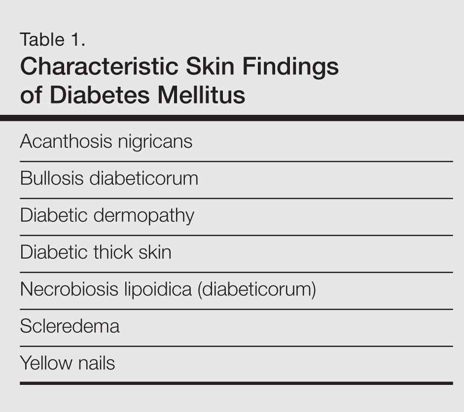
Diabetes mellitus is a morbid and costly condition that carries a high burden of disease for patients (both with and without a diagnosis) and for society as a whole. The economic burden of diabetes in the United States recently was estimated at nearly $250 billion annually,1 and this number continues to rise. The cutaneous manifestations of diabetes are diverse and far-reaching, ranging from benign cosmetic concerns to severe dermatologic conditions. Given the wide range of etiology for diabetes mellitus and its existence on a spectrum of severity, it is perhaps not surprising that some of these entities are the subject of debate (vis-à-vis the strength of association between these skin conditions and diabetes) and can manifest in different forms. However, it is clear that the cutaneous manifestations of diabetes are equally as important to consider and manage as the systemic complications of the disease. In analyzing associations with diabetes, it is important to note that given such a high incidence of diabetes among the general population and its close association with other disease states, such as the metabolic syndrome, studies aimed at determining direct relationships to this entity must be well controlled for confounding factors, which may not even always be possible. Regardless, it is important for dermatologists and dermatology residents to recognize and understand the protean cutaneous manifestations of diabetes mellitus, and this column will explore skin findings that are characteristic of diabetes (Table 1) as well as other dermatoses with a reported but less clear association with diabetes (Table 2).

Skin Findings Characteristic of Diabetes
Diabetic Thick Skin
The association between diabetes and thick skin is well described as either a mobility-limiting affliction of the joints of the hands (cheiroarthropathy) or as an asymptomatic thickening of the skin. It has been estimated that 8% to 36% of patients with insulin-dependent diabetes develop some form of skin thickening2; one series also found this association to be true for patients with non–insulin-dependent diabetes mellitus (NIDDM).3 Skin thickening is readily observable on clinical presentation or ultrasonography, with increasing thickness in many cases associated with long-term disease progression. This increasing thickness was shown histopathologically to be a direct result of activated fibroblasts and increased collagen polymerization, with some similar features to progressive systemic sclerosis.4 Interestingly, even clinically normal skin showed some degree of fibroblast activation in diabetic patients, but collagen fibers in each case were smaller in diameter than those found in progressive systemic sclerosis. This finding clearly has implications on quality of life, as a lack of hand mobility due to the cheiroarthropathy can be severely disabling. Underlining the need for strict glycemic control, it has been suggested that tight control of blood sugar levels can lead to improvement in diabetic thick skin; however, reports of improvement are based on a small sample population.5 Huntley papules are localized to areas on the dorsum of the hands overlying the joints, demonstrating hyperkeratosis and enlarged dermal papillae.6 They also can be found in a minority of patients without diabetes. Interestingly, diabetic thick skin also has been associated with neurologic disorders in diabetes.7 Diabetic thick skin was found to be significantly (P<.05) correlated with diabetic neuropathy, independent of duration of diabetes, age, or glycosylated hemoglobin levels, though no causal or etiologic link between these entities has been proven.
Yellow Nails
Nail changes are well described in diabetes, ranging from periungual telangiectases to complications from infections, such as paronychia; however, a well-recognized finding, especially in elderly diabetic patients, is a characteristic yellowing of the nails, reported to affect up to 40% of patients with diabetes.8 The mechanism behind it likely includes accumulation of glycation end products, which also has been thought to lead to yellowing of the skin, and vascular impairment.9 These nails tend to exhibit slow growth, likely resulting from a nail matrix that is poorly supplied with blood, and also can be more curved than normal with longitudinal ridges (onychorrhexis).10 It is important, however, not to attribute yellow nails to diabetes without considering other causes of yellow nails, such as onychomycosis, yellow nail syndrome, and yellow nails associated with lymphedema or respiratory tract disease (eg, pleural effusion, bronchiectasis).11
Diabetic Dermopathy
Colloquially known as shin spots, diabetic dermopathy is perhaps the most common skin finding in this patient population, though it also can occur in up to 1 in 5 individuals without diabetes.12 Although it is very common, it is not a condition that should be overlooked, as numerous studies have shown an increase in microangiopathic complications such as retinopathy in patients with diabetic dermopathy.13,14 Although follow-up studies may be necessary to fully characterize the relationship between shin spots and diabetes, it certainly is reasonable to be more wary of diabetic patients presenting with many shin spots, as the general consensus is that these areas represent postinflammatory hyperpigmentation and cutaneous atrophy in the setting of poor vascular supply, which should prompt analysis of other areas that might be affected by poor vasculature, such as an ophthalmologic examination. Antecedent and perhaps unnoticed trauma has been implicated given a possible underlying neuropathy, but this theory has not been supported by studies.
Bullosis Diabeticorum
Bullosis diabeticorum is a rare but well-described occurrence of self-resolving, nonscarring blisters that arise on the extremities of diabetic patients. This entity should be distinguished from other primary autoimmune blistering disorders and from simple mechanobullous lesions. Several types of bullosis diabeticorum have been described, with the most classic form showing an intraepidermal cleavage plane.15 These lesions tend to resolve in weeks but can be recurrent. The location of the pathology underlines its nonscarring nature, though similar lesions have been reported showing a cleavage plane in the lamina lucida of the dermoepidermal junction, which underlines the confusion in the literature surrounding diabetic bullae.16 Some may even use this term interchangeably with trauma or friction-induced blisters, which diabetics may be prone to develop due to peripheral neuropathy. Confounding reports have stated there is a correlation between bullosis diabeticorum and neuropathy as well as the acral location of these blisters. Although many authors cite the incidence of bullosis diabeticorum being 0.5%,17 no population-based studies have confirmed this figure and some have speculated that the actual incidence is higher.18 In the end, the term bullosis diabeticorum is probably best reserved for a rapidly appearing blister on the extremities of diabetic patients with at most minimal trauma, with a lesion containing sterile fluid and negative immunofluorescence. The mechanism for these blisters is thought to be microangiopathy, with scant blood supply to the skin causing it to be more prone to acantholysis and blister formation.19 This theory was reinforced in a study showing a reduced threshold for suction blister formation in diabetic patients.20 Care should be taken to prevent secondary infections at these sites.
Acanthosis Nigricans
Acanthosis nigricans, which consists of dark brown plaques in the flexural areas, especially the posterior neck and axillae, is a common finding in diabetic patients and is no doubt familiar to clinicians. The pathophysiology of these lesions has been well studied and is a prototype for the effects of insulin resistance in the skin. In this model, high concentrations of insulin binding to insulinlike growth factor receptor in the skin stimulate keratinocyte proliferation,21 leading to the clinical appearance and the histologic finding of hyperkeratosis and papillomatosis, which in turn is responsible for the observed hyperpigmentation. It is an important finding, especially in those without a known history of diabetes, as it can also signal an underlying endocrinopathy (eg, Cushing syndrome, acromegaly, polycystic ovary syndrome) or malignancy (ie, adenocarcinoma of the gastrointestinal tract). Several distinct mechanisms of insulin resistance have been described, including insulin resistance due to receptor defects, such as those seen with insulin resistance in NIDDM; autoimmune processes; and postreceptor defects in insulin action.22 Keratolytics and topical retinoids have been used to ameliorate the appearance of these lesions.
Necrobiosis Lipoidica (Diabeticorum)
Necrobiosis lipoidica diabeticorum was first described by Urback23 in 1932, but reports of similar lesions were described in nondiabetic patients soon after. The dermatologic community has since come to realize that perhaps a more accurate nomenclature is necrobiosis lipoidica to fully encompass this entity. Clinically, lesions appear as erythematous papules and plaques that expand into a larger well-circumscribed plaque with a waxy yellowish atrophic center, often with telangiectases, and usually presenting in the pretibial area. Lesions can become ulcerated in up to one-third of cases. Necrobiosis lipoidica also is defined by characteristic histologic findings, including important features such as palisaded granulomas arranged in a tierlike fashion, necrotizing vasculitis, collagen degradation, and panniculitis. Necrobiosis lipoidica is still relatively rare, developing in approximately 0.3% of patients with diabetes,24 though its relationship with insulin resistance and diabetes is strong. Approximately two-thirds of patients with necrobiosis lipoidica have diabetes and an even higher number go on to develop diabetes or have a positive family history of diabetes.
Although these figures are interesting, the data are nearly a half-century-old, and it is unclear if these findings still hold true today. The etiology of necrobiosis lipoidica also remains elusive, with theories focusing on the role of microangiopathy, immunoglobulin deposition leading to vasculitis, structural abnormalities in collagen or fibroblasts, and trauma; however, the true nature of this condition is likely some combination of these factors.25 These lesions are difficult to treat, especially at an advanced stage. Management with topical steroids to limit the inflammatory progression of the lesions is the mainstay of therapy.
Scleredema
Scleredema adultorum (Buschke disease) refers to indurated plaques over the posterior neck and upper back. It is usually thought of as 3 distinct forms. The form that is known to occur in diabetic patients is sometimes referred to as scleredema diabeticorum; the other 2 occur as postinfectious, usually Streptococcus, or malignancy-related forms. The prevalence of scleredema diabeticorum among diabetic individuals most frequently is reported as 2.5%26; however, it is worth noting that other estimates have been as high as 14%.27 Although there has been some correlation between poorly controlled NIDDM, treatment and tight glucose control does not seem to readily resolve these lesions with only few conflicting case studies serving as evidence for and against this premise.28-30 The lesions often are recalcitrant toward a wide variety of treatment approaches. Histopathologic analysis generally reveals a thickened dermis with large collagen bundles, with clear spaces between the collagen representing mucin and increased numbers of mast cells. Proposed mechanisms include stimulation of collagen synthesis by fibroblasts and retarded collagen degradation, likely due to excess glucose.31
Dermatoses Demonstrating an Association With Diabetes
Granuloma Annulare
Granuloma annulare (GA) is a dermatologic condition existing in numerous forms. The generalized form has been suggested to have some association with diabetes. The lesions of GA are classically round, flesh-colored to erythematous papules arising in the dermis that may start on the dorsal extremities where the localized form typically presents, though larger annular plaques or patches may exist in the generalized form. Histologically, GA has a characteristic granulomatous infiltrate and palisaded granulomatous dermatitis, depending on the stage of the evolution. Many studies dating back to the mid-20th century have attempted to elucidate a link between GA and diabetes, with numerous reports showing conflicting results across their study populations.32-36 This issue is further muddled by links between generalized GA and a host of other diseases, such as malignancy, thyroid disease, hepatitis, and human immunodeficiency virus infection. The usual course of GA is spontaneous resolution, including a peculiar phenomenon noted in the literature whereby biopsy of one of the lesions led to clearance of other lesions on the body.37 However, the generalized form may be more difficult to treat, with therapeutic approaches including topical steroids, light therapy, and systemic immunomodulators.
Lichen Planus
A recent small population study in Turkey demonstrated a strong relationship between lichen planus and abnormal glucose tolerance. In this study of 30 patients with lichen planus, approximately half (14/30) had abnormal glucose metabolism and a quarter (8/30) had known diabetes, but larger studies are needed to clarify this relationship.38 Prior to this report, a link between oral lichen planus and diabetes had been shown in larger case series.39,40 Clinically, one may see white plaques with a characteristic lacy reticular pattern in the mouth. At other cutaneous sites, lichen planus generally appears as pruritic, purple, flat-topped polygonal papules. The clinical finding of lichen planus also is linked with many other disease states, most notably hepatitis C virus, but also thymoma, liver disease, and inflammatory bowel disease, among other associations.41
Vitiligo
As an autoimmune entity, it stands to reason that vitiligo may be seen more commonly associated with insulin-dependent diabetes, which has been shown to hold true in one study, while no association was found between later-onset NIDDM and vitiligo.42 Given the nonspecific nature of this association and the relatively common presentation of vitiligo, no special consideration is likely needed when examining a patient with vitiligo, but its presence should remind the clinician that these autoimmune entities tend to travel together.
Acquired Perforating Dermatosis
Although the classic presentation of acquired perforating dermatosis (Kyrle disease) is linked to renal failure, diabetes also has been connected to its presentation. Extremely rare outside of the setting of chronic renal failure, acquired perforating dermatosis occurs in up to 10% of dialysis patients.43,44 It is characterized by papules with a central keratin plug, representing transepidermal elimination of keratin, collagen, and other cellular material; its etiology has not been elucidated. The connection between acquired perforating dermatosis and diabetes is not completely clear; it would seem that renal failure is a prerequisite for itspresentation. A large proportion of renal failure necessitating hemodialysis occurs in patients with diabetic nephropathy, which may explain the coincidence of diabetes, renal failure, and acquired perforating dermatosis.45 The presentation of this cutaneous finding should not, however, affect treatment of the underlying conditions. Symptom relief in the form of topical steroids can be used as a first-line treatment of these often pruritic lesions.
Eruptive Xanthomas
The link between diabetes and eruptive xanthomas seems to be a rather tenuous one, hinging on the fact that many diabetic patients have abnormalities in carbohydrate and lipid metabolism. A central feature of eruptive xanthomas is an elevation in triglycerides, which can occur in diabetes. Indeed, it has been estimated that only 0.1% of diabetics will develop eruptive xanthomas,46 and its main importance may be to prompt the physician to treat the hypertriglyceridemia and consider other concerning possibilities such as acute pancreatitis.
Psoriasis
Psoriasis is a common dermatologic condition that has been shown to have a far-reaching impact both on patients’ quality of life and cardiovascular risk profiles. Data have emerged linking psoriasis with diabetes as an independent risk factor47; although this retrospective study had its limitations, it certainly is interesting to note that patients with psoriasis may have an increased risk for developing diabetes. Perhaps more importantly, though, this study also implied that patients with severe psoriasis may present with diabetes that is more difficult to control, evidenced by increased treatment with systemic therapies as opposed to milder forms of intervention such as diet control.47 There almost certainly are other confounding factors and further studies would serve to reveal the strength of this association, but it is certainly an intriguing concept. Echoing these findings, a more recent nationwide study from Denmark demonstrated that psoriasis is associated with increased incidence of new-onset diabetes, adjusting for numerous confounding factors.48 The relationship between psoriasis and diabetes is worth noting as evidence continues to emerge.
Conclusion
Given the diverse cutaneous manifestations of diabetes, it is important to distinguish those that are directly related to diabetes from those that suggest there may be another underlying process. For example, a new patient presenting to a primary care physician with acanthosis nigricans and yellow nails should immediately trigger a test for a hemoglobin A1c (glycated hemoglobin) level to investigate for diabetes; however, clinicians also should be wary of patients with acanthosis nigricans who report early satiety, as this asso-ciation may be a sign of underlying malignancy. Conversely, the presence of yellow nails in a patient with chronic diabetes should not be ignored. The physician should consider onychomycosis and query the patient about possible respiratory symptoms. In the case of a multisystem disease such as diabetes, it may be challenging to reconcile seemingly disparate skin findings, but having a framework to approach the cutaneous manifestations of diabetes can help to properly identify and treat an individual patient’s afflictions.
- American Diabetes Association. Economic costs of diabetes in the U.S. in 2012 [published online instead of print March 16, 2013]. Diabetes Care. 2013;36:1033-1046.
- Collier A, Matthews DM, Kellett HA, et al. Change in skin thickness associated with cheiroarthropathy in insulin dependent diabetes mellitus. Br Med J (Clin Res Ed). 1986;292:936.
- Fitzcharles MA, Duby S, Waddell RW, et al. Limitation of joint mobility (cheiroarthropathy) in adult noninsulin-dependent diabetic patients. Ann Rheum Dis. 1984;43:251-254.
- Hanna W, Friesen D, Bombardier C, et al. Pathologic features of diabetic thick skin. J Am Acad Dermatol. 1987;16:546-553.
- Lieberman LS, Rosenbloom AL, Riley WJ, et al. Reduced skin thickness with pump administration of insulin. N Engl J Med. 1980;303:940-941.
- Guarneri C, Guarneri F, Borgia F, et al. Finger pebbles in a diabetic patient: Huntley's papules. Int J Dermatol. 2005;44:755-756.
- Forst T, Kann P, Pfützner A, et al. Association between "diabetic thick skin syndrome" and neurological disorders in diabetes mellitus. Acta Diabetol. 1994;31:73-77.
- Nikoleishvili LR, Kurashvili RB, Virsaladze DK, et al. Characteristic changes of skin and its accessories in type 2 diabetes mellitus [in Russian]. Georgian Med News. 2006:43-46.
- Lithner F. Purpura, pigmentation and yellow nails of the lower extremities in diabetics. Acta Med Scand. 1976;199:203-208.
- Greene RA, Scher RK. Nail changes associated with diabetes mellitus. J Am Acad Dermatol. 1987;16:1015-1021.
- Hiller E, Rosenow EC 3rd, Olsen AM. Pulmonary manifestations of the yellow nail syndrome. Chest. 1972;61:452-458.
- Feingold KR, Elias PM. Endocrine-skin interactions. cutaneous manifestations of pituitary disease, thyroid disease, calcium disorders, and diabetes. J Am Acad Dermatol. 1987;17:921-940.
- Abdollahi A, Daneshpazhooh M, Amirchaghmaghi E, et al. Dermopathy and retinopathy in diabetes: is there an association? Dermatology, 2007;214:133-136.
- Morgan AJ, Schwartz RA. Diabetic dermopathy: A subtle sign with grave implications. J Am Acad Dermatol. 2008;58:447-451.
- Perez MI, Kohn SR. Cutaneous manifestations of diabetes mellitus. J Am Acad Dermatol. 1994;30:519-531.
- Cantwell AR, Martz W. Idiopathic bullae in diabetics. Bullosis diabeticorum. Arch Dermatol. 1967;96:42-44.
- Larsen K, Jensen T, Karlsmark T, Holstein PE. Incidence of bullosis diabeticorum – a controversial cause of chronic foot ulceration. Int Wound J. 2008;5:591-596.
- Lipsky BA, Baker PD, Ahroni JH. Diabetic bullae: 12 cases of a purportedly rare cutaneous disorder. Int J Dermatol. 2000;39:196-200.
- Basarab T, Munn SE, McGrath J, et al. Bullosis diabeticorum. a case report and literature review. Clin Exp Dermatol. 1995;20:218-220.
- Bernstein JE, Levine LE, Medenica MM, et al. Reduced threshold to suction-induced blister formation in insulin-dependent diabetics. J Am Acad Dermatol. 1983;8:790-791.
- Cruz PD Jr, Hud JA Jr. Excess insulin binding to insulin-like growth factor receptors: proposed mechanism for acanthosis nigricans. J Invest Dermatol. 1992;98(suppl 6):S82-S85.
- Romano G, Moretti G, Di Benedetto A, et al. Skin lesions in diabetes mellitus: prevalence and clinical correlations. Diabetes Res Clin Pract. 1998;39:101-106.
- Urback E. Eine neue diabetische Stoffwechseldermatose: Nekrobiosis lipoidica diabeticorum. Arch. f. Dermat. u Syph. 1932;166:273.
- Muller SA, Winkelmann RK. Necrobiosis lipoidica diabeticorum. a clinical and pathological investigation of 171 cases. Arch Dermatol. 1966;93:272-281.
- Engel MF, Smith JG Jr. The pathogenesis of necrobiosis lipoidica. necrobiosis lipoidica, a form fruste of diabetes mellitus. Arch Dermatol. 1960;82:791-797.
- Cole GW, Headley J, Skowsky R. Scleredema diabeticorum: a common and distinct cutaneous manifestation of diabetes mellitus. Diabetes Care. 1983;6:189-192.
- Sattar MA, Diab S, Sugathan TN, et al. Scleroedema diabeticorum: a minor but often unrecognized complication of diabetes mellitus. Diabet Med. 1988;5:465-468.
- Rho YW, Suhr KB, Lee JH, et al. A clinical observation of scleredema adultorum and its relationship to diabetes. J Dermatol. 1998;25:103-107.
- Baillot-Rudoni S, Apostol D, Vaillant G, et al. Implantable pump therapy restores metabolic control and quality of life in type 1 diabetic patients with Buschke's nonsystemic scleroderma. Diabetes Care. 2006;29:1710.
- Meguerditchian C, Jacquet P, Béliard S, et al. Scleredema adultorum of Buschke: an under recognized skin complication of diabetes. Diabetes Metab. 2006;32:481-484.
- Behm B, Schreml S, Landthaler M, et al. Skin signs in diabetes mellitus. J Eur Acad Dermatol Venereol. 2012;26:1203-1211.
- Nebesio CL, Lewis C, Chuang TY. Lack of an association between granuloma annulare and type 2 diabetes mellitus. Br J Dermatol. 2002;146:122-124.
- Stankler L, Leslie G. Generalized granuloma annulare. a report of a case and review of the literature. Arch Dermatol. 1976;95:509-513.
- Williamson DM, Dykes JR. Carbohydrate metabolism in granuloma annulare. J Invest Dermatol. 1972;58:400-404.
- Dabski K, Winkelmann RK. Generalized granuloma annulare: clinical and laboratory findings in 100 patients. J Am Acad Dermatol. 1989;20:39-47.
- Veraldi S, Bencini PL, Drudi E, et al. Laboratory abnormalities in granuloma annulare: a case-control study. Br J Dermatol. 1997;136:652-653.
- Levin NA, Patterson JW, Yao LL, et al. Resolution of patch-type granuloma annulare lesions after biopsy. J Am Acad Dermatol. 2002;46:426-429.
- Seyhan M, Ozcan H, Sahin I, et al. High prevalence of glucose metabolism disturbance in patients with lichen planus. Diabetes Res Clin Pract. 2007;77:198-202.
- Romero MA, Seoane J, Varela-Centelles P, et al. Prevalence of diabetes mellitus amongst oral lichen planus patients. clinical and pathological characteristics. Med Oral. 2002;7:121-129.
- Albrecht M, Banoczy J, Dinya E, et al. Occurrence of oral leukoplakia and lichen planus in diabetes mellitus. J Oral Pathol Med. 1992;21:364-366.
- Lehman JS, Tollefson MM, Gibson LE. Lichen planus. Int J Dermatol. 2009;48:682-694.
- Gould IM, Gray RS, Urbaniak SJ, et al. Vitiligo in diabetes mellitus. Br J Dermatol. 1985;113:153-155.
- White CR Jr, Heskel NS, Pokorny DJ. Perforating folliculitis of hemodialysis. Am J Dermatopathol. 1982;4:109-116.
- Hurwitz RM, Melton ME, Creech FT 3rd, et al. Perforating folliculitis in association with hemodialysis. Am J Dermatopathol. 1982;4:101-108.
- Rapini RP, Herbert AA, Drucker CR. Acquired perforating dermatosis. evidence for combined transepidermal elimination of both collagen and elastic fibers. Arch Dermatol. 1989;125:1074-1078.
- Muller SA. Dermatologic disorders associated with diabetes mellitus. Mayo Clin Proc. 1966;41:689-703.
- Azfar RS, Seminara NM, Shin DB, et al. Increased risk of diabetes mellitus and likelihood of receiving diabetes mellitus treatment in patients with psoriasis. Arch Dermatol. 2012;148:995-1000.
- Khalid U, Hansen PR, Gislason GH, et al. Psoriasis and new-onset diabetes: a Danish nationwide cohort study. Diabetes Care. 2013;36:2402-2407.
Diabetes mellitus is a morbid and costly condition that carries a high burden of disease for patients (both with and without a diagnosis) and for society as a whole. The economic burden of diabetes in the United States recently was estimated at nearly $250 billion annually,1 and this number continues to rise. The cutaneous manifestations of diabetes are diverse and far-reaching, ranging from benign cosmetic concerns to severe dermatologic conditions. Given the wide range of etiology for diabetes mellitus and its existence on a spectrum of severity, it is perhaps not surprising that some of these entities are the subject of debate (vis-à-vis the strength of association between these skin conditions and diabetes) and can manifest in different forms. However, it is clear that the cutaneous manifestations of diabetes are equally as important to consider and manage as the systemic complications of the disease. In analyzing associations with diabetes, it is important to note that given such a high incidence of diabetes among the general population and its close association with other disease states, such as the metabolic syndrome, studies aimed at determining direct relationships to this entity must be well controlled for confounding factors, which may not even always be possible. Regardless, it is important for dermatologists and dermatology residents to recognize and understand the protean cutaneous manifestations of diabetes mellitus, and this column will explore skin findings that are characteristic of diabetes (Table 1) as well as other dermatoses with a reported but less clear association with diabetes (Table 2).
Skin Findings Characteristic of Diabetes
Diabetic Thick Skin
The association between diabetes and thick skin is well described as either a mobility-limiting affliction of the joints of the hands (cheiroarthropathy) or as an asymptomatic thickening of the skin. It has been estimated that 8% to 36% of patients with insulin-dependent diabetes develop some form of skin thickening2; one series also found this association to be true for patients with non–insulin-dependent diabetes mellitus (NIDDM).3 Skin thickening is readily observable on clinical presentation or ultrasonography, with increasing thickness in many cases associated with long-term disease progression. This increasing thickness was shown histopathologically to be a direct result of activated fibroblasts and increased collagen polymerization, with some similar features to progressive systemic sclerosis.4 Interestingly, even clinically normal skin showed some degree of fibroblast activation in diabetic patients, but collagen fibers in each case were smaller in diameter than those found in progressive systemic sclerosis. This finding clearly has implications on quality of life, as a lack of hand mobility due to the cheiroarthropathy can be severely disabling. Underlining the need for strict glycemic control, it has been suggested that tight control of blood sugar levels can lead to improvement in diabetic thick skin; however, reports of improvement are based on a small sample population.5 Huntley papules are localized to areas on the dorsum of the hands overlying the joints, demonstrating hyperkeratosis and enlarged dermal papillae.6 They also can be found in a minority of patients without diabetes. Interestingly, diabetic thick skin also has been associated with neurologic disorders in diabetes.7 Diabetic thick skin was found to be significantly (P<.05) correlated with diabetic neuropathy, independent of duration of diabetes, age, or glycosylated hemoglobin levels, though no causal or etiologic link between these entities has been proven.
Yellow Nails
Nail changes are well described in diabetes, ranging from periungual telangiectases to complications from infections, such as paronychia; however, a well-recognized finding, especially in elderly diabetic patients, is a characteristic yellowing of the nails, reported to affect up to 40% of patients with diabetes.8 The mechanism behind it likely includes accumulation of glycation end products, which also has been thought to lead to yellowing of the skin, and vascular impairment.9 These nails tend to exhibit slow growth, likely resulting from a nail matrix that is poorly supplied with blood, and also can be more curved than normal with longitudinal ridges (onychorrhexis).10 It is important, however, not to attribute yellow nails to diabetes without considering other causes of yellow nails, such as onychomycosis, yellow nail syndrome, and yellow nails associated with lymphedema or respiratory tract disease (eg, pleural effusion, bronchiectasis).11
Diabetic Dermopathy
Colloquially known as shin spots, diabetic dermopathy is perhaps the most common skin finding in this patient population, though it also can occur in up to 1 in 5 individuals without diabetes.12 Although it is very common, it is not a condition that should be overlooked, as numerous studies have shown an increase in microangiopathic complications such as retinopathy in patients with diabetic dermopathy.13,14 Although follow-up studies may be necessary to fully characterize the relationship between shin spots and diabetes, it certainly is reasonable to be more wary of diabetic patients presenting with many shin spots, as the general consensus is that these areas represent postinflammatory hyperpigmentation and cutaneous atrophy in the setting of poor vascular supply, which should prompt analysis of other areas that might be affected by poor vasculature, such as an ophthalmologic examination. Antecedent and perhaps unnoticed trauma has been implicated given a possible underlying neuropathy, but this theory has not been supported by studies.
Bullosis Diabeticorum
Bullosis diabeticorum is a rare but well-described occurrence of self-resolving, nonscarring blisters that arise on the extremities of diabetic patients. This entity should be distinguished from other primary autoimmune blistering disorders and from simple mechanobullous lesions. Several types of bullosis diabeticorum have been described, with the most classic form showing an intraepidermal cleavage plane.15 These lesions tend to resolve in weeks but can be recurrent. The location of the pathology underlines its nonscarring nature, though similar lesions have been reported showing a cleavage plane in the lamina lucida of the dermoepidermal junction, which underlines the confusion in the literature surrounding diabetic bullae.16 Some may even use this term interchangeably with trauma or friction-induced blisters, which diabetics may be prone to develop due to peripheral neuropathy. Confounding reports have stated there is a correlation between bullosis diabeticorum and neuropathy as well as the acral location of these blisters. Although many authors cite the incidence of bullosis diabeticorum being 0.5%,17 no population-based studies have confirmed this figure and some have speculated that the actual incidence is higher.18 In the end, the term bullosis diabeticorum is probably best reserved for a rapidly appearing blister on the extremities of diabetic patients with at most minimal trauma, with a lesion containing sterile fluid and negative immunofluorescence. The mechanism for these blisters is thought to be microangiopathy, with scant blood supply to the skin causing it to be more prone to acantholysis and blister formation.19 This theory was reinforced in a study showing a reduced threshold for suction blister formation in diabetic patients.20 Care should be taken to prevent secondary infections at these sites.
Acanthosis Nigricans
Acanthosis nigricans, which consists of dark brown plaques in the flexural areas, especially the posterior neck and axillae, is a common finding in diabetic patients and is no doubt familiar to clinicians. The pathophysiology of these lesions has been well studied and is a prototype for the effects of insulin resistance in the skin. In this model, high concentrations of insulin binding to insulinlike growth factor receptor in the skin stimulate keratinocyte proliferation,21 leading to the clinical appearance and the histologic finding of hyperkeratosis and papillomatosis, which in turn is responsible for the observed hyperpigmentation. It is an important finding, especially in those without a known history of diabetes, as it can also signal an underlying endocrinopathy (eg, Cushing syndrome, acromegaly, polycystic ovary syndrome) or malignancy (ie, adenocarcinoma of the gastrointestinal tract). Several distinct mechanisms of insulin resistance have been described, including insulin resistance due to receptor defects, such as those seen with insulin resistance in NIDDM; autoimmune processes; and postreceptor defects in insulin action.22 Keratolytics and topical retinoids have been used to ameliorate the appearance of these lesions.
Necrobiosis Lipoidica (Diabeticorum)
Necrobiosis lipoidica diabeticorum was first described by Urback23 in 1932, but reports of similar lesions were described in nondiabetic patients soon after. The dermatologic community has since come to realize that perhaps a more accurate nomenclature is necrobiosis lipoidica to fully encompass this entity. Clinically, lesions appear as erythematous papules and plaques that expand into a larger well-circumscribed plaque with a waxy yellowish atrophic center, often with telangiectases, and usually presenting in the pretibial area. Lesions can become ulcerated in up to one-third of cases. Necrobiosis lipoidica also is defined by characteristic histologic findings, including important features such as palisaded granulomas arranged in a tierlike fashion, necrotizing vasculitis, collagen degradation, and panniculitis. Necrobiosis lipoidica is still relatively rare, developing in approximately 0.3% of patients with diabetes,24 though its relationship with insulin resistance and diabetes is strong. Approximately two-thirds of patients with necrobiosis lipoidica have diabetes and an even higher number go on to develop diabetes or have a positive family history of diabetes.
Although these figures are interesting, the data are nearly a half-century-old, and it is unclear if these findings still hold true today. The etiology of necrobiosis lipoidica also remains elusive, with theories focusing on the role of microangiopathy, immunoglobulin deposition leading to vasculitis, structural abnormalities in collagen or fibroblasts, and trauma; however, the true nature of this condition is likely some combination of these factors.25 These lesions are difficult to treat, especially at an advanced stage. Management with topical steroids to limit the inflammatory progression of the lesions is the mainstay of therapy.
Scleredema
Scleredema adultorum (Buschke disease) refers to indurated plaques over the posterior neck and upper back. It is usually thought of as 3 distinct forms. The form that is known to occur in diabetic patients is sometimes referred to as scleredema diabeticorum; the other 2 occur as postinfectious, usually Streptococcus, or malignancy-related forms. The prevalence of scleredema diabeticorum among diabetic individuals most frequently is reported as 2.5%26; however, it is worth noting that other estimates have been as high as 14%.27 Although there has been some correlation between poorly controlled NIDDM, treatment and tight glucose control does not seem to readily resolve these lesions with only few conflicting case studies serving as evidence for and against this premise.28-30 The lesions often are recalcitrant toward a wide variety of treatment approaches. Histopathologic analysis generally reveals a thickened dermis with large collagen bundles, with clear spaces between the collagen representing mucin and increased numbers of mast cells. Proposed mechanisms include stimulation of collagen synthesis by fibroblasts and retarded collagen degradation, likely due to excess glucose.31
Dermatoses Demonstrating an Association With Diabetes
Granuloma Annulare
Granuloma annulare (GA) is a dermatologic condition existing in numerous forms. The generalized form has been suggested to have some association with diabetes. The lesions of GA are classically round, flesh-colored to erythematous papules arising in the dermis that may start on the dorsal extremities where the localized form typically presents, though larger annular plaques or patches may exist in the generalized form. Histologically, GA has a characteristic granulomatous infiltrate and palisaded granulomatous dermatitis, depending on the stage of the evolution. Many studies dating back to the mid-20th century have attempted to elucidate a link between GA and diabetes, with numerous reports showing conflicting results across their study populations.32-36 This issue is further muddled by links between generalized GA and a host of other diseases, such as malignancy, thyroid disease, hepatitis, and human immunodeficiency virus infection. The usual course of GA is spontaneous resolution, including a peculiar phenomenon noted in the literature whereby biopsy of one of the lesions led to clearance of other lesions on the body.37 However, the generalized form may be more difficult to treat, with therapeutic approaches including topical steroids, light therapy, and systemic immunomodulators.
Lichen Planus
A recent small population study in Turkey demonstrated a strong relationship between lichen planus and abnormal glucose tolerance. In this study of 30 patients with lichen planus, approximately half (14/30) had abnormal glucose metabolism and a quarter (8/30) had known diabetes, but larger studies are needed to clarify this relationship.38 Prior to this report, a link between oral lichen planus and diabetes had been shown in larger case series.39,40 Clinically, one may see white plaques with a characteristic lacy reticular pattern in the mouth. At other cutaneous sites, lichen planus generally appears as pruritic, purple, flat-topped polygonal papules. The clinical finding of lichen planus also is linked with many other disease states, most notably hepatitis C virus, but also thymoma, liver disease, and inflammatory bowel disease, among other associations.41
Vitiligo
As an autoimmune entity, it stands to reason that vitiligo may be seen more commonly associated with insulin-dependent diabetes, which has been shown to hold true in one study, while no association was found between later-onset NIDDM and vitiligo.42 Given the nonspecific nature of this association and the relatively common presentation of vitiligo, no special consideration is likely needed when examining a patient with vitiligo, but its presence should remind the clinician that these autoimmune entities tend to travel together.
Acquired Perforating Dermatosis
Although the classic presentation of acquired perforating dermatosis (Kyrle disease) is linked to renal failure, diabetes also has been connected to its presentation. Extremely rare outside of the setting of chronic renal failure, acquired perforating dermatosis occurs in up to 10% of dialysis patients.43,44 It is characterized by papules with a central keratin plug, representing transepidermal elimination of keratin, collagen, and other cellular material; its etiology has not been elucidated. The connection between acquired perforating dermatosis and diabetes is not completely clear; it would seem that renal failure is a prerequisite for itspresentation. A large proportion of renal failure necessitating hemodialysis occurs in patients with diabetic nephropathy, which may explain the coincidence of diabetes, renal failure, and acquired perforating dermatosis.45 The presentation of this cutaneous finding should not, however, affect treatment of the underlying conditions. Symptom relief in the form of topical steroids can be used as a first-line treatment of these often pruritic lesions.
Eruptive Xanthomas
The link between diabetes and eruptive xanthomas seems to be a rather tenuous one, hinging on the fact that many diabetic patients have abnormalities in carbohydrate and lipid metabolism. A central feature of eruptive xanthomas is an elevation in triglycerides, which can occur in diabetes. Indeed, it has been estimated that only 0.1% of diabetics will develop eruptive xanthomas,46 and its main importance may be to prompt the physician to treat the hypertriglyceridemia and consider other concerning possibilities such as acute pancreatitis.
Psoriasis
Psoriasis is a common dermatologic condition that has been shown to have a far-reaching impact both on patients’ quality of life and cardiovascular risk profiles. Data have emerged linking psoriasis with diabetes as an independent risk factor47; although this retrospective study had its limitations, it certainly is interesting to note that patients with psoriasis may have an increased risk for developing diabetes. Perhaps more importantly, though, this study also implied that patients with severe psoriasis may present with diabetes that is more difficult to control, evidenced by increased treatment with systemic therapies as opposed to milder forms of intervention such as diet control.47 There almost certainly are other confounding factors and further studies would serve to reveal the strength of this association, but it is certainly an intriguing concept. Echoing these findings, a more recent nationwide study from Denmark demonstrated that psoriasis is associated with increased incidence of new-onset diabetes, adjusting for numerous confounding factors.48 The relationship between psoriasis and diabetes is worth noting as evidence continues to emerge.
Conclusion
Given the diverse cutaneous manifestations of diabetes, it is important to distinguish those that are directly related to diabetes from those that suggest there may be another underlying process. For example, a new patient presenting to a primary care physician with acanthosis nigricans and yellow nails should immediately trigger a test for a hemoglobin A1c (glycated hemoglobin) level to investigate for diabetes; however, clinicians also should be wary of patients with acanthosis nigricans who report early satiety, as this asso-ciation may be a sign of underlying malignancy. Conversely, the presence of yellow nails in a patient with chronic diabetes should not be ignored. The physician should consider onychomycosis and query the patient about possible respiratory symptoms. In the case of a multisystem disease such as diabetes, it may be challenging to reconcile seemingly disparate skin findings, but having a framework to approach the cutaneous manifestations of diabetes can help to properly identify and treat an individual patient’s afflictions.
- American Diabetes Association. Economic costs of diabetes in the U.S. in 2012 [published online instead of print March 16, 2013]. Diabetes Care. 2013;36:1033-1046.
- Collier A, Matthews DM, Kellett HA, et al. Change in skin thickness associated with cheiroarthropathy in insulin dependent diabetes mellitus. Br Med J (Clin Res Ed). 1986;292:936.
- Fitzcharles MA, Duby S, Waddell RW, et al. Limitation of joint mobility (cheiroarthropathy) in adult noninsulin-dependent diabetic patients. Ann Rheum Dis. 1984;43:251-254.
- Hanna W, Friesen D, Bombardier C, et al. Pathologic features of diabetic thick skin. J Am Acad Dermatol. 1987;16:546-553.
- Lieberman LS, Rosenbloom AL, Riley WJ, et al. Reduced skin thickness with pump administration of insulin. N Engl J Med. 1980;303:940-941.
- Guarneri C, Guarneri F, Borgia F, et al. Finger pebbles in a diabetic patient: Huntley's papules. Int J Dermatol. 2005;44:755-756.
- Forst T, Kann P, Pfützner A, et al. Association between "diabetic thick skin syndrome" and neurological disorders in diabetes mellitus. Acta Diabetol. 1994;31:73-77.
- Nikoleishvili LR, Kurashvili RB, Virsaladze DK, et al. Characteristic changes of skin and its accessories in type 2 diabetes mellitus [in Russian]. Georgian Med News. 2006:43-46.
- Lithner F. Purpura, pigmentation and yellow nails of the lower extremities in diabetics. Acta Med Scand. 1976;199:203-208.
- Greene RA, Scher RK. Nail changes associated with diabetes mellitus. J Am Acad Dermatol. 1987;16:1015-1021.
- Hiller E, Rosenow EC 3rd, Olsen AM. Pulmonary manifestations of the yellow nail syndrome. Chest. 1972;61:452-458.
- Feingold KR, Elias PM. Endocrine-skin interactions. cutaneous manifestations of pituitary disease, thyroid disease, calcium disorders, and diabetes. J Am Acad Dermatol. 1987;17:921-940.
- Abdollahi A, Daneshpazhooh M, Amirchaghmaghi E, et al. Dermopathy and retinopathy in diabetes: is there an association? Dermatology, 2007;214:133-136.
- Morgan AJ, Schwartz RA. Diabetic dermopathy: A subtle sign with grave implications. J Am Acad Dermatol. 2008;58:447-451.
- Perez MI, Kohn SR. Cutaneous manifestations of diabetes mellitus. J Am Acad Dermatol. 1994;30:519-531.
- Cantwell AR, Martz W. Idiopathic bullae in diabetics. Bullosis diabeticorum. Arch Dermatol. 1967;96:42-44.
- Larsen K, Jensen T, Karlsmark T, Holstein PE. Incidence of bullosis diabeticorum – a controversial cause of chronic foot ulceration. Int Wound J. 2008;5:591-596.
- Lipsky BA, Baker PD, Ahroni JH. Diabetic bullae: 12 cases of a purportedly rare cutaneous disorder. Int J Dermatol. 2000;39:196-200.
- Basarab T, Munn SE, McGrath J, et al. Bullosis diabeticorum. a case report and literature review. Clin Exp Dermatol. 1995;20:218-220.
- Bernstein JE, Levine LE, Medenica MM, et al. Reduced threshold to suction-induced blister formation in insulin-dependent diabetics. J Am Acad Dermatol. 1983;8:790-791.
- Cruz PD Jr, Hud JA Jr. Excess insulin binding to insulin-like growth factor receptors: proposed mechanism for acanthosis nigricans. J Invest Dermatol. 1992;98(suppl 6):S82-S85.
- Romano G, Moretti G, Di Benedetto A, et al. Skin lesions in diabetes mellitus: prevalence and clinical correlations. Diabetes Res Clin Pract. 1998;39:101-106.
- Urback E. Eine neue diabetische Stoffwechseldermatose: Nekrobiosis lipoidica diabeticorum. Arch. f. Dermat. u Syph. 1932;166:273.
- Muller SA, Winkelmann RK. Necrobiosis lipoidica diabeticorum. a clinical and pathological investigation of 171 cases. Arch Dermatol. 1966;93:272-281.
- Engel MF, Smith JG Jr. The pathogenesis of necrobiosis lipoidica. necrobiosis lipoidica, a form fruste of diabetes mellitus. Arch Dermatol. 1960;82:791-797.
- Cole GW, Headley J, Skowsky R. Scleredema diabeticorum: a common and distinct cutaneous manifestation of diabetes mellitus. Diabetes Care. 1983;6:189-192.
- Sattar MA, Diab S, Sugathan TN, et al. Scleroedema diabeticorum: a minor but often unrecognized complication of diabetes mellitus. Diabet Med. 1988;5:465-468.
- Rho YW, Suhr KB, Lee JH, et al. A clinical observation of scleredema adultorum and its relationship to diabetes. J Dermatol. 1998;25:103-107.
- Baillot-Rudoni S, Apostol D, Vaillant G, et al. Implantable pump therapy restores metabolic control and quality of life in type 1 diabetic patients with Buschke's nonsystemic scleroderma. Diabetes Care. 2006;29:1710.
- Meguerditchian C, Jacquet P, Béliard S, et al. Scleredema adultorum of Buschke: an under recognized skin complication of diabetes. Diabetes Metab. 2006;32:481-484.
- Behm B, Schreml S, Landthaler M, et al. Skin signs in diabetes mellitus. J Eur Acad Dermatol Venereol. 2012;26:1203-1211.
- Nebesio CL, Lewis C, Chuang TY. Lack of an association between granuloma annulare and type 2 diabetes mellitus. Br J Dermatol. 2002;146:122-124.
- Stankler L, Leslie G. Generalized granuloma annulare. a report of a case and review of the literature. Arch Dermatol. 1976;95:509-513.
- Williamson DM, Dykes JR. Carbohydrate metabolism in granuloma annulare. J Invest Dermatol. 1972;58:400-404.
- Dabski K, Winkelmann RK. Generalized granuloma annulare: clinical and laboratory findings in 100 patients. J Am Acad Dermatol. 1989;20:39-47.
- Veraldi S, Bencini PL, Drudi E, et al. Laboratory abnormalities in granuloma annulare: a case-control study. Br J Dermatol. 1997;136:652-653.
- Levin NA, Patterson JW, Yao LL, et al. Resolution of patch-type granuloma annulare lesions after biopsy. J Am Acad Dermatol. 2002;46:426-429.
- Seyhan M, Ozcan H, Sahin I, et al. High prevalence of glucose metabolism disturbance in patients with lichen planus. Diabetes Res Clin Pract. 2007;77:198-202.
- Romero MA, Seoane J, Varela-Centelles P, et al. Prevalence of diabetes mellitus amongst oral lichen planus patients. clinical and pathological characteristics. Med Oral. 2002;7:121-129.
- Albrecht M, Banoczy J, Dinya E, et al. Occurrence of oral leukoplakia and lichen planus in diabetes mellitus. J Oral Pathol Med. 1992;21:364-366.
- Lehman JS, Tollefson MM, Gibson LE. Lichen planus. Int J Dermatol. 2009;48:682-694.
- Gould IM, Gray RS, Urbaniak SJ, et al. Vitiligo in diabetes mellitus. Br J Dermatol. 1985;113:153-155.
- White CR Jr, Heskel NS, Pokorny DJ. Perforating folliculitis of hemodialysis. Am J Dermatopathol. 1982;4:109-116.
- Hurwitz RM, Melton ME, Creech FT 3rd, et al. Perforating folliculitis in association with hemodialysis. Am J Dermatopathol. 1982;4:101-108.
- Rapini RP, Herbert AA, Drucker CR. Acquired perforating dermatosis. evidence for combined transepidermal elimination of both collagen and elastic fibers. Arch Dermatol. 1989;125:1074-1078.
- Muller SA. Dermatologic disorders associated with diabetes mellitus. Mayo Clin Proc. 1966;41:689-703.
- Azfar RS, Seminara NM, Shin DB, et al. Increased risk of diabetes mellitus and likelihood of receiving diabetes mellitus treatment in patients with psoriasis. Arch Dermatol. 2012;148:995-1000.
- Khalid U, Hansen PR, Gislason GH, et al. Psoriasis and new-onset diabetes: a Danish nationwide cohort study. Diabetes Care. 2013;36:2402-2407.

Experts disagree on skin endpoint in psoriatic arthritis
NEW YORK – For the skin component of psoriatic arthritis, consensus about how high to set the bar for disease control continues to elude experts, judging from a debate about the value of "clear" or "almost clear" as the most acceptable treatment endpoints.
Current therapies in psoriatic arthritis (PsA) are sufficiently effective to support a treat-to-target approach for both skin lesions and joint inflammation, but there is lack of consensus about which metrics should be employed to define any specific target for skin lesions, according to Dr. Joel Gelfand, medical director of the clinical studies unit in the department of dermatology at the University of Pennsylvania, Philadelphia. He noted that even if the goal is the absence of skin involvement, no one agrees on what method should be used to document this endpoint.

"No one in clinical practice uses PASI," maintained Dr. Gelfand, referring to the Psoriasis and Area and Severity Index, which is one of the most widely accepted standards for judging efficacy in treatment trials. Speaking at the joint meetings of the Group for Research and Assessment of Psoriasis and Psoriatic Arthritis (GRAPPA) and the Spondyloarthritis Research & Treatment Network, Dr. Gelfand said most objective scoring systems, such as PASI, are too labor intensive. Clinicians tend to use their own system for documenting relative control of skin lesions in the medical record.
Evaluating disease control without applying a reproducible, broadly used metric has numerous potential problems, according to Dr. Gelfand. Most importantly, it prevents objective evaluation of whether treatment targets are being met. This obscures efforts to document benefit relative to either clinical goals or optimal care. Dr. Gelfand made the analogy to the treatment goals created for blood pressure, blood glucose, or cholesterol, where the targets are clear and a change in treatment is warranted if the target is not reached.
This point of view resonated with the audience of dermatologists, rheumatologists, and PsA patients attending the meeting. In an electronic audience poll, there was nearly 100% agreement that a treat-to-target approach is appropriate for skin lesions but no strong consensus on what the target should be. In the poll attempting to define consensus, 32% agreed with a target of less than 3% body surface area (BSA) while 48% agreed the BSA target should be less than 1%. It is notable that dermatologists were more likely to accept a less rigorous BSA than rheumatologists. The majority of patients agreed the target should be less than 1% BSA, although this group represented less than 10% of the audience.
Not surprisingly, health-related quality of life studies show measurable differences between "clear" and "almost clear" skin lesions in PsA patients whether measured by PASI, BSA, or other measures, according to Dr. Gelfand, but he cautioned that it is important to be realistic about goals. He suggested that a target that exposes patients to unacceptable side effects is the wrong target, and this may be the source of dissension among experts.
Asked for a comment, Dr. Wolf-Henning Boehncke, professor and chair of the department of dermatology at the University of Geneva agreed in principle with the treat-to-target approach. However, Dr. Boehncke, who is the current president of GRAPPA, also emphasized simple and practical assessment tools and achievable endpoints.
"We must define targets that are reasonable," Dr. Boehncke said. While complex patient assessment tools are not likely to be widely used by clinicians, unrealistic targets can be counterproductive if they are not considered in the overall context of tolerability and patient satisfaction.
Dr. Gelfand has or has had financial relationships with AbbVie, Amgen, Celgene, Eli Lilly, Genentech, Janssen, Merck, Novartis, and Pfizer.
NEW YORK – For the skin component of psoriatic arthritis, consensus about how high to set the bar for disease control continues to elude experts, judging from a debate about the value of "clear" or "almost clear" as the most acceptable treatment endpoints.
Current therapies in psoriatic arthritis (PsA) are sufficiently effective to support a treat-to-target approach for both skin lesions and joint inflammation, but there is lack of consensus about which metrics should be employed to define any specific target for skin lesions, according to Dr. Joel Gelfand, medical director of the clinical studies unit in the department of dermatology at the University of Pennsylvania, Philadelphia. He noted that even if the goal is the absence of skin involvement, no one agrees on what method should be used to document this endpoint.
"No one in clinical practice uses PASI," maintained Dr. Gelfand, referring to the Psoriasis and Area and Severity Index, which is one of the most widely accepted standards for judging efficacy in treatment trials. Speaking at the joint meetings of the Group for Research and Assessment of Psoriasis and Psoriatic Arthritis (GRAPPA) and the Spondyloarthritis Research & Treatment Network, Dr. Gelfand said most objective scoring systems, such as PASI, are too labor intensive. Clinicians tend to use their own system for documenting relative control of skin lesions in the medical record.
Evaluating disease control without applying a reproducible, broadly used metric has numerous potential problems, according to Dr. Gelfand. Most importantly, it prevents objective evaluation of whether treatment targets are being met. This obscures efforts to document benefit relative to either clinical goals or optimal care. Dr. Gelfand made the analogy to the treatment goals created for blood pressure, blood glucose, or cholesterol, where the targets are clear and a change in treatment is warranted if the target is not reached.
This point of view resonated with the audience of dermatologists, rheumatologists, and PsA patients attending the meeting. In an electronic audience poll, there was nearly 100% agreement that a treat-to-target approach is appropriate for skin lesions but no strong consensus on what the target should be. In the poll attempting to define consensus, 32% agreed with a target of less than 3% body surface area (BSA) while 48% agreed the BSA target should be less than 1%. It is notable that dermatologists were more likely to accept a less rigorous BSA than rheumatologists. The majority of patients agreed the target should be less than 1% BSA, although this group represented less than 10% of the audience.
Not surprisingly, health-related quality of life studies show measurable differences between "clear" and "almost clear" skin lesions in PsA patients whether measured by PASI, BSA, or other measures, according to Dr. Gelfand, but he cautioned that it is important to be realistic about goals. He suggested that a target that exposes patients to unacceptable side effects is the wrong target, and this may be the source of dissension among experts.
Asked for a comment, Dr. Wolf-Henning Boehncke, professor and chair of the department of dermatology at the University of Geneva agreed in principle with the treat-to-target approach. However, Dr. Boehncke, who is the current president of GRAPPA, also emphasized simple and practical assessment tools and achievable endpoints.
"We must define targets that are reasonable," Dr. Boehncke said. While complex patient assessment tools are not likely to be widely used by clinicians, unrealistic targets can be counterproductive if they are not considered in the overall context of tolerability and patient satisfaction.
Dr. Gelfand has or has had financial relationships with AbbVie, Amgen, Celgene, Eli Lilly, Genentech, Janssen, Merck, Novartis, and Pfizer.
AT THE 2014 GRAPPA AND SPARTAN ANNUAL MEETINGS
Key clinical point: Experts in psoriatic arthritis agree that a treat-to-target approach should be applied to skin involvement but remain divided about how the targets should be defined and measured.
Major finding: In the poll attempting to define consensus, 32% agreed with a target of less than 3% body surface area (BSA) while 48% agreed the BSA target should be less than 1%.
Data source: Consensus review of outcome tools.
Disclosures: Dr. Joel Gelfand has or has had financial relationships with AbbVie, Amgen, Celgene, Eli Lilly, Genentech, Janssen, Merck, Novartis, and Pfizer.

Secukinumab bests etanercept, placebo for sustained improvement of psoriasis
At least two-thirds of adults with plaque psoriasis treated with the fully human anti-interleukin-17A monoclonal antibody secukinumab achieved PASI 75 at 12 weeks in two related randomized phase III trials. The findings were reported online July 9 in the New England Journal of Medicine.
In the ERASURE (Efficacy of Response and Safety of Two Fixed Secukinumab Regimens in Psoriasis) trial, a multicenter, double-blind study involving 738 patients, the proportions of patients who achieved a 75% or greater reduction in Psoriasis Area and Severity Index (PASI 75) score at 12 weeks were 81.6%, 71.6%, and 4.5% with 300 mg of secukinumab, 150 mg of secukinumab, and placebo, respectively.

In the FIXTURE (Full Year Investigative Examination of Secukinumab vs. Etanercept Using Two Dosing Regimens) study, a multicenter, double-blind, active-controlled study involving 1,306 patients, the proportions meeting PASI 75 were 77.1% and 67% for 300 and 150 mg, respectively, compared with 44% for etanercept and 4.9% for placebo.
The difference between each secukinumab dose and its comparators was statistically significant in both studies, Dr. Richard G. Langley of Dalhousie University, Halifax, N.S., Dr. Boni Elewski of the University of Alabama, Birmingham, and their colleagues reported on behalf of the ERASURE and FIXTURE Study Groups.
The investigators also found that the proportions of patients with a response of 0 or 1 on the modified investigator’s global assessment at 12 weeks were 65.3%, 51.2%, and 2.4% for the 300- and 150-mg secukinumab doses and placebo, respectively, in ERASURE; and 62.5% and 51.1% for the 300- and 150-mg doses, 27.2% for etanercept, and 2.8% for placebo in FIXTURE (N. Engl. J. Med. 2014 July 9 [doi: 10.1056/NE/NEJMoa314258]).
The differences between each secukinumab dose and its comparators were significant for this measure as well, the investigators reported.
Secukinumab at the 300- and 150-mg doses in both ERASURE and FIXTURE was also superior to placebo (and in FIXTURE, superior to etanercept) for all secondary efficacy endpoints, including PASI 90 and PASI 100 response at week 12; patient reports of itching, pain, and scaling on the Psoriasis Symptom Diary at week 12; and Dermatology Life Quality Index scores.
Both ERASURE and FIXTURE were designed to evaluate the safety of secukinumab as induction therapy with assessment at week 12, and as maintenance therapy with assessment at week 52. Subjects were aged 18 years or older with poorly controlled, moderate to severe plaque psoriasis that had been diagnosed at least 6 months prior, and with a PASI score of 12 or higher, a modified investigator’s global assessment score of 3 or 4, and involvement of 10% or more of their body surface area.
ERASURE was conducted from June 2011 to April 2013 at 88 sites worldwide, and FIXTURE was conducted from June 2011 to June 2013 at 231 sites. Patients in the 300-mg groups received two 150-mg subcutaneous injections of secukinumab, and those in the 150-mg group received one 150-mg injection of secukinumab, and one placebo injection. Etanercept was given at a dose of 50 mg administered subcutaneously twice weekly from baseline until week 12, and then once weekly through week 51.
Adverse events were more common in patients treated with secukinumab in ERASURE; 55% of patients in the 300-mg group, 60.4% in the 150-mg group, and 47% in the placebo group experienced at least one adverse event during the induction period, and 29.4%, 26.9%, and 16.2% in those groups, respectively, experienced infections and/or infestations. The most common adverse events were nasopharyngitis, headache, and upper respiratory tract infection.
The incidence of adverse events in FIXTURE was similar in those receiving secukinumab and etanercept, and was greater in both groups than in the placebo group. Infections and infestations occurred in 26.7%, 30.9%, 24.5%, and 19.3% of those receiving 300 mg of secukinumab, 150 mg of secukinumab, etanercept, and placebo, respectively. However, candida infections were more common with secukinumab than with etanercept (4.7%, 2.3%, and 1.2% of patients in the 300- and 150-mg secukinumab groups and the etanercept group, respectively, had candida infections). All candida infections in the secukinumab group were mild or moderate, and two of four infections in the etanercept group were severe.
Grade 3 neutropenia occurred in nine patients receiving secukinumab and in no patients receiving etanercept.
"The results of these phase 3 studies validate interleukin-17A as an important therapeutic target in moderate to severe plaque psoriasis, confirming earlier findings from basic research and phase 2 trials of secukinumab that suggested that interleukin-17A plays a role in the pathogenesis of psoriasis," the investigators said.
Interleukin-17A plays a key role in host defense, specifically in mucocutaneous microbial surveillance, they added.
"Continued vigilance with respect to the potential for candida infection will be necessary for interleukin-17A inhibitors. Neutropenia may also be of potential concern because of the reported role of interleukin-17A in the stimulation of granulopoiesis and neutrophil trafficking," they noted.
A limitation of both ERASURE and FIXTURE was that few patients continued to receive placebo after week 12, thus limiting the comparisons with the placebo group during the maintenance periods. Also, the study populations may have been too small to detect rare adverse events, the authors wrote, noting that extension studies to evaluate long-term efficacy are ongoing.
ERASURE and FIXTURE were supported by Novartis Pharmaceuticals. Dr. Langley reported no conflicts related to these studies. Dr. Elewski disclosed funding from Novartis during the study period.
At least two-thirds of adults with plaque psoriasis treated with the fully human anti-interleukin-17A monoclonal antibody secukinumab achieved PASI 75 at 12 weeks in two related randomized phase III trials. The findings were reported online July 9 in the New England Journal of Medicine.
In the ERASURE (Efficacy of Response and Safety of Two Fixed Secukinumab Regimens in Psoriasis) trial, a multicenter, double-blind study involving 738 patients, the proportions of patients who achieved a 75% or greater reduction in Psoriasis Area and Severity Index (PASI 75) score at 12 weeks were 81.6%, 71.6%, and 4.5% with 300 mg of secukinumab, 150 mg of secukinumab, and placebo, respectively.
In the FIXTURE (Full Year Investigative Examination of Secukinumab vs. Etanercept Using Two Dosing Regimens) study, a multicenter, double-blind, active-controlled study involving 1,306 patients, the proportions meeting PASI 75 were 77.1% and 67% for 300 and 150 mg, respectively, compared with 44% for etanercept and 4.9% for placebo.
The difference between each secukinumab dose and its comparators was statistically significant in both studies, Dr. Richard G. Langley of Dalhousie University, Halifax, N.S., Dr. Boni Elewski of the University of Alabama, Birmingham, and their colleagues reported on behalf of the ERASURE and FIXTURE Study Groups.
The investigators also found that the proportions of patients with a response of 0 or 1 on the modified investigator’s global assessment at 12 weeks were 65.3%, 51.2%, and 2.4% for the 300- and 150-mg secukinumab doses and placebo, respectively, in ERASURE; and 62.5% and 51.1% for the 300- and 150-mg doses, 27.2% for etanercept, and 2.8% for placebo in FIXTURE (N. Engl. J. Med. 2014 July 9 [doi: 10.1056/NE/NEJMoa314258]).
The differences between each secukinumab dose and its comparators were significant for this measure as well, the investigators reported.
Secukinumab at the 300- and 150-mg doses in both ERASURE and FIXTURE was also superior to placebo (and in FIXTURE, superior to etanercept) for all secondary efficacy endpoints, including PASI 90 and PASI 100 response at week 12; patient reports of itching, pain, and scaling on the Psoriasis Symptom Diary at week 12; and Dermatology Life Quality Index scores.
Both ERASURE and FIXTURE were designed to evaluate the safety of secukinumab as induction therapy with assessment at week 12, and as maintenance therapy with assessment at week 52. Subjects were aged 18 years or older with poorly controlled, moderate to severe plaque psoriasis that had been diagnosed at least 6 months prior, and with a PASI score of 12 or higher, a modified investigator’s global assessment score of 3 or 4, and involvement of 10% or more of their body surface area.
ERASURE was conducted from June 2011 to April 2013 at 88 sites worldwide, and FIXTURE was conducted from June 2011 to June 2013 at 231 sites. Patients in the 300-mg groups received two 150-mg subcutaneous injections of secukinumab, and those in the 150-mg group received one 150-mg injection of secukinumab, and one placebo injection. Etanercept was given at a dose of 50 mg administered subcutaneously twice weekly from baseline until week 12, and then once weekly through week 51.
Adverse events were more common in patients treated with secukinumab in ERASURE; 55% of patients in the 300-mg group, 60.4% in the 150-mg group, and 47% in the placebo group experienced at least one adverse event during the induction period, and 29.4%, 26.9%, and 16.2% in those groups, respectively, experienced infections and/or infestations. The most common adverse events were nasopharyngitis, headache, and upper respiratory tract infection.
The incidence of adverse events in FIXTURE was similar in those receiving secukinumab and etanercept, and was greater in both groups than in the placebo group. Infections and infestations occurred in 26.7%, 30.9%, 24.5%, and 19.3% of those receiving 300 mg of secukinumab, 150 mg of secukinumab, etanercept, and placebo, respectively. However, candida infections were more common with secukinumab than with etanercept (4.7%, 2.3%, and 1.2% of patients in the 300- and 150-mg secukinumab groups and the etanercept group, respectively, had candida infections). All candida infections in the secukinumab group were mild or moderate, and two of four infections in the etanercept group were severe.
Grade 3 neutropenia occurred in nine patients receiving secukinumab and in no patients receiving etanercept.
"The results of these phase 3 studies validate interleukin-17A as an important therapeutic target in moderate to severe plaque psoriasis, confirming earlier findings from basic research and phase 2 trials of secukinumab that suggested that interleukin-17A plays a role in the pathogenesis of psoriasis," the investigators said.
Interleukin-17A plays a key role in host defense, specifically in mucocutaneous microbial surveillance, they added.
"Continued vigilance with respect to the potential for candida infection will be necessary for interleukin-17A inhibitors. Neutropenia may also be of potential concern because of the reported role of interleukin-17A in the stimulation of granulopoiesis and neutrophil trafficking," they noted.
A limitation of both ERASURE and FIXTURE was that few patients continued to receive placebo after week 12, thus limiting the comparisons with the placebo group during the maintenance periods. Also, the study populations may have been too small to detect rare adverse events, the authors wrote, noting that extension studies to evaluate long-term efficacy are ongoing.
ERASURE and FIXTURE were supported by Novartis Pharmaceuticals. Dr. Langley reported no conflicts related to these studies. Dr. Elewski disclosed funding from Novartis during the study period.
FROM THE NEW ENGLAND JOURNAL OF MEDICINE
Key clinical point: The study findings validate interleukin-17A as a therapeutic target for moderate to severe plaque psoriasis, and a majority of patients maintained a clinical response for up to a year with treatments every 4 weeks.
Major finding: In ERASURE, the proportions of patients achieving PASI 75 at 12 weeks were 81.6%, 71.6%, and 4.5% with 300 mg of secukinumab, 150 mg of secukinumab, and placebo, respectively. In FIXTURE, they were 77.1% and 67% for 300 and 150 mg of secukinumab, respectively, compared with 44% for etanercept and 4.9% for placebo.
Data source: Two randomized, controlled trials: ERASURE, which included 738 patients, and FIXTURE, which included 1,306 patients.
Disclosures: ERASURE and FIXTURE were supported by Novartis Pharmaceuticals. Dr. Langley reported no conflicts related to these studies. Dr. Elewski disclosed funding from Novartis during the study period.

Assessing Attributes of Topical Vehicles for the Treatment of Acne, Atopic Dermatitis, and Plaque Psoriasis
The skin care market includes topical product formulations (eg, foams, lotions, ointments, creams) for a number of dermatologic therapeutic targets; however, there is limited information available regarding patient preference for product vehicles by specific dermatologic disease. There are few studies in the literature examining patient adherence to topical medications.1 In the current study, 6 focus groups comprised of patients with 3 dermatologic conditions of interest—acne, atopic dermatitis (AD), or plaque psoriasis (PP)—were surveyed to gain a better understanding of treatment preferences among patients and attributes of various formulations that are most desirable and important to this patient population.
Patient preference for a vehicle is relevant to treatment adherence in patients with conditions such as acne, AD, and PP because noncompliance is a major factor in the high rates of failure that have been associated with topical dermatologic treatments.2 The aesthetic attributes of a given vehicle formulation depend on the disease state being treated, the site of application, and the length of treatment.3 A limited number of studies have linked patient preferences to the attributes of topical medications. In one study, participants preferred an aqueous gel formulation compared with previously used topical treatments for AD.3 In another study, participants ranked the following properties of a hypothetical topical medication as most important: gel formulation, room temperature storage, product life of up to 18 months once opened, application with fingers, and a once-daily regimen.2 A third study found that foams were preferred among other formulations (ie, creams, gels, and ointments) for a variety of disease states, including AD, PP, and seborrheic dermatitis.4 The current study uses a qualitative analysis to further explore patient preferences of vehicle attributes for the topical treatment of acne, AD, and PP.
Methods
Study Participants
Six focus groups were conducted with patients who were using topical prescription medications for the treatment of acne, AD, or PP from March to April 2012. Participants were recruited from Raleigh, North Carolina, and New York, New York, with 3 focus groups (1 for each condition) from each city.
Following institutional review board approval of the study processes and recruitment materials, participants meeting the following criteria were included in the study: 18 years or older; diagnosed with acne, AD, or PP by a physician (participants who currently reported more than 1 of these conditions were not eligible); diagnosed with the respective skin condition at least 6 months prior to screening; current or prior use of topical prescription medications in at least 2 different vehicle formulations (eg, cream and foam, ointment and gel); use of at least 1 topical prescription treatment 5 or more times per month; and English speaking and able to provide written consent.
Study Design
A semistructured discussion guide was developed to ensure consistency in the topics surveyed among all 6 focus groups, and the same 2 moderators conducted the discussion for all 6 groups. At the beginning of the study, after providing written informed consent, participants were given an overview of the study and were asked general questions intended to get the participants talking about their experiences with their respective conditions. To avoid or minimize bias, participants were only asked open-ended questions designed to ascertain what symptoms they experienced in relation to their respective conditions. Finally, the discussions were focused on the topical prescription treatments that participants had tried and the properties of each treatment they liked and disliked.
The focus groups were recorded (audio) and transcribed; transcriptions were then verified through an iterative process of technical and editorial review. Analysis of the results was conducted by evaluation and review of the field notes as well as the transcripts from the focus groups.
Results
A total of 54 participants were surveyed (average age, 40.9 years). Although the average age of participants in the acne and AD groups was generally the same (35.2 and 35.4 years, respectively), the average age of participants in the PP group was higher (52.2 years). The majority of participants were white females who had at least a college degree. On average, participants had been diagnosed with their respective condition approximately 15.5 years prior to screening. Participant demographics and clinical characteristics are presented in more detail in Table 1.
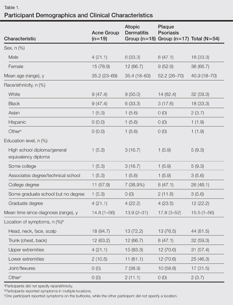
At the time of screening, participants reported prior or present use of topical prescription medications in various formulations for treatment of their respective conditions. The most commonly reported vehicles across all 3 conditions were creams and ointments, followed by lotions, gels, and foams.
Symptoms Across Conditions
At the beginning of the study, participants reported symptoms they experienced in association with their respective skin conditions. Itching and redness were the only symptoms reported across all 3 conditions in all 6 focus groups. Dry skin/dryness, flaking/scaling/peeling, and pain were reported by at least 1 participant in 5 of 6 focus groups. Other common symptoms reported in at least 3 focus groups included pimples/bumps/boils, sensitivity, bleeding, discoloration of skin, cracking/cuts/skin breaking, and buildup of dead or thickened skin/chunks of skin/plaque. As shown in Table 2, there was more variability in the symptoms reported across the 2 acne groups compared with those reported by the AD and PP groups. Table 2 displays all symptoms spontaneously reported across each of the 6 focus groups. Symptoms are listed according to the terms/descriptions provided by focus group participants.
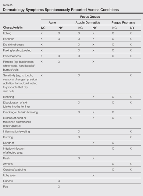
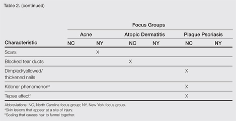
Results by Condition
Unless otherwise noted, participant responses generally were consistent across the North Carolina and New York focus groups. Results from the study, which included a total of 54 participants, suggested a high degree of similarity among preferences for topical treatment attributes across the 3 conditions. Moisturizing was the single attribute that was mentioned across all 3 conditions in all 6 focus groups as an important characteristic in a topical dermatologic treatment. Other attributes mentioned by at least 1 participant in 5 of 6 focus groups included the following: absorbs/disappears/dries quickly, available in various formulations, does not bleach or stain skin/hair/clothing, is not greasy/oily, is not sticky/tacky, is long lasting/long acting/stays on/lasts through sweating or hand washing, is fragrance or odor free, is easy to apply/simple to use, and can use all the time.
A few condition-specific attributes also were noted. An attribute was considered condition specific if it was mentioned by at least 1 participant in both groups for any condition. Preferred properties for topical medications that were specific to acne patients included the following: easy to dispense/dispenses right amount, nondrying, product goes on/spreads smoothly, container (eg, tube, pump) is not easily broken/does not leak, and creamy. Preferred properties that were specific to AD patients included the following: is not noticeable to others/conceals area, good consistency, and cooling. Preferred properties that were specific to 2 conditions included no residue among acne and AD patients and soothing among AD and PP patients. Preferred properties for topical medications across all focus groups are described in further detail in Table 3.
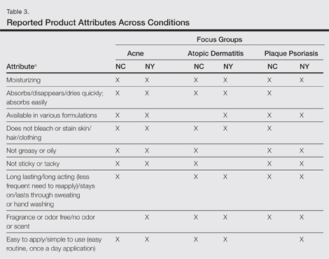
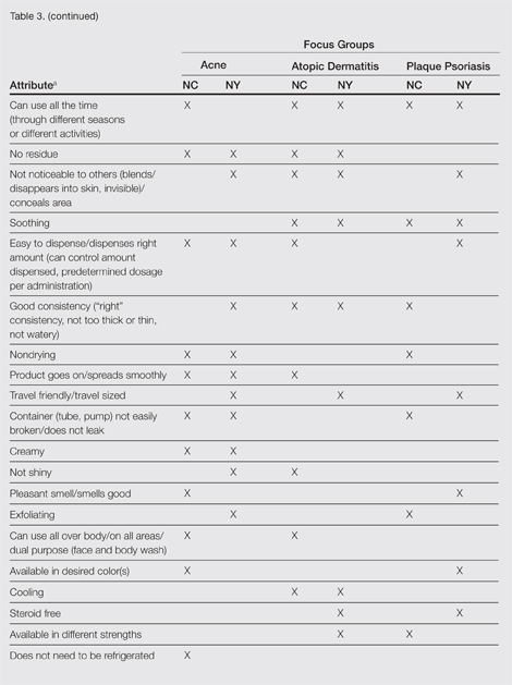
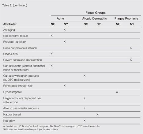
Regarding vehicle-type preferences, acne patients tended to prefer washes, creams, and lotions; AD patients preferred creams; and psoriasis patients preferred creams, ointments, and foams (particularly for the scalp).
Participants across all 3 conditions reported that during daytime hours (versus at night), they would be less likely to use products that are oily, shiny, thick (eg, ointments, oils); bleach or stain the skin/clothing; interfere with makeup, work, or other activities; or are visible to others. Rather, a majority of participants noted that they were more likely to use thinner, less oily products (eg, creams, lotions) during daytime hours, in social situations, or during certain activities (eg, exercise). Participants across all 3 conditions noted that they only used prescription shampoos when they had enough time to repeatedly rinse their hair to eliminate the smell. Participants across all 6 groups noted that they tended to choose creams or lotions over ointments (when available) during the summer because they considered these products to be lighter and less greasy.
Overall, participants indicated that condition-specific symptoms did not influence their preference for topical formulations; rather, they used whatever prescriptions they currently had. However, in some instances, participants noted that the location of the affected area might influence the type of product selected. For example, some participants tended not to use ointments on the scalp or in locations where their clothes might come into contact with the medicated area to avoid clothes sticking to medicated areas. Other participants used lighter, less stringent products on the face versus other locations. Participants noted that foams were preferred for more discrete localized areas.
Comment
Despite the variability in symptoms and conditions across the study population as well as participants’ varying experience with and access to topical treatments, the attributes participants valued most in topical prescription treatments were relatively consistent across the 3 conditions: moisturizing, absorbs/disappears/dries quickly, available in various formulations, does not bleach or stain skin/hair/clothing, is not greasy or oily, is not sticky or tacky, is long lasting/long acting, is fragrance or odor free, is easy to apply/simple to use, and can use all the time. Preferred vehicle attributes were generally consistent across the 3 conditions, but preferences for vehicle types tended to vary by condition. Although some vehicles were more closely associated with specific attributes (eg, the majority considered lotions to be moisturizing), no one vehicle was associated with the complete list of attributes for the ideal topical medication. Regardless of the symptoms experienced or particular situations/activities, participants noted that they tended to use the topical medications that were available to them, even if intended for different locations of the body, and nearly all reported using more than 1 type of topical treatment.
Caution should be used in interpreting these findings, as participant preferences are largely dependent on prior experience with different vehicle types. The small number of participants across the 3 conditions in this analysis also limits conclusions that can be drawn from the data sets, as each condition has different though somewhat overlapping treatment algorithms. A focused inquiry into each of these conditions through separate evaluations may provide for a more robust analysis. Future research might employ questionnaire methodology to develop items assessing the attributes of interest (eg, moisturizing ability, rate of absorption, greasiness/oiliness, stickiness/tackiness, fragrance/odor). The final set of desirable attributes may vary depending on the condition (eg, a moisturizing product may be more important to PP and AD patients than acne patients) as well as comparator products.
Acknowledgment—The authors would like to thank Jennifer Gwazdauskas, MBA, Research Triangle Park, North Carolina, of Stiefel, a GSK company, for her assistance with manuscript review.
- Tan X, Feldman SR, Chang J, et al. Topical drug delivery systems in dermatology: a review of patient adherence issues. Expert Opin Drug Deliv. 2012;9:1263-1271.
- Kellett N, West F, Finlay AY. Conjoint analysis: a novel, rigorous tool for determining patient p for topical antibiotic treatment for acne. a randomised controlled trial. Br J Dermatol. 2006;154:524-532.
- Trookman NS, Rizer RL, Ho ET, et al. The importance of vehicle properties to patients with atopic dermatitis. Cutis. 2011;88:13-17.
- Weiss S, Wyres M, Brundage T. A novel foam vehicle is consistently preferred by patients for dermatologic conditions. J Am Acad Dermatol. 2011;64(suppl 1):AB50.
The skin care market includes topical product formulations (eg, foams, lotions, ointments, creams) for a number of dermatologic therapeutic targets; however, there is limited information available regarding patient preference for product vehicles by specific dermatologic disease. There are few studies in the literature examining patient adherence to topical medications.1 In the current study, 6 focus groups comprised of patients with 3 dermatologic conditions of interest—acne, atopic dermatitis (AD), or plaque psoriasis (PP)—were surveyed to gain a better understanding of treatment preferences among patients and attributes of various formulations that are most desirable and important to this patient population.
Patient preference for a vehicle is relevant to treatment adherence in patients with conditions such as acne, AD, and PP because noncompliance is a major factor in the high rates of failure that have been associated with topical dermatologic treatments.2 The aesthetic attributes of a given vehicle formulation depend on the disease state being treated, the site of application, and the length of treatment.3 A limited number of studies have linked patient preferences to the attributes of topical medications. In one study, participants preferred an aqueous gel formulation compared with previously used topical treatments for AD.3 In another study, participants ranked the following properties of a hypothetical topical medication as most important: gel formulation, room temperature storage, product life of up to 18 months once opened, application with fingers, and a once-daily regimen.2 A third study found that foams were preferred among other formulations (ie, creams, gels, and ointments) for a variety of disease states, including AD, PP, and seborrheic dermatitis.4 The current study uses a qualitative analysis to further explore patient preferences of vehicle attributes for the topical treatment of acne, AD, and PP.
Methods
Study Participants
Six focus groups were conducted with patients who were using topical prescription medications for the treatment of acne, AD, or PP from March to April 2012. Participants were recruited from Raleigh, North Carolina, and New York, New York, with 3 focus groups (1 for each condition) from each city.
Following institutional review board approval of the study processes and recruitment materials, participants meeting the following criteria were included in the study: 18 years or older; diagnosed with acne, AD, or PP by a physician (participants who currently reported more than 1 of these conditions were not eligible); diagnosed with the respective skin condition at least 6 months prior to screening; current or prior use of topical prescription medications in at least 2 different vehicle formulations (eg, cream and foam, ointment and gel); use of at least 1 topical prescription treatment 5 or more times per month; and English speaking and able to provide written consent.
Study Design
A semistructured discussion guide was developed to ensure consistency in the topics surveyed among all 6 focus groups, and the same 2 moderators conducted the discussion for all 6 groups. At the beginning of the study, after providing written informed consent, participants were given an overview of the study and were asked general questions intended to get the participants talking about their experiences with their respective conditions. To avoid or minimize bias, participants were only asked open-ended questions designed to ascertain what symptoms they experienced in relation to their respective conditions. Finally, the discussions were focused on the topical prescription treatments that participants had tried and the properties of each treatment they liked and disliked.
The focus groups were recorded (audio) and transcribed; transcriptions were then verified through an iterative process of technical and editorial review. Analysis of the results was conducted by evaluation and review of the field notes as well as the transcripts from the focus groups.
Results
A total of 54 participants were surveyed (average age, 40.9 years). Although the average age of participants in the acne and AD groups was generally the same (35.2 and 35.4 years, respectively), the average age of participants in the PP group was higher (52.2 years). The majority of participants were white females who had at least a college degree. On average, participants had been diagnosed with their respective condition approximately 15.5 years prior to screening. Participant demographics and clinical characteristics are presented in more detail in Table 1.
At the time of screening, participants reported prior or present use of topical prescription medications in various formulations for treatment of their respective conditions. The most commonly reported vehicles across all 3 conditions were creams and ointments, followed by lotions, gels, and foams.
Symptoms Across Conditions
At the beginning of the study, participants reported symptoms they experienced in association with their respective skin conditions. Itching and redness were the only symptoms reported across all 3 conditions in all 6 focus groups. Dry skin/dryness, flaking/scaling/peeling, and pain were reported by at least 1 participant in 5 of 6 focus groups. Other common symptoms reported in at least 3 focus groups included pimples/bumps/boils, sensitivity, bleeding, discoloration of skin, cracking/cuts/skin breaking, and buildup of dead or thickened skin/chunks of skin/plaque. As shown in Table 2, there was more variability in the symptoms reported across the 2 acne groups compared with those reported by the AD and PP groups. Table 2 displays all symptoms spontaneously reported across each of the 6 focus groups. Symptoms are listed according to the terms/descriptions provided by focus group participants.
Results by Condition
Unless otherwise noted, participant responses generally were consistent across the North Carolina and New York focus groups. Results from the study, which included a total of 54 participants, suggested a high degree of similarity among preferences for topical treatment attributes across the 3 conditions. Moisturizing was the single attribute that was mentioned across all 3 conditions in all 6 focus groups as an important characteristic in a topical dermatologic treatment. Other attributes mentioned by at least 1 participant in 5 of 6 focus groups included the following: absorbs/disappears/dries quickly, available in various formulations, does not bleach or stain skin/hair/clothing, is not greasy/oily, is not sticky/tacky, is long lasting/long acting/stays on/lasts through sweating or hand washing, is fragrance or odor free, is easy to apply/simple to use, and can use all the time.
A few condition-specific attributes also were noted. An attribute was considered condition specific if it was mentioned by at least 1 participant in both groups for any condition. Preferred properties for topical medications that were specific to acne patients included the following: easy to dispense/dispenses right amount, nondrying, product goes on/spreads smoothly, container (eg, tube, pump) is not easily broken/does not leak, and creamy. Preferred properties that were specific to AD patients included the following: is not noticeable to others/conceals area, good consistency, and cooling. Preferred properties that were specific to 2 conditions included no residue among acne and AD patients and soothing among AD and PP patients. Preferred properties for topical medications across all focus groups are described in further detail in Table 3.
Regarding vehicle-type preferences, acne patients tended to prefer washes, creams, and lotions; AD patients preferred creams; and psoriasis patients preferred creams, ointments, and foams (particularly for the scalp).
Participants across all 3 conditions reported that during daytime hours (versus at night), they would be less likely to use products that are oily, shiny, thick (eg, ointments, oils); bleach or stain the skin/clothing; interfere with makeup, work, or other activities; or are visible to others. Rather, a majority of participants noted that they were more likely to use thinner, less oily products (eg, creams, lotions) during daytime hours, in social situations, or during certain activities (eg, exercise). Participants across all 3 conditions noted that they only used prescription shampoos when they had enough time to repeatedly rinse their hair to eliminate the smell. Participants across all 6 groups noted that they tended to choose creams or lotions over ointments (when available) during the summer because they considered these products to be lighter and less greasy.
Overall, participants indicated that condition-specific symptoms did not influence their preference for topical formulations; rather, they used whatever prescriptions they currently had. However, in some instances, participants noted that the location of the affected area might influence the type of product selected. For example, some participants tended not to use ointments on the scalp or in locations where their clothes might come into contact with the medicated area to avoid clothes sticking to medicated areas. Other participants used lighter, less stringent products on the face versus other locations. Participants noted that foams were preferred for more discrete localized areas.
Comment
Despite the variability in symptoms and conditions across the study population as well as participants’ varying experience with and access to topical treatments, the attributes participants valued most in topical prescription treatments were relatively consistent across the 3 conditions: moisturizing, absorbs/disappears/dries quickly, available in various formulations, does not bleach or stain skin/hair/clothing, is not greasy or oily, is not sticky or tacky, is long lasting/long acting, is fragrance or odor free, is easy to apply/simple to use, and can use all the time. Preferred vehicle attributes were generally consistent across the 3 conditions, but preferences for vehicle types tended to vary by condition. Although some vehicles were more closely associated with specific attributes (eg, the majority considered lotions to be moisturizing), no one vehicle was associated with the complete list of attributes for the ideal topical medication. Regardless of the symptoms experienced or particular situations/activities, participants noted that they tended to use the topical medications that were available to them, even if intended for different locations of the body, and nearly all reported using more than 1 type of topical treatment.
Caution should be used in interpreting these findings, as participant preferences are largely dependent on prior experience with different vehicle types. The small number of participants across the 3 conditions in this analysis also limits conclusions that can be drawn from the data sets, as each condition has different though somewhat overlapping treatment algorithms. A focused inquiry into each of these conditions through separate evaluations may provide for a more robust analysis. Future research might employ questionnaire methodology to develop items assessing the attributes of interest (eg, moisturizing ability, rate of absorption, greasiness/oiliness, stickiness/tackiness, fragrance/odor). The final set of desirable attributes may vary depending on the condition (eg, a moisturizing product may be more important to PP and AD patients than acne patients) as well as comparator products.
Acknowledgment—The authors would like to thank Jennifer Gwazdauskas, MBA, Research Triangle Park, North Carolina, of Stiefel, a GSK company, for her assistance with manuscript review.
- Tan X, Feldman SR, Chang J, et al. Topical drug delivery systems in dermatology: a review of patient adherence issues. Expert Opin Drug Deliv. 2012;9:1263-1271.
- Kellett N, West F, Finlay AY. Conjoint analysis: a novel, rigorous tool for determining patient p for topical antibiotic treatment for acne. a randomised controlled trial. Br J Dermatol. 2006;154:524-532.
- Trookman NS, Rizer RL, Ho ET, et al. The importance of vehicle properties to patients with atopic dermatitis. Cutis. 2011;88:13-17.
- Weiss S, Wyres M, Brundage T. A novel foam vehicle is consistently preferred by patients for dermatologic conditions. J Am Acad Dermatol. 2011;64(suppl 1):AB50.
- Patient preference for topical product formulation varies by dermatologic condition being treated.
- Most important vehicle attributes cited across study conditions include moisturizing, absorbs/disappears/dries quickly, available in various formulations, does not bleach or stain skin/hair/clothing, is not greasy or oily, is not sticky or tacky, is long lasting/long acting, is fragrance or odor free, is easy to apply/simple to use, and can use all the time.
- Patient preference for product vehicle is relevant to treatment adherence and may vary depending on the condition being treated.