User login
Fingolimod Reduces Annualized Relapse Rate in Pediatric MS
PARIS—Among children and adolescents with multiple sclerosis (MS), patients treated with fingolimod have a lower annualized relapse rate, compared with patients treated with interferon beta-1a, according to phase III trial results presented at the Seventh Joint ECTRIMS–ACTRIMS Meeting. In addition, fingolimod results in consistently favorable MRI outcomes and may delay disability progression, compared with interferon beta-1a. Fingolimod’s safety profile in children is generally consistent with that in adults.

Between 3% and 5% of patients with MS have disease onset before age 18, but no disease-modifying therapies are FDA-approved specifically for children and adolescents with the disease. “The current treatment recommendations are based on open-label or retrospective studies,” said Tanuja Chitnis, MD, Director of the Partners Pediatric MS Center at Massachusetts General Hospital and Scientist at the Ann Romney Center for Neurological Diseases at Brigham and Women’s Hospital, in Boston.
To investigate the safety and efficacy of fingolimod versus interferon beta-1a in children and adolescents with MS, Dr. Chitnis and colleagues conducted the PARADIGMS study. “PARADIGMS is the first successfully completed global, randomized, clinical trial in pediatric MS,” said Dr. Chitnis, the principal investigator for PARADIGMS.
The PARADIGMS Study
The two-year, double-blind, double-dummy, active-controlled, parallel-group, multicenter study included children and adolescents with a confirmed diagnosis of MS. The study enrolled 215 children between the ages of 10 and 17 with an Expanded Disability Status Scale score between 0 and 5.5. The study included patients who had experienced at least one relapse in the past year or two relapses in the previous two years, or who had evidence of one or more gadolinium-enhancing lesions on MRI within six months prior to randomization.
Patients’ mean age at randomization was about 15. About two-thirds of patients were female, about two-thirds were treatment naïve, and patients were predominantly Caucasian or white. Patients had an average of 1.5 relapses in the prior 12 months and about 2.5 relapses in the prior two years.
Patients were randomized 1:1 to receive once-daily oral fingolimod (0.5 mg or 0.25 mg, dependent on patients’ body weight) or interferon beta-1a 30 μg intramuscular once weekly.
The primary end point was the frequency of relapses in patients treated up to 24 months (ie, annualized relapse rate). Secondary end points included the number of new or newly enlarged T2 lesions, gadolinium-enhancing T1 lesions, safety, and the pharmacokinetic properties of fingolimod.
Researchers conducted the study at 87 sites in more than 25 countries. Investigators designed the study with the FDA, the European Medicines Agency, and the International Pediatric MS Study Group. Basel, Switzerland-based Novartis, which markets fingolimod as Gilenya, supported the study.
Freedom From Relapse
Oral fingolimod reduced the relapse rate by 82% over a period of two years, compared with interferon beta-1a intramuscular injections, Dr. Chitnis said.
About 85% of fingolimod-treated patients were free of confirmed relapse at month 24, versus 39% of patients in the interferon beta-1a arm.
In addition, fingolimod-treated patients had a significant reduction in the number of new or newly enlarging T2 lesions and gadolinium-enhancing T1 lesions, and significantly less brain volume loss, compared with patients who received interferon beta-1a.
In a post hoc analysis, fingolimod significantly delayed confirmed disability progression, compared with interferon beta-1a.
Adverse Events
“The safety profile of fingolimod is generally consistent with that seen in the adult clinical trials,” Dr. Chitnis said. Adverse events occurred more often in the interferon beta-1a arm, whereas serious adverse events occurred more often in the fingolimod arm. The serious adverse events observed in the fingolimod arm included leukopenia in two patients, granulomatosis in one patient, hypersensitivity reaction in one patient, and four patients with seizure events, Dr. Chitnis said.
In all, 92% of fingolimod-treated patients completed the study, compared with 75% of patients in the interferon beta-1a arm. All patients had the option of transferring to an open-label fingolimod arm in an extension phase, and 171 patients entered this extension study.
—Jake Remaly
PARIS—Among children and adolescents with multiple sclerosis (MS), patients treated with fingolimod have a lower annualized relapse rate, compared with patients treated with interferon beta-1a, according to phase III trial results presented at the Seventh Joint ECTRIMS–ACTRIMS Meeting. In addition, fingolimod results in consistently favorable MRI outcomes and may delay disability progression, compared with interferon beta-1a. Fingolimod’s safety profile in children is generally consistent with that in adults.
Between 3% and 5% of patients with MS have disease onset before age 18, but no disease-modifying therapies are FDA-approved specifically for children and adolescents with the disease. “The current treatment recommendations are based on open-label or retrospective studies,” said Tanuja Chitnis, MD, Director of the Partners Pediatric MS Center at Massachusetts General Hospital and Scientist at the Ann Romney Center for Neurological Diseases at Brigham and Women’s Hospital, in Boston.
To investigate the safety and efficacy of fingolimod versus interferon beta-1a in children and adolescents with MS, Dr. Chitnis and colleagues conducted the PARADIGMS study. “PARADIGMS is the first successfully completed global, randomized, clinical trial in pediatric MS,” said Dr. Chitnis, the principal investigator for PARADIGMS.
The PARADIGMS Study
The two-year, double-blind, double-dummy, active-controlled, parallel-group, multicenter study included children and adolescents with a confirmed diagnosis of MS. The study enrolled 215 children between the ages of 10 and 17 with an Expanded Disability Status Scale score between 0 and 5.5. The study included patients who had experienced at least one relapse in the past year or two relapses in the previous two years, or who had evidence of one or more gadolinium-enhancing lesions on MRI within six months prior to randomization.
Patients’ mean age at randomization was about 15. About two-thirds of patients were female, about two-thirds were treatment naïve, and patients were predominantly Caucasian or white. Patients had an average of 1.5 relapses in the prior 12 months and about 2.5 relapses in the prior two years.
Patients were randomized 1:1 to receive once-daily oral fingolimod (0.5 mg or 0.25 mg, dependent on patients’ body weight) or interferon beta-1a 30 μg intramuscular once weekly.
The primary end point was the frequency of relapses in patients treated up to 24 months (ie, annualized relapse rate). Secondary end points included the number of new or newly enlarged T2 lesions, gadolinium-enhancing T1 lesions, safety, and the pharmacokinetic properties of fingolimod.
Researchers conducted the study at 87 sites in more than 25 countries. Investigators designed the study with the FDA, the European Medicines Agency, and the International Pediatric MS Study Group. Basel, Switzerland-based Novartis, which markets fingolimod as Gilenya, supported the study.
Freedom From Relapse
Oral fingolimod reduced the relapse rate by 82% over a period of two years, compared with interferon beta-1a intramuscular injections, Dr. Chitnis said.
About 85% of fingolimod-treated patients were free of confirmed relapse at month 24, versus 39% of patients in the interferon beta-1a arm.
In addition, fingolimod-treated patients had a significant reduction in the number of new or newly enlarging T2 lesions and gadolinium-enhancing T1 lesions, and significantly less brain volume loss, compared with patients who received interferon beta-1a.
In a post hoc analysis, fingolimod significantly delayed confirmed disability progression, compared with interferon beta-1a.
Adverse Events
“The safety profile of fingolimod is generally consistent with that seen in the adult clinical trials,” Dr. Chitnis said. Adverse events occurred more often in the interferon beta-1a arm, whereas serious adverse events occurred more often in the fingolimod arm. The serious adverse events observed in the fingolimod arm included leukopenia in two patients, granulomatosis in one patient, hypersensitivity reaction in one patient, and four patients with seizure events, Dr. Chitnis said.
In all, 92% of fingolimod-treated patients completed the study, compared with 75% of patients in the interferon beta-1a arm. All patients had the option of transferring to an open-label fingolimod arm in an extension phase, and 171 patients entered this extension study.
—Jake Remaly
Oligoclonal Bands Could Be a Valuable Criterion for the Diagnosis of MS
PARIS—Oligoclonal bands, together with symptomatic lesions disseminated in space, increase the risk of multiple sclerosis (MS), according to data presented at the Seventh Joint ECTRIMS–ACTRIMS Meeting. MRI dissemination in space (DIS) at any time plus positive oligoclonal bands should be considered as an additional criterion for MS diagnosis, according to the researchers.
Previous research has suggested that the presence of oligoclonal bands in typical clinically isolated syndromes (CIS) increases the risk of a second attack independently of MRI findings. Georgina Arrambide, MD, PhD, a neurologist at Vall d’Hebron University Hospital in Barcelona, and colleagues studied an ongoing CIS cohort to explore whether oligoclonal bands would be a valuable criterion for MS diagnosis in the context of the 2010 McDonald criteria.

An Examination of MRIs
The investigators obtained MRIs at three to five months after CIS diagnosis, at one year, and at every five years. Oligoclonal bands were determined by isoelectric focusing combined with immunoblotting. Dr. Arrambide and colleagues selected 565 patients with oligoclonal band determination and sufficient data on baseline brain MRI to assess 2010 DIS and dissemination in time (DIT) considering the symptomatic lesions. They excluded 167 participants (29.6%) who already fulfilled DIS and DIT criteria and divided the remaining 398 participants into groups with no DIS and no DIT (n = 218), DIS only (n = 164), and DIT only (n = 16).
Next, the researchers performed Cox proportional hazards regression models with 2010 McDonald as the outcome, using no DIS no DIT with no lesions (n = 107) as the reference for no DIS no DIT with one or more lesion, DIS only, and DIT only. To assess performance, Dr. Arrambide’s group selected cases with a follow-up of three or more years or a second attack within three years of the CIS (n = 305). These participants were divided into groups with no DIS and no DIT (n = 165), DIS only (n = 129), and DIT only (n = 11). The investigators classified participants with no DIS and no DIT with one or more lesion (n = 93) and DIS only according to their oligoclonal band status. They assessed sensitivity, specificity, accuracy, positive predictive value, and negative predictive value with 2010 McDonald at three years as the outcome.
Oligoclonal Bands Increased Risk of Conversion to MS
The adjusted hazard ratios of second attack were 2.8 for no DIS and no DIT with one or more lesion and negative oligoclonal bands, 6.4 for no DIS and no DIT with one or more lesion and positive oligoclonal bands, 9.7 for DIS only with negative oligoclonal bands, 14.8 for DIS only with positive oligoclonal bands, and 7.9 for DIT only. Regarding performance, specificity was 77.6 for no DIS no DIT with one or more lesion and negative oligoclonal bands, 89.1 for no DIS no DIT with one or more lesion and positive oligoclonal bands, 92.5 for DIS only and negative oligoclonal bands, 88.1 for DIS only and positive oligoclonal bands, and 97.8 for DIT only. DIS only with positive oligoclonal bands had the highest sensitivity (46.2), accuracy (64.6), and positive predictive value (83.2).
PARIS—Oligoclonal bands, together with symptomatic lesions disseminated in space, increase the risk of multiple sclerosis (MS), according to data presented at the Seventh Joint ECTRIMS–ACTRIMS Meeting. MRI dissemination in space (DIS) at any time plus positive oligoclonal bands should be considered as an additional criterion for MS diagnosis, according to the researchers.
Previous research has suggested that the presence of oligoclonal bands in typical clinically isolated syndromes (CIS) increases the risk of a second attack independently of MRI findings. Georgina Arrambide, MD, PhD, a neurologist at Vall d’Hebron University Hospital in Barcelona, and colleagues studied an ongoing CIS cohort to explore whether oligoclonal bands would be a valuable criterion for MS diagnosis in the context of the 2010 McDonald criteria.
An Examination of MRIs
The investigators obtained MRIs at three to five months after CIS diagnosis, at one year, and at every five years. Oligoclonal bands were determined by isoelectric focusing combined with immunoblotting. Dr. Arrambide and colleagues selected 565 patients with oligoclonal band determination and sufficient data on baseline brain MRI to assess 2010 DIS and dissemination in time (DIT) considering the symptomatic lesions. They excluded 167 participants (29.6%) who already fulfilled DIS and DIT criteria and divided the remaining 398 participants into groups with no DIS and no DIT (n = 218), DIS only (n = 164), and DIT only (n = 16).
Next, the researchers performed Cox proportional hazards regression models with 2010 McDonald as the outcome, using no DIS no DIT with no lesions (n = 107) as the reference for no DIS no DIT with one or more lesion, DIS only, and DIT only. To assess performance, Dr. Arrambide’s group selected cases with a follow-up of three or more years or a second attack within three years of the CIS (n = 305). These participants were divided into groups with no DIS and no DIT (n = 165), DIS only (n = 129), and DIT only (n = 11). The investigators classified participants with no DIS and no DIT with one or more lesion (n = 93) and DIS only according to their oligoclonal band status. They assessed sensitivity, specificity, accuracy, positive predictive value, and negative predictive value with 2010 McDonald at three years as the outcome.
Oligoclonal Bands Increased Risk of Conversion to MS
The adjusted hazard ratios of second attack were 2.8 for no DIS and no DIT with one or more lesion and negative oligoclonal bands, 6.4 for no DIS and no DIT with one or more lesion and positive oligoclonal bands, 9.7 for DIS only with negative oligoclonal bands, 14.8 for DIS only with positive oligoclonal bands, and 7.9 for DIT only. Regarding performance, specificity was 77.6 for no DIS no DIT with one or more lesion and negative oligoclonal bands, 89.1 for no DIS no DIT with one or more lesion and positive oligoclonal bands, 92.5 for DIS only and negative oligoclonal bands, 88.1 for DIS only and positive oligoclonal bands, and 97.8 for DIT only. DIS only with positive oligoclonal bands had the highest sensitivity (46.2), accuracy (64.6), and positive predictive value (83.2).
Does Concussion Increase the Risk of MS?
PARIS—Head trauma in adolescence, particularly if it is repeated, is associated with an increased risk of subsequent multiple sclerosis (MS), according to data described at the Seventh Joint ECTRIMS-ACTRIMS Meeting. The increased risk may result from the initiation of an autoimmune process in the CNS. This finding underscores the importance of protecting young people from head injuries, according to the researchers.
Previous studies have suggested an association between head trauma and MS risk, but they have had methodologic limitations such as retrospective data collection and small study populations. Tomas Olsson, MD, Professor of Clinical Neuroscience and Senior Physician at Karolinska Institutet in Stockholm, and colleagues used prospectively recorded data to assess whether concussion in childhood or adolescence is associated with subsequent MS risk.

The investigators used the national Swedish Patient and MS registers to identify all diagnoses of MS up to 2012 among people born in 1964 (when the Patient Register was established) or later. They identified 7,292 patients with MS and matched each with 10 people without MS by sex, year of birth, age or vital status at MS diagnosis, and region of residence. This matching resulted in a study population of 80,212. Diagnoses of concussion and control diagnoses of broken limb bones were identified using the Patient Register from birth to age 10 or from age 11 to 20. Dr. Olsson and colleagues used conditional logistic regression to examine associations between broken bones, concussions, and MS.
Concussion in adolescence was associated with an increased risk of MS, producing adjusted odds ratios of 1.22 for one diagnosis of concussion and 2.33 for more than one diagnosis of concussion, compared with none. No notable association with MS was observed for concussion in childhood, or for broken limb bones in childhood or in adolescence.
PARIS—Head trauma in adolescence, particularly if it is repeated, is associated with an increased risk of subsequent multiple sclerosis (MS), according to data described at the Seventh Joint ECTRIMS-ACTRIMS Meeting. The increased risk may result from the initiation of an autoimmune process in the CNS. This finding underscores the importance of protecting young people from head injuries, according to the researchers.
Previous studies have suggested an association between head trauma and MS risk, but they have had methodologic limitations such as retrospective data collection and small study populations. Tomas Olsson, MD, Professor of Clinical Neuroscience and Senior Physician at Karolinska Institutet in Stockholm, and colleagues used prospectively recorded data to assess whether concussion in childhood or adolescence is associated with subsequent MS risk.
The investigators used the national Swedish Patient and MS registers to identify all diagnoses of MS up to 2012 among people born in 1964 (when the Patient Register was established) or later. They identified 7,292 patients with MS and matched each with 10 people without MS by sex, year of birth, age or vital status at MS diagnosis, and region of residence. This matching resulted in a study population of 80,212. Diagnoses of concussion and control diagnoses of broken limb bones were identified using the Patient Register from birth to age 10 or from age 11 to 20. Dr. Olsson and colleagues used conditional logistic regression to examine associations between broken bones, concussions, and MS.
Concussion in adolescence was associated with an increased risk of MS, producing adjusted odds ratios of 1.22 for one diagnosis of concussion and 2.33 for more than one diagnosis of concussion, compared with none. No notable association with MS was observed for concussion in childhood, or for broken limb bones in childhood or in adolescence.

What Is the Prevalence of Truly Benign MS?
PARIS—Benign multiple sclerosis (MS) appears to be rare. Its estimated prevalence is less than 4%, according to a study described at the Seventh Joint ECTRIMS–ACTRIMS Meeting.
The existence of benign MS has been proposed, but it remains controversial. Neurologists are uncertain about the frequency and pathologic explanation for a favorable outcome in MS. Identifying and studying individuals with benign MS would have “considerable implications for patient management and for our understanding of the biology of the disease,” said Emma Tallantyre, BMBS, PhD, Clinical Senior Lecturer in the Division of Psychological Medicine and Clinical Neurosciences at Cardiff University in the United Kingdom, and colleagues.

Most definitions of benign MS are focused on walking ability after 10 or 15 years, despite the far wider effects of MS on ability. Dr. Tallantyre and colleagues screened a prevalent population of more than 2,000 people with MS and found 275 individuals who had unlimited walking ability after 15 or more years from onset. The investigators undertook detailed assessments of 56 of the individuals within this group (ie, those recorded to have unlimited walking ability after the longest disease durations). Assessment incorporated scores of cognition, fatigue, mood, vision, bladder symptoms, and arm and leg function.
All patients were considered to have relapsing-remitting MS, but they showed a wide range of relapse frequency and severity. In a group of 32 patients who fulfilled a contemporary definition of benign MS based on the Expanded Disability Status Scale, the researchers considered less than 25% to be truly benign, which was defined as having normal function in all domains. Patient-reported scores of MS impact correlated strongly with the outcomes of clinical assessment, but patients’ own perceptions of their condition was more benign than clinicians’ perceptions.
MR imaging was used to explore the biology underlying benign MS using a global approach and a tract-based approach. The study provides early insights into the phenotypic and imaging characteristics of benign MS and could provide information about the biologic mechanisms of a favorable outcome in MS, said Dr. Tallantyre.
PARIS—Benign multiple sclerosis (MS) appears to be rare. Its estimated prevalence is less than 4%, according to a study described at the Seventh Joint ECTRIMS–ACTRIMS Meeting.
The existence of benign MS has been proposed, but it remains controversial. Neurologists are uncertain about the frequency and pathologic explanation for a favorable outcome in MS. Identifying and studying individuals with benign MS would have “considerable implications for patient management and for our understanding of the biology of the disease,” said Emma Tallantyre, BMBS, PhD, Clinical Senior Lecturer in the Division of Psychological Medicine and Clinical Neurosciences at Cardiff University in the United Kingdom, and colleagues.
Most definitions of benign MS are focused on walking ability after 10 or 15 years, despite the far wider effects of MS on ability. Dr. Tallantyre and colleagues screened a prevalent population of more than 2,000 people with MS and found 275 individuals who had unlimited walking ability after 15 or more years from onset. The investigators undertook detailed assessments of 56 of the individuals within this group (ie, those recorded to have unlimited walking ability after the longest disease durations). Assessment incorporated scores of cognition, fatigue, mood, vision, bladder symptoms, and arm and leg function.
All patients were considered to have relapsing-remitting MS, but they showed a wide range of relapse frequency and severity. In a group of 32 patients who fulfilled a contemporary definition of benign MS based on the Expanded Disability Status Scale, the researchers considered less than 25% to be truly benign, which was defined as having normal function in all domains. Patient-reported scores of MS impact correlated strongly with the outcomes of clinical assessment, but patients’ own perceptions of their condition was more benign than clinicians’ perceptions.
MR imaging was used to explore the biology underlying benign MS using a global approach and a tract-based approach. The study provides early insights into the phenotypic and imaging characteristics of benign MS and could provide information about the biologic mechanisms of a favorable outcome in MS, said Dr. Tallantyre.

Interferon Beta May Reduce Mortality in MS
PARIS—Interferon beta is associated with a lower risk of all-cause mortality among patients with multiple sclerosis (MS) treated in clinical practice, according to data presented at the Seventh Joint ECTRIMS–ACTRIMS Meeting. Participants in the study were observed for as long as 18 years. The findings were consistent across two geographically distinct regions (ie, Canada and France), said the investigators.
Interferon beta has benefits for patients with MS, but whether the treatment prolongs survival in this population is unclear. Elaine Kingwell, PhD, a research associate in the Division of Neurology at the University of British Columbia in Vancouver, and colleagues investigated the association between interferon beta treatment and all-cause mortality in the clinical setting.

The researchers accessed prospectively collected data on patients with relapsing-remitting MS at onset, including sex, birth date, Expanded Disability Status Scale (EDSS) scores, and disease modifying treatments (DMTs), from British Columbia, Canada, and Rennes, France. Data were linked to population-based administrative databases capturing death dates in both countries. In Canada, data about drug prescriptions filled, universal health care registration, hospital admissions, and physician visits also were available. Patients were treatment-naïve at study entry, and investigators followed them from the earliest of first MS clinic visit, 18th birthday, or January 1, 1996, to the earliest of death, emigration, or December 31, 2013.
Using a nested case–control design, the researchers randomly selected as many as 20 MS controls by incidence density sampling and matched them to MS cases (ie, patients who died during follow-up) by sex, age (± 5 years), calendar year, and EDSS score at study entry. The association between all-cause mortality and interferon beta exposure (greater than six months) was estimated by conditional logistic regression, expressed as odds ratios (OR), and adjusted for glatiramer acetate (greater than six months exposure), any exposure to another DMT, age, and comorbidity burden. Analyses were stratified by sex and country. The researchers examined the association with cumulative exposure to interferon beta (up to three years or more than three years).
The cohort included 7,009 patients (75% women) with relapsing-remitting MS with a median age at study entry of 42. During follow-up (median, 12.3 years), 30% of participants were exposed to interferon beta, 11% were exposed to glatiramer acetate, and 12% were exposed to other DMTs, including MS-specific immunosuppressants. As many as 20 controls were successfully matched to each of 649 cases (mean age at death, 60). The odds of exposure to interferon beta was 32% lower among cases than controls. Stratification by sex or country did not change the interpretation of findings. Compared with no or minimal exposure, a longer cumulative time on interferon beta (ie, more than three years) was associated with increased survival (OR, 0.44), whereas exposure of less than three years was not (OR, 1.00).
PARIS—Interferon beta is associated with a lower risk of all-cause mortality among patients with multiple sclerosis (MS) treated in clinical practice, according to data presented at the Seventh Joint ECTRIMS–ACTRIMS Meeting. Participants in the study were observed for as long as 18 years. The findings were consistent across two geographically distinct regions (ie, Canada and France), said the investigators.
Interferon beta has benefits for patients with MS, but whether the treatment prolongs survival in this population is unclear. Elaine Kingwell, PhD, a research associate in the Division of Neurology at the University of British Columbia in Vancouver, and colleagues investigated the association between interferon beta treatment and all-cause mortality in the clinical setting.
The researchers accessed prospectively collected data on patients with relapsing-remitting MS at onset, including sex, birth date, Expanded Disability Status Scale (EDSS) scores, and disease modifying treatments (DMTs), from British Columbia, Canada, and Rennes, France. Data were linked to population-based administrative databases capturing death dates in both countries. In Canada, data about drug prescriptions filled, universal health care registration, hospital admissions, and physician visits also were available. Patients were treatment-naïve at study entry, and investigators followed them from the earliest of first MS clinic visit, 18th birthday, or January 1, 1996, to the earliest of death, emigration, or December 31, 2013.
Using a nested case–control design, the researchers randomly selected as many as 20 MS controls by incidence density sampling and matched them to MS cases (ie, patients who died during follow-up) by sex, age (± 5 years), calendar year, and EDSS score at study entry. The association between all-cause mortality and interferon beta exposure (greater than six months) was estimated by conditional logistic regression, expressed as odds ratios (OR), and adjusted for glatiramer acetate (greater than six months exposure), any exposure to another DMT, age, and comorbidity burden. Analyses were stratified by sex and country. The researchers examined the association with cumulative exposure to interferon beta (up to three years or more than three years).
The cohort included 7,009 patients (75% women) with relapsing-remitting MS with a median age at study entry of 42. During follow-up (median, 12.3 years), 30% of participants were exposed to interferon beta, 11% were exposed to glatiramer acetate, and 12% were exposed to other DMTs, including MS-specific immunosuppressants. As many as 20 controls were successfully matched to each of 649 cases (mean age at death, 60). The odds of exposure to interferon beta was 32% lower among cases than controls. Stratification by sex or country did not change the interpretation of findings. Compared with no or minimal exposure, a longer cumulative time on interferon beta (ie, more than three years) was associated with increased survival (OR, 0.44), whereas exposure of less than three years was not (OR, 1.00).
Siponimod Improves MRI Outcomes in Patients With Secondary Progressive MS
PARIS—Siponimod significantly reduces MRI activity and slows brain volume loss in patients with secondary progressive multiple sclerosis (MS), according to a study described at the Seventh Joint ECTRIMS–ACTRIMS Meeting. Effects are observable at month 12 and sustained at month 24. “These results, together with the clinically observed reduction in confirmed disability progression reported previously, corroborate the positive impact of treatment with siponimod in patients with secondary progressive MS,” said Robert Fox, MD, a researcher at the Mellen Center for Treatment and Research in MS in the Cleveland Clinic, and colleagues.

The EXPAND study demonstrated the benefits of siponimod, a selective modulator of sphingosine 1-phosphate receptor subtypes 1 and 5, on confirmed disability progression. Dr. Fox and colleagues examined these data to evaluate the effect of siponimod versus placebo on predefined MRI outcomes in patients with secondary progressive MS.
The researchers randomized patients 2:1 to receive siponimod or placebo. MRI scans were performed at baseline and every 12 months thereafter. Radiologists at a central reading center analyzed the scans. Key MRI outcomes included T2 lesion volume, number of new or enlarging T2 lesions, number of gadolinium-enhancing lesions, and brain volume loss assessed by percent brain volume change.
The full analysis set comprised patients who received one or more doses of study drug as per original randomization. The per-protocol analysis set consisted of all full-analysis-set patients without major protocol deviations and included efficacy data only up to discontinuation of double-blinded treatment.
The investigators randomized 1,651 patients. A total of 1,099 patients received siponimod (2 mg), and 546 received placebo. Dr. Fox and colleagues observed treatment benefits in favor of siponimod for all key outcomes and analysis sets investigated. Post-baseline MRI data were available for more than 80% of participants.
At month 12, the adjusted mean differences in the change in T2 lesion volume from baseline versus placebo were −613 mm3 in the full analysis set and −634 mm3 in the per protocol analysis set. At month 24, the differences were −778 mm3 in the full analysis set and −830 mm3 in the per protocol analysis set.
At month 12, the adjusted mean differences in percent brain volume change were 0.175 in the full analysis set and 0.221 in the per protocol analysis set. At month 24, the differences were 0.128 in the full analysis set and 0.277 in the per protocol analysis set.
Siponimod reduced the average T1 gadolinium-enhancing lesion count over months 12 and 24 by 86.6% in the full analysis set and 91.1% in the per protocol analysis set. Siponimod reduced the average count of new or enlarging T2 lesions by 80.6% in the full analysis set and 85.3% in the per protocol analysis set.
This study was funded by Novartis Pharma, which is headquartered in Basel, Switzerland.
PARIS—Siponimod significantly reduces MRI activity and slows brain volume loss in patients with secondary progressive multiple sclerosis (MS), according to a study described at the Seventh Joint ECTRIMS–ACTRIMS Meeting. Effects are observable at month 12 and sustained at month 24. “These results, together with the clinically observed reduction in confirmed disability progression reported previously, corroborate the positive impact of treatment with siponimod in patients with secondary progressive MS,” said Robert Fox, MD, a researcher at the Mellen Center for Treatment and Research in MS in the Cleveland Clinic, and colleagues.
The EXPAND study demonstrated the benefits of siponimod, a selective modulator of sphingosine 1-phosphate receptor subtypes 1 and 5, on confirmed disability progression. Dr. Fox and colleagues examined these data to evaluate the effect of siponimod versus placebo on predefined MRI outcomes in patients with secondary progressive MS.
The researchers randomized patients 2:1 to receive siponimod or placebo. MRI scans were performed at baseline and every 12 months thereafter. Radiologists at a central reading center analyzed the scans. Key MRI outcomes included T2 lesion volume, number of new or enlarging T2 lesions, number of gadolinium-enhancing lesions, and brain volume loss assessed by percent brain volume change.
The full analysis set comprised patients who received one or more doses of study drug as per original randomization. The per-protocol analysis set consisted of all full-analysis-set patients without major protocol deviations and included efficacy data only up to discontinuation of double-blinded treatment.
The investigators randomized 1,651 patients. A total of 1,099 patients received siponimod (2 mg), and 546 received placebo. Dr. Fox and colleagues observed treatment benefits in favor of siponimod for all key outcomes and analysis sets investigated. Post-baseline MRI data were available for more than 80% of participants.
At month 12, the adjusted mean differences in the change in T2 lesion volume from baseline versus placebo were −613 mm3 in the full analysis set and −634 mm3 in the per protocol analysis set. At month 24, the differences were −778 mm3 in the full analysis set and −830 mm3 in the per protocol analysis set.
At month 12, the adjusted mean differences in percent brain volume change were 0.175 in the full analysis set and 0.221 in the per protocol analysis set. At month 24, the differences were 0.128 in the full analysis set and 0.277 in the per protocol analysis set.
Siponimod reduced the average T1 gadolinium-enhancing lesion count over months 12 and 24 by 86.6% in the full analysis set and 91.1% in the per protocol analysis set. Siponimod reduced the average count of new or enlarging T2 lesions by 80.6% in the full analysis set and 85.3% in the per protocol analysis set.
This study was funded by Novartis Pharma, which is headquartered in Basel, Switzerland.
Highlights From the 7th Joint ECTRIMS‑ACTRIMS Meeting
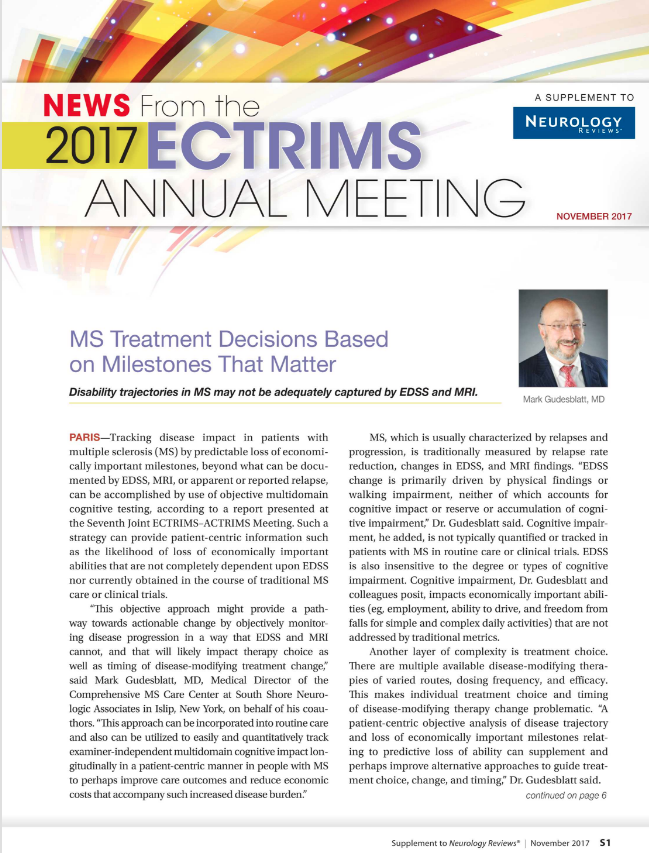
MS Treatment Decisions Based on Milestones That Matter
PARIS—Tracking disease impact in patients with multiple sclerosis (MS) by predictable loss of economically important milestones trajectory, beyond what can be documented by EDSS, MRI, or apparent or reported relapse, can be accomplished by use of objective multidomain cognitive testing, according to a report presented at the Seventh Joint ECTRIMS–ACTRIMS Meeting. Such a strategy can provide patient-centric information such as predicting likely loss of economically impactful abilities that are not completely dependent upon EDSS nor currently obtained in the course of traditional MS care or clinical trials. “This objective approach might provide a pathway towards actionable change by objectively monitoring disease progression in a way that EDSS and MRI are unable and that will likely impact therapy choice as well as timing of disease-modifying treatment change,” said Mark Gudesblatt, MD, Medical Director of the Comprehensive MS Care Center at South Shore Neurologic Associates in Islip, New York, on behalf of his coauthors. “This approach can be incorporated into routine care and also can be utilized to easily and quantitatively track examiner independent multidomain cognitive impact longitudinally in a patient-centric manner in people with MS to perhaps improve care outcomes and reduce economic costs that accompany such increased disease burden.”

MS, which is usually characterized by relapses and progression, is traditionally measured by relapse rate reduction, changes in EDSS, and MRI findings. “EDSS change is primarily driven by physical findings or walking impairment, neither of which accounts for cognitive impact or reserve or accumulation of cognitive impairment,” Dr. Gudesblatt said. Cognitive impairment, he added, is not typically quantified or tracked in patients with MS in routine care or clinical trials. EDSS is also insensitive to the degree or types of cognitive impairment. Cognitive impairment, Dr. Gudesblatt and colleagues posit, impacts economically important abilities (eg, employment, ability to drive, and freedom from falls for both simple and complex daily activities) that are not addressed by traditional metrics.
Another layer of complexity is treatment choice. There are multiple available disease-modifying therapies of varied routes, dosing frequency, and efficacy. This makes individual treatment choice and timing of disease-modifying therapy change problematic. “A patient-centric objective analysis of disease trajectory and loss of economically important milestones relating to predictive loss of ability can supplement and perhaps improve alternative approaches to guide treatment choice, change, and timing,” Dr. Gudesblatt said.
An objective, quantitative, patient-centric, and granular EDSS-independent approach of likely disability trajectory might improve decision making regarding disease-modifying therapy choice and timing of change, offer a path to compare outcome measures across clinical trials, and possibly provide an opportunity to preempt the appearance of important disabilities that result in significantly increased cost of care and reduced quality of life. “Objective comprehensive analytics documenting unseen disease impact and change offer unique opportunities to improve care,” Dr. Gudesblatt said.
Toward this end, Dr. Gudesblatt and colleagues conducted a cross-sectional review of a prospective digital MS registry obtained in the course of routine care utilizing standardized computerized cognitive testing (NeuroTrax) to evaluate the relationship of cognitive impairment to disability. Cognitive impairment was defined as number of cognitive domains impaired (CDI) more than one standard deviation from age/education normal. Disability domains assessed were unemployment, loss of driving, and freedom from falls. Patients with an EDSS of less than 6 were included in the study cohort (ie, no one was included who was disabled to the point of requiring a cane to ambulate).
The researchers found that increasing accumulated number of CDI in patients with MS and an EDSS less than 6 is associated with likely progressive loss of:
- Employment (n = 543, CDI-0 = 61%, CDI-1 = 50%, CDI-2 = 43%, CDI-3 = 32%)
- Driving (n = 115, CDI-0 = 100%, CDI-1 = 66%, CDI-2 = 53%, CDI-3 = 21%)
- Freedom from falls (n = 159) for simple daily activities (CDI-0 = 77%, CDI-1 = 65%, CDI-2 = 37%, CDI-3 = 39%) and reduced freedom from falls for complex daily activities (CDI-0 = 72%, CDI-1 = 58%, CDI-2 = 36%, CDI-3 = 33%).
Increased risk of falls and reduced likelihood of employment and driving all represent significant impact on quality of life and result in increased economic burden and long-term costs of the disease, Dr. Gudesblatt and colleagues said.
PARIS—Tracking disease impact in patients with multiple sclerosis (MS) by predictable loss of economically important milestones trajectory, beyond what can be documented by EDSS, MRI, or apparent or reported relapse, can be accomplished by use of objective multidomain cognitive testing, according to a report presented at the Seventh Joint ECTRIMS–ACTRIMS Meeting. Such a strategy can provide patient-centric information such as predicting likely loss of economically impactful abilities that are not completely dependent upon EDSS nor currently obtained in the course of traditional MS care or clinical trials. “This objective approach might provide a pathway towards actionable change by objectively monitoring disease progression in a way that EDSS and MRI are unable and that will likely impact therapy choice as well as timing of disease-modifying treatment change,” said Mark Gudesblatt, MD, Medical Director of the Comprehensive MS Care Center at South Shore Neurologic Associates in Islip, New York, on behalf of his coauthors. “This approach can be incorporated into routine care and also can be utilized to easily and quantitatively track examiner independent multidomain cognitive impact longitudinally in a patient-centric manner in people with MS to perhaps improve care outcomes and reduce economic costs that accompany such increased disease burden.”
MS, which is usually characterized by relapses and progression, is traditionally measured by relapse rate reduction, changes in EDSS, and MRI findings. “EDSS change is primarily driven by physical findings or walking impairment, neither of which accounts for cognitive impact or reserve or accumulation of cognitive impairment,” Dr. Gudesblatt said. Cognitive impairment, he added, is not typically quantified or tracked in patients with MS in routine care or clinical trials. EDSS is also insensitive to the degree or types of cognitive impairment. Cognitive impairment, Dr. Gudesblatt and colleagues posit, impacts economically important abilities (eg, employment, ability to drive, and freedom from falls for both simple and complex daily activities) that are not addressed by traditional metrics.
Another layer of complexity is treatment choice. There are multiple available disease-modifying therapies of varied routes, dosing frequency, and efficacy. This makes individual treatment choice and timing of disease-modifying therapy change problematic. “A patient-centric objective analysis of disease trajectory and loss of economically important milestones relating to predictive loss of ability can supplement and perhaps improve alternative approaches to guide treatment choice, change, and timing,” Dr. Gudesblatt said.
An objective, quantitative, patient-centric, and granular EDSS-independent approach of likely disability trajectory might improve decision making regarding disease-modifying therapy choice and timing of change, offer a path to compare outcome measures across clinical trials, and possibly provide an opportunity to preempt the appearance of important disabilities that result in significantly increased cost of care and reduced quality of life. “Objective comprehensive analytics documenting unseen disease impact and change offer unique opportunities to improve care,” Dr. Gudesblatt said.
Toward this end, Dr. Gudesblatt and colleagues conducted a cross-sectional review of a prospective digital MS registry obtained in the course of routine care utilizing standardized computerized cognitive testing (NeuroTrax) to evaluate the relationship of cognitive impairment to disability. Cognitive impairment was defined as number of cognitive domains impaired (CDI) more than one standard deviation from age/education normal. Disability domains assessed were unemployment, loss of driving, and freedom from falls. Patients with an EDSS of less than 6 were included in the study cohort (ie, no one was included who was disabled to the point of requiring a cane to ambulate).
The researchers found that increasing accumulated number of CDI in patients with MS and an EDSS less than 6 is associated with likely progressive loss of:
- Employment (n = 543, CDI-0 = 61%, CDI-1 = 50%, CDI-2 = 43%, CDI-3 = 32%)
- Driving (n = 115, CDI-0 = 100%, CDI-1 = 66%, CDI-2 = 53%, CDI-3 = 21%)
- Freedom from falls (n = 159) for simple daily activities (CDI-0 = 77%, CDI-1 = 65%, CDI-2 = 37%, CDI-3 = 39%) and reduced freedom from falls for complex daily activities (CDI-0 = 72%, CDI-1 = 58%, CDI-2 = 36%, CDI-3 = 33%).
Increased risk of falls and reduced likelihood of employment and driving all represent significant impact on quality of life and result in increased economic burden and long-term costs of the disease, Dr. Gudesblatt and colleagues said.
Durable Improvements in Clinical Outcomes With Alemtuzumab: Seven-Year Follow-Up
PARIS—Clinical efficacy of alemtuzumab was maintained for seven years in patients who had inadequate response to prior therapy, despite 47% receiving no additional treatment since the initial two courses of alemtuzumab, according to study data presented at the Seventh Joint ECTRIMS–ACTRIMS Meeting. In addition, 44% of patients showed improvement in disability, researchers reported. “These findings suggest that alemtuzumab may provide a unique treatment approach for patients with relapsing-remitting multiple sclerosis (RRMS), offering durable efficacy in the absence of continuous treatment,” said Barry Singer, MD, Director of the MS Center for Innovations in Care, Missouri Baptist Medical Center, St. Louis.

Alemtuzumab Treatment: Then
In the CARE-MS II trial, alemtuzumab significantly improved clinical outcomes compared with subcutaneous interferon beta-1a over two years in patients with active RRMS and an inadequate response to prior therapy. Durable efficacy of alemtuzumab was demonstrated over six years in a completed extension study in the absence of continuous treatment. Patients in the CARE-MS II study received two courses of alemtuzumab 12 mg/day (five days of therapy at baseline and three days of therapy 12 months later). In the extension study, patients could receive as-needed alemtuzumab retreatment (12 mg/day on three consecutive days at least 12 months after a previous course for relapse or MRI activity) or other disease-modifying therapy per investigator discretion. Patients completing at least 48 months of the extension could enroll in the five-year TOPAZ study for further long-term evaluation.
Alemtuzumab Treatment: Now
The goal of the TOPAZ study was to evaluate the seven-year efficacy and safety of alemtuzumab in patients with RRMS who received alemtuzumab in the CARE-MS II trial. In TOPAZ, patients could receive alemtuzumab retreatment 12 months or more after a previous course or other disease-modifying therapy at any time point (both per investigator discretion; no criteria). MRI scans were done annually. Annualized relapse rate, six-month confirmed disability worsening, six-month confirmed disability improvement, no evidence of disease activity (NEDA), and adverse events were analyzed in TOPAZ.
In total, 338 of 393 (86%) CARE-MS II patients who entered the extension remained on study until the end of year six and then entered TOPAZ; 317 (94%) remained on study through year seven. Annualized release rate remained low (0.14 at year seven) and the proportion of patients with stable or improved Expanded Disability Status Scale score remained high (73% at year seven). Through year seven, 69% of patients were free from six-month confirmed disability worsening, 44% achieved six-month confirmed disability improvement, and the majority achieved NEDA each year. These effects were achieved with 47% of patients receiving no additional treatment (alemtuzumab or other disease-modifying treatment) after their initial two courses of alemtuzumab. Incidences of overall adverse events, infusion-associated reactions, and infections decreased over time and were reduced, compared with those in the two-year core study. Thyroid adverse events incidence peaked at year three and then declined.
PARIS—Clinical efficacy of alemtuzumab was maintained for seven years in patients who had inadequate response to prior therapy, despite 47% receiving no additional treatment since the initial two courses of alemtuzumab, according to study data presented at the Seventh Joint ECTRIMS–ACTRIMS Meeting. In addition, 44% of patients showed improvement in disability, researchers reported. “These findings suggest that alemtuzumab may provide a unique treatment approach for patients with relapsing-remitting multiple sclerosis (RRMS), offering durable efficacy in the absence of continuous treatment,” said Barry Singer, MD, Director of the MS Center for Innovations in Care, Missouri Baptist Medical Center, St. Louis.
Alemtuzumab Treatment: Then
In the CARE-MS II trial, alemtuzumab significantly improved clinical outcomes compared with subcutaneous interferon beta-1a over two years in patients with active RRMS and an inadequate response to prior therapy. Durable efficacy of alemtuzumab was demonstrated over six years in a completed extension study in the absence of continuous treatment. Patients in the CARE-MS II study received two courses of alemtuzumab 12 mg/day (five days of therapy at baseline and three days of therapy 12 months later). In the extension study, patients could receive as-needed alemtuzumab retreatment (12 mg/day on three consecutive days at least 12 months after a previous course for relapse or MRI activity) or other disease-modifying therapy per investigator discretion. Patients completing at least 48 months of the extension could enroll in the five-year TOPAZ study for further long-term evaluation.
Alemtuzumab Treatment: Now
The goal of the TOPAZ study was to evaluate the seven-year efficacy and safety of alemtuzumab in patients with RRMS who received alemtuzumab in the CARE-MS II trial. In TOPAZ, patients could receive alemtuzumab retreatment 12 months or more after a previous course or other disease-modifying therapy at any time point (both per investigator discretion; no criteria). MRI scans were done annually. Annualized relapse rate, six-month confirmed disability worsening, six-month confirmed disability improvement, no evidence of disease activity (NEDA), and adverse events were analyzed in TOPAZ.
In total, 338 of 393 (86%) CARE-MS II patients who entered the extension remained on study until the end of year six and then entered TOPAZ; 317 (94%) remained on study through year seven. Annualized release rate remained low (0.14 at year seven) and the proportion of patients with stable or improved Expanded Disability Status Scale score remained high (73% at year seven). Through year seven, 69% of patients were free from six-month confirmed disability worsening, 44% achieved six-month confirmed disability improvement, and the majority achieved NEDA each year. These effects were achieved with 47% of patients receiving no additional treatment (alemtuzumab or other disease-modifying treatment) after their initial two courses of alemtuzumab. Incidences of overall adverse events, infusion-associated reactions, and infections decreased over time and were reduced, compared with those in the two-year core study. Thyroid adverse events incidence peaked at year three and then declined.
How Does Cladribine Compare With Other MS Therapies?
New data confirm cladribine’s efficacy as a treatment for relapsing-remitting multiple sclerosis (MS), according to results published online ahead of print August 1 in Multiple Sclerosis Journal. The drug’s effect on relapses is comparable to that of fingolimod, and its effect on disability accumulation is comparable to those of interferon β and fingolimod. Compared with interferon, fingolimod, and natalizumab, cladribine may be associated with superior recovery from disability.
A phase III trial demonstrated that, compared with placebo, cladribine reduced relapse rate and increased the likelihood of remaining free from three-month confirmed disability progression in patients with relapsing-remitting MS. The European Medicines Agency approved the therapy in August 2017. Cladribine’s potential position in the treatment landscape is unclear, however, because no direct comparisons of cladribine with other MS therapies are available.
An Analysis of Matched Cohorts
Tomas Kalincik, MD, PhD, Professor of Medicine at Royal Melbourne Hospital in Australia, and colleagues conducted a propensity score–matched analysis of observational data from MSBase, including patients from the Australian Cladribine Product Familiarization Program, to compare the effectiveness of cladribine to that of interferon β, fingolimod, and natalizumab. Eligible participants had relapsing-remitting MS, received one of the study medications as monotherapy for one or more years, and had no prior exposure to alemtuzumab, mitoxantrone, rituximab, or hematopoietic stem cell transplantation.

Patients received 3.5 mg/kg of oral cladribine, 44 μg of subcutaneous interferon β-1a three times weekly, 0.5 mg/day of oral fingolimod, or 300 μg of IV natalizumab every four weeks. Data were recorded during routine clinical practice. The primary end points were the proportion of patients free from relapses, disability accumulation, and disability improvement while on study therapy.
Cladribine Was Associated With Superior Disability Improvement
The researchers included 37 patients treated with cladribine, 1,940 patients treated with interferon β, 1,892 patients treated with fingolimod, and 1,410 patients treated with natalizumab in their analysis. The investigators noted only small differences in baseline characteristics between the matched cohorts.
Compared with participants receiving interferon β, patients receiving cladribine were less likely to have a relapse during the first year of treatment (hazard ratio [HR], 0.6). The proportion of relapse-free patients was 86% in the cladribine group and 70% in the interferon β group. The probability of disability accumulation was similar for these drugs (HR, 0.41), but the cladribine group was more likely to have disability improvement (HR, 15).
The proportion of relapse-free patients at one year was 79% in the cladribine and fingolimod groups, and cumulative hazards of a relapse did not differ between the two groups (HR, 1.2). The probability of disability accumulation was similar for cladribine and fingolimod (HR, 1.8), but the probability of disability improvement was greater for cladribine (HR, 3.9).
The probability of relapse was higher with cladribine than with natalizumab (HR, 1.8), but the proportions of relapse-free patients at the end of year one were 80% and 81%, respectively. The probability of disability accumulation was greater in the cladribine group than in the natalizumab group (HR, 2.5). The probability of disability improvement was greater among patients receiving cladribine than among those receiving natalizumab (HR, 4). Sensitivity analyses largely confirmed the results of the primary analyses.
“Six-month confirmed improvement of disability was observed in 10%–20% of the cladribine cohort during the first year, which was superior to all three comparator therapies. This [finding] is of interest in the context of the comparison to natalizumab, which is known to be associated with a marked improvement in disability early after its commencement,” said Dr. Kalincik and colleagues. “Improvement in disability in a cohort with this profile is unexpected.”
The study’s main limitation is the small size of the cladribine cohort, said the authors. Another limitation is the brief duration of follow-up for the cladribine group, which precludes conclusions about long-term outcomes. Nevertheless, the comparative effectiveness results “represent timely information about the role of cladribine in the management of MS,” Dr. Kalincik concluded.
—Erik Greb
Suggested Reading
Kalincik T, Jokubaitis V, Spelman T, et al. Cladribine versus fingolimod, natalizumab and interferon β for multiple sclerosis. Mult Scler. 2017 Aug 1 [Epub ahead of print].
New data confirm cladribine’s efficacy as a treatment for relapsing-remitting multiple sclerosis (MS), according to results published online ahead of print August 1 in Multiple Sclerosis Journal. The drug’s effect on relapses is comparable to that of fingolimod, and its effect on disability accumulation is comparable to those of interferon β and fingolimod. Compared with interferon, fingolimod, and natalizumab, cladribine may be associated with superior recovery from disability.
A phase III trial demonstrated that, compared with placebo, cladribine reduced relapse rate and increased the likelihood of remaining free from three-month confirmed disability progression in patients with relapsing-remitting MS. The European Medicines Agency approved the therapy in August 2017. Cladribine’s potential position in the treatment landscape is unclear, however, because no direct comparisons of cladribine with other MS therapies are available.
An Analysis of Matched Cohorts
Tomas Kalincik, MD, PhD, Professor of Medicine at Royal Melbourne Hospital in Australia, and colleagues conducted a propensity score–matched analysis of observational data from MSBase, including patients from the Australian Cladribine Product Familiarization Program, to compare the effectiveness of cladribine to that of interferon β, fingolimod, and natalizumab. Eligible participants had relapsing-remitting MS, received one of the study medications as monotherapy for one or more years, and had no prior exposure to alemtuzumab, mitoxantrone, rituximab, or hematopoietic stem cell transplantation.
Patients received 3.5 mg/kg of oral cladribine, 44 μg of subcutaneous interferon β-1a three times weekly, 0.5 mg/day of oral fingolimod, or 300 μg of IV natalizumab every four weeks. Data were recorded during routine clinical practice. The primary end points were the proportion of patients free from relapses, disability accumulation, and disability improvement while on study therapy.
Cladribine Was Associated With Superior Disability Improvement
The researchers included 37 patients treated with cladribine, 1,940 patients treated with interferon β, 1,892 patients treated with fingolimod, and 1,410 patients treated with natalizumab in their analysis. The investigators noted only small differences in baseline characteristics between the matched cohorts.
Compared with participants receiving interferon β, patients receiving cladribine were less likely to have a relapse during the first year of treatment (hazard ratio [HR], 0.6). The proportion of relapse-free patients was 86% in the cladribine group and 70% in the interferon β group. The probability of disability accumulation was similar for these drugs (HR, 0.41), but the cladribine group was more likely to have disability improvement (HR, 15).
The proportion of relapse-free patients at one year was 79% in the cladribine and fingolimod groups, and cumulative hazards of a relapse did not differ between the two groups (HR, 1.2). The probability of disability accumulation was similar for cladribine and fingolimod (HR, 1.8), but the probability of disability improvement was greater for cladribine (HR, 3.9).
The probability of relapse was higher with cladribine than with natalizumab (HR, 1.8), but the proportions of relapse-free patients at the end of year one were 80% and 81%, respectively. The probability of disability accumulation was greater in the cladribine group than in the natalizumab group (HR, 2.5). The probability of disability improvement was greater among patients receiving cladribine than among those receiving natalizumab (HR, 4). Sensitivity analyses largely confirmed the results of the primary analyses.
“Six-month confirmed improvement of disability was observed in 10%–20% of the cladribine cohort during the first year, which was superior to all three comparator therapies. This [finding] is of interest in the context of the comparison to natalizumab, which is known to be associated with a marked improvement in disability early after its commencement,” said Dr. Kalincik and colleagues. “Improvement in disability in a cohort with this profile is unexpected.”
The study’s main limitation is the small size of the cladribine cohort, said the authors. Another limitation is the brief duration of follow-up for the cladribine group, which precludes conclusions about long-term outcomes. Nevertheless, the comparative effectiveness results “represent timely information about the role of cladribine in the management of MS,” Dr. Kalincik concluded.
—Erik Greb
Suggested Reading
Kalincik T, Jokubaitis V, Spelman T, et al. Cladribine versus fingolimod, natalizumab and interferon β for multiple sclerosis. Mult Scler. 2017 Aug 1 [Epub ahead of print].