User login
Clinical Characteristics Predictive of High Costs Among Patients With MS
DALLAS—Baseline use of corticosteroids and documentation of other brain MRI results may be significantly associated with higher costs for patients with multiple sclerosis (MS), according to data presented at the 2014 Cooperative Meeting of CMSC and ACTRIMS. “This study provides insight into factors associated with high-cost MS patients and may help to prospectively identify potential high-cost MS patients who may benefit from cost-effective proactive clinical management,” the researchers said. “Additionally, while most patients have documentation of brain MRI in their medical records, many of the additional clinical characteristics needed to assess disease severity are not documented in the medical record.”
Debra F. Eisenberg, MS, PhD, and colleagues sought to assess patient demographics, clinical characteristics, medication utilization, and resource use among patients with MS stratified as low, medium, and high cost through administrative claims review and patient medical record review. For their observational, retrospective cohort study, the researchers used data drawn from the HealthCore Integrated Research Database (HIRD), which includes medical and pharmacy claims data. Patients age 18 or older newly diagnosed with MS during the period from January 1, 2007, to April 30, 2011, were identified in the database. Annualized MS-related cost was computed, and patients were classified into high-, medium-, and low-cost strata. A total of 400 patients with a confirmed diagnosis of MS and documentation of brain MRI were selected for medical record review. Bivariate analyses and multivariate logistic regression models were used to identify factors associated with high-cost patients.
Among the 400 patient medical records abstracted, 84, 132, and 184 patients fell in the low-, medium-, and high-cost groups, respectively. Patients had a mean age of 41 at diagnosis, and 70% were female. Nearly all (97%) of the patients had brain MRI results documented in their medical records. Of the 389 patients with MRI results, 31.7% of the low-, 53.6% of the medium-, and 35.2% of the high-cost patients had active brain lesions. Common symptoms reported were numbness (63%), fatigue (59%), and pain (59%). Relapsing-remitting disease was documented in 14% of the low-, 40% of the medium-, and 33% of the high-cost patients. Approximately 50% of the patients had gait impairment, ranging from 38% of the low-, 44% of the medium-, and 64% of the high-cost patients. Other brain MRI results not related to T2 imaging, active lesions, demyelination, black holes, and brain atrophy were seen to a greater extent among high-cost patients. In addition, high-cost patients were more likely to use antidepressants (31.5%), corticosteroids (43.5%), narcotics (38.6%), and stimulants (6.5%). High-cost patients also were more likely to have electrocardiogram (36.4%) and spinal tap (20.1%) procedures.
Lead author Dr. Eisenberg is affiliated with HealthCore, which is headquartered in Wilmington, Delaware.
—Glenn S. Williams
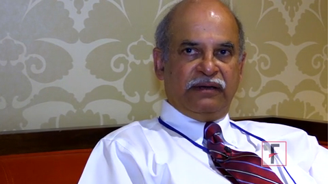
VIDEO: New MS database could change practice
DALLAS – The North American Registry for Care and Research in MS, a national database of multiple sclerosis patients, and the first of its kind, is expected to change multiple sclerosis care in the next decade.
The NARCRMS, which acts as both a database and a registry, will collect patient information from MS centers across the United States over time. It is modeled after the Alzheimer’s Disease Neuroimaging Initiative (ADNI), and it’s a collaboration between the industry and MS centers to create an open source database, "available in real time to patients, physicians, and industry," Dr. Kottil W. Rammohan, professor of clinical neurology and director of the MS center of excellence at the University of Miami, said at a meeting of the Consortium of Multiple Sclerosis Centers and the Americas Committee for Treatment and Research in Multiple Sclerosis.
The NARCRMS, not to be confused with NARCOMS (North American Research Committee on Multiple Sclerosis), hasn’t launched yet – it will be established later this year – but the leaders have great hopes for it, and say that it will help find answers to questions that exist because of a lack of data.
For instance, there are very few head-to-head trials comparing drugs with each other. The database could provide insight and the answer, said Dr. Rammohan.
More information and details will be available once the database is launched, but Dr. Rammohan provided an overview of the NARCRMS, and explained why a database hasn’t been established until now.
Dr. Rammohan said he had no relevant financial disclosures.
On Twitter @naseemmiller
The video associated with this article is no longer available on this site. Please view all of our videos on the MDedge YouTube channel
AT THE CMSC/ACTRIMS ANNUAL MEETING
iPad app could change how MS is measured, treated
DALLAS – The superior performance of an iPad-based app for the self-administration of the multiple sclerosis performance test, when compared with a technician-administered one, could mean big changes in how data are collected and interpreted for the purposes of clinical trials and disease management, according to an expert.
"There are some important implications of this," said Dr. Richard Rudick, who was director of the Mellen Center for Multiple Sclerosis Treatment and Research at the Cleveland Clinic until recently accepting a position with Biogen Idec as vice president, development sciences, Value-Based Medicine Group.

Among the considerations implicated by the findings is that unfiltered, accurate patient data could be transferred in real time to the "cloud" where it would be available for immediate viewing, as well as kept for future study. This would give clinicians new ways to "collect, display, aggregate, and analyze neurological performance," Dr. Rudick said at a meeting of the Consortium of Multiple Sclerosis Centers and the Americas Committee for Treatment and Research in Multiple Sclerosis.
iPad app bested technician performance
The app-based multiple sclerosis performance test (MSPT) was developed by Dr. Rudick and his colleagues to simulate the technician-based one in all aspects and comprises the walking speed test, the manual dexterity test, the low-contrast visual acuity test, and the processing speed test. These approximate the MSPT’s timed 25-foot walk test, the 9-hole peg test, the Sloan low-contrast visual acuity test, and the Symbol Digit Modalities Test.
The industry-sponsored, cross-sectional validation study matched 49 healthy controls with 51 patients according to age, sex, and education. Roughly three-quarters of the study arm had relapsing MS, and a quarter had the progressive form of the disease.
Participants were tested at a single site via each modality, once in the morning and then again in the afternoon. The test/retest results were consistent and correlative, according to Dr. Rudick. "They were highly reliable, whether the technician did it, or the iPad," he said.
The question was whether the two tests were measuring the same thing. Because data for all aspects of each test were comparable, Dr. Rudick concluded that the tests were comparable.
The most important measure was how well the app version separated the two study groups, when compared with the ability of the technician-based test, according to Dr. Rudick. "In virtually every case, except for the visual, the iPad actually does a little bit better than the technician in distinguishing the MS patients from the healthy controls," he said.
For example, in the timed 25-foot walk test administered by the technician, the mean score in the MS group was 7 (P less than .001; standard deviation, 4.28), while the mean score for the walking speed test in the MS group was 7.26 (P less than .001; SD, 4.25). In the healthy controls group, the mean score for the technician-given test was 4.24 (P less than .001). That group’s mean score for the self-given walking speed test was 4.27 (P less than .001; SD, 4.27).
Still need humans
Patient-reported outcomes were also correlative to both forms of the tests. However, in an interview after the presentation, Dr. Rudick said that patient-reported cognitive impairment doesn’t usually correlate with the actual measurements used in neurocognitive testing. "What does seem to correlate with patients reporting cognitive impairment is if they are depressed. Then the depression score matches the patient-reported cognitive impairment better than the actual cognitive test score does," he said.
When Dr. Rudick asked the audience, which included many physician assistants and registered nurses in addition to physicians, whether they would embrace the use of this technology, the majority assented. However, during the discussion following the presentation, Neil Jouvenant, a physician assistant at the University of Nebraska Medical Center in Omaha, said there are some patients for whom this technology would not be appropriate, such as those who walk with difficulty.
In an interview, Mr. Jouvenant said that in addition, "you still need a technician to instruct and encourage patients. If the iPad were to instruct a patient to ‘get up now, strap this to your back, and walk 25 feet,’ they won’t because they don’t really think they can. There is a fine line between someone who can walk a certain distance and someone who can’t." The technician can help in those situations, he said.
More inclusive and comprehensive
Although Dr. Rudick agreed that at least for now, this technology is not appropriate for all patients, the technology does hold promise for those who would have been excluded in the past, such as patients who live in rural areas but would like to participate in clinical trials.
The collection of normative data from healthy adults will also mean that clinical interpretations of MSPT scores will have broader utility in MS patients and groups, and the technology can be adapted to yield additional data such as specific measurements for balance and speed.
Dr. Patricia Coyle, professor of psychiatry and neurology at the State University of New York at Stony Brook, and director of the MS comprehensive care center there, said in an interview that technology such as this has the power to "revolutionize" disease management, particularly if it is collected into a central database accessible to any clinician or researcher.
"There are only so many MS patients, and we don’t have a good idea of their disease activity. They’re not tracked. No one’s trying to pull that data together," she said. But having these data "potentially would mean revolutionizing" the field.
Novartis funded the study on the MSPT app. Dr. Rudick said that he has received consulting fees from Genzyme and Novartis. Dr. Coyle reported she has financial relationships with Biogen Idec, Genentech, and Genzyme, among others.
On Twitter @whitneymcknight
DALLAS – The superior performance of an iPad-based app for the self-administration of the multiple sclerosis performance test, when compared with a technician-administered one, could mean big changes in how data are collected and interpreted for the purposes of clinical trials and disease management, according to an expert.
"There are some important implications of this," said Dr. Richard Rudick, who was director of the Mellen Center for Multiple Sclerosis Treatment and Research at the Cleveland Clinic until recently accepting a position with Biogen Idec as vice president, development sciences, Value-Based Medicine Group.
Among the considerations implicated by the findings is that unfiltered, accurate patient data could be transferred in real time to the "cloud" where it would be available for immediate viewing, as well as kept for future study. This would give clinicians new ways to "collect, display, aggregate, and analyze neurological performance," Dr. Rudick said at a meeting of the Consortium of Multiple Sclerosis Centers and the Americas Committee for Treatment and Research in Multiple Sclerosis.
iPad app bested technician performance
The app-based multiple sclerosis performance test (MSPT) was developed by Dr. Rudick and his colleagues to simulate the technician-based one in all aspects and comprises the walking speed test, the manual dexterity test, the low-contrast visual acuity test, and the processing speed test. These approximate the MSPT’s timed 25-foot walk test, the 9-hole peg test, the Sloan low-contrast visual acuity test, and the Symbol Digit Modalities Test.
The industry-sponsored, cross-sectional validation study matched 49 healthy controls with 51 patients according to age, sex, and education. Roughly three-quarters of the study arm had relapsing MS, and a quarter had the progressive form of the disease.
Participants were tested at a single site via each modality, once in the morning and then again in the afternoon. The test/retest results were consistent and correlative, according to Dr. Rudick. "They were highly reliable, whether the technician did it, or the iPad," he said.
The question was whether the two tests were measuring the same thing. Because data for all aspects of each test were comparable, Dr. Rudick concluded that the tests were comparable.
The most important measure was how well the app version separated the two study groups, when compared with the ability of the technician-based test, according to Dr. Rudick. "In virtually every case, except for the visual, the iPad actually does a little bit better than the technician in distinguishing the MS patients from the healthy controls," he said.
For example, in the timed 25-foot walk test administered by the technician, the mean score in the MS group was 7 (P less than .001; standard deviation, 4.28), while the mean score for the walking speed test in the MS group was 7.26 (P less than .001; SD, 4.25). In the healthy controls group, the mean score for the technician-given test was 4.24 (P less than .001). That group’s mean score for the self-given walking speed test was 4.27 (P less than .001; SD, 4.27).
Still need humans
Patient-reported outcomes were also correlative to both forms of the tests. However, in an interview after the presentation, Dr. Rudick said that patient-reported cognitive impairment doesn’t usually correlate with the actual measurements used in neurocognitive testing. "What does seem to correlate with patients reporting cognitive impairment is if they are depressed. Then the depression score matches the patient-reported cognitive impairment better than the actual cognitive test score does," he said.
When Dr. Rudick asked the audience, which included many physician assistants and registered nurses in addition to physicians, whether they would embrace the use of this technology, the majority assented. However, during the discussion following the presentation, Neil Jouvenant, a physician assistant at the University of Nebraska Medical Center in Omaha, said there are some patients for whom this technology would not be appropriate, such as those who walk with difficulty.
In an interview, Mr. Jouvenant said that in addition, "you still need a technician to instruct and encourage patients. If the iPad were to instruct a patient to ‘get up now, strap this to your back, and walk 25 feet,’ they won’t because they don’t really think they can. There is a fine line between someone who can walk a certain distance and someone who can’t." The technician can help in those situations, he said.
More inclusive and comprehensive
Although Dr. Rudick agreed that at least for now, this technology is not appropriate for all patients, the technology does hold promise for those who would have been excluded in the past, such as patients who live in rural areas but would like to participate in clinical trials.
The collection of normative data from healthy adults will also mean that clinical interpretations of MSPT scores will have broader utility in MS patients and groups, and the technology can be adapted to yield additional data such as specific measurements for balance and speed.
Dr. Patricia Coyle, professor of psychiatry and neurology at the State University of New York at Stony Brook, and director of the MS comprehensive care center there, said in an interview that technology such as this has the power to "revolutionize" disease management, particularly if it is collected into a central database accessible to any clinician or researcher.
"There are only so many MS patients, and we don’t have a good idea of their disease activity. They’re not tracked. No one’s trying to pull that data together," she said. But having these data "potentially would mean revolutionizing" the field.
Novartis funded the study on the MSPT app. Dr. Rudick said that he has received consulting fees from Genzyme and Novartis. Dr. Coyle reported she has financial relationships with Biogen Idec, Genentech, and Genzyme, among others.
On Twitter @whitneymcknight
EXPERT ANALYSIS AT THE CMSC/ACTRIMS ANNUAL MEETING

Functional Electrical Stimulation Cycling May Be Beneficial in Moderate to Severe MS
DALLAS—Functional electrical stimulation (FES) cycling may be an effective exercise option in people who have moderate to severe multiple sclerosis (MS), reported Deborah Backus, PhD, and colleagues at the 2014 Cooperative Meeting of CMSC and ACTRIMS.
Her study included 16 people with MS who had an Expanded Disability Status Scale score of greater than 6.5. Subjects trained two to three times per week for about one month (ie, in a total of 12 sessions) on the RT-300 FES cycle, and the intensity of FES was adjusted for each participant’s comfort level. “The goal was to cycle at 40 to 50 rpm for 30 minutes, either actively or with electrical stimulation for assistance,” noted Dr. Backus, who is the Director of MS Research at the Eula C. and Andrew C. Carlos MS Rehabilitation and Wellness Program at the Shepherd Center in Atlanta.
The investigators analyzed data collected immediately before and after the four-week training period using the MS Quality of Life Inventory (MSQLI) subscales, Modified Ashworth Scale (MAS, spasticity), and manual muscle test (MMT, strength). The study authors also collected data from each training session to monitor subjects’ progress on the cycle and any status changes.
Fourteen participants (six females) completed the training. The researchers found that all persons maintained or increased the amount of time that they could cycle, and seven increased the resistance against which they cycled.
“The most important finding is that there were no adverse events, and no increase in any MS-related symptoms,” said Dr. Backus. “Participants demonstrated a significant increase in one measure of cognitive processing speed and a significant decrease in fatigue. There was no significant change in the other subscales of the MSQLI. There was neither a significant increase nor a decrease in MAS and MMT scores.”
The investigators also found that the type of MS and use of antispasticity medications, disease-modifying therapies, or dalfampridine or 4-aminopyridine “did not appear to influence the response to training.”
“Further study is required to examine the parameters of FES cycling that are most effective for people with different constellations of MS symptoms and to fully explore the potential benefits for optimizing function and improving health in people with moderate to severe MS,” Dr. Backus concluded.
—Colby Stong
Alemtuzumab found to reduce brain lesions, volume loss
PHILADELPHIA – The rates of new or growing brain lesions slowed in both treatment-naive and relapsed patients with relapse-remitting multiple sclerosis who took the monoclonal antibody alemtuzumab in two phase III trials with follow-up data out to 3 years.
Loss of brain volume also slowed over the 3-year period based on changes in brain parenchymal fraction (BPF), reported Dr. Douglas Arnold, a neurologist at the Montreal Neurological Institute and Hospital of McGill University.
The data are consistent with clinical disease activity endpoints previously reported in the CARE-MS I and II (Comparison of Alemtuzumab and Rebif Efficacy in Multiple Sclerosis) studies indicating that alemtuzumab could be used to reduce relapse rates in patients with first-line treatment-refractory relapsing-remitting multiple sclerosis (RRMS), and in those who are treatment naive.
At 3 years follow-up in the CARE-MS I study, 336 of 370 patients with RRMS who were treatment-naive and had received alemtuzumab 12 mg at baseline and again at 12 months in the randomized, controlled phase III trial were found to have no gadolinium-enhancing (Gd-enhancing) lesions. In this same cohort, 325 patients had no new or enlarging T2 lesions at year 3. Overall, no MRI activity was observed in 354 patients at 2 years and in 326 at 3 years.
The CARE-MS II study included 421 RRMS patients who had relapsed after undergoing another first-line therapy. At year3, 364 patients were free from Gd-enhancing lesions, and 361 had no new or enlarging T2 lesions. No MRI activity could be detected in 402 patients at 2 years and in 361 at 3 years.
In the CARE-MS I trial, 18% were re-treated with alemtuzumab in year 3; 20% of the CARE-MS II patients were re-treated with the monoclonal antibody during the third year, while less than 3% of all participants were treated with another disease-modifying therapy in year 3.
"Given that the majority of patients received no treatment for 2 years, the results support the durable efficacy of alemtuzumab in RRMS," Dr. Arnold said at the annual meeting of the American Academy of Neurology.
The treatment was also associated with a reduction in loss of brain volume over the 3-year study period, according to BPF data. In both patient populations, the median percentage reduction in BPF in the third year was less than observed in years 1 and 2: 0.19% and 0.10%, respectively, for year 3, compared with 0.59% and 0.48%, respectively, for the first year, and 0.25% and 0.22% in the second.
The data "support the positive benefit-risk profile of alemtuzumab in RRMS," Dr. Arnold said. Previous studies have shown that alemtuzumab had a slightly higher risk of infusion-associated reactions when compared with interferon beta-1a treatment: 77% of the alemtuzumab group vs. 66% in the interferon beta-1a group, although the infections were reported as mild to moderate.
Dr. Arnold disclosed he has received compensation from Bayer, Eli Lilly, Genentech, Genzyme, GlaxoSmithKline, Novartis, and others. Genzyme and Bayer supported CARE-MS I and II.
On Twitter @whitneymcknight
AT THE AAN 2014
Key clinical point: The benefit of alemtuzumab on MRI disease activity appears to extend for 2 years beyond the last dose.
Major finding: More than three-fourths of treatment-naive RRMS and relapsed RRMS patients showed activity-free MRIs at 3 years post treatment with alemtuzumab.
Data source: 370 and 421 patients, respectively, in the 3-year follow-up of the phase III, randomized, controlled CARE-MS I and II studies.
Disclosures: Dr. Arnold disclosed he has received compensation from Bayer, Eli Lilly, Genentech, Genzyme, GlaxoSmithKline, Novartis, and others. Genzyme and Bayer supported CARE-MS I and II.
Proposed Diagnostic Criteria Reflect New Understanding of Neuromyelitis Optica
PHILADELPHIA—A proposed revision of the diagnostic criteria for neuromyelitis optica takes into account newly appreciated variations in the disease’s clinical presentation. The new criteria, which were presented at the 66th Annual Meeting of the American Academy of Neurology, would offer potential diagnoses for patients who have symptoms but who do not have the serum antibodies usually associated with the disorder.
The document reflects the current understanding of neuromyelitis optica as a spectrum of clinical symptoms, said Dean Wingerchuk, MD, Professor of Neurology at the Mayo Clinic in Scottsdale, Arizona. Neuromyelitis optica spectrum disorder (NMOSD) was identified in 2007, one year after the existing diagnostic criteria were published.
“We wanted to encompass all patients who would have previously been diagnosed as having neuromyelitis optica or NMOSD,” in the new guidelines, said Dr. Wingerchuk. A new stratification of patients as antibody-positive or antibody-negative reflects the fact that not all patients are seropositive at presentation, particularly if they present early in the course of the disease; that antibody testing is not available or reliable everywhere; and that as-yet-unidentified antibodies might be implicated in the disorder.
The workgroup that authored the document consisted of 18 members from nine countries. It began its work in 2011. The proposed criteria need to be validated prospectively before they can be adopted widely, noted Dr. Wingerchuk, who was a primary author of the 2006 criteria.
The existing criteria require the presence of transverse myelitis, optic neuritis, and at least two of the following:
- Brain MRI findings that are nondiagnostic for multiple sclerosis (MS)
- A spinal cord lesion extending over three or more vertebral segments
- Seropositivity for NMO-IgG.
The newly proposed criteria include six core characteristics: optic neuritis, acute myelitis, area postrema syndrome (ie, nausea, vomiting, and hiccups), other brainstem syndromes, symptomatic narcolepsy or acute diencephalic syndrome with MRI findings, and symptomatic cerebral syndrome with MRI findings. Antibody-positive patients must have at least one core characteristic to be diagnosed with neuromyelitis optica, and no better explanation for their symptoms should be apparent.
Antibody-negative patients must meet stricter criteria to receive a diagnosis. They must have at least two of the core characteristics and meet the following requirements:
- At least one of the core symptoms must be optic neuritis, myelitis, or area postrema syndrome.
- The core characteristics must be disseminated in space.
- MRI findings must distinguish NMOSD from MS or other demyelinating disorders.
Prospective validation will require follow-up of patients who are seropositive at diagnosis but present with less common syndromes. Validation also will require detailed descriptions of seronegative groups to determine whether they eventually convert to a clinical NMOSD, concluded Dr. Wingerchuk.
—Michele G. Sullivan
Suggested Reading
Tackley G, Kuker W, Palace J. Magnetic resonance imaging in neuromyelitis optica. Mult Scler. 2014 May 14 [Epub ahead of print].
Wingerchuk DM, Weinshenker BG. Neuromyelitis optica (Devic’s syndrome). Handb Clin Neurol. 2014;122:581-599.
PHILADELPHIA—A proposed revision of the diagnostic criteria for neuromyelitis optica takes into account newly appreciated variations in the disease’s clinical presentation. The new criteria, which were presented at the 66th Annual Meeting of the American Academy of Neurology, would offer potential diagnoses for patients who have symptoms but who do not have the serum antibodies usually associated with the disorder.
The document reflects the current understanding of neuromyelitis optica as a spectrum of clinical symptoms, said Dean Wingerchuk, MD, Professor of Neurology at the Mayo Clinic in Scottsdale, Arizona. Neuromyelitis optica spectrum disorder (NMOSD) was identified in 2007, one year after the existing diagnostic criteria were published.
“We wanted to encompass all patients who would have previously been diagnosed as having neuromyelitis optica or NMOSD,” in the new guidelines, said Dr. Wingerchuk. A new stratification of patients as antibody-positive or antibody-negative reflects the fact that not all patients are seropositive at presentation, particularly if they present early in the course of the disease; that antibody testing is not available or reliable everywhere; and that as-yet-unidentified antibodies might be implicated in the disorder.
The workgroup that authored the document consisted of 18 members from nine countries. It began its work in 2011. The proposed criteria need to be validated prospectively before they can be adopted widely, noted Dr. Wingerchuk, who was a primary author of the 2006 criteria.
The existing criteria require the presence of transverse myelitis, optic neuritis, and at least two of the following:
- Brain MRI findings that are nondiagnostic for multiple sclerosis (MS)
- A spinal cord lesion extending over three or more vertebral segments
- Seropositivity for NMO-IgG.
The newly proposed criteria include six core characteristics: optic neuritis, acute myelitis, area postrema syndrome (ie, nausea, vomiting, and hiccups), other brainstem syndromes, symptomatic narcolepsy or acute diencephalic syndrome with MRI findings, and symptomatic cerebral syndrome with MRI findings. Antibody-positive patients must have at least one core characteristic to be diagnosed with neuromyelitis optica, and no better explanation for their symptoms should be apparent.
Antibody-negative patients must meet stricter criteria to receive a diagnosis. They must have at least two of the core characteristics and meet the following requirements:
- At least one of the core symptoms must be optic neuritis, myelitis, or area postrema syndrome.
- The core characteristics must be disseminated in space.
- MRI findings must distinguish NMOSD from MS or other demyelinating disorders.
Prospective validation will require follow-up of patients who are seropositive at diagnosis but present with less common syndromes. Validation also will require detailed descriptions of seronegative groups to determine whether they eventually convert to a clinical NMOSD, concluded Dr. Wingerchuk.
—Michele G. Sullivan
Suggested Reading
Tackley G, Kuker W, Palace J. Magnetic resonance imaging in neuromyelitis optica. Mult Scler. 2014 May 14 [Epub ahead of print].
Wingerchuk DM, Weinshenker BG. Neuromyelitis optica (Devic’s syndrome). Handb Clin Neurol. 2014;122:581-599.
Suggested Reading
Tackley G, Kuker W, Palace J. Magnetic resonance imaging in neuromyelitis optica. Mult Scler. 2014 May 14 [Epub ahead of print].
Wingerchuk DM, Weinshenker BG. Neuromyelitis optica (Devic’s syndrome). Handb Clin Neurol. 2014;122:581-599.
Medical Marijuana May Alleviate MS Symptoms
PHILADELPHIA—Certain forms of medical marijuana can help treat symptoms of multiple sclerosis (MS), but they may not be helpful in treating levodopa-induced movements in Parkinson’s disease, researchers reported at the 66th Annual Meeting of the American Academy of Neurology (AAN).
The researchers did not find enough evidence to show whether medical marijuana is helpful in treating motor problems in Huntington’s disease, tics in Tourette syndrome, cervical dystonia, and seizures in epilepsy. These conclusions are part of AAN’s review of scientific research on the use of medical marijuana in brain diseases. The findings were published in the April 29, 2014, print issue of Neurology.
“This review by the world’s largest association for neurologists is intended to help neurologists and their patients understand the current research on medical marijuana for the treatment of certain brain diseases,” said review author Barbara S. Koppel, MD, of New York Medical College in Valhalla, and Fellow of the AAN. “The AAN review also highlights the need for more high-quality studies of the long-term efficacy and safety of medical marijuana in the treatment of neurologic diseases.”
The AAN review concluded that only the pill or oral spray forms of medical marijuana can help treat symptoms of MS, including spasticity, certain types of pain (ie, pain related to spasticity, including painful spasms, and painful burning and numbness), and overactive bladder. Most of the MS studies examined pill or oral spray forms of medical marijuana. Two studies examined smoked medical marijuana for treating MS symptoms, but these studies did not provide enough information to show whether smoked medical marijuana is effective. “It’s important to note that medical marijuana can worsen thinking and memory problems, and this is a concern, since many people with MS suffer from these problems already due to the disease itself,” said Dr. Koppel.
In addition, the AAN review concluded that medical marijuana, in the form of synthetic tetrahydrocannabinol (THC) pills, likely does not help relieve abnormal movements that can develop in the late stages of Parkinson’s disease as a result of levodopa.
Medical marijuana use does entail safety concerns. In at least two studies, participants reported side effects such as nausea, increased weakness, behavioral or mood changes, suicidal thoughts or hallucinations, dizziness or fainting symptoms, fatigue, and feelings of intoxication. There were reports of two seizures. Mood changes and suicidal thoughts are of special concern for people with neurologic illness, who are at an increased risk for depression and suicide. The risk of serious psychologic effects is approximately 1%, according to the studies.
The only two medical marijuana pills with FDA approval contain THC. “The psychoactive side effects, such as feeling intoxicated, seemed to come from the THC,” said Dr. Koppel. “They’re trying to create pills that are leaning more toward the cannabinoid, which has the therapeutic effects with fewer psychoactive side effects.”
Clinicians who practice in states where medical marijuana is legal may recommend the drug for the MS symptoms that respond to it, Dr. Koppel added. “The problem is that the studies are done very rigorously with a certain amount of THC versus cannabinoid. The type of marijuana you can buy so far is not so rigorously defined.” Clinicians also should warn their patients about marijuana’s potential side effects.
In general, medical marijuana is prescribed as a treatment only when standard treatment has not helped. In most of the studies reviewed, patients were allowed to continue their standard treatments. The AAN review is endorsed by the American Autonomic Society, the American Epilepsy Society, and the International Rett Syndrome Foundation.
—Erik Greb
New Drugs May Offer Hope for Migraine Prevention
Two new studies may offer hope for patients with migraine. Both studies involve drugs for migraine prophylaxis rather than acute treatment. These studies are among the first to test monoclonal antibodies for the prevention of migraine, and both are directed against calcitonin gene-related peptide (CGRP), a relatively new target in migraine prevention. Both are phase II studies.
One study involved 163 patients who had migraine from five to 14 days per month. The patients received either a placebo or a single IV dose of a drug called ALD403. After the initial injection, study participants were followed for 24 weeks. Those who received the drug had an average of 5.6 fewer migraine days per month, a 66% decrease, compared with 4.6 fewer days per month for those who received a placebo, or a 52% decrease. Sixteen percent of those who received the drug had no migraine days at 12 weeks, while none of those who received the placebo were free from migraine at that point.
There were no differences in side effects between those receiving the drug and those receiving the placebo.
“These results may potentially represent a new era in preventive therapy for migraine,” said Peter Goadsby, MD, PhD, of the University of California, San Francisco, who was an investigator in both studies.
“Migraine remains poorly treated, and there are few effective and well-tolerated treatments approved that prevent attacks from occurring,” said coinvestigator David Dodick, MD, of Mayo Clinic Arizona in Phoenix. “There is a huge treatment need for migraine—the third most common and seventh most disabling medical disorder in the world.”
In the second study, 217 patients who had migraine four to 14 days per month received biweekly subcutaneous injections of either a placebo or a drug called LY2951742 for 12 weeks.
Those who received the drug had an average of 4.2 fewer migraine days per month at 12 weeks, or a 63% decrease, while those who received placebo had three fewer migraine days per month, or a 42% decrease. Those who received the drug were more likely to have side effects including pain at the injection site, upper respiratory tract infections, and abdominal pain, but overall the drug was considered to be safe and well tolerated.
“We’re cautiously optimistic that a new era of mechanism-based migraine prevention is beginning,” Dr. Dodick said.
Botulinum Toxin for Poststroke Spasticity—Could a Higher Dosage Yield Greater Benefit?
Although patients with poststroke spasticity are generally satisfied with their botulinum toxin treatment, many individuals may derive additional benefit with higher dosages and shorter injection intervals than the standard 12-week regimen, researchers reported at the 66th Annual Meeting of the American Academy of Neurology.
Djamel Bensmail, MD, PhD, and colleagues conducted two cross-sectional surveys among patients and physicians in Canada, France, Germany, and the United States. A total of 79 eligible patients had received two or more treatments of abobotulinumtoxinA, incobotulinumtoxinA, or onabotulinumtoxinA for poststroke spasticity. The investigators gathered data regarding current and prior botulinum toxin treatments along with physician and patient treatment satisfaction. All 105 participating physicians had treated patients with poststroke spasticity and had three or more years’ experience of injecting botulinum toxin.
“Botulinum toxin injections are the first-line treatment for patients with poststroke spasticity,” stated Dr. Bensmail, from the Department of Physical Medicine and Rehabilitation, R. Poincaré Hospital, AP-HP, University of Versailles Saint Quentin in Garches, France. “However, for some patients, treatment at the standard-of-care 12‑week interval may result in re-emergence of symptoms before reinjection.”
Sixty-one patients (77%) received onabotulinumtoxinA, 15 patients (19%) received abobotulinumtoxinA, and three patients (4%) received incobotulinumtoxinA. The researchers found that 40.5% of patients were overall very satisfied with the treatment, and 48.1% were somewhat satisfied with their current treatment.
“Satisfaction was highest at the time of peak effect and lowest just before reinjection,” stated the investigators. “While 78.9% of patients preferred injection intervals of less than or equal to 12 weeks, only 45.6% received such intervals. Intervals of 10 weeks or fewer were preferred by 43.4% of patients but received by just 6.3%. The mean interval was 13.7 weeks.”
Among the physicians surveyed, most (57.7%) were moderately or very satisfied (36.5%) with botulinum toxin treatment for poststroke spasticity. However, the physicians also believed, on average, that 16.2% of patients would benefit from shorter injection intervals and that 24.6% would benefit from higher doses than those that are approved.
—Colby Stong
Potential Targets and Interventions for Parkinson’s Disease
A trio of studies from researchers at the Perelman School of Medicine at the University of Pennsylvania in Philadelphia demonstrate new approaches to understanding, treating, and potentially staving off Parkinson’s disease. Studies show that factors such as estrogen exposure and statin use have an impact on the onset of Parkinson’s disease. And a new look at telemedicine demonstrates feasibility in providing care for patients with Parkinson’s disease using remote video visits to expand access and center care around the needs of the patients.
Statins May Delay Onset of Parkinson’s Disease
Research presented by Yosef Berlyand, an undergraduate in the laboratory of Alice Chen-Plotkin, MD, MSc, Assistant Professor of Neurology, suggests that statins may be beneficial in Parkinson’s disease. In collaboration with Roy Alcalay, MD, and colleagues at the Columbia University School of Medicine, members of Dr. Chen-Plotkin’s research group demonstrated that blood levels of the protein apolipoprotein A1 (ApoA1) are lower in people with Parkinson’s disease than in those without the disease. Patients with Parkinson’s disease taking statins, which can elevate levels of ApoA1, had an older age of disease onset, which appears to be driven by taking statins.
Previous work led by Dr. Chen-Plotkin suggested that ApoA1 levels may be a new biomarker for Parkinson’s disease risk. The team is now conducting a follow-up study on plasma ApoA1 and statins, evaluating participants in the Michael J. Fox Foundation’s Parkinson’s Progression Marker Initiative cohort to confirm whether ApoA1-modifying drugs such as statins may be a promising neuroprotective therapy for Parkinson’s disease.
Estrogen Investigated for Protection From Parkinson’s Disease
In another study, an analysis by Kara Smith, MD, a movement disorders fellow in Neurology at the Perelman School of Medicine, and colleagues investigated the role that estrogen plays in decreasing the lifetime risk of Parkinson’s disease, in light of the fact that men have a relative risk of 1.5 for having Parkinson’s disease compared with women. In a systematic review of studies using animal models of Parkinson’s disease, the research team found consistent evidence that 17β-estradiol, in particular, may play a key role in binding to the estrogen receptor and protecting cells from Parkinson’s pathology. The team said that further research needs to look at 17β-estradiol in more accurate models of Parkinson’s disease before results can be translated to clinical trials in patients with Parkinson’s disease.
Telemedicine Improves Access to Specialty Parkinson’s Care
To help remove barriers to specialty care experienced by many patients who live far from care or have disabilities that make it difficult to travel, University of Pennsylvania researchers examined use of telemedicine visits to increase access to specialty care for patients with Parkinson’s disease.
A research team led by Jayne Wilkinson, MD, and Meredith Spinder, MD, conducted a randomized controlled trial using video telemedicine in the patient’s home or at a facility near the patient. In the case of this study, that location was VA Community-Based Outpatient Clinics, which connected them to a neurologist specializing in movement disorders and Parkinson’s disease who was based at the Parkinson’s Disease Research, Education, and Clinical Center at the Philadelphia VA Medical Center.
Early results demonstrate that the process of using telemedicine for Parkinson’s disease care is feasible, provided similar quality of life, care, and communication, and significantly decreased travel.
Acetazolamide May Improve Vision in Patients With Idiopathic Intracranial Hypertension
When administered with a low-sodium weight-reduction diet, acetazolamide modestly improves visual field function in patients with idiopathic intracranial hypertension, according to research presented at the 66th Annual Meeting of the American Academy of Neurology. The drug and diet together may be more effective than the diet alone for improving vision. Acetazolamide also may prevent further progression of visual loss in these patients.
Michael Wall, MD, Professor of Neurology at the University of Iowa Carver College of Medicine in Iowa City, and colleagues enrolled 165 participants with idiopathic intracranial hypertension and mild visual loss in a multicenter study. Patients’ mean age was 29, and all but four participants were women. All patients received a low-sodium weight-reduction diet. Participants were randomized to as much as 4 g/day of acetazolamide or matching placebo and followed up for six months.
The trial’s primary end point was the change in perimetric mean deviation (PMD) from baseline to month six in the most affected eye, as measured by Humphrey Field Analyzer. PMD is a measure of global visual field loss with a range of 2 to −32 dB. Larger negative values indicate greater vision loss.
At six months, the mean improvement in PMD was greater with acetazolamide (1.43 dB) than with placebo (0.71 dB). In addition, papilledema grade improved from 2.76 to 1.45 for patients receiving acetazolamide, compared with an improvement from 2.76 to 2.15 for patients receiving placebo. Vision-related quality of life, as measured by the Visual Function Questionnaire 25, improved from 82.97 to 91.30 for patients receiving acetazolamide, compared with an improvement from 82.97 to 84.95 for patients receiving placebo. Participants assigned to acetazolamide also lost 7.50 kg, compared with 3.45 kg for patients receiving placebo.
Idiopathic intracranial hypertension primarily affects young women who are overweight or obese. The disease’s incidence ranges from 10 to 20 patients per 100,000, and incidence increases with increasing BMI. The condition presents with persistent debilitating headaches and visual loss.
Acetazolamide previously has been used to treat idiopathic and secondary intracranial hypertension syndromes. The drug has been on the market “for a long time” and “is a fairly benign medication,” said Natalia Rost, MD, Associate Director of Acute Stroke Services at Massachusetts General Hospital in Boston. Some patients who take acetazolamide report tremors, added Dr. Rost, who described Dr. Wall’s study.
—Erik Greb
A Link Between Caffeinated Soda and Huntington’s Disease?
Total lifetime caffeinated soda consumption may be associated with an earlier onset of Huntington’s disease, reported Caroline Tanner, MD, PhD, and colleagues at the 66th Annual Meeting of the American Academy of Neurology.
The researchers sought to assess the role of various health and lifestyle factors on phenoconversion in persons who are at risk for Huntington’s disease with the CAG expansion (CAG >37). All participants were enrolled in the Prospective Huntington at Risk Observational Study. Previous research has found that the environment may cause 60% of the variance in disease onset age that is not due to CAGn, as well as an association between caffeine use and an earlier age of Huntington’s disease onset.
With the use of self-report questionnaires, the investigators analyzed participants’ exposure to tobacco, caffeine, alcohol, nonsteroidal anti-inflammatory drugs, vitamins, estrogen in women, head injury, physical activity, and cognitive activity. The researchers compared the time from baseline to motor phenoconversion between exposure groups in CAG >37 subjects by using Cox proportional hazards analyses that were adjusted for age, gender, and CAGn.
A total of 247 persons with CAG >37 completed the questionnaire. Some risk factors, such as caffeine, were “very common,” occurring in 99% of subjects, noted Dr. Tanner, who is the Director of Clinical Research at the Parkinson’s Institute and Clinical Center in Sunnyvale, California. The mean follow-up was 4.2 years (SD, 2), during which 36 persons phenoconverted.
“Drinking larger amounts of caffeinated soda predicted shorter time to phenoconversion,” said Dr. Tanner. “Six percent of low lifetime caffeinated soda consumers phenoconverted, compared with 13% of moderate consumers and 22% of high consumers.”
The hazard ratios for phenoconversion were 3.08 for moderate caffeinated soda consumers and 5.86 for high caffeinated soda consumers. Dr. Tanner noted that the association between lifetime caffeinated soda consumption and earlier onset of Huntington’s disease “was not seen with other caffeinated beverages and may be spurious. Other lifestyle risk factors associated with Parkinson’s disease or Alzheimer’s disease did not modify time to phenoconversion in Huntington’s disease.”
—Colby Stong
Lesion length, number correspond to neuromyelitis optica Dx
PHILADELPHIA – Magnetic resonance imaging of the optic nerve may help differentiate the causes of a first optic neuritis event, a study at Johns Hopkins University showed.
Optic neuritis can be a presenting symptom of both neuromyelitis optica and multiple sclerosis, Maureen Mealy, M.S.C.N., said at the annual meeting of the American Academy of Neurology. Serum antibodies can differentiate the two disorders as root causes. But early in the neuromyelitis optica (NMO) disease process, less than 20% of patients are positive for NMO immunoglobulin.

"It would be helpful if we could know whether the first incidence of optic neuritis was the presenting symptom of MS or NMO," said Ms. Mealy, clinical manager of the Johns Hopkins Transverse Myelitis Center and Neuromyelitis Optica Clinic, Baltimore.
To investigate the usefulness of MRI as a possible diagnostic tool, Ms. Mealy and her colleagues reviewed the records of 52 patients who presented with optic neuritis. Of these, 26 were later found to have NMO and 26 to have MS. The incident was a first event in 35% of the NMO patients and 61% of the MS patients.
Those with MS were a median of 32 years old, while those with NMO were a median of 36 years. At the time of the MRI, a test for NMO IgG was positive in 54% of those with NMO. None of the MS patients were positive for the antibody. Ms. Mealy noted that positive antibodies were more common among this NMO group than what is normally found in published studies, probably because the patients were seen at a major regional referral center.
The MRI was performed within 30 days of symptom onset. Investigators looked at some very specific regions of the optic nerve, Ms. Mealy said: the retrobulbar, canalicular, and prechiasmal segments; the chiasm; and the optic tract extending into the brain. Neuroradiologists measured the length of each lesion, and also scored each lesion by the number of involved segments. "There were a total of nine segments examined in each image – two retrobulbar, two canalicular, two prechiasmal, the chiasm, and the two optic tracts," she said.
Lesions were significantly longer among patients with NMO than among those with MS (30.5 vs. 13.5 mm). In addition, NMO-related events were significantly more likely to have multisegment lesions. Most MS-related events (19) had only one segment involved, whereas just 6 NMO events had only one segment involved. Eleven NMO events had three segments involved, two had four segments, and four had five or more segments. "There were no MS events with three or more involved segments," said Ms. Mealy, a multiple sclerosis certified nurse.
NMO-associated lesions were also more likely to be bilateral (25% vs. 0% MS lesions), and to involve the optic chiasm (25% vs. 7%) and optic tracts (18% vs. 4%).
MS lesions were more likely to be localized anteriorly in the retrobulbar and/or canalicular optic nerve (70% vs. 36% in NMO). "In contrast, 64% of NMO lesions involved the posterior pathways," she noted. "It seems that a long or extensive lesion that occurs more posteriorly could be an early biomarker for NMO."
She cautioned that the neuroradiologists were not blinded when reading the MRIs. This adds to the necessity for prospective blinded studies before imaging can be used as a predictor or diagnostic tool, she said.
Ms. Mealy said she had no financial disclosures.
On Twitter @Alz_Gal
PHILADELPHIA – Magnetic resonance imaging of the optic nerve may help differentiate the causes of a first optic neuritis event, a study at Johns Hopkins University showed.
Optic neuritis can be a presenting symptom of both neuromyelitis optica and multiple sclerosis, Maureen Mealy, M.S.C.N., said at the annual meeting of the American Academy of Neurology. Serum antibodies can differentiate the two disorders as root causes. But early in the neuromyelitis optica (NMO) disease process, less than 20% of patients are positive for NMO immunoglobulin.
"It would be helpful if we could know whether the first incidence of optic neuritis was the presenting symptom of MS or NMO," said Ms. Mealy, clinical manager of the Johns Hopkins Transverse Myelitis Center and Neuromyelitis Optica Clinic, Baltimore.
To investigate the usefulness of MRI as a possible diagnostic tool, Ms. Mealy and her colleagues reviewed the records of 52 patients who presented with optic neuritis. Of these, 26 were later found to have NMO and 26 to have MS. The incident was a first event in 35% of the NMO patients and 61% of the MS patients.
Those with MS were a median of 32 years old, while those with NMO were a median of 36 years. At the time of the MRI, a test for NMO IgG was positive in 54% of those with NMO. None of the MS patients were positive for the antibody. Ms. Mealy noted that positive antibodies were more common among this NMO group than what is normally found in published studies, probably because the patients were seen at a major regional referral center.
The MRI was performed within 30 days of symptom onset. Investigators looked at some very specific regions of the optic nerve, Ms. Mealy said: the retrobulbar, canalicular, and prechiasmal segments; the chiasm; and the optic tract extending into the brain. Neuroradiologists measured the length of each lesion, and also scored each lesion by the number of involved segments. "There were a total of nine segments examined in each image – two retrobulbar, two canalicular, two prechiasmal, the chiasm, and the two optic tracts," she said.
Lesions were significantly longer among patients with NMO than among those with MS (30.5 vs. 13.5 mm). In addition, NMO-related events were significantly more likely to have multisegment lesions. Most MS-related events (19) had only one segment involved, whereas just 6 NMO events had only one segment involved. Eleven NMO events had three segments involved, two had four segments, and four had five or more segments. "There were no MS events with three or more involved segments," said Ms. Mealy, a multiple sclerosis certified nurse.
NMO-associated lesions were also more likely to be bilateral (25% vs. 0% MS lesions), and to involve the optic chiasm (25% vs. 7%) and optic tracts (18% vs. 4%).
MS lesions were more likely to be localized anteriorly in the retrobulbar and/or canalicular optic nerve (70% vs. 36% in NMO). "In contrast, 64% of NMO lesions involved the posterior pathways," she noted. "It seems that a long or extensive lesion that occurs more posteriorly could be an early biomarker for NMO."
She cautioned that the neuroradiologists were not blinded when reading the MRIs. This adds to the necessity for prospective blinded studies before imaging can be used as a predictor or diagnostic tool, she said.
Ms. Mealy said she had no financial disclosures.
On Twitter @Alz_Gal
AT THE AAN 2014 ANNUAL MEETING
Key clinical point: Patients who present with optic neuritis and have multisegment lesions on MRI, particularly involving posterior pathways of the optic nerve, may be more likely to have NMO than MS.
Major finding: Optic nerve lesions associated with neuromyelitis optica were a mean of 30.5 mm long, compared with 13.5 mm for lesions associated with multiple sclerosis.
Data source: A retrospective study of 52 patients.
Disclosures: Ms. Mealy said she had no financial disclosures.

Dalfampridine’s effects on walking ability in MS extend to 24 weeks
PHILADELPHIA – Dalfampridine was shown to improve walking ability and balance in patients with progressive and relapsing-remitting multiple sclerosis, according to data presented at the annual meeting of the American Academy of Neurology.
For a more integrated assessment of dalfampridine’s effect on patients’ walking abilities over a longer period of time, Dr. Jan Lycke of the University of Gothenberg (Sweden) and his associates randomized 132 patients with MS and an Expanded Disability Status Scale score of 4-7 to 24 weeks of dalfampridine or placebo in the double-blind trial called MOBILE (Exploratory Study to Assess the Effect of Fampridine on Walking Ability and Balance in Patients With Multiple Sclerosis).
The study was conducted in Canada and European countries with prolonged-release fampridine (Fampyra), known as dalfampridine (Ampyra) in the United States. Dalfampridine was approved in the United States in 2010 as the first oral medication for MS based on its ability to improve walking speed, although it has since been associated with seizures in some patients in doses above the recommended 10 mg twice daily.
The 68 patients who took dalfampridine 10 mg twice daily and the 64 in the placebo group were assessed using the Multiple Sclerosis Walking Scale-12 (MSWS-12), the Multiple Sclerosis Impact Scale-29 (MSIS-29), the Timed Up and Go (TUG), and the Berg Balance Scale (BBS).
Nearly half of patients in the treatment arm (49% vs. 28.1% on placebo; P = .015) reached or exceeded the clinically meaningful threshold of at least an 8-point improvement on the MSWS-12, beginning in weeks 2-4 and then sustained throughout the study period, Dr. Lycke said.
"There was a median improvement change in the [TUG] measurement of at least 10%-15% [47.1% vs. 30.2% placebo; P = .026], which was sustained throughout the treatment period," he told the audience. A 15% or greater improvement of the TUG score was considered clinically meaningful. This cohort had similar improvement rates over baseline for BBS scores, he said.
Dalfampridine treatment led to greater median treatment differences from baseline to week 24, compared with placebo, on the MSWS-12 (–3.27; 95% confidence interval, –7.59-1.19), TUG speed (9.64%; 95% CI, 2.05%-16.48%), BBS score (1.50; 95% CI, 0.00-2.93), and MSIS-29 physical subscale (–3.30; 95% CI, –7.68-0.98). After treatment discontinuation at week 24, the measurements returned to baseline levels.
No seizures were recorded during the trial, Dr. Lycke said.
The trial was sponsored by Biogen Idec, which holds a licensing agreement with Acorda Therapeutics to market prolonged-released fampridine outside of the United States. Dr. Lycke had numerous disclosures, including Biogen Idec, Genzyme, Novartis, and Teva.
On Twitter @whitneymcknight
AT THE AAN 2014 ANNUAL MEETING
Key clinical point: The improvements in walking ability that are observed with dalfampridine extend to at least 24 weeks.
Major finding: Nearly half of dalfampridine-treated patients improved at least 8 or more points on the MSWS-12 scale and saw a 15% or greater improvement in their TUG speed, compared with only a third of the placebo arm.
Data source: A 24-week, double-blind, placebo-controlled, randomized study of 132 patients with MS.
Disclosures: The trial was sponsored by Biogen Idec. Dr. Lycke had numerous disclosures, including Biogen Idec, Genzyme, Novartis, and Teva.
Rhonda Voskuhl, MD