User login
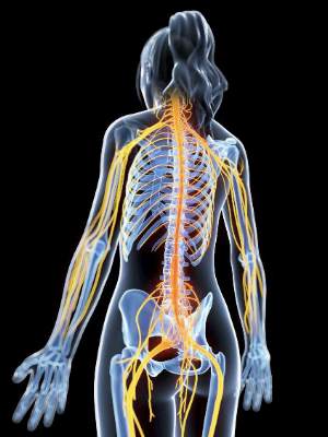
Lower GABA levels mean greater physical disability for MS patients
Physical disabilities in patients with progressive multiple sclerosis may be correlated with lowered gamma-aminobutyric acid (GABA) concentrations, according to Dr. Niamh Cawley of Queen Square Multiple Sclerosis Centre, University College London Institute of Neurology.
In the patient group of 30 individuals with secondary progressive MS, GABA concentrations were 0.403 mmol lower in the hippocampus than in the control group, and were 0.385 mmol lower in the sensorimotor cortex. There was no significant difference in prefrontal cortex GABA levels between the patient group and the control group (Brain. 2015 Sep;138[pt 9]:2584-95. doi: 10.1093/brain/awv209).

A decreased GABA concentration in the sensorimotor cortex was associated with diminished motor function in right upper and lower limbs, and the patient group scored worse than did the control group on all motor and sensory tests performed. Grip strength dropped by nearly 11 (kg force) for each GABA-unit decrease, and muscle strength in the right upper and lower limbs decreased by nearly 9.
“These findings raise the possibility that altered GABA neurotransmission may be a marker of neurodegeneration, but it may also suggest that GABA is a mechanism of neurodegeneration in progressive multiple sclerosis patients. If we put these findings together with the evidence that GABA may mediate neuroprotection, targeting GABA may be a productive strategy that should be further explored in multiple sclerosis,” wrote Dr. Cawley and her associates.
The work was funded by the Multiple Sclerosis Society of Great Britain and Northern Ireland, Philips Healthcare, and the U.K. National Institute for Health Research.
Read the full study in Brain.
Physical disabilities in patients with progressive multiple sclerosis may be correlated with lowered gamma-aminobutyric acid (GABA) concentrations, according to Dr. Niamh Cawley of Queen Square Multiple Sclerosis Centre, University College London Institute of Neurology.
In the patient group of 30 individuals with secondary progressive MS, GABA concentrations were 0.403 mmol lower in the hippocampus than in the control group, and were 0.385 mmol lower in the sensorimotor cortex. There was no significant difference in prefrontal cortex GABA levels between the patient group and the control group (Brain. 2015 Sep;138[pt 9]:2584-95. doi: 10.1093/brain/awv209).
A decreased GABA concentration in the sensorimotor cortex was associated with diminished motor function in right upper and lower limbs, and the patient group scored worse than did the control group on all motor and sensory tests performed. Grip strength dropped by nearly 11 (kg force) for each GABA-unit decrease, and muscle strength in the right upper and lower limbs decreased by nearly 9.
“These findings raise the possibility that altered GABA neurotransmission may be a marker of neurodegeneration, but it may also suggest that GABA is a mechanism of neurodegeneration in progressive multiple sclerosis patients. If we put these findings together with the evidence that GABA may mediate neuroprotection, targeting GABA may be a productive strategy that should be further explored in multiple sclerosis,” wrote Dr. Cawley and her associates.
The work was funded by the Multiple Sclerosis Society of Great Britain and Northern Ireland, Philips Healthcare, and the U.K. National Institute for Health Research.
Read the full study in Brain.
Blood-brain barrier permeability predicts MS in optic neuritis patients
Permeability of the blood-brain barrier may provide prognostic information for later multiple sclerosis in patients with optic neuritis, according to Dr. Stig P. Cramer of Rigshospitalet in Glostrup, Denmark.
In a study of 39 patients with monosymptomatic optic neuritis and 18 controls, investigators used dynamic contrast-enhanced MRI to measure blood-brain barrier permeability. MRI and lumbar puncture were performed within 4 weeks of onset, and information on multiple sclerosis (MS) conversion was acquired from hospital records 2 years later.
A multivariate logistic regression analysis found that baseline white matter permeability significantly improved prediction of MS, compared with T2 lesion count alone (P = .007). A receiver operating characteristic curve analysis of permeability in normal-appearing white matter gave a cut-off of 0.13 mL/100 g per min, which predicted conversion to MS with a sensitivity of 88% and specificity of 72%, the authors reported (Brain 2015:138;2571-83).
The findings suggest that “a new marker of low-grade multiple sclerosis disease activity could have many useful applications, i.e. subclinical disease activity monitoring or identification of inadequate treatment response before the next relapse occurs,” Dr. Cramer and his colleagues wrote.
“Quantification of [blood-brain barrier] permeability may constitute an early prognostic factor in the pathogenesis of multiple sclerosis,” they added.
The Research Foundation of the Capital Region of Denmark, Foundation for Health Research, Biogen Idec, and the Danish Multiple Sclerosis Society funded the work.
Read the full report here.
Breastfeeding protects against postpartum MS relapse
New mothers with MS are less likely to have a relapse within 6 months of delivery if they breastfeed their babies exclusively for at least 2 months, according to the findings of a prospective study of 201 women published online Aug. 31 in JAMA Neurology.
“Our findings indicate that women with MS should be supported if they choose to breastfeed exclusively since it clearly does not increase the risk of postpartum relapse. Relapse in the first 6 months post partum may be diminished by exclusive breastfeeding, but once regular feedings are introduced, disease activity is likely to return,” wrote Dr. Kerstin Hellwig of Ruhr-University Bochum (Germany) and her colleagues (JAMA Neurol. 2015 Aug 31. doi: 10.1001/jamaneurol.2015.1806).

The effect of breastfeeding on postpartum MS relapse has been controversial. Some studies have found that exclusive breastfeeding for at least the first 2 months might be beneficial, while others – studies that “defined breastfeeding crudely and/or measured breastfeeding retrospectively,” the authors said – found no protective effect.
The 201 women in the study had relapsing-remitting MS for a median of about 4.5 years and had voluntarily enrolled while pregnant in the German MS and pregnancy registry. They completed a series of questionnaires during pregnancy and the first post partum year.
Overall, 120 women (59.7%) intended to breastfeed exclusively for at least 2 months; 42 (20.9%) combined breastfeeding with supplemental feedings within the first 2 months; and 39 (19.4%) did not breastfeed; 178 (88.6%) reported using disease-modifying therapies before pregnancy, most often glatiramer acetate or interferon beta.
Among the 81 women who did not breastfeed or who supplemented breastfeeding early on, 31 (38.3%) had an MS relapse within the first 6 post partum months, compared with 29 women (24.2%) among the 120 who breastfed their children exclusively (adjusted hazard ratio, 1.70; 95% confidence interval, 1.02-2.85; P = 0.04).
The researchers described exclusively breastfeeding as acting like a “modestly effective treatment with a natural end date.”
“During exclusive breastfeeding, the pulsatile release of gonadotropin-releasing hormone and luteinizing hormone is suppressed with a corresponding suppression of the growth of ovarian follicles resulting in lactational amenorrhea and anovulation. Shortly after the breastfeeding frequency is reduced (1 or 2 regularly replaced breastfeeding meals are sufficient to interrupt this cycle), the ovarian activity resumes with the return of menses,” Dr. Hellwig and her associates wrote.
They also speculated that the “hormonal changes leading to anovulation might play a key role since women with MS are less likely to receive the diagnosis during their anovulatory years (childhood or after menopause) and women with MS were found to be more likely to experience relapse shortly before menstruation.”
The mean age in the study was about 31 years, but women who breastfed exclusively tended to be older than their peers and less likely to have received disease-modifying therapies before or at the time of conception. The first postpartum menses among exclusive breastfeeders came at a median of 185 days vs. 64 days for other women.
“In the present study, we observed that an earlier return of menses was associated with a higher risk of relapse in the first 6 months post-partum,” the authors wrote.
The work was funded by the German Research Foundation. The German MS and pregnancy registry was partly supported by Bayer Healthcare, Biogen Idec, Merck Serono, Novartis Pharma, and Genzyme Pharmaceuticals. Five of the researchers reported receiving speaker honoraria or other financial support from pharmaceutical companies.
New mothers with MS are less likely to have a relapse within 6 months of delivery if they breastfeed their babies exclusively for at least 2 months, according to the findings of a prospective study of 201 women published online Aug. 31 in JAMA Neurology.
“Our findings indicate that women with MS should be supported if they choose to breastfeed exclusively since it clearly does not increase the risk of postpartum relapse. Relapse in the first 6 months post partum may be diminished by exclusive breastfeeding, but once regular feedings are introduced, disease activity is likely to return,” wrote Dr. Kerstin Hellwig of Ruhr-University Bochum (Germany) and her colleagues (JAMA Neurol. 2015 Aug 31. doi: 10.1001/jamaneurol.2015.1806).
The effect of breastfeeding on postpartum MS relapse has been controversial. Some studies have found that exclusive breastfeeding for at least the first 2 months might be beneficial, while others – studies that “defined breastfeeding crudely and/or measured breastfeeding retrospectively,” the authors said – found no protective effect.
The 201 women in the study had relapsing-remitting MS for a median of about 4.5 years and had voluntarily enrolled while pregnant in the German MS and pregnancy registry. They completed a series of questionnaires during pregnancy and the first post partum year.
Overall, 120 women (59.7%) intended to breastfeed exclusively for at least 2 months; 42 (20.9%) combined breastfeeding with supplemental feedings within the first 2 months; and 39 (19.4%) did not breastfeed; 178 (88.6%) reported using disease-modifying therapies before pregnancy, most often glatiramer acetate or interferon beta.
Among the 81 women who did not breastfeed or who supplemented breastfeeding early on, 31 (38.3%) had an MS relapse within the first 6 post partum months, compared with 29 women (24.2%) among the 120 who breastfed their children exclusively (adjusted hazard ratio, 1.70; 95% confidence interval, 1.02-2.85; P = 0.04).
The researchers described exclusively breastfeeding as acting like a “modestly effective treatment with a natural end date.”
“During exclusive breastfeeding, the pulsatile release of gonadotropin-releasing hormone and luteinizing hormone is suppressed with a corresponding suppression of the growth of ovarian follicles resulting in lactational amenorrhea and anovulation. Shortly after the breastfeeding frequency is reduced (1 or 2 regularly replaced breastfeeding meals are sufficient to interrupt this cycle), the ovarian activity resumes with the return of menses,” Dr. Hellwig and her associates wrote.
They also speculated that the “hormonal changes leading to anovulation might play a key role since women with MS are less likely to receive the diagnosis during their anovulatory years (childhood or after menopause) and women with MS were found to be more likely to experience relapse shortly before menstruation.”
The mean age in the study was about 31 years, but women who breastfed exclusively tended to be older than their peers and less likely to have received disease-modifying therapies before or at the time of conception. The first postpartum menses among exclusive breastfeeders came at a median of 185 days vs. 64 days for other women.
“In the present study, we observed that an earlier return of menses was associated with a higher risk of relapse in the first 6 months post-partum,” the authors wrote.
The work was funded by the German Research Foundation. The German MS and pregnancy registry was partly supported by Bayer Healthcare, Biogen Idec, Merck Serono, Novartis Pharma, and Genzyme Pharmaceuticals. Five of the researchers reported receiving speaker honoraria or other financial support from pharmaceutical companies.
FROM JAMA NEUROLOGY
Key clinical point: Don’t discourage new mothers with multiple sclerosis from breastfeeding.
Major finding: Among 81 women who did not breastfeed or who supplemented breastfeeding early on, 31 (38.3%) had an MS relapse within the first 6 postpartum months, compared with 29 women (24.2%) among the 120 who intended to breastfeed their children exclusively for at least 2 months (adjusted HR, 1.70).
Data source: A prospective study of 201 pregnant women with relapsing-remitting MS who were followed for 1 year post partum.
Disclosures: The work was funded by the German Research Foundation. The German MS and pregnancy registry was partly supported by Bayer Healthcare, Biogen Idec, Merck Serono, Novartis Pharma, and Genzyme Pharmaceuticals. Five of the researchers reported receiving speaker honoraria or other financial support from pharmaceutical companies.

PML Observed in Three Patients With MS Treated With Fingolimod
For the first time, progressive multifocal leukoencephalopathy (PML) has been reported in patients with multiple sclerosis (MS) treated with fingolimod who had not previously been treated with an immunosuppressant, according to an FDA warning issued on August 4.
The FDA described two cases of PML—one is considered definite and the second is considered probable—in patients who had not received prior treatment with an immunosupressant drug for MS or any other medical condition. Prior cases of PML in patients taking fingolimod, including a case in a European patient in 2013, were confounded by immunosuppressant use before or concurrent with the drug.
In addition, Novartis, the manufacturer of the drug, reported on August 17 that it had been informed of another case of PML in a patient who did not have prior exposure to natalizumab. This patient has a history of colorectal cancer that was treated with chemotherapy and radiation treatment, and Crohn’s disease.
Clinicians who suspect that a patient on fingolimod may have PML should stop treatment and evaluate the patient for a diagnosis of PML, according to the FDA. People on fingolimod should contact their health care professionals immediately “if they experience symptoms such as new or worsening weakness; increased trouble using their arms or legs; or changes in thinking, eyesight, strength, or balance,” said the agency.
Symptoms in the patient with definite PML, a 54-year-old who had had MS for about 14 years and had been treated with fingolimod for about 2.5 years, included instability while walking, clumsiness, inattention, somnolence, and mental sluggishness. John Cunningham virus (JCV) DNA was present in the patient’s CSF, and MRI findings were characteristic of PML. The probable case was in a 49-year-old who had been treated with fingolimod for about four years and had no symptoms, but had new MRI lesions consistent with PML and a CSF test that was positive for JCV DNA.
Fingolimod, taken once per day by mouth, is a sphingosine 1-phosphate receptor modulator marketed by Novartis as Gilenya. It was approved in 2010 and is indicated for treating relapsing forms of MS to reduce the frequency of clinical exacerbations and to delay the accumulation of physical disability. The fingolimod prescribing information and Medication Guide are being updated to include information about cases of PML. Possible cases of PML associated with fingolimod should be reported to the FDA’s MedWatch program website or by calling 1-800-332-1088.
—Elizabeth Mechcatie
Are Effective Therapies for Progressive MS on the Horizon?
INDIANAPOLIS—Although many drugs have failed in clinical trials to provide benefits to patients with progressive multiple sclerosis (MS), neurologists have reason to be optimistic about the quest for effective therapies, according to an overview presented at the 2015 CMSC Annual Meeting. Ongoing studies are investigating agents that appear to offer neuroprotection. An international collaboration is helping to advance research into mesenchymal stem cells (MSCs), which may promote remyelination. “There are lots of things we can look at when we think about how we would approach progressive MS,” said Alan J. Thompson, MD, consultant neurologist at the National Hospital for Neurology and Neurosurgery in London.
Agents That May Offer Neuroprotection
One approach to providing neuroprotection is to block sodium channels on the axon and on the microglia. This technique has proven effective in animal models, but human trials have yielded mixed results. In 2010, Kapoor et al randomized 120 patients with secondary progressive MS to 400 mg of lamotrigine, a sodium-channel blocker, or placebo. At two years, participants who received lamotrigine had greater cerebral volume loss than controls. Brain volume partly recovered when treatment was stopped, however.
When the researchers performed a post hoc analysis of their data, they made two observations that supported a benefit of lamotrigine. The first was that lamotrigine was associated with improvements on the timed 25-foot walk. The other observation was that when patients who stopped taking lamotrigine were excluded from the analysis, lamotrigine reduced serum neurofilament levels, compared with placebo. Serum neurofilament levels were correlated with disability, and the researchers concluded that lamotrigine might protect axons against degeneration. “There’s some indication that this [drug] needs to be pursued a little bit further,” said Dr. Thompson.
Kapoor et al later studied phenytoin, a neuroprotective agent with mechanism of action similar to that of lamotrigine, in a phase II randomized controlled trial of 86 patients with acute optic neuritis. The trial’s primary end point was retinal nerve fiber layer thickness. After six months, participants who received phenytoin had a 30% reduction in atrophy, compared with controls.
Biotin, a coenzyme involved in fatty acid synthesis and energy production, also has shown potential as a neuroprotective agent. In a phase III study of 154 patients with primary and secondary progressive MS, biotin met the primary end point of improvement on EDSS or timed 25-foot walk at nine months that was confirmed at 12 months. The drug’s effect on EDSS score was the greater contributor to the positive result. The data are “exciting,” but should be interpreted cautiously because of the study’s relatively small number of patients, according to Dr. Thompson. The results of a trial of biotin in patients with optic neuritis will be reported later this year, he added.
Ongoing Trials of Neuroprotective Agents
Research into agents that may provide neuroprotection is ongoing. One study, the MS-Secondary Progressive Multi-Arm Randomization Trial (MS-SMART), uses an adaptive trial design that will allow investigators to compare several agents at the same time. In MS-SMART, equal numbers of participants will receive amiloride (5 mg bid), riluzole (50 mg bid), fluoxetine (20 mg bid), or placebo.
Amiloride blocks the neuronal proton-gated acid-sensing ion channel, which is increased within axons and oligodendrocytes in MS lesions. Researchers at the University of Oxford in the United Kingdom found that the drug reduced the rates of atrophy and white and gray matter damage in people with primary progressive MS.
Riluzole is a treatment for motor neuron disease. A Dutch pilot study in 2005 suggested that riluzole reduced the rate of cervical cord atrophy and decreased the development of new T1 hypointense lesions on MRI.
Fluoxetine, a selective serotonin reuptake inhibitor, is an antidepressant. The drug might protect against axonal loss because it stimulates glycogenolysis, which is a necessary energy source for axons, and enhances the production of brain-derived neurotrophic factor in rodent astrocyte cultures. Recruitment into MS-SMART will continue through 2015.
In addition, the Secondary and Primary Progressive Ibudilast NeuroNEXT Trial in MS (SPRINT-MS) will examine the potential neuroprotective effects of ibudilast. The randomized, placebo-controlled study will last for 96 weeks and focus on ibudilast’s effect on whole-brain atrophy. Secondary end points will include diffusion tensor imaging of descending pyramidal tracts, magnetization transfer ratio of the whole brain, and optical coherence tomography of the retinal nerve fiber layer.
Stem Cells Could Promote Remyelination
Neurologists will continue to seek therapies that can stimulate remyelination in patients with progressive MS. Various studies since 2009 have suggested that MSCs could produce this outcome. Despite their small patient populations, these trials can produce meaningful results, said Dr. Thompson.
He and his colleagues published a proof-of-concept study of autologous MSCs for the treatment of MS in Lancet Neurology in 2012. They examined 10 patients with secondary progressive MS and previous optic neuritis before and after the latter received MSCs. The treatment significantly improved visual acuity, and the researchers found evidence that it might promote remyelination.
Organizations including the Consortium of MS Centers have supported the International MSC Transplantation Study Group, which will guide future research into MSCs. The study group published a consensus paper on MSCs in Multiple Sclerosis in 2010. The consensus paper drew on the current literature to set guidelines for phase I and phase II clinical trials of MSCs in patients with MS. “Useful collaborative work going on in this area [provides] a lot of cause for optimism,” said Dr. Thompson.
—Erik Greb
Suggested Reading
Arun T, Tomassini V, Sbardella E, et al. Targeting ASIC1 in primary progressive multiple sclerosis: evidence of neuroprotection with amiloride. Brain. 2013;136(Pt 1):106-115.
Connick P, Kolappan M, Crawley C, et al. Autologous mesenchymal stem cells for the treatment of secondary progressive multiple sclerosis: an open-label phase 2a proof-of-concept study. Lancet Neurol. 2012;11(2):150-156.
Gnanapavan S, Grant D, Morant S, et al. Biomarker report from the phase II lamotrigine trial in secondary progressive MS - neurofilament as a surrogate of disease progression. PLoS One. 2013;8(8):e70019.
Sedel F, Papeix C, Bellanger A, et al. High doses of biotin in chronic progressive multiple sclerosis: a pilot study. Mult Scler Relat Disord. 2015;4(2):159-169.
INDIANAPOLIS—Although many drugs have failed in clinical trials to provide benefits to patients with progressive multiple sclerosis (MS), neurologists have reason to be optimistic about the quest for effective therapies, according to an overview presented at the 2015 CMSC Annual Meeting. Ongoing studies are investigating agents that appear to offer neuroprotection. An international collaboration is helping to advance research into mesenchymal stem cells (MSCs), which may promote remyelination. “There are lots of things we can look at when we think about how we would approach progressive MS,” said Alan J. Thompson, MD, consultant neurologist at the National Hospital for Neurology and Neurosurgery in London.
Agents That May Offer Neuroprotection
One approach to providing neuroprotection is to block sodium channels on the axon and on the microglia. This technique has proven effective in animal models, but human trials have yielded mixed results. In 2010, Kapoor et al randomized 120 patients with secondary progressive MS to 400 mg of lamotrigine, a sodium-channel blocker, or placebo. At two years, participants who received lamotrigine had greater cerebral volume loss than controls. Brain volume partly recovered when treatment was stopped, however.
When the researchers performed a post hoc analysis of their data, they made two observations that supported a benefit of lamotrigine. The first was that lamotrigine was associated with improvements on the timed 25-foot walk. The other observation was that when patients who stopped taking lamotrigine were excluded from the analysis, lamotrigine reduced serum neurofilament levels, compared with placebo. Serum neurofilament levels were correlated with disability, and the researchers concluded that lamotrigine might protect axons against degeneration. “There’s some indication that this [drug] needs to be pursued a little bit further,” said Dr. Thompson.
Kapoor et al later studied phenytoin, a neuroprotective agent with mechanism of action similar to that of lamotrigine, in a phase II randomized controlled trial of 86 patients with acute optic neuritis. The trial’s primary end point was retinal nerve fiber layer thickness. After six months, participants who received phenytoin had a 30% reduction in atrophy, compared with controls.
Biotin, a coenzyme involved in fatty acid synthesis and energy production, also has shown potential as a neuroprotective agent. In a phase III study of 154 patients with primary and secondary progressive MS, biotin met the primary end point of improvement on EDSS or timed 25-foot walk at nine months that was confirmed at 12 months. The drug’s effect on EDSS score was the greater contributor to the positive result. The data are “exciting,” but should be interpreted cautiously because of the study’s relatively small number of patients, according to Dr. Thompson. The results of a trial of biotin in patients with optic neuritis will be reported later this year, he added.
Ongoing Trials of Neuroprotective Agents
Research into agents that may provide neuroprotection is ongoing. One study, the MS-Secondary Progressive Multi-Arm Randomization Trial (MS-SMART), uses an adaptive trial design that will allow investigators to compare several agents at the same time. In MS-SMART, equal numbers of participants will receive amiloride (5 mg bid), riluzole (50 mg bid), fluoxetine (20 mg bid), or placebo.
Amiloride blocks the neuronal proton-gated acid-sensing ion channel, which is increased within axons and oligodendrocytes in MS lesions. Researchers at the University of Oxford in the United Kingdom found that the drug reduced the rates of atrophy and white and gray matter damage in people with primary progressive MS.
Riluzole is a treatment for motor neuron disease. A Dutch pilot study in 2005 suggested that riluzole reduced the rate of cervical cord atrophy and decreased the development of new T1 hypointense lesions on MRI.
Fluoxetine, a selective serotonin reuptake inhibitor, is an antidepressant. The drug might protect against axonal loss because it stimulates glycogenolysis, which is a necessary energy source for axons, and enhances the production of brain-derived neurotrophic factor in rodent astrocyte cultures. Recruitment into MS-SMART will continue through 2015.
In addition, the Secondary and Primary Progressive Ibudilast NeuroNEXT Trial in MS (SPRINT-MS) will examine the potential neuroprotective effects of ibudilast. The randomized, placebo-controlled study will last for 96 weeks and focus on ibudilast’s effect on whole-brain atrophy. Secondary end points will include diffusion tensor imaging of descending pyramidal tracts, magnetization transfer ratio of the whole brain, and optical coherence tomography of the retinal nerve fiber layer.
Stem Cells Could Promote Remyelination
Neurologists will continue to seek therapies that can stimulate remyelination in patients with progressive MS. Various studies since 2009 have suggested that MSCs could produce this outcome. Despite their small patient populations, these trials can produce meaningful results, said Dr. Thompson.
He and his colleagues published a proof-of-concept study of autologous MSCs for the treatment of MS in Lancet Neurology in 2012. They examined 10 patients with secondary progressive MS and previous optic neuritis before and after the latter received MSCs. The treatment significantly improved visual acuity, and the researchers found evidence that it might promote remyelination.
Organizations including the Consortium of MS Centers have supported the International MSC Transplantation Study Group, which will guide future research into MSCs. The study group published a consensus paper on MSCs in Multiple Sclerosis in 2010. The consensus paper drew on the current literature to set guidelines for phase I and phase II clinical trials of MSCs in patients with MS. “Useful collaborative work going on in this area [provides] a lot of cause for optimism,” said Dr. Thompson.
—Erik Greb
Suggested Reading
Arun T, Tomassini V, Sbardella E, et al. Targeting ASIC1 in primary progressive multiple sclerosis: evidence of neuroprotection with amiloride. Brain. 2013;136(Pt 1):106-115.
Connick P, Kolappan M, Crawley C, et al. Autologous mesenchymal stem cells for the treatment of secondary progressive multiple sclerosis: an open-label phase 2a proof-of-concept study. Lancet Neurol. 2012;11(2):150-156.
Gnanapavan S, Grant D, Morant S, et al. Biomarker report from the phase II lamotrigine trial in secondary progressive MS - neurofilament as a surrogate of disease progression. PLoS One. 2013;8(8):e70019.
Sedel F, Papeix C, Bellanger A, et al. High doses of biotin in chronic progressive multiple sclerosis: a pilot study. Mult Scler Relat Disord. 2015;4(2):159-169.

Researchers Seek Drugs to Promote Remyelination in Progressive MS
INDIANAPOLIS—Investigators are conducting clinical trials of various drugs that may slow clinical worsening in patients with progressive multiple sclerosis (MS), according to an overview provided at the 2015 CMSC Annual Meeting. Two such agents, anti-LINGO-1 antibody and rHIgM22 monoclonal antibody, are under active development in early clinical trials.
“Treatments that promote remyelination are going to prove to be the most effective way to prevent or treat progressive MS,” said Dennis Bourdette, MD, Roy and Eulalia Swank Family Research Professor and Chairman of the Department of Neurology at Oregon Health & Science University (OHSU) School of Medicine in Portland. Investigators are developing cell-based, large-molecule (ie, biologic), and small-molecule (ie, chemically manufactured) therapies that could promote remyelination.
Cell-Based Therapies
One potential treatment is to inject oligodendrocyte precursor cells (OPCs) into the brain and spinal cord. These OPCs then could become oligodendrocytes and produce myelin. Researchers in New York state under the leadership of Drs. Steven Goldman and Burk Jubelt are preparing for a phase I trial of this technique. They plan to use embryonic OPCs and are working with the FDA to establish the appropriate controls for the study.
Current surgical technology should allow the team to inject human OPCs into the brain safely, said Dr. Bourdette. “Researchers have shown that human embryonic-derived OPCs injected into the brains of rodents that have hypomyelination caused by a genetic defect will migrate long distances and myelinate axons.” The goal is for the OPCs to migrate into areas of demyelination in the brain of people with MS and remyelinate axons.
But how far the OPCs will migrate and whether this technique will succeed are open questions, added Dr. Bourdette. The brain of a patient with MS already has OPCs, and adding more OPCs may not promote remyelination, he said. “It might be an approach that is particularly useful, though, in people who are disabled from large plaques in the cervical cord.”
Large-Molecule Approaches
Another approach to promoting remyelination is to block the protein LINGO, which inhibits the differentiation of OPCs into oligodendrocytes. Biogen has developed an anti-LINGO-1 monoclonal antibody that appeared to promote remyelination in patients with optic neuritis during a small phase II trial. Investigators currently are studying this monoclonal antibody in patients with relapsing MS.
A group of investigators at the Mayo Clinic have developed recombinant human IgM22 antibody that promotes OPC differentiation and remyelination in animal models. The drug’s safety has been assessed in a phase I trial. Because the anti-LINGO antibody and the human IgM22 are large molecules, they will not penetrate the brain easily, said Dr. Bourdette. Large doses of these medicines thus will be required to achieve a therapeutic effect.
Small Molecules in Development
Small molecules also could stimulate OPC differentiation. These drugs can be modified to enhance their ability to penetrate the CNS, in contrast with large molecules.
One class of drugs that researchers are studying is thyromimetic medicines, which seek to simulate the effects of thyroid hormone. Thyroid hormone is necessary for OPCs to differentiate into oligodendrocytes and to promote myelination. Patients with hypothyroidism do not produce the normal amount of myelin. Investigators use thyroid hormone to promote OPC differentiation in vitro, and the drug is a potential treatment for patients with MS.
Peter Calabresi, MD, Director of the Division of Neuroimmunology at Johns Hopkins University in Baltimore, and colleagues are preparing to begin a trial of thyroid hormone for promoting remyelination in patients with MS. Although the approach seems promising, “the problem is that you may have to make patients thyrotoxic in order to get enough of thyroid to be beneficial,” said Dr. Bourdette. Thyroid hormone also can be cardiotoxic.
Thyromimetic drugs may represent a better approach, said Dr. Bourdette. These drugs are modifications of the thyroid hormone that have preferential activity for the thyroid hormone receptors on oligodendrocytes, which differ from those on the heart. Investigators thus can make thyromimetic drugs with differential affinities for the brain and OPC. Thomas Scanlan, PhD, Director of the Program in Chemical Biology at OHSU, and colleagues created a thyromimetic drug as a means of lowering cholesterol with minimal effects on the heart. This drug can stimulate OPCs. Researchers at OHSU now are studying the agent in a mouse model of demyelination. “The drug does accelerate remyelination in the mouse model, but not as well as the thyroid hormone,” said Dr. Bourdette. The investigators hypothesize that the thyromimetic drug does not penetrate the brain effectively, and they are modifying the drug to improve its ability to enter the brain.
Other investigators are working on developing small-molecule drugs that can stimulate remyelination. There is thus reason to be optimistic that the small-molecule approach to promoting remyelination will prove to be beneficial, said Dr. Bourdette. Providing a strong stimulus to the OPCs may allow them to overcome the toxic environment that they inhabit, he concluded.
—Erik Greb
Suggested Reading
Franklin RJ. Regenerative medicines for remyelination: from aspiration to reality. Cell Stem Cell. 2015;16(6):576-577.
Naim FJ, Madhavan M, Zaremba A, et al. Drug-based modulation of endogenous stem cells promotes functional remyelination in vivo. Nature. 2015;522(7555):216-220.
Olsen JA, Akirav EM. Remyelination in multiple sclerosis: cellular mechanisms and novel therapeutic approaches. J Neurosci Res. 2015;93(5):687-696.
Silvestroff L, Bartucci S, Pasquini J, Franco P. Cuprizone-induced demyelination in the rat cerebral cortex and thyroid hormone effects on cortical remyelination. Exp Neurol. 2012;235(1):357-367.
INDIANAPOLIS—Investigators are conducting clinical trials of various drugs that may slow clinical worsening in patients with progressive multiple sclerosis (MS), according to an overview provided at the 2015 CMSC Annual Meeting. Two such agents, anti-LINGO-1 antibody and rHIgM22 monoclonal antibody, are under active development in early clinical trials.
“Treatments that promote remyelination are going to prove to be the most effective way to prevent or treat progressive MS,” said Dennis Bourdette, MD, Roy and Eulalia Swank Family Research Professor and Chairman of the Department of Neurology at Oregon Health & Science University (OHSU) School of Medicine in Portland. Investigators are developing cell-based, large-molecule (ie, biologic), and small-molecule (ie, chemically manufactured) therapies that could promote remyelination.
Cell-Based Therapies
One potential treatment is to inject oligodendrocyte precursor cells (OPCs) into the brain and spinal cord. These OPCs then could become oligodendrocytes and produce myelin. Researchers in New York state under the leadership of Drs. Steven Goldman and Burk Jubelt are preparing for a phase I trial of this technique. They plan to use embryonic OPCs and are working with the FDA to establish the appropriate controls for the study.
Current surgical technology should allow the team to inject human OPCs into the brain safely, said Dr. Bourdette. “Researchers have shown that human embryonic-derived OPCs injected into the brains of rodents that have hypomyelination caused by a genetic defect will migrate long distances and myelinate axons.” The goal is for the OPCs to migrate into areas of demyelination in the brain of people with MS and remyelinate axons.
But how far the OPCs will migrate and whether this technique will succeed are open questions, added Dr. Bourdette. The brain of a patient with MS already has OPCs, and adding more OPCs may not promote remyelination, he said. “It might be an approach that is particularly useful, though, in people who are disabled from large plaques in the cervical cord.”
Large-Molecule Approaches
Another approach to promoting remyelination is to block the protein LINGO, which inhibits the differentiation of OPCs into oligodendrocytes. Biogen has developed an anti-LINGO-1 monoclonal antibody that appeared to promote remyelination in patients with optic neuritis during a small phase II trial. Investigators currently are studying this monoclonal antibody in patients with relapsing MS.
A group of investigators at the Mayo Clinic have developed recombinant human IgM22 antibody that promotes OPC differentiation and remyelination in animal models. The drug’s safety has been assessed in a phase I trial. Because the anti-LINGO antibody and the human IgM22 are large molecules, they will not penetrate the brain easily, said Dr. Bourdette. Large doses of these medicines thus will be required to achieve a therapeutic effect.
Small Molecules in Development
Small molecules also could stimulate OPC differentiation. These drugs can be modified to enhance their ability to penetrate the CNS, in contrast with large molecules.
One class of drugs that researchers are studying is thyromimetic medicines, which seek to simulate the effects of thyroid hormone. Thyroid hormone is necessary for OPCs to differentiate into oligodendrocytes and to promote myelination. Patients with hypothyroidism do not produce the normal amount of myelin. Investigators use thyroid hormone to promote OPC differentiation in vitro, and the drug is a potential treatment for patients with MS.
Peter Calabresi, MD, Director of the Division of Neuroimmunology at Johns Hopkins University in Baltimore, and colleagues are preparing to begin a trial of thyroid hormone for promoting remyelination in patients with MS. Although the approach seems promising, “the problem is that you may have to make patients thyrotoxic in order to get enough of thyroid to be beneficial,” said Dr. Bourdette. Thyroid hormone also can be cardiotoxic.
Thyromimetic drugs may represent a better approach, said Dr. Bourdette. These drugs are modifications of the thyroid hormone that have preferential activity for the thyroid hormone receptors on oligodendrocytes, which differ from those on the heart. Investigators thus can make thyromimetic drugs with differential affinities for the brain and OPC. Thomas Scanlan, PhD, Director of the Program in Chemical Biology at OHSU, and colleagues created a thyromimetic drug as a means of lowering cholesterol with minimal effects on the heart. This drug can stimulate OPCs. Researchers at OHSU now are studying the agent in a mouse model of demyelination. “The drug does accelerate remyelination in the mouse model, but not as well as the thyroid hormone,” said Dr. Bourdette. The investigators hypothesize that the thyromimetic drug does not penetrate the brain effectively, and they are modifying the drug to improve its ability to enter the brain.
Other investigators are working on developing small-molecule drugs that can stimulate remyelination. There is thus reason to be optimistic that the small-molecule approach to promoting remyelination will prove to be beneficial, said Dr. Bourdette. Providing a strong stimulus to the OPCs may allow them to overcome the toxic environment that they inhabit, he concluded.
—Erik Greb
Suggested Reading
Franklin RJ. Regenerative medicines for remyelination: from aspiration to reality. Cell Stem Cell. 2015;16(6):576-577.
Naim FJ, Madhavan M, Zaremba A, et al. Drug-based modulation of endogenous stem cells promotes functional remyelination in vivo. Nature. 2015;522(7555):216-220.
Olsen JA, Akirav EM. Remyelination in multiple sclerosis: cellular mechanisms and novel therapeutic approaches. J Neurosci Res. 2015;93(5):687-696.
Silvestroff L, Bartucci S, Pasquini J, Franco P. Cuprizone-induced demyelination in the rat cerebral cortex and thyroid hormone effects on cortical remyelination. Exp Neurol. 2012;235(1):357-367.
Tissue-Loss Markers
Gray matter fraction (GMF) change is more meaningful than white matter fraction (WMF) as a marker of tissue loss and may be useful to augment whole brain atrophy measurements in MS clinical trials, according to a study of 140 patients with relapsing-remitting MS who were randomized to intramuscular interferon (IFN) beta-1a or placebo. Researchers found:
• Significantly less GM atrophy (during year 2), but not WM atrophy (at any point), was seen with IFN beta-1a compared with placebo.
• Pseudoatrophy effects were more apparent in WM than in GM; in year 1, greater WM volume loss was seen with IFN beta-1a than with placebo, but GM volume loss was similar between groups.
• Risk of sustained disability progression was significantly associated with GM atrophy, but not WM atrophy.
Citation: Fisher E, Nakamura K, Lee JC, You X, Sperling B, Rudick RA. Effect of intramuscular interferon beta-1a on gray matter atrophy in relapsing−remitting multiple sclerosis: a retrospective analysis. [Published online ahead of print August 3, 2015]. Mult Scler. doi: 10.1177/1352458515599072.
Commentary: Understanding a disease requires thorough analysis of its impact. Multiple sclerosis is classically known as a white matter disease. It is also known that MS white matter plaques are in typical subcortical locations. This has been the tenet of MS care and diagnosis by MRI. However, it is also a tenet that MRI changes do not always correlate with clinical exam or disease impact. Many people involved in MS care and research have explored the reason for this discrepancy. This important article is another step along the path of understanding. MS plaques have now been identified within the white matter of the gray matter. This article provides more evidence that the disease impacts gray matter as well as white matter. The atrophy in gray matter correlates more with disease impact and burden than the atrophy in white matter. MS should no longer be considered a disease of white matter or white matter atrophy. Perhaps it’s time to update our diagnostic approach, monitoring of disease impact and progression, and therapeutic intervention efficacy measures as well as the metrics of how we measure disease impact. —Mark Gudesblatt, MD, Medical Director of the Comprehensive MS Care Center at South Shore Neurologic Associates in Islip, NY
Patient Characteristics as Prognosticators
Patient characteristics that may help predict disease outcomes include number of relapses in the past 2 years, new MRI lesions in year 1, higher number of relapses in year 1, MRI activity, and the presence of neutralizing antibodies, according to a study of 857 patients with relapsing-remitting MS who were treated with interferon beta-1b. All 857 patients were included in the analysis of clinical outcomes and 765 of those were included in the MRI outcomes analysis. Researchers found:
• Future occurrence of relapses was predicted by a higher number of relapses in the past 2 years, ≥ 3 new MRI lesions in year 1, and, especially, a higher number of relapses in year 1.
• Future MRI activity was principally predicted by age, MRI activity, and the presence of neutralizing antibodies in year 1.
Citation: Hartung HP, Kappos L, Goodin DS, et al. Predictors of disease activity in 857 patients with MS treated with interferon beta-1b. [Published online ahead of print August 5, 2015]. J Neurol. doi: 10.1007/s00415-015-7862-9.
Lipoprotein Levels in BBB Injury
Higher serum high-density lipoprotein (HDL) is associated with lower levels of blood brain barrier (BBB) injury and decreased CD80+ and CD80+CD19+ cell extravasation into the cerebrospinal fluid (CSF), according to a study of 154 patients with MS who were receiving interferon beta-1a therapy following their first clinical demyelinating event. Researchers found:
• Higher levels of serum HDL cholesterol (HDL-C) and apolipoprotein A-1 were associated with lower CSF total protein level, CSF albumin level, albumin quotient, and CSF IgC level.
• HDL-C was also associated with CSF CD80+ amd with CSF CD80+CD19+ cell frequencies.
Citation: Fellows K, Uher T, Browne RW, et al. Protective associations of HDL with blood brain barrier injury in multiple sclerosis patients. [Published online ahead of print August 4, 2015]. J Lipid Res. doi: 10.1194/jlr.M060970.
Alcohol and Smoking in MS
Alcohol dependence and smoking were associated with anxiety and depression, according to a study of 949 patients with MS. Researchers found:
• Alcohol dependence was associated with an increased odds ratio of anxiety (OR=1.84) and depression (OR=1.53).
• Smoking was associated with increased odds of anxiety (OR=1.29) and depression (OR=1.37).
• Alcohol dependence was associated with increased incidence of depression but not anxiety.
• Depression was associated with increased incidence of alcohol dependence.
Citation: McKay KA, Tremlett H, Fisk JD, et al. Adverse health behaviours are associated with depression and anxiety in multiple sclerosis: a prospective multisite study. [Published online ahead of print August 5, 2015]. Mult Scler. doi: 10.1177/1352458515599073.