User login
Patients with Ph-like ALL may benefit from TKIs
Credit: St Jude Children’s
Research Hospital
New research indicates that Philadelphia chromosome-like acute lymphoblastic leukemia (Ph-like ALL) becomes more common with age and is associated with poor prognosis.
The study also showed that Ph-like ALL is characterized by genomic alterations that might make patients receptive to treatment with tyrosine kinase inhibitors (TKIs).
Initial tests in a small number of patients seem to support this theory, but trials are needed to verify and expand upon these results, according to researchers.
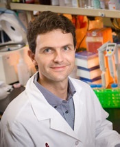
Charles Mullighan, MD, MBBS, of the St Jude Children’s Research Hospital in Memphis, Tennessee, and his colleagues reported the results in The New England Journal of Medicine.
Age and prognosis
The researchers performed genomic profiling of 1725 patients with precursor B-cell ALL, detailed genomic analyses of 154 patients with Ph-like ALL, and transcriptome sequencing for 160 patients with non-Ph-like ALL.
The team found the prevalence of Ph-like ALL increased significantly with age, from 10% among children with standard-risk B-ALL (ages 1 to 9) and 13% among those with high-risk ALL (ages 10 to 15) to 21% among adolescents (ages 16 to 20) and 27% among young adults with ALL (ages 21 to 39).
Regardless of their age, patients with Ph-like ALL were less likely than other B-ALL patients to be alive and leukemia-free 5 years after diagnosis.
Overall survival for children, adolescents, and young adults with Ph-like ALL was 62%, compared to 91% for other B-ALL patients of the same age. Leukemia-free survival was about 47% for patients with Ph-like ALL and about 83% for other patients.
Genomic alterations and TKI treatment
The researchers found that 91% of patients with Ph-like ALL had chromosomal rearrangements or other genetic alterations that activate cytokine receptor or kinase signaling.
“We identified several new subgroups of Ph-like ALL that were distinguished by the type of cytokine receptor or kinase gene alteration,” said Kathryn Roberts, PhD, also of St Jude Children’s Research Hospital.
Evidence suggests that several of these subtypes would be vulnerable to TKIs and other targeted therapies. For example, about 12% of patients had rearrangements involving the genes ABL1, ABL2, CSF1R, and PDGFRB, which are known to respond to dasatinib and related TKIs.
Other Ph-like ALL patients had gene rearrangements involving JAK2, EPOR, and other genes that can be targeted by the drug ruxolitinib.
To determine if TKIs are effective in these patients, the researchers administered TKIs to 12 patients with Ph-ALL. Follow-up is not sufficient for all of the patients, but 5 achieved remission following TKI treatment (alone, with chemotherapy, or followed by transplant), and 1 patient has been in remission for more than a year.
“We showed that Ph-like ALL is a common disease that spans the age spectrum, and we identified new genomic alterations that converge on a handful of signaling pathways that are vulnerable to treatment with tyrosine kinase inhibitors,” Dr Mullighan said. “The findings lead the way for clinical trials that could help to transform the outlook for patients, regardless of age.”
A study testing TKI therapy in children with Ph-like ALL is scheduled to begin later this year or early in 2015. ![]()
Credit: St Jude Children’s
Research Hospital
New research indicates that Philadelphia chromosome-like acute lymphoblastic leukemia (Ph-like ALL) becomes more common with age and is associated with poor prognosis.
The study also showed that Ph-like ALL is characterized by genomic alterations that might make patients receptive to treatment with tyrosine kinase inhibitors (TKIs).
Initial tests in a small number of patients seem to support this theory, but trials are needed to verify and expand upon these results, according to researchers.
Charles Mullighan, MD, MBBS, of the St Jude Children’s Research Hospital in Memphis, Tennessee, and his colleagues reported the results in The New England Journal of Medicine.
Age and prognosis
The researchers performed genomic profiling of 1725 patients with precursor B-cell ALL, detailed genomic analyses of 154 patients with Ph-like ALL, and transcriptome sequencing for 160 patients with non-Ph-like ALL.
The team found the prevalence of Ph-like ALL increased significantly with age, from 10% among children with standard-risk B-ALL (ages 1 to 9) and 13% among those with high-risk ALL (ages 10 to 15) to 21% among adolescents (ages 16 to 20) and 27% among young adults with ALL (ages 21 to 39).
Regardless of their age, patients with Ph-like ALL were less likely than other B-ALL patients to be alive and leukemia-free 5 years after diagnosis.
Overall survival for children, adolescents, and young adults with Ph-like ALL was 62%, compared to 91% for other B-ALL patients of the same age. Leukemia-free survival was about 47% for patients with Ph-like ALL and about 83% for other patients.
Genomic alterations and TKI treatment
The researchers found that 91% of patients with Ph-like ALL had chromosomal rearrangements or other genetic alterations that activate cytokine receptor or kinase signaling.
“We identified several new subgroups of Ph-like ALL that were distinguished by the type of cytokine receptor or kinase gene alteration,” said Kathryn Roberts, PhD, also of St Jude Children’s Research Hospital.
Evidence suggests that several of these subtypes would be vulnerable to TKIs and other targeted therapies. For example, about 12% of patients had rearrangements involving the genes ABL1, ABL2, CSF1R, and PDGFRB, which are known to respond to dasatinib and related TKIs.
Other Ph-like ALL patients had gene rearrangements involving JAK2, EPOR, and other genes that can be targeted by the drug ruxolitinib.
To determine if TKIs are effective in these patients, the researchers administered TKIs to 12 patients with Ph-ALL. Follow-up is not sufficient for all of the patients, but 5 achieved remission following TKI treatment (alone, with chemotherapy, or followed by transplant), and 1 patient has been in remission for more than a year.
“We showed that Ph-like ALL is a common disease that spans the age spectrum, and we identified new genomic alterations that converge on a handful of signaling pathways that are vulnerable to treatment with tyrosine kinase inhibitors,” Dr Mullighan said. “The findings lead the way for clinical trials that could help to transform the outlook for patients, regardless of age.”
A study testing TKI therapy in children with Ph-like ALL is scheduled to begin later this year or early in 2015. ![]()
Antifungal shows promise in hematologic conditions

WASHINGTON, DC—A new antifungal agent is as effective as an existing drug against invasive mold infection in patients with hematologic disorders, results of a phase 3 trial suggest.
Overall response and all-cause mortality rates were comparable with the newer drug, isavuconazole (ISA), and the existing drug, voriconazole (VRC).
The overall rates of treatment-emergent adverse events were comparable as well, but ISA was associated with a significantly lower incidence of several events.
Kieren Marr, MD, of Johns Hopkins University in Baltimore, Maryland, and her colleagues presented these results in a subset of patients from the SECURE trial at the 54th Interscience Conference on Antimicrobial Agents and Chemotherapy (poster M-1757).
Patient characteristics and safety outcomes
Of the 433 patients with a hematologic disorder enrolled in the trial, 217 had proven or probable invasive mold infection. The researchers divided patients into 2 groups according to disease: those with acute myeloid leukemia (AML) and those with acute lymphoblastic leukemia (ALL) or other conditions.
In all, 102 patients had AML, and 115 had ALL (n=28) or other conditions, including non-Hodgkin lymphoma (n=19), chronic lymphocytic leukemia (n=15), refractory anemia with excess blasts (n=9), myelodysplastic syndrome (n=8), chronic myeloid leukemia (n=6), and “other” underlying conditions (n=30).
Patients were randomized to receive VRC (n=105) or ISA (N=112). Thirty patients in the ISA arm and 26 in the VRC arm had undergone allogeneic transplant prior to therapy.
The primary outcome was all-cause mortality at day 42. Overall response and safety were secondary endpoints.
The overall rates of treatment-emergent adverse events were similar between ISA and VRC arms. Ninety-seven percent of patients in the ISA arm and 98% of patients in the VRC arm reported at least 1 treatment-emergent adverse event.
However, patients in the ISA arm had significantly fewer (P<0.05) events for the cardiac disorders, eye, psychiatric disorders, renal and urinary, and vascular system organ classes.
Response and mortality
All-cause mortality rates were comparable between the ISA and VRC arms—22% and 24%, respectively—as were overall response rates—39% and 34%, respectively—and complete response rates—13% and 10%, respectively.
All-cause mortality rates among patients with AML were 18% in the ISA arm and 15% in the VRC arm. Overall response rates were 36% and 48%, respectively.
For patients with ALL or other hematologic conditions, all-cause mortality rates were 26% in the ISA arm and 32% in the VRC arm. Overall response rates were 42% and 21%, respectively.
In transplant patients, the all-cause mortality rate was 27% for both the ISA and VRC arms. The overall response rate was 27% for both arms as well.
“These results show the potential of isavuconazole as a potent antifungal in the fight against invasive mold disease,” Dr Marr said.
ISA is an investigational antifungal under development by Astellas and Basilea Pharmaceutica International Ltd. The SECURE trial was funded by Astellas.
The US Food and Drug Administration recently accepted a new drug application seeking approval for ISA for the treatment of invasive aspergillosis and invasive mucormycosis. ![]()
Discovery could lead to better proteasome inhibitors

showing multiple myeloma
A newly discovered mechanism has paved the way for the next generation of proteasome inhibitors, according to a paper published in Chemistry & Biology.
Investigators developed a series of molecules that employ this mechanism, inhibiting the proteasome in 2 ways.
They are now planning to synthesize related compounds that may offer improved proteasome inhibition, target cancer cells more selectivity, and eliminate the resistance problems that occur with current drugs.
The group’s research began with epoxyketone, a molecule isolated from a cyanobacterium called carmaphycin, whose reactive group is the same as that of the proteasome inhibitor carfilzomib.
“Epoxyketones are very potent, selective inhibitors of the proteasome because they interact with this enzyme in 2 stages—the first reversible, and the second irreversible,” noted study author Daniela Trivella, PhD, of the Brazilian Biosciences National Laboratory at the Brazilian Center for Research in Energy and Materials in Campinas.
To optimize epoxyketone’s effects and find new reactive groups, the investigators developed and tested a series of synthetic analogs with slight structural modifications.
One of the molecules had an enone as a reactive group and had characteristics of carmaphycin and another natural molecule called syringolin, which was isolated from plant pathogens.
By investigating the reaction mechanisms of the new molecule, called carmaphycin-syringolin enone, the team verified that the enone interacts with the proteasome in 2 stages, with the second stage being irreversible.
The investigators also observed that, in the case of the enone, the second reaction occurs more slowly, increasing the duration of the reversible phase of carmaphycin-syringolin enone inhibition.
“Because the irreversible inactivation of the proteasome has toxic effects, the best window of reversibility observed for the carmaphycin-syringolin enone will potentially reduce the toxicity of this new class of proteasome inhibitors,” Dr Trivella said. “The compound would therefore present a balance between selectivity and potency.”
Toxicity tests are still underway. But the investigators have already conducted studies to determine exactly how the interaction between the enzyme target and the carmaphycin-syringolin enone target occurs.
“We discovered that a chemical reaction called hydroamination occurs, which had never before [been] seen under physiological conditions,” Dr Trivella said.
“This type of reaction is frequently used by synthetic chemists in preparing substances, but, normally, it requires very specific temperature and pH conditions and the use of catalysts to occur. It has never been reported as a mechanism of enzyme inhibition.”
Inspired by this new mechanism for proteasome inhibition, the investigators plan to synthesize and test a new series of carmaphycin-syringolin enone analogs to determine their effects on the therapeutic window and assess whether they are also capable of reacting with proteasomes that are resistant to traditional inhibitors. ![]()
Cancer centers may not allow for dignified deaths

Credit: NCI and
Mathews Media Group
A new study suggests many patients in cancer centers do not experience a dignified death.
Study investigators surveyed physicians and nurses in 16 hospitals belonging to 10 cancer centers in Baden-Württemberg, Germany.
The results revealed a need for cancer centers to invest more in palliative care services, adequate rooms for dying patients, staff training in end-of-life care, and advance-care-planning standards.
Karin Jors, of the University Medical Center Freiburg, and her colleagues reported these findings in Cancer.
Previous research has shown that hospitals are often ill-prepared to provide care for dying patients.
To investigate whether the circumstances for dying on cancer center wards allow for a dignified death, Jors and her colleagues surveyed physicians and nurses in German cancer centers.
Among 1131 survey respondents, 57% believed that patients could die with dignity on their ward.
Half of the surveyed staff members indicated that they rarely have enough time to care for dying patients, and 55% found the rooms available for dying patients unsatisfactory.
Only 19% of respondents felt they had been well-prepared to care for dying patients, and only 6% of physicians felt that way.
On the other hand, physicians perceived the circumstances for dying patients much more positively than nurses, especially regarding communication and life-prolonging measures.
While 72% of physicians reported that patients can usually die a dignified death on their ward, only 52% of nurses shared this opinion.
Palliative care staff reported much better conditions for dying patients than staff from other wards, with 95% of palliative care staff indicating that patients die with dignity on their wards.
“In our aging society, it is predicted that the number of hospital deaths will continue to rise in the coming years, and many of these deaths will be attributable to cancer,” Jors said.
“For this reason, it is particularly important that cancer centers strive to create a comfortable, dignified experience for dying patients and their families. Above all, this requires that staff members are provided with the adequate resources to care for these patients.”
The investigators therefore encourage the integration of palliative care into standard oncology care, beginning as early as diagnosis. They also believe physicians and nurses would benefit from increased education and training in end-of-life care. ![]()
Drug shows early promise for hematologic malignancies
A drug that targets mitochondrial function is largely safe and can be active in heavily pretreated patients with hematologic malignancies, a phase 1 trial indicates.
The drug, CPI-613, prompted responses in only 4 of 21 evaluable patients. However, 2 of those responses lasted more than 2 years.
CPI-613 was generally well-tolerated and did not induce bone marrow suppression. Four patients experienced renal failure, but it was reversed in 3 of them.
These results appear in Clinical Cancer Research.
“This drug is selectively taken up by cancer cells and then shuts down the production of energy in the mitochondria,” said study author Timothy Pardee, MD, PhD, of the Comprehensive Cancer Center of Wake Forest University in Winston-Salem, North Carolina.
“This is the first drug to inhibit mitochondria in this way, and, if it proves effective in further clinical trials, it will open up a whole new approach to fighting cancer.”
Dr Pardee and his colleagues evaluated CPI-613 in 26 patients with relapsed or refractory hematologic malignancies—11 with acute myeloid leukemia, 6 with non-Hodgkin lymphoma, 4 with multiple myeloma, 4 with myelodysplastic syndrome (MDS), and 1 with Hodgkin lymphoma.
The median patient age was 65 years (range, 19-81), and the median number of prior therapies was 3 (range, 1-9).
Treatment dosing and toxicity
Patients received CPI-613 as a 2-hour infusion on days 1 and 4 for 3 weeks every 28 days.
When the infusion time was shortened to 1 hour, renal failure occurred in 2 patients. At 3780 mg/m2, there were 2 dose-limiting toxicities. There were no such toxicities at a dose of 2940 mg/m2 over 2 hours, so this was considered the maximum-tolerated dose.
The following grade 2 or higher toxicities were probably or definitely related to treatment: nausea (1 grade 2), vomiting (1 grade 3), diarrhea (3 grade 2), proteinuria (1 grade 2), renal failure (4 grade 3), hypotension (1 grade 2), hypocalcemia (1 grade 2), hypoalbuminemia (1 grade 2), and hyperkalemia (1 grade 3).
Renal failure was resolved in 3 of the 4 patients. The remaining patient chose hospice care.
Response data
Five patients discontinued treatment—1 refused therapy, 1 acquired an infection, and 3 developed acute kidney failure.
Of the 21 patients evaluable for response, 4 had an objective response following CPI-613 treatment, and 2 had prolonged stable disease.
One patient with MDS achieved a complete response that has been maintained for more than 3 years. A patient with acute myeloid leukemia achieved a morphologically leukemia-free state, went on to transplant, and is still alive and leukemia-free.
A patient with Burkitt lymphoma achieved a partial response after 3 cycles of therapy that was maintained for 17 cycles. She discontinued CPI-613 to have her residual disease resected, and has not received any treatment since. She is now disease-free more than 12 months later.
A patient with cutaneous T-cell lymphoma achieved a partial response that has been sustained for more than 2 years. At her request, she started to receive continuous therapy (no 1-week rest period), and she remains on treatment without significant toxicities and no evidence of marrow suppression.
The 2 patients with prolonged stable disease had MDS. Their disease was stable for 8 and 12 cycles, respectively. Two patients with multiple myeloma also initially had stable disease, but they progressed after 2 and 4 cycles, respectively.
Two patients died from disease progression while on study.
The researchers said these results suggest that agents targeting mitochondrial metabolism can be safe and active in hematologic malignancies. A phase 2 trial of CPI-613 is now underway.
Support for the phase 1 trial was provided by National Cancer Institute grants P30CA012197 and 1K08CA169809, the Doug Coley Foundation for Leukemia Research, the Frances P. Tutwiler Fund, The MacKay Foundation for Cancer Research, and Cornerstone Pharmaceuticals, which manufactured and provided CPI-613. ![]()
A new treatment strategy for high-risk MDS/AML

Credit: Rhoda Baer
Preclinical research has revealed a potential therapeutic strategy for high-risk acute myeloid leukemia (AML) and myelodysplastic syndrome (MDS).
In experiments with human cells and mouse models of del(5q) AML/MDS, researchers found that an NF-κB signaling network fueled the survival and growth of leukemic cells.
But the team could inhibit this network by targeting p62, thereby impeding leukemic cell expansion and inducing apoptosis.
Daniel Starczynowski PhD, of Cincinnati Children’s Hospital Medical Center in Ohio, and his colleagues described this work in Cell Reports.
“Unfortunately, a large portion of del(5q) AML and MDS patients have an increased number of bone marrow blasts and additional chromosomal mutations,” Dr Starczynowski said.
“These patients have very poor prognosis because the disease is very resistant to available treatments such as chemotherapy and radiation. Finding new therapies is important, and this study identifies new therapeutic possibilities.”
Dr Starczynowski and his colleagues began this research by focusing on miR-146a, a microRNA previously shown to be involved in the pathogenesis of del(5q) MDS/AML.
The team found the loss of miR-146a in leukemic cells results in derepression of TRAF6, a mediator of NF-κB activation, which implicates this molecular complex in the aggressive nature of del(5q) MDS/AML.
So the researchers theorized that inhibiting the TRAF6/NF-κB axis might help treat aggressive forms of del(5q) MDS/AML with low miR-146a expression. The problem was that past attempts to directly inhibit NF-κB had not exactly proven successful.
Fortunately, chromosome deletions that target tumor suppressor genes also involve multiple neighboring genes. So the team examined the expression of all genes residing within chromosome 5q from del(5q) and control CD34+ cells, with the goal of finding a more suitable therapeutic target.
To determine which of the genes they identified are necessary for del(5q) leukemic cell function, the researchers knocked down each gene and examined leukemic progenitor function. They found that only knockdown of SQSTM1/p62 resulted in reduced colony formation.
So the team tested inhibition/knockdown of p62 as an experimental treatment strategy in miR-146alow MDS/AML cell lines, primary del(5q) AML samples, and mouse models of AML/MDS.
They found that targeting p62 reduced the number of leukemic cell colonies by 80% in human AML/MDS cells. And in mice, targeting p62 prevented the expansion of leukemic cells and significantly delayed mortality.
These results suggest that interfering with the p62-TRAF6 signaling complex represents a therapeutic option in miR-146a-deficient and aggressive del(5q) MDS/AML.
However, Dr Starczynowski noted that additional research is needed to further verify these findings and learn more about the molecular processes involved. ![]()
Deaths from childhood cancer on decline in UK

Credit: Logan Tuttle
The rate of children dying from cancer in the UK has dropped 22% in the last decade, according to new figures published by Cancer Research UK.
From 2001 to 2003, 328 children died from cancer each year. But from 2010 to 2012, the annual death toll from childhood cancers decreased to 258.
The steepest decline in mortality was among leukemia patients. Death rates across all forms of leukemia combined dropped by 47%, from 102 to 53 deaths each year.
For acute lymphoblastic leukemia, the annual mortality rate decreased by 52%, falling from 63 to 29 deaths per year. For acute myeloid leukemia, the death rate fell by 33%, from 30 to 20 deaths per year. And for chronic myeloid leukemia, the death rate decreased by 74%, from 2 deaths per year to 1.
Annual mortality rates decreased for lymphoma patients as well. For all lymphomas, the death rate decreased by 31%, falling from 15 to 11 deaths per year. And for non-Hodgkin lymphomas, the rate dropped 35%, from 14 to 10 deaths per year.
Much of this success is due to new combinations of chemotherapy drugs, but efforts to improve imaging and radiotherapy techniques has also played a part, according to Cancer Research UK.
“It’s very encouraging to see that fewer children are dying of cancer, but a lot more needs to be done,” said Pam Kearns, director of the Cancer Research UK Clinical Trials Unit in Birmingham.
“Many children who survive cancer will live with the long-term side effects of their treatment that can have an impact throughout their adult lives, so it’s vital that we find kinder and even more effective treatments for them.”
Around 1600 children are diagnosed with cancer every year in the UK. Overall survival for childhood cancer has tripled since the 1960s. The proportion of children surviving their cancer for at least 10 years increased from 24% in 1966-1970 to 76% in 2001-2005. ![]()
Note: The above figures are age-standardized mortality rates, which take the age and size of the population into account, providing a figure for the number of children who die from cancer per million individuals. Looking at the numbers of children dying from the disease does not adjust for the increasing size of the UK population over the last 10 years, so changes in the numbers of deaths will not match the changes in rates.
Credit: Logan Tuttle
The rate of children dying from cancer in the UK has dropped 22% in the last decade, according to new figures published by Cancer Research UK.
From 2001 to 2003, 328 children died from cancer each year. But from 2010 to 2012, the annual death toll from childhood cancers decreased to 258.
The steepest decline in mortality was among leukemia patients. Death rates across all forms of leukemia combined dropped by 47%, from 102 to 53 deaths each year.
For acute lymphoblastic leukemia, the annual mortality rate decreased by 52%, falling from 63 to 29 deaths per year. For acute myeloid leukemia, the death rate fell by 33%, from 30 to 20 deaths per year. And for chronic myeloid leukemia, the death rate decreased by 74%, from 2 deaths per year to 1.
Annual mortality rates decreased for lymphoma patients as well. For all lymphomas, the death rate decreased by 31%, falling from 15 to 11 deaths per year. And for non-Hodgkin lymphomas, the rate dropped 35%, from 14 to 10 deaths per year.
Much of this success is due to new combinations of chemotherapy drugs, but efforts to improve imaging and radiotherapy techniques has also played a part, according to Cancer Research UK.
“It’s very encouraging to see that fewer children are dying of cancer, but a lot more needs to be done,” said Pam Kearns, director of the Cancer Research UK Clinical Trials Unit in Birmingham.
“Many children who survive cancer will live with the long-term side effects of their treatment that can have an impact throughout their adult lives, so it’s vital that we find kinder and even more effective treatments for them.”
Around 1600 children are diagnosed with cancer every year in the UK. Overall survival for childhood cancer has tripled since the 1960s. The proportion of children surviving their cancer for at least 10 years increased from 24% in 1966-1970 to 76% in 2001-2005.
Note: The above figures are age-standardized mortality rates, which take the age and size of the population into account, providing a figure for the number of children who die from cancer per million individuals. Looking at the numbers of children dying from the disease does not adjust for the increasing size of the UK population over the last 10 years, so changes in the numbers of deaths will not match the changes in rates.
Blastic Plasmacytoid Dendritic Cell Neoplasm: A Case Successfully Treated With HyperCVAD Followed by Allogeneic Stem Cell Transplantation
Introduction: Blastic plasmacytoid dendritic cell neoplasm (BPDCN) is a rare, clinically aggressive tumor derived from the precursors of palsmacytoid dendritic cells with a high frequency of cutaneous and bone marrow involvement and leukaemic dissemination. The prognosis is poor, and an optimal treatment approach has not been defined. We describe a case of BPDCN that has been successfully treated with acute leukaemia-type induction (hyperCVAD) followed by allogeneic stem cell transplantation (ASCT).
Purpose: To evaluate the efficacy of hyperCVAD and ASCT in treatment of BPDCN.
Method: A case report and literature review. Result: An African American male, aged 49 years, presented with firm violaceous nodules on the left calf, back, and shoulders, accompanied by 40 lb weight loss. Patient did not experience night sweats, fevers, and chill. Complete blood cell count and serum lactate dehydrogenase were normal. A skin biopsy revealed dense dermal infiltrates consisting of intermediate-sized cells with high N:C ratio. The tumor cells were strongly positive for CD4, CD56; partially positive for TdT; weakly positive for CD45 and CD43; and negative for CD3, CD20, CD30, MPO, CD34, CD117. Bone marrow biopsy and aspirate smear showed hypercellular marrow with predominant blastic cells with high nuclear-cytoplasmic ratio, finely chromatin, and prominent nucleoli. Flow cytometric analysis demonstrated 85% blasts that were positive for HLA-DR, CD4, CD56, CD38, and TdT (partial); but negative for CD34, CD117, CD33, CD13, CD14, CD15, CD2, CD3, CD5, CD11c, CD7, CD19, CD10, CD20, CD22, CD24, Kappa, Lambda, CD25, CD52, and MPO. Cyotogenetic analysis reported an abnormal complex chromosome abnormality: 46, XY, add (7)(q22), add (8)(p11.2), add (9)(q13), psu dic(13:6)(p12;q16), del(13)(q12q22), -17, +21, +mar[9]. The peripheral blood smear revealed rare blasts. The patient responded well to hyperCVAD chemo followed by ASCT. He has remained disease free for > 5 years.
Conclusions: Aggressive chemotherapy followed by ASCT is a favorable treatment plan for BPDCN.
Liver grafts donated after circulatory death increase early risk of diabetes
SAN FRANCISCO – The type of liver graft used in transplantation plays a large role in early development of new-onset diabetes, according to a retrospective study of 430 patients from the United Kingdom.
A team led by Dr. Hermien Hartog, an honorary clinical fellow in the Liver Unit, Queen Elizabeth Hospital, Birmingham, England, studied patients undergoing primary liver transplant between 2008 and 2012. Patients were excluded from the study if they had preexisting diabetes, had died, or had undergone retransplantation within 90 days.
The investigators assessed both the development of new-onset diabetes after transplant (NODAT), using criteria adapted from a published article (Transplantation 2013;96:58-64), and its resolution, defined as the date of cessation of antihyperglycemic therapy or the last episode of hyperglycemia.
Seventy-nine percent of the patients received grafts donated after brain death (DBD), Dr. Hartog reported at the annual meeting of the 2014 World Transplant Congress. Among the recipients of grafts donated after circulatory death (DCD), the mean warm ischemic time was 21 minutes.
With a median follow-up of 2.5 years, the cumulative 1-year incidence of NODAT was 19% in the entire cohort, with a median time to onset of 30 days. In the 44% of affected patients whose NODAT resolved, the median time to resolution was 150 days post transplantation, Dr. Hartog reported at the congress, which was sponsored by the American Society of Transplant Surgeons.
The cumulative 1-year incidence of NODAT was 23% in DCD graft recipients and 18% in DBD graft recipients, a nonsignificant difference. But when patients were stratified by graft type, "we saw an early occurrence and high peak incidence of NODAT in DCD graft recipients. Also, a larger proportion of these patients resolved their NODAT over time," she commented.
The overall temporal pattern suggested that "the effect that we see of graft type seems to be temporary and [lessens] over time when multifactorial factors come into play," according to Dr. Hartog.
In multivariate analyses, the risk of NODAT within 90 days of transplantation was higher for patients who received a DCD graft (hazard ratio, 1.8). More detailed analysis showed that the elevation of risk was greatest within the first 15 days.
"Our study confirms known associations with NODAT after liver transplantation but identifies DCD graft as a novel risk factor. This causes a temporary effect in the early post-transplant period that is independent from known risk factors," Dr. Hartog commented.
"Based on our observations, we hypothesize that hyperglycemia may be related to liver graft function through ischemia-reperfusion–induced hepatic insulin resistance," she added. "We are currently trying to confirm our data in an independent data set, which will also include postreperfusion glucose levels and correlation with the insulin receptor pathway in time-zero liver biopsies."
"The clinical relevance of our findings is as yet unknown," she acknowledged. However, they may help inform new approaches for graft optimization and selection.
Session cochair Dr. Darius Mirza, also of the University of Birmingham, asked, "Why does the pattern of recovery seem to be different in the DCDs versus the DBDs? Also, why are the cumulative incidence and the time frame so different?"
"Actually, in the literature, I have not seen any reports looking at the early post-transplant period. So most reports look at one time point, normally 1 year," Dr. Hartog replied. "What I think is that there is an early peak caused by DCD grafts that would explain why there is an early peak, but also why those patients recover later on. I think this peak is a bit obscure because there are also other factors that come into play, maybe after a while, that will obscure that first peak. If you would take those other factors out of the equation, I think you would just see a peak in the early period."
Dr. Mirza also wondered about the role of using DCD grafts that are accepted under extended criteria. "So you start off using mainly young, fit DCD livers. Now, the vast majority are extended-criteria DCD livers. Do you think that plays a role, or is it too early to say?"
"Yes, I think so," Dr. Hartog said, while adding that this phenomenon is likely not restricted to DCD grafts. "From earlier literature, there is a clear difference between a living donated graft and deceased donation. And it might also be that the extended grafts or the more steatotic grafts may exhibit this effect more than the better grafts."
Dr. Hartog disclosed no conflicts of interest relevant to the research.
AT THE 2014 WORLD TRANSPLANT CONGRESS
Key clinical point: Recipients of liver grafts donated after circulatory death are at a slightly higher risk for post-transplant new-onset diabetes.
Major finding: The risk of new-onset diabetes within 90 days of transplantation was 1.8-fold higher for patients who received a DCD graft than for peers who received a DBD graft.
Data source: A retrospective cohort study of 430 primary liver transplant recipients
Disclosures: Dr. Hartog disclosed no relevant conflicts of interest.

A Familial Cluster of Myelodysplasia and Myelofibrosis
Purpose: Familial clusters of either myelodysplasia (MDS) or myelofibrosis (MF) are well documented although uncommon. The inheritance of a somatic driver mutation presumably accounts for these kindreds, and DNA sequencing has revealed multiple candidate mutations (JAK2, ASXL1, TET2, EZH2, SRSF2) that are shared across the spectrum of these disorders. Given this overlap of nonrandom mutations in MDS and MF, it is surprising that clusters of both MDS and MF within the same family seem to be very rare. Recently, however, we observed a woman with MDS who reported a deceased sibling with MDS and a deceased paternal uncle with MF.
Methods: A careful family history and a review of the medical records, archived pathology, and clinical course were performed and compared with that of the index case.
Results: The index case is a woman aged 49 years presenting with severe anemia in July 2013. A bone marrow (BM) biopsy was mildly hypocellular with reduced erythroid maturation, dyspoietic hypolobated megakaryocytes and no increase in blasts. Cytogenetics revealed an isolated del(5)(q13q31) (in 20/20 cells) with del(7q) in 3/20 cells. Lenalidomide therapy resulted in initial transfusion independence. The deceased brother presented April 1994 with weakness at age 39. A CBC showed pancytopenia with a few blasts and nucleated rbc. A BM biopsy revealed predominantly extreme erythroid megaloblastosis with marked nuclear atypia, hypolobated megakaryocytes without fibrosis, mildly dyspoietic myeloid maturation, and 5% nonerythroid CD34+ blasts. Complex cytogenetic changes included monosomy 7 and der(5), likely a functional 5q deletion or duplication. Despite transfusion support, the patient died of infection and CNS hemorrhage after several months. The paternal uncle presented in June 1996 at age 60 with anemia. The CBC showed leukoerythro-blastosis with prominent dacrocytes, mild thrombo-cytopenia but no dyspoiesis. A BM biopsy revealed marked fibrosis, prominent osteosclerosis, and large hyperlobated hyperchromatic megakaryo-cytes. Overall the history, blood, and biopsy findings were consistent with primary MF but not MDS with fibrosis. Death occurred after 3 years of transfusion support.
Conclusions: Despite the extreme rarity of reported MDS and MF cases within a single family, the kindred reported here suggests the existence of an inherited gene defect that increases the risk of developing either MDS or MF. Presumably, the onset of clinically evident disease and its eventual phenotype is determined by the accumulation of additional different secondary genetic changes. The lack of disease in 6 other siblings and the deceased father aged 75 years, however, argues that any hypothetical driver mutation has incomplete penetrance, ie, a reduced likelihood of either disorder developing within a lifetime. Moreover, since this report cannot rule out either chance alone or a common environmental etiology despite substantial age and household differences, DNA sequencing studies will be necessary to identify a putative inherited gene mutation driving the development of both MDS or MF in this unusual kindred.