User login
Selinexor dose lowered due to sepsis in AML patients

Photo by Esther Dyson
An excess of sepsis cases has prompted dose reductions in a phase 2 trial of selinexor in older patients with relapsed/refractory acute myeloid leukemia (AML).
For this study, known as SOPRA, researchers are comparing selinexor to physician’s choice of treatment in AML patients age 60 and older.
There have only been 8 cases of sepsis in the selinexor arm thus far, but this is more than observed in the physician’s
choice arm.
So the company developing selinexor, Karyopharm Therapeutics Inc., said it has reduced the selinexor dose used in this study.
The company decided to reduce the dose from 55 mg/m2 to a fixed dose of 60 mg, which corresponds to approximately 35 mg/m2. Dosing will remain twice weekly.
The change was implemented based on ongoing safety and tolerability evaluations in the SOPRA study, as well as maturing data from AML patients in the phase 1, first-in-human trial of selinexor.
The SOPRA study uses a 2 to 1 randomization of AML patients to selinexor or physician’s choice and, therefore, approximately twice as many cases of sepsis would be expected on the selinexor arm compared with the physician’s choice arm.
As of the end of July 2015, there have been 8 reports of sepsis in 7 patients receiving selinexor at 55 mg/m2, compared with 2 reports of sepsis in 2 patients receiving physician’s choice.
Karyopharm pointed out that these numbers are small, and sepsis is often observed in patients with AML, but the incidence of sepsis appears to be higher in the patients receiving selinexor.
In addition, the company noted an apparent increase in the incidence of sepsis in patients with relapsed or refractory AML receiving high doses of selinexor twice weekly in the phase 1 trial of patients with hematologic malignancies.
Karyopharm said selinexor doses of 60 mg twice weekly do not appear to be associated with any increase in sepsis or other infection-related events in patients with hematologic malignancies or solid tumors.
In addition, most patients with AML in the phase 1 study who showed a response to selinexor, including complete responses, received the drug at doses of approximately 60 mg or lower.
As a result of the change in dose, the SOPRA study will now have an interim assessment in mid-2016.
Karyopharm is actively enrolling patients in the SOPRA study, as well as a study of selinexor in patients with relapsed/refractory diffuse large B-cell lymphoma (SADAL trial), and a study of the drug in patients with Richter’s transformation (SIRRT trial).
Preliminary top-line data from all 3 studies are anticipated in the fourth quarter of 2016.
The company has also initiated a single-arm trial of selinexor plus dexamethasone in patients with multiple myeloma (STORM trial), which will initially include 80 patients. Preliminary top-line data from this study are anticipated in mid-2016. ![]()
Febuxostat better than allopurinol for preventing tumor lysis syndrome
Febuxostat achieved significantly better serum uric acid control, compared with allopurinol, important for the prevention of tumor lysis syndrome (TLS), reported Dr. Michele Spina and coauthors from the division of medical oncology at the National Cancer Institute in Aviano, Italy.
Serum uric acid control is of key relevance in tumor lysis syndrome prevention as it correlates with both tumor lysis syndrome (TLS) and renal events risk, the investigators said.
In a randomized trial of 346 patients with hematologic malignancies and intermediate to high TLS risk, patients were given either a fixed dose of 120 mg of febuxostat or a dose of allopurinol at either 200, 300, or 600 mg. Febuxostat performed significantly better than allopurinol in reducing serum uric acid (P less than .0001). Both drugs achieved similar renal function preservation (P = .0903) and drug-related adverse events were reported in 6.4% of patients in both treatment arms.
The study is the “largest adult trial performed in TLS prevention,” Dr. Spina and her colleagues said in the paper. “Febuxostat achieved a significant superior serum uric acid control with one fixed dose in comparison to allopurinol with comparable renal function preservation and safety profile,” they concluded.
Read the full report in Annals of Oncology here.
How religion affects well-being in cancer patients

Photo by Petr Kratochvil
Three meta-analyses shed new light on the role religion and spirituality play in cancer patients’ mental, social, and physical well-being.
The analyses, published in Cancer, indicate that religion and spirituality have significant associations with patients’ health.
But investigators observed wide variability among studies with regard to how different dimensions of religion and spirituality relate to different aspects of health.
In the first analysis, the investigators focused on physical health. Patients reporting greater overall religiousness and spirituality reported better physical health, greater ability to perform their usual daily tasks, and fewer physical symptoms of cancer and treatment.
“These relationships were particularly strong in patients who experienced greater emotional aspects of religion and spirituality, including a sense of meaning and purpose in life as well as a connection to a source larger than oneself,” said study author Heather Jim, PhD, of the Moffitt Cancer Center in Tampa, Florida.
Dr Jim noted that patients who reported greater cognitive aspects of religion and spirituality, such as the ability to integrate the cancer into their religious or spiritual beliefs, also reported better physical health. However, physical health was not related to behavioral aspects of religion and spiritualty, such as church attendance, prayer, or meditation.
In the second analysis, the investigators examined patients’ mental health. The team discovered that emotional aspects of religion and spirituality were more strongly associated with positive mental health than behavioral or cognitive aspects of religion and spirituality.
“Spiritual well-being was, unsurprisingly, associated with less anxiety, depression, or distress,” said study author John Salsman, PhD, of Wake Forest School of Medicine in Winston-Salem, North Carolina.
“Also, greater levels of spiritual distress and a sense of disconnectedness with God or a religious community was associated with greater psychological distress or poorer emotional well-being.”
The third analysis pertained to social health, or patients’ capacity to retain social roles and relationships in the face of illness. Religion and spirituality, as well as each of its dimensions, had modest but reliable links with social health.
“When we took a closer look, we found that patients with stronger spiritual well-being, more benign images of God (such as perceptions of a benevolent God rather than an angry or distant God), or stronger beliefs (such as convictions that a personal God can be called upon for assistance) reported better social health,” said study author Allen Sherman, PhD, of the University of Arkansas for Medical Sciences in Little Rock. “In contrast, those who struggled with their faith fared more poorly.”
The investigators believe future research should focus on how relationships between religious or spiritual involvement and health change over time and whether support services designed to enhance particular aspects of religion and spirituality in interested patients might help improve their well-being.
“In addition, some patients struggle with the religious or spiritual significance of their cancer, which is normal,” Dr Jim said. “How they resolve their struggle may impact their health, but more research is needed to better understand and support these patients.” ![]()
Pretransplant support helps cancer patients sleep better
WASHINGTON – Fatigue and sleep disturbances are common among patients who have undergone hematopoietic stem cell transplants, but those who feel they have good social support can rest a little easier, say investigators.
Patients who prior to undergoing HSCT had a sense of worth, felt integrated in a social network, and had close attachments were more likely to have better-quality sleep than were their counterparts who felt more isolated and detached, reported Savitri Viozat, a research assistant at the University of Wisconsin Carbone Cancer Center in Madison.
“Devising interventions to optimize social support during the pretransplant period may help to improve sleep quality,” she said at the joint congress of the International Psycho-Oncology Society and the American Psychosocial Oncology Society.
The investigators asked 431 adults who were scheduled to undergo either autologous or allogeneic HSCT to complete before transplant the Social Provisions Scale, a validated instrument measuring social support, and to complete the Pittsburgh Sleep Quality Inventory and Fatigue Symptom Inventory before transplant and at 1, 3, 6, and 12 months post HSCT.
To gauge the effects of support on sleep, the researchers created mixed-effects linear regression models controlling for transplant regimen, age, and time since transplant.
They found that in the 12 months following HSCT, patients who reported having better social support before transplant had significantly better sleep quality, fewer nighttime sleep disturbances, less sleep-related dysfunction during waking hours, and longer sleep duration (P less than .05 for all comparisons). In addition, patients with better support had shorter sleep latency (time to fall alseep, P less than .01).
There were no significant associations between social support and either sleep efficiency or use of sleep-aid medications, the authors found.
As Ms. Viozat mentioned, the association between support and better sleep was strongest at the 1- and 3-month post-HSCT intervals.
The three dimensions of support most strongly associated with better quality sleep were worth, social integration, and attachment. The dimension of reliable alliance (assurance of continued assistance) also was associated with better sleep duration and lower disturbance, and the dimension of guidance was significantly associated with lower daytime dysfunction and shorter sleep latency.
Nurturance, the only dimension of social support that includes caring for others as well as being cared for by others, was not associated with any improvements in posttransplant sleep quality.
Insomnia interventions
Two other studies presented at the meeting looked at insomnia interventions in patients with cancer.
Kevin Hochard, Ph.D., and colleagues at the University of Chester, England, conducted a systematic review of studies on insomnia interventions for patients with cancer being treated with curative intent. They found that cognitive behavioral therapy (CBT) and mindfulness interventions were common and generally resulted in small but significant improvement in sleep, quality of life, and mood.
A cognitive behavioral therapy intervention for cancer survivors was the focus of a different study, led by Eric Zhou, Ph.D., of the Dana-Farber Cancer Institute in Boston.
Twenty-five of 34 participants completed a program consisting of three CBT sessions over 4 weeks emphasizing sleep restriction and stimulus control, with brief discussions of cognitive factors related to insomnia and sleep hygiene.
There were overall improvements in both sleep efficiency (from 77.8% to 88.7%), and reductions in the mean Insomnia Severity Index total (16.5 to 10.6) from preintervention to postintervention (P less than .01 for all comparisons).
“All participants believed the program helped to improve their understanding of insomnia and all but one reported overall satisfaction with the program. Only 1 in 3 had discussed their insomnia symptoms with medical providers in the prior year,” Dr. Zhou and colleagues wrote.
AT THE WORLD CONGRESS OF PSYCHO-ONCOLOGY
Key clinical point: Social support before stem-cell transplants helps patients sleep better.
Major finding: The social support dimensions of worth, social integration, and attachment prior to transplant were most strongly associated with posttransplant sleep quality.
Data source: Prospective study of 431 patients scheduled to undergo autologous or allogeneic hematopoietic stem cell transplants.
Disclosures: The University of Wisconsin study was supported by the National Institutes of Health and Forward Lymphoma Foundation. The authors reported no conflicts of interest. The study by Hochard et al. was internally funded. The authors did not report conflicts of interest. Dr. Zhou and colleagues did not report funding sources or conflicts of interest.
Lenalidomide can treat pulmonary sarcoidosis in MDS

Treatment with lenalidomide can have a significant effect on pulmonary sarcoidosis in myelodysplastic syndrome (MDS), according to a case study.
The case was a 71-year-old woman with newly diagnosed 5q-MDS and a long-standing history of refractory pulmonary sarcoidosis.
After 2 cycles of treatment with lenalidomide, the patient had substantial improvements in lung function, fatigue, daily activity, and quality of life.
This case is the first of its kind to show the potential effects of lenalidomide as a therapeutic option in patients with pulmonary sarcoidosis.
Ali Bazargan, MD, of St. Vincent’s Hospital in Melbourne, Victoria, Australia, and his colleagues described this case in CHEST.
The patient had a 12-year history of stage IV pulmonary sarcoidosis with no extrapulmonary organ involvement. She had never smoked but had a history of hypertension that was managed with perindopril.
The patient presented with refractory and worsening dyspnea, despite receiving long-term therapy with methotrexate and inhaled and systemic corticosteroids. Before she began receiving lenalidomide, the patient was taking 15 mg of prednisolone and 400 mg of inhaled budesonide daily.
Blood tests revealed the patient had macrocytic anemia (hemoglobin level, 81 g/L; mean corpuscular volume, 114 fL).
A subsequent bone marrow biopsy revealed hypocellular marrow with trilineage dysplasia consistent with 5q-MDS but no evidence of noncaseating granulomas. So the patient began receiving lenalidomide at 10 mg daily.
While the researchers were trying to establish her diagnosis of 5q-MDS, the patient became transfusion-dependent and experienced severe dyspnea, fatigue, and a considerable decline in quality of life.
A chest CT scan revealed irregular masses in her lung, with bibasal alveolar infiltrates that had developed within a 12-month period.
However, after 2 cycles of lenalidomide, the patient had significant improvements in dyspnea, fatigue, daily activity, and quality of life. Lung function testing showed an increase in vital capacity from 1.73 L to 1.93 L.
And a chest CT scan performed 4 months after the patient began taking lenalidomide showed that the bibasal alveolar infiltrates had completely cleared.
During this period, the patient’s dose of prednisolone was reduced from 15 mg daily to 5 mg on alternate days, but she continues to receive the same dose of lenalidomide. ![]()
Physical activity can benefit kids with cancer

Photo by Bill Branson
Children with cancer can benefit from “adapted” physical activities, according to a pilot study published in ecancermedicalscience.
Investigators followed 11 cancer patients, ages 10 to 18, on a dog sledding expedition, which involved adapted training activities.
All of the patients completed the training and the expedition, and they exhibited significant improvements in physical and psychological health after completing the program.
“What I learned from this study is that we doctors have the false belief that kids with cancer cannot practice sport because they are too tired or weak from their treatments,” said study author Nicolas André, MD, PhD, of Hôpital d'Enfants de la Timone in Marseille, France.
“These perceptions are at least partly wrong. Adapted physical activities can be performed by most children with cancer, even during their treatment, and can bring a lot to children.”
To arrive at these conclusions, Dr André and his colleagues measured the effects of a 6-week long adapted physical activity program on children and adolescents with cancer.
The study included 11 patients—4 girls and 7 boys—with a mean age of 14.3 ± 2.9 years. Seven of the patients were still receiving treatment.
About the program
The patients first completed a physical preparation program consisting of general conditioning to improve their strength, speed, endurance, flexibility, skill, and ability to handle greater workloads.
Typically, these activities lasted from 60 to 120 minutes and took place 1 to 5 times a week. The intensity of physical activity was adjusted to each subject.
After completing the preparation, the patients began a 5-day long expedition in Quebec, Canada. They completed a session of physical training in the snow and had their first contact with the pack of dogs and the sleds the day before departure.
Overall, the subjects engaged in physical activity 4 to 5 hours per day during the expedition.
Results
The patients performed a series of physical tests and completed psychological questionnaires before and after the program. The results showed improvements in all physical and psychological parameters.
After completing the program, the subjects reported significant improvements in global self-esteem (P=0.02), perceived sport competence (P=0.02), and perceived physical strength (P=0.001).
They also demonstrated significant improvements in their ability to do sit-ups (P=0.01), their muscle tone (P=0.01), and their resting heart rate (P=0.03).
“Based on our work over the last 8 years, we all are convinced that practicing adapted physical activity is very positive for children with cancer,” said study author Laurent Grélot, PhD, of Aix Marseille University in France.
“It avoids cardiovascular and muscular deconditioning, can decrease treatment-induced fatigue, and can help maintaining social integration.”
Based on the success of this pilot study, the investigators are conducting a randomized trial to evaluate the benefits of adapted physical activities for children with cancer. ![]()
R-ISS identifies three survival patterns in multiple myeloma
A single tool that combines the International Staging System (ISS) with testing for chromosomal abnormalities (CA) and serum lactate dehydrogenase (LDH) affords a superior staging system for multiple myeloma, according to a study published in the Journal of Clinical Oncology.
The new staging system, called R-ISS, was able to identify three survival patterns in patients with newly diagnosed multiple myeloma:

• The first, R-ISS I (n = 871), included ISS stage I (serum 2-microglobulin level less than 3.5 mg/L and serum albumin level of 3.5 g/dL or more), no high-risk chromosomal abnormalities [del(17p) and/or t(4;14) and/or t(14;16)], and normal serum lactate dehydrogenase level (less than the upper limit of normal range).
• R-ISS III (n = 295) included ISS stage III (serum-beta-2-microglobulin level greater than 5.5 mg/L) and high-risk chromosomal abnormalities or high serum lactate dehydrogenase level.
• R-ISS II (n = 1,894) included all other possible combinations.
Corresponding rates of 5-year overall survival were 82% for R-ISS stage I, 62% for R-ISS stage II, and 40% for R-ISS stage III at a median follow-up of 46 months. The 5-year progression-free survival rates were 55%, 36%, and 24%, respectively.
Multiple myeloma “can no longer be considered a single disease, but a mix of different disease entities,” wrote Dr. Antonio Palumbo, chief of the myeloma unit in the department of oncology, division of hematology, University of Torino (Italy) and his colleagues (J Clin Oncol. 2015 Aug 3. doi: 10.1200/JCO.2015.61.2267).
“The combination of three different prognostic tools in the R-ISS allows a better evaluation of patient prognosis; approximately 26% of patients would have been wrongly allocated to a good-prognosis group if we had considered only one of these three factors,” the authors emphasized.
“In clinical practice, a better definition of [multiple myeloma] subgroups is essential to provide more effective personalized therapies,” they said. “The R-ISS staging system is a new risk stratification algorithm with an improved prognostic power compared with the individual ISS, CA, and LDH parameters.”
The individual roles of the three predictors in R-ISS were initially evaluated in a population of 4,445 patients with newly diagnosed multiple myeloma who were enrolled in 11 international, multicenter clinical trials from 2005 to 2012. The authors used the K-adaptive partitioning algorithm to define the most appropriate subgroups in 3,060 patients who had complete ISS, CA, and LDH data.
Subgroup analyses for overall survival and progression-free survival also were conducted. The risk of death was higher for patients older than 65 years (hazard ratio, 1.32; 95% confidence interval, 1.14-1.52; P <.001), male sex (HR, 1.16; 95% CI, 1.02-1.33; P = .029), R-ISS stage II versus I (HR, 3.59; 95% CI, 2.68-4.80; P <.001), and R-ISS stage III versus I (HR, 9.64; 95% CI, 6.24-14.88; P <.001).
The risk of progression also was higher for those older than 65 years (HR, 1.57; 95% CI, 1.42-1.75; P <.001), R-ISS stage II versus I (HR, 1.99; 95% CI, 1.61-2.37; P <.001), and R-ISS stage III versus I (HR, 3.37; 95% CI, 2.54-4.56; P <.001).
Almost all (95%) of the patients received novel agents, and these therapies improved overall survival across prognostic subgroups.
There is no disclosure of outside funding for this study. Several of the coauthors have disclosed financial relationships with industry, as noted in the paper.
A single tool that combines the International Staging System (ISS) with testing for chromosomal abnormalities (CA) and serum lactate dehydrogenase (LDH) affords a superior staging system for multiple myeloma, according to a study published in the Journal of Clinical Oncology.
The new staging system, called R-ISS, was able to identify three survival patterns in patients with newly diagnosed multiple myeloma:
• The first, R-ISS I (n = 871), included ISS stage I (serum 2-microglobulin level less than 3.5 mg/L and serum albumin level of 3.5 g/dL or more), no high-risk chromosomal abnormalities [del(17p) and/or t(4;14) and/or t(14;16)], and normal serum lactate dehydrogenase level (less than the upper limit of normal range).
• R-ISS III (n = 295) included ISS stage III (serum-beta-2-microglobulin level greater than 5.5 mg/L) and high-risk chromosomal abnormalities or high serum lactate dehydrogenase level.
• R-ISS II (n = 1,894) included all other possible combinations.
Corresponding rates of 5-year overall survival were 82% for R-ISS stage I, 62% for R-ISS stage II, and 40% for R-ISS stage III at a median follow-up of 46 months. The 5-year progression-free survival rates were 55%, 36%, and 24%, respectively.
Multiple myeloma “can no longer be considered a single disease, but a mix of different disease entities,” wrote Dr. Antonio Palumbo, chief of the myeloma unit in the department of oncology, division of hematology, University of Torino (Italy) and his colleagues (J Clin Oncol. 2015 Aug 3. doi: 10.1200/JCO.2015.61.2267).
“The combination of three different prognostic tools in the R-ISS allows a better evaluation of patient prognosis; approximately 26% of patients would have been wrongly allocated to a good-prognosis group if we had considered only one of these three factors,” the authors emphasized.
“In clinical practice, a better definition of [multiple myeloma] subgroups is essential to provide more effective personalized therapies,” they said. “The R-ISS staging system is a new risk stratification algorithm with an improved prognostic power compared with the individual ISS, CA, and LDH parameters.”
The individual roles of the three predictors in R-ISS were initially evaluated in a population of 4,445 patients with newly diagnosed multiple myeloma who were enrolled in 11 international, multicenter clinical trials from 2005 to 2012. The authors used the K-adaptive partitioning algorithm to define the most appropriate subgroups in 3,060 patients who had complete ISS, CA, and LDH data.
Subgroup analyses for overall survival and progression-free survival also were conducted. The risk of death was higher for patients older than 65 years (hazard ratio, 1.32; 95% confidence interval, 1.14-1.52; P <.001), male sex (HR, 1.16; 95% CI, 1.02-1.33; P = .029), R-ISS stage II versus I (HR, 3.59; 95% CI, 2.68-4.80; P <.001), and R-ISS stage III versus I (HR, 9.64; 95% CI, 6.24-14.88; P <.001).
The risk of progression also was higher for those older than 65 years (HR, 1.57; 95% CI, 1.42-1.75; P <.001), R-ISS stage II versus I (HR, 1.99; 95% CI, 1.61-2.37; P <.001), and R-ISS stage III versus I (HR, 3.37; 95% CI, 2.54-4.56; P <.001).
Almost all (95%) of the patients received novel agents, and these therapies improved overall survival across prognostic subgroups.
There is no disclosure of outside funding for this study. Several of the coauthors have disclosed financial relationships with industry, as noted in the paper.
FROM THE JOURNAL OF CLINICAL ONCOLOGY
Key clinical point: The R-ISS staging system allows for improved risk stratification in newly diagnosed patients with multiple myeloma.
Major finding: Combining three common prognostic tools into one staging system allowed for a better evaluation of patient prognosis.
Data source: Data from 4,445 patients with newly diagnosed multiple myeloma patients who were enrolled in 11 international trials were pooled together, and the K-adaptive partitioning algorithm was used to define subgroups based on ISS, CA, and LDH data.
Disclosures: There is no disclosure of outside funding for this study. Several of the coauthors have disclosed financial relationships with industry, as noted in the paper.

How a molecule turns B cells into macrophages

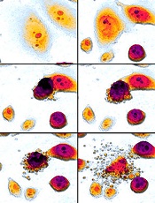
pseudopodia to engulf particles
The transcription factor C/EBPα reprograms B cells into macrophages by “short-circuiting” the cells so they re-express genes reserved for embryonic development, according to research published in Stem Cell Reports.
Over the past 28 years, researchers have shown that a number of specialized cell types can be forcibly converted into other cell types, but the science of how this change takes place is still emerging.
“For a long time, it was unclear whether forcing cell fate decisions by expressing transcription factors in the wrong cell type could teach us something about what happens normally during physiological differentiation,” said Thomas Graf, PhD, of the Center for Genomic Regulation in Barcelona, Spain.
“What we have now found is that the two processes are actually surprisingly similar.”
The researchers found that B-cell transdifferentiation takes place when C/EBPα binds to two regions of DNA that act as gene expression enhancers. One of these regions is normally active in immune cells, and the other is only turned on when macrophage precursors are ready to differentiate.
This indicates that the convergence of these two enhancer pathways can cause the B cell to act like a macrophage precursor, thus triggering the unnatural transdifferentiation.
“This has taught us a great deal about how a transcription factor can activate a new gene expression program (in our case, that of macrophages) but has left us in the dark about the other part of the equation; namely, how the factor silences the B-cell program, something that must happen if transdifferentiation is to work,” Dr Graf said. “This is one of the questions we are focusing on now.”
Dr Graf is interested in this pathway because C/EBPα-induced, B cell-to-macrophage transdifferentiation can convert both human B-cell lymphoma and leukemia cells into functional, non-cancerous macrophages.
He believes that induced transdifferentiation could become therapeutically relevant if a drug could be found that can replace the transcription factor. And understanding the mechanisms of the process could help labs that use this transdifferentiation approach to generate cells for regenerative purposes. ![]()
Newfound mechanism could be used to fight cancers
apoptosis in cancer cells
Researchers say they have identified a new mechanism by which the tumor suppressor protein p53 triggers apoptosis, and they believe this process could be harnessed to kill cancer cells.
The team discovered how p53 acts in the cytoplasm to trigger cell death by binding to and activating the BAX protein.
The process involves a shape change in one of p53’s amino acids that serves as the “switch” to activate BAX and trigger the apoptotic pathway.
The team also identified the enzyme in the cytoplasm that promotes the change that controls the “switch.”
Richard Kriwacki, PhD, of St. Jude Children’s Research Hospital in Memphis, Tennessee, and his colleagues described these findings in Molecular Cell.
Like up to half of all proteins, p53 includes both structured and disordered regions. A disordered region is a segment that does not adopt a single shape but
remains flexible and constantly switches between different shapes until
it encounters a partner.
Dr Kriwacki and his colleagues showed that both structured and disordered regions of p53 play a role in BAX activation in the cytoplasm.
The process starts when a structured region of p53 known as the DNA-binding domain binds to BAX. That sets the stage for the unstructured region of p53 to form a second bond, which activates BAX and triggers apoptosis.
“There were no previous reports of this disordered region of p53 binding to BAX, so the finding that this region was the key to BAX activation was a total surprise,” Dr Kriwacki said.
The disordered p53 segment included the amino acid proline, which can change between two shapes, particularly in the presence of the enzyme Pin1.
Using NMR spectroscopy, the researchers showed that the proline shape change promotes p53 binding and activation of BAX.
“These results expand our understanding of the different ways p53 modulates cell behavior,” Dr Kriwacki said. “The findings also raise the possibility of killing tumor cells using small molecules to trigger BAX-dependent apoptosis.” ![]()
Delayed cancer diagnosis tied to care dissatisfaction

Photo courtesy of NIH
Patients who make 3 or more trips to the general practitioner (GP) before they are referred for cancer tests are more likely to be dissatisfied with subsequent care, according to research published in the European Journal of Cancer Care.
Researchers analyzed survey responses from nearly 60,000 cancer patients and found that about 23% had visited their GP 3 or more times before they were referred for cancer tests.
These patients were more likely than patients with 1 or 2 GP visits to report negative experiences with regard to many different aspects of their care.
“This research shows that first impressions go a long way in determining how cancer patients view their experience of cancer treatment,” said study author Georgios Lyratzopoulos, MD, of University College London in the UK.
“A negative experience of diagnosis can trigger loss of confidence in their care throughout the cancer journey. When they occur, diagnostic delays are largely due to cancer symptoms being extremely hard to distinguish from other diseases, combined with a lack of accurate and easy-to-use tests. New diagnostic tools to help doctors decide which patients need referring are vital to improve the care experience for even more cancer patients.”
Dr Lyratzopoulos and his colleagues initially analyzed survey data from 73,462 cancer patients, but 15,355 of those patients (21%) had not seen a GP prior to their cancer diagnosis.
Of the 58,107 patients who had at least 1 primary care consultation, 44,827 (77%) had seen their GP once or twice before referral for cancer tests, and 13,280 (23%) had seen the GP 3 or more times.
The researchers analyzed patients’ responses to 12 survey questions that assessed satisfaction with various aspects of their cancer care.
And results showed that patients who had seen their GP 3 or more times before referral were significantly more likely than patients with 1 to 2 GP visits to report negative experiences across 10 of the 12 aspects of care.
The 12 aspects of care and patient responses (for 3+ GP visits and 1-2 visits, respectively) were as follows:
- Dissatisfied with overall care: 16% vs 9% (adjusted* odds ratio [aOR]=1.44, P<0.001)
- Dissatisfied with the way they were informed of their cancer: 22% vs 14% (aOR=1.38, P<0.001)
- Dissatisfied with their involvement in decision-making: 32% vs 25% (aOR=1.13, P<0.001)
- Lack of confidence/trust in their doctors: 19% vs 12% (aOR=1.22, P<0.001)
- Lack of confidence/trust in their nurses: 36% vs 27% (aOR=1.22, P<0.001)
- Thought doctors/nurses deliberately withheld information: 16% vs 10% (aOR=1.27, P<0.001)
- Dissatisfied with staff support: 41% vs 27% (aOR=1.68, P<0.001)
- Dissatisfied with integration of various caregivers (GP, hospital doctors/nurses, specialist nurses, etc): 45% vs 32% (aOR=1.48, P<0.001)
- Dissatisfied with the amount of time they had to wait at appointments with their hospital doctor: 31% vs 28% (aOR=1.10, P=0.007)
- Said their GP did not receive enough information about their condition or hospital treatment: 8% vs 5% (aOR=1.36, P<0.001)
- Said they did not receive clear written information about what they should or should not do after leaving the hospital: 20% vs 14% (aOR=1.06, P=0.100)
- Said they did not receive the name of a Clinical Nurse Specialist who would be in charge of their care: 11% vs 9% (aOR=1.00, P=0.894).
“This is the first time we’ve had direct feedback from patients on such a large scale to show how the timeliness of their diagnosis colors their experience of the care they later receive,” said Sara Hiom, of Cancer Research UK, which supported this study.
“It’s another good reason to highlight the importance of diagnosing cancer as quickly as possible, not just to give patients the best chances of survival, but also to improve their experience of the care they receive throughout their cancer journey.” ![]()
*The odds ratio was adjusted for patient age, sex, ethnicity, cancer diagnosis, and response tendency (likelihood of giving a positive or negative response to questions).
Photo courtesy of NIH
Patients who make 3 or more trips to the general practitioner (GP) before they are referred for cancer tests are more likely to be dissatisfied with subsequent care, according to research published in the European Journal of Cancer Care.
Researchers analyzed survey responses from nearly 60,000 cancer patients and found that about 23% had visited their GP 3 or more times before they were referred for cancer tests.
These patients were more likely than patients with 1 or 2 GP visits to report negative experiences with regard to many different aspects of their care.
“This research shows that first impressions go a long way in determining how cancer patients view their experience of cancer treatment,” said study author Georgios Lyratzopoulos, MD, of University College London in the UK.
“A negative experience of diagnosis can trigger loss of confidence in their care throughout the cancer journey. When they occur, diagnostic delays are largely due to cancer symptoms being extremely hard to distinguish from other diseases, combined with a lack of accurate and easy-to-use tests. New diagnostic tools to help doctors decide which patients need referring are vital to improve the care experience for even more cancer patients.”
Dr Lyratzopoulos and his colleagues initially analyzed survey data from 73,462 cancer patients, but 15,355 of those patients (21%) had not seen a GP prior to their cancer diagnosis.
Of the 58,107 patients who had at least 1 primary care consultation, 44,827 (77%) had seen their GP once or twice before referral for cancer tests, and 13,280 (23%) had seen the GP 3 or more times.
The researchers analyzed patients’ responses to 12 survey questions that assessed satisfaction with various aspects of their cancer care.
And results showed that patients who had seen their GP 3 or more times before referral were significantly more likely than patients with 1 to 2 GP visits to report negative experiences across 10 of the 12 aspects of care.
The 12 aspects of care and patient responses (for 3+ GP visits and 1-2 visits, respectively) were as follows:
- Dissatisfied with overall care: 16% vs 9% (adjusted* odds ratio [aOR]=1.44, P<0.001)
- Dissatisfied with the way they were informed of their cancer: 22% vs 14% (aOR=1.38, P<0.001)
- Dissatisfied with their involvement in decision-making: 32% vs 25% (aOR=1.13, P<0.001)
- Lack of confidence/trust in their doctors: 19% vs 12% (aOR=1.22, P<0.001)
- Lack of confidence/trust in their nurses: 36% vs 27% (aOR=1.22, P<0.001)
- Thought doctors/nurses deliberately withheld information: 16% vs 10% (aOR=1.27, P<0.001)
- Dissatisfied with staff support: 41% vs 27% (aOR=1.68, P<0.001)
- Dissatisfied with integration of various caregivers (GP, hospital doctors/nurses, specialist nurses, etc): 45% vs 32% (aOR=1.48, P<0.001)
- Dissatisfied with the amount of time they had to wait at appointments with their hospital doctor: 31% vs 28% (aOR=1.10, P=0.007)
- Said their GP did not receive enough information about their condition or hospital treatment: 8% vs 5% (aOR=1.36, P<0.001)
- Said they did not receive clear written information about what they should or should not do after leaving the hospital: 20% vs 14% (aOR=1.06, P=0.100)
- Said they did not receive the name of a Clinical Nurse Specialist who would be in charge of their care: 11% vs 9% (aOR=1.00, P=0.894).
“This is the first time we’ve had direct feedback from patients on such a large scale to show how the timeliness of their diagnosis colors their experience of the care they later receive,” said Sara Hiom, of Cancer Research UK, which supported this study.
“It’s another good reason to highlight the importance of diagnosing cancer as quickly as possible, not just to give patients the best chances of survival, but also to improve their experience of the care they receive throughout their cancer journey.”
*The odds ratio was adjusted for patient age, sex, ethnicity, cancer diagnosis, and response tendency (likelihood of giving a positive or negative response to questions).