User login
Home pesticide exposure linked to childhood cancers

in his garden
Researchers have linked residential pesticide exposure to childhood cancers, but the estimated risks vary according to the cancer type, the type of pesticide, and where it is applied.
The investigators conducted a meta-analysis of published studies and found that childhood exposure to indoor pesticides was associated with a
significantly increased risk of all the cancers analyzed, as well as leukemia and lymphoma individually.
Overall, exposure to outdoor pesticides was not associated with an increased risk of childhood cancers. However, herbicide exposure was linked to an increased risk of leukemia and all cancers combined.
Mei Chen, PhD, of the Harvard T.H. Chan School of Public Health in Boston, Massachusetts, and colleagues conducted this meta-analysis and reported the results in Pediatrics.
The team searched for observational studies published in PubMed before February 2014 and ultimately included 16 studies in their analysis.
They assessed exposure to indoor pesticides and indoor insecticides (a subgroup of indoor pesticides), as well as exposure to outdoor pesticides, which included outdoor insecticides, herbicides, and fungicides.
The cancer types analyzed were leukemia, lymphoma, brain tumors, neuroblastoma, Wilms tumor, and soft tissue sarcoma.
Indoor pesticides
When the investigators analyzed all cancer types together, they found a significantly increased risk of childhood cancers associated with exposure to indoor pesticides (odds ratio [OR]=1.40).
Likewise, there was a significantly increased risk for leukemia (OR=1.48), acute leukemia (OR=1.59), lymphoma (OR=1.43), and all hematopoietic malignancies (leukemias and lymphomas, OR=1.47).
But the increased risk of childhood brain tumors was not statistically significant, and the other cancers were not analyzed separately.
Exposure to indoor insecticides was associated with a significant increase in the risk of leukemia (OR=1.47), acute leukemia (OR=1.59), lymphoma (OR=1.43), and all hematopoietic malignancies (OR=1.46).
Outdoor pesticides
There was no significant association between exposure to outdoor pesticides or outdoor insecticides and any of the cancer types. And there were not enough studies on fungicides to assess the risk of cancers associated with their use.
However, there was a significant association between exposure to herbicide and all childhood cancers (OR=1.35) as well as leukemia (OR=1.26).
The investigators said these results suggest cancer risks are related to the type of pesticides used and where they are applied.
And although additional research is needed to confirm the association between pesticide exposure and childhood cancers, steps should be taken to limit exposure to pesticides during childhood. ![]()
Studies help explain resistance to BET inhibitors
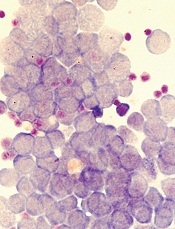
Image by Robert Paulson
Two groups of researchers have reported results that help explain how leukemia resists treatment with BET inhibitors.
One group was able to grow and maintain leukemia stem cells (LSCs) in vitro, and their subsequent experiments showed how LSCs react to BET inhibition.
The other group found evidence to suggest that by measuring Wnt signaling markers, we might be able to predict which patients will respond to BET inhibition.
Both groups described their research in letters to Nature.
“[T]he risk of resistance developing is common in any cancer treatment,” said Mark Dawson, PhD, a researcher at Peter MacCallum Cancer Centre in East Melbourne, Victoria, Australia and an author of the LSC study.
“Knowing precisely how that happens in advance puts us one step ahead in outmaneuvering the disease. Being able to grow and maintain leukemia
stem cells in vitro also gives us unprecedented access and insight into how they work so we can find new and better ways to target and destroy them.”
In their study, Dr Dawson and his colleagues assessed BET inhibitor resistance in a model of acute myeloid leukemia.
The team transduced murine hematopoietic stem and progenitor cells with MLL–AF9 and treated the cells with the BET inhibitor I-BET or dimethylsulfoxide (vehicle). They then isolated individual blast colonies to generate 4 vehicle-treated and 5 I-BET-resistant cell lines.
The researchers found that resistance to I-BET also conferred resistance to the chemically distinct BET inhibitor JQ1 and to genetic knockdown of BET proteins.
Further investigation revealed that resistance to BET inhibitors emerges from LSCs, both ex vivo and in vivo. And that resistance is, in part, a result of increased Wnt/β-catenin signaling.
The researchers noted that not all LSCs are intrinsically resistant to BET inhibition, but a small proportion of them are either transcriptionally primed or display rapid transcriptional plasticity to survive the initial BET inhibitor challenge. The team said these cells then thrive and become the dominant population.
These findings are consistent with results of the other study, conducted by Johannes Zuber, MD, of the Research Institute of Molecular Pathology in Vienna, Austria, and his colleagues.
With this study, the researchers set out to determine why only certain leukemia subtypes are sensitive to BET inhibitors. Their experiments revealed that loss of the PRC2 complex, which is known to inactivate genes during normal development, can render leukemia cells resistant to BET inhibitors.
By further characterizing these resistant cells, the team found that MYC and other BRD4-regulated genes (which are turned off by BET inhibitors) were back on again. So the leukemia cells had found a way to activate these genes in the absence of BRD4.
The researchers then compared leukemia cells that had acquired resistance during BET inhibitor treatment to leukemia cells that were resistant in the first place.
In both cases, the cells used similar pathways to turn critical genes such as MYC back on and thereby escape the effects of BET inhibition. A pathway that proved particularly important was the Wnt signaling pathway, which is known to activate MYC in cancers.
To determine whether this knowledge could be used to predict which patients will respond to BET inhibitors, the researchers measured Wnt signaling markers in samples from leukemia patients.
The team found that cells with low Wnt activity were sensitive to BET inhibitors, while high Wnt activity was associated with resistance.
Specifically, the researchers quantified 9 Wnt-associated transcripts in sensitive and resistant samples. Three of these transcripts—TCF4, CCND2, and HOXB4—were significantly overexpressed in resistant samples.
So the team used these 3 transcripts to establish a “resistance index” that, they believe, may provide a first step toward developing a predictive biomarker.
The researchers said, collectively, their study reveals that leukemia cells can become resistant to BET inhibitors by rewiring the regulation of critical BRD4 target genes. This transcriptional plasticity highlights an emerging mode of drug resistance that is distinct from established resistance mechanisms such as mutations in binding pockets or drug elimination through efflux pumps.
Dr Zuber and his colleagues believe that a better understanding of these adaptation mechanisms will lead to combination therapies that will ultimately “outsmart” cancer cells.
“We now have learned that cancer cells can adapt to targeted therapies, but their repertoire of escape routes is quite limited,” Dr Zuber said. “A better understanding of the common escape routes will allow us to predict the next effective targeted therapy so that we are always one step ahead of the cancer cell.” ![]()
Gene linked to aggressive AML
Photo courtesy of NIH
The gene FOXC1 is associated with aggressive acute myeloid leukemia (AML), according to research published in Cancer Cell.
Researchers said tissue-inappropriate derepression of FOXC1 has functional consequences and prognostic significance in AML.
They found evidence suggesting that FOXC1 enhances clonogenic potential, helps block monocyte/macrophage differentiation, accelerates leukemia onset in mice, and leads to inferior survival in AML patients.
“This is an important finding which helps us understand how acute myeloid leukemia develops and why some cases of AML are more aggressive than others,” said study author Tim Somervaille, MBBS, PhD, of The University of Manchester in the UK.
“Here, instead of being faulty or mutated, this normal gene is turned on in the wrong place at the wrong time, which makes the cancer grow more rapidly. There are certain situations where this gene is necessary, as in the development of the eye and skeleton before birth, but when it’s switched on in the wrong tissue, it causes more aggressive forms of leukemia.”
Dr Somervaille and his colleagues said FOXC1 is expressed in at least 20% of human AML cases but not in normal hematopoietic populations.
The researchers analyzed levels of transcription factor genes in data from published studies to identify transcription regulators expressed in human AML hematopoietic stem and progenitor cells (HSPCs) but not normal HSPCs. In these studies, FOXC1 was among the genes that were most highly upregulated in AML HSPCs.
Further investigation revealed that FOXC1 expression is associated with mutations in NPM1 and t(6;9) but no other recurring mutations in AML.
When Dr Somervaille and his colleagues conducted experiments with human AML cells, they found that FOXC1 “contributes to oncogenic potential by maintaining differentiation block and clonogenic activity.”
In vitro experiments with normal HSPCs showed that FOXC1 expression temporarily impairs myeloid differentiation. In mice, expression of FOXC1 in normal HSPCs reduced donor:recipient chimerism in the blood and skewed differentiation toward the myeloid lineage and away from the B-cell lineage.
By comparing samples from AML patients, the researchers found that FOXC1 expression is associated with high HOX gene expression.
Subsequent experiments showed that FOXC1 collaborates with HOXA9 to enhance clonogenic potential and cell-cycle progression, help block monocyte/macrophage and B-lineage differentiation, and accelerate the onset of symptomatic leukemia in mice.
To determine if the same effects occur in humans, the researchers again analyzed data from AML patients. The results indicated that FOXC1 expression helps block monocyte/macrophage differentiation and leads to inferior survival.
Dr Somervaille and his colleagues said these findings may have therapeutic implications, as previous research has shown that, in basal-like breast cancer, high FOXC1 expression renders cells more susceptible to pharmacological inhibition of NF-kB. But additional research is needed to investigate therapeutic implications for AML. ![]()
Social factors may impact survival in AML

Photo by Rhoda Baer
A new study indicates that certain social factors may impact survival in adults with acute myelogenous leukemia (AML) who are under 65.
The research showed associations between patient survival and insurance status, marital status, and county-level income.
“We believe these 3 factors indicate lack of material and social support preventing young patients from successfully walking the long and difficult road towards a cure,” said Uma Borate, MD, of the University of Alabama at Birmingham.
To conduct this study, Dr Borate and her colleagues analyzed data on 5541 patients, ages 19 to 64, who were diagnosed with AML between 2007 and 2011.
The team reported their findings in Cancer.
Multivariable analysis showed that AML subtype, age, and sex were independently associated with patients’ survival. And the non-biological factors independently associated with survival were insurance status, marital status, and county-level median household income.
Specifically, there was a significantly increased risk of premature death among patients who were uninsured (P=0.005) or Medicaid beneficiaries (P<0.001), compared to patients with private insurance.
Single (P<0.001) or divorced (P=0.011) patients had a significantly higher risk of premature death than married patients. But there was no significant difference between married and widowed patients (P=0.206).
And patients who lived in areas with lower income—the lowest 3 of 5 income groups—had a significantly increased risk of premature death.
Compared to patients in the fifth income quintile ($58.3K-$79.9K), there was an increased risk of death in the first quintile ($16.2K-$38.8K, P=0.001), second quintile ($38.8K-$42.2K, P<0.001), and third quintile ($42.2K-$47.9K, P<0.001).
Early and late mortality
The researchers wanted to determine if the impact of non-biological factors on survival was related to early mortality (a possible surrogate for access to care or late presentation) or late mortality (a possible surrogate for access to post-remission therapy and hematopoietic stem cell transplant).
So they conducted an exploratory analysis of factors influencing the risk of death within the first 2 months of diagnosis.
Being a Medicaid beneficiary (P=0.01) or uninsured (P<0.001) was independently associated with an increased risk of death within the first 2 months.
The same was true for patients belonging to the first income quartile (P=0.001), second quartile (P=0.003), third quartile (P=0.02), and fourth quartile (P=0.028).
On the other hand, there was no significant difference in early death according to marital status.
The researchers also performed a landmark survival analysis including only patients who survived at least 2 months from diagnosis.
In this analysis, marital status (P<0.001), insurance status (P=0.001), and income (P=0.021) were all independent predictors of survival.
Implications
“As physicians, we often emphasize more of the biology of the cancer, especially with the recent focus on personalized medicine,” said study author Luciano Jose Costa, MD, PhD, also of the University of Alabama at Birmingham.
“But we need to pay the same attention to resources available to our patients, as this greatly impacts their chances to survive leukemia.”
The researchers believe this will be especially important as the US transitions to a healthcare system that ties physician and hospital payments to patient outcomes.
“Taking from the results of this study, factors that have nothing to do with quality of care need to be accounted for when comparing predicted with actual outcomes,” Dr Borate said. “Otherwise, we will create a disincentive for hospitals and doctors to care for less privileged patients.” ![]()
Scientists describe new way to create etoposide

Scientists have reported a new way to produce the chemotherapeutic agent etoposide, and they believe this discovery could lead to a more stable supply of the drug.
Currently, producing etoposide requires isolating one of its precursors, (–)-podophyllotoxin, from the endangered, slow-growing, Himalayan Mayapple plant (Podophyllum hexandrum).
But researchers found they could generate the immediate precursor to etoposide—(–)-4’-desmethyl-epipodophyllotoxin—in a more easily accessible, faster-growing tobacco plant (Nicotiana benthamiana).
Elizabeth Sattely, PhD, of Stanford University in California, and her graduate student, Warren Lau, described this work in Science.
The pair noted that there are 4 known genes behind (–)-podophyllotoxin production, but the full recipe for this compound has eluded researchers, in part because of the Mayapple’s immense genome.
To tap into the Mayapple’s chemotherapeutic potential, Lau and Dr Sattely first focused on the 4 known genes—PLR, SDH, CYP719A23, and DIR. Then, they analyzed RNA sequencing data from the Mayapple to identify similar genes.
Next, the pair manipulated the tobacco plant to express multiple gene candidates at once and identified the resulting compounds in leaf tissue using mass spectrometry.
Dr Sattely and Lau identified 6 new genes—OMT3, CYP71CU1, OMT1, 2-ODD, CYP71BE54, and CYP82D61.
These genes, when expressed with the original 4, produce the immediate etoposide precursor (–)-4′-desmethyl-epipodophyllotoxin, which outperforms (–)-podophyllotoxin as a chemotherapy ingredient.
The researchers said this work has revealed a simpler and more direct route to etoposide that circumvents the semisynthetic epimerization and demethylation required to produce etoposide from (–)-podophyllotoxin.
However, Dr Sattely said the eventual goal is to use yeast instead of plants to produce etoposide. Yeast can be grown in large vats and may therefore provide a more stable source of drugs.
In addition, yeast provides the opportunity to modify genes to produce proteins with slightly different functions. And it may be possible to feed the yeast a slightly different starting product, thereby changing the chemical a molecular assembly line churns out.
These approaches could provide a way of tweaking existing drugs in an effort to improve them. ![]()
T-cell exhaustion may be therapeutic target for AML

Image courtesy of NIAID
New research has revealed a population of T cells associated with relapse of acute myeloid leukemia (AML) after allogeneic stem cell transplant (allo-SCT).
Patients experienced an increase in this cell population before their relapse was diagnosed clinically.
The cells also appear to be markers of T-cell exhaustion, which suggests therapies targeting T-cell exhaustion might be able to treat or prevent AML relapse after
allo-SCT.
Hong Zheng, MD, PhD, of Penn State University College of Medicine in Hershey, Pennsylvania, and her colleagues conducted this research and reported the results in Blood Cancer Journal.
The investigators noted that T cells are major players in the graft-vs-leukemia effect, but persistent antigen stimulation can lead to T-cell exhaustion.
To determine whether T-cell exhaustion is involved in relapse after allo-SCT, the team performed phenotypic and functional studies on T cells from the peripheral blood of AML transplant recipients.
Dr Zheng and her colleagues discovered that patients who relapsed after allo-SCT had significantly elevated levels of PD-1hiTIM-3+ T cells.
These T cells produced fewer cytokines—interleukin 2, tumor necrosis factor-α, and interferon-γ—than normal T cells, which is a sign of T-cell exhaustion.
In addition, while other T cells mostly consisted of all 4 T-cell subsets, PD-1hiTIM-3+ T cells had no naive T cells (CCR7+CD45RA+) and significantly decreased levels of terminally differentiated effector T cells (CCR7-CD45RA+).
The investigators said this suggests PD-1hiTIM-3+ cells are phenotypically antigen-experienced T cells that have lost functional subsets, which is consistent with a state of T-cell exhaustion.
The team believes their findings could pave the way for new treatments for AML patients undergoing allo-SCT.
The PD-1 inhibitor nivolumab has been shown to block T-cell exhaustion in patients with solid tumors. So Dr Zheng is planning a clinical trial to see if such treatment could work for AML patients as well.
She also believes that PD-1hiTIM-3+ T cells could be used to diagnose relapses earlier than is currently possible.
“Two months before we were able to clinically diagnose relapse in these patients, we found these markers elevated in them,” Dr Zheng said. “If we can have an early diagnostic marker, we will potentially be able to improve clinical outcome significantly.”
Dr Zheng is currently investigating the trigger for T-cell exhaustion in transplant recipients with AML.
“The hypothesis is that when you have chronic antigen stimulation, the T cell can get exhausted,” she said. “We think that residual leukemia in our patients is the chronic stimulator that causes this.” ![]()
Drug granted orphan designation for AML

Image by Lance Liotta
The investigational therapy SL-401 has received orphan designation to treat acute myeloid leukemia (AML) in the European Union (EU).
SL-401 targets the interleukin-3 receptor (IL-3R). It is comprised of human IL-3 coupled to a truncated diphtheria toxin payload that inhibits protein synthesis.
SL-401 is under development to treat a range of hematologic malignancies, as IL-3R is overexpressed on cancer stem cells in multiple hematologic malignancies.
SL-401 has orphan designation from the US Food and Drug Administration (FDA) for the treatment of AML and blastic plasmacytoid dendritic cell neoplasm (BPDCN).
SL-401 research
At ASH 2012 (abstract 3625), researchers reported results with SL-401 in a study of patients with AML, BPDCN, and myelodysplastic syndromes (MDS).
At that time, the study had enrolled 80 patients, including 59 with relapsed or refractory AML, 11 with de novo AML unfit for chemotherapy, 7 with high-risk MDS, and 3 with relapsed/refractory BPDCN.
Patients received a single cycle of SL-401 as a 15-minute intravenous infusion in 1 of 2 dosing regimens to determine the maximum tolerated dose (MTD) and assess antitumor activity.
With regimen A, 45 patients received doses ranging from 4 to 12.5 μg/kg every other day for up to 6 doses. With regimen B, 35 patients received doses ranging from 7.1 to 22.1 μg/kg daily for up to 5 doses.
Of the 59 patients with relapsed/refractory AML, 2 achieved complete responses (CRs), 5 had partial responses (PRs), and 8 had minor responses (MRs). One CR lasted more than 8 months, and the other lasted more than 25 months.
Of the 11 patients with AML who were not candidates for chemotherapy, 2 had PRs and 1 had an MR. Among the 7 patients with high-risk MDS, there was 1 PR and 1 MR.
And among the 3 patients with BPDCN, there were 2 CRs. One CR lasted more than 2 months, and the other lasted more than 4 months.
The MTD was not achieved with regimen A, but the MTD for regimen B was 16.6 μg/kg/day. The dose-limiting toxicities were a gastrointestinal bleed (n=1), transaminase and creatinine kinase elevations (n=1), and capillary leak syndrome (n=3). There was no evidence of treatment-related bone marrow suppression.
Last year, researchers reported additional results in BPDCN patients (Frankel et al, Blood 2014).
Eleven BPDCN patients received a single course of SL-401 (at 12.5 μg/kg intravenously over 15 minutes) daily for up to 5 doses. Three patients who had initial responses to SL-401 received a second course while in relapse.
Seven of 9 evaluable (78%) patients responded to a single course of SL-401. There were 5 CRs and 2 PRs. The median duration of responses was 5 months (range, 1-20+ months).
The most common adverse events were transient and included fever, chills, hypotension, edema, hypoalbuminemia, thrombocytopenia, and transaminasemia.
Three multicenter clinical trials of SL-401 are currently open in the following indications:
- BPDCN and relapsed/refractory AML
- AML patients in first complete remission with minimal residual disease
- Four types of advanced, high-risk myeloproliferative neoplasms, including systemic mastocytosis, advanced symptomatic hypereosinophilic disorder, myelofibrosis, and chronic myelomonocytic leukemia.
Additional SL-401 studies are planned for patients with myeloma, lymphomas, and other leukemias.
About orphan designation
In the EU, orphan designation is granted to therapies intended to treat a life-threatening or chronically debilitating condition that affects no more than 5 in 10,000 persons and where no satisfactory treatment is available.
Companies that obtain orphan designation for a drug in the EU benefit from a number of incentives, including protocol assistance, a type of scientific advice specific for designated orphan medicines, and 10 years of market exclusivity once the medicine is on the market. Fee reductions are also available, depending on the status of the sponsor and the type of service required.
The FDA grants orphan designation to drugs that are intended to treat diseases or conditions affecting fewer than 200,000 patients in the US.
In the US, orphan designation provides the sponsor of a drug with various development incentives, including opportunities to apply for research-related tax credits and grant funding, assistance in designing clinical trials, and 7 years of US market exclusivity if the drug is approved. ![]()
Image by Lance Liotta
The investigational therapy SL-401 has received orphan designation to treat acute myeloid leukemia (AML) in the European Union (EU).
SL-401 targets the interleukin-3 receptor (IL-3R). It is comprised of human IL-3 coupled to a truncated diphtheria toxin payload that inhibits protein synthesis.
SL-401 is under development to treat a range of hematologic malignancies, as IL-3R is overexpressed on cancer stem cells in multiple hematologic malignancies.
SL-401 has orphan designation from the US Food and Drug Administration (FDA) for the treatment of AML and blastic plasmacytoid dendritic cell neoplasm (BPDCN).
SL-401 research
At ASH 2012 (abstract 3625), researchers reported results with SL-401 in a study of patients with AML, BPDCN, and myelodysplastic syndromes (MDS).
At that time, the study had enrolled 80 patients, including 59 with relapsed or refractory AML, 11 with de novo AML unfit for chemotherapy, 7 with high-risk MDS, and 3 with relapsed/refractory BPDCN.
Patients received a single cycle of SL-401 as a 15-minute intravenous infusion in 1 of 2 dosing regimens to determine the maximum tolerated dose (MTD) and assess antitumor activity.
With regimen A, 45 patients received doses ranging from 4 to 12.5 μg/kg every other day for up to 6 doses. With regimen B, 35 patients received doses ranging from 7.1 to 22.1 μg/kg daily for up to 5 doses.
Of the 59 patients with relapsed/refractory AML, 2 achieved complete responses (CRs), 5 had partial responses (PRs), and 8 had minor responses (MRs). One CR lasted more than 8 months, and the other lasted more than 25 months.
Of the 11 patients with AML who were not candidates for chemotherapy, 2 had PRs and 1 had an MR. Among the 7 patients with high-risk MDS, there was 1 PR and 1 MR.
And among the 3 patients with BPDCN, there were 2 CRs. One CR lasted more than 2 months, and the other lasted more than 4 months.
The MTD was not achieved with regimen A, but the MTD for regimen B was 16.6 μg/kg/day. The dose-limiting toxicities were a gastrointestinal bleed (n=1), transaminase and creatinine kinase elevations (n=1), and capillary leak syndrome (n=3). There was no evidence of treatment-related bone marrow suppression.
Last year, researchers reported additional results in BPDCN patients (Frankel et al, Blood 2014).
Eleven BPDCN patients received a single course of SL-401 (at 12.5 μg/kg intravenously over 15 minutes) daily for up to 5 doses. Three patients who had initial responses to SL-401 received a second course while in relapse.
Seven of 9 evaluable (78%) patients responded to a single course of SL-401. There were 5 CRs and 2 PRs. The median duration of responses was 5 months (range, 1-20+ months).
The most common adverse events were transient and included fever, chills, hypotension, edema, hypoalbuminemia, thrombocytopenia, and transaminasemia.
Three multicenter clinical trials of SL-401 are currently open in the following indications:
- BPDCN and relapsed/refractory AML
- AML patients in first complete remission with minimal residual disease
- Four types of advanced, high-risk myeloproliferative neoplasms, including systemic mastocytosis, advanced symptomatic hypereosinophilic disorder, myelofibrosis, and chronic myelomonocytic leukemia.
Additional SL-401 studies are planned for patients with myeloma, lymphomas, and other leukemias.
About orphan designation
In the EU, orphan designation is granted to therapies intended to treat a life-threatening or chronically debilitating condition that affects no more than 5 in 10,000 persons and where no satisfactory treatment is available.
Companies that obtain orphan designation for a drug in the EU benefit from a number of incentives, including protocol assistance, a type of scientific advice specific for designated orphan medicines, and 10 years of market exclusivity once the medicine is on the market. Fee reductions are also available, depending on the status of the sponsor and the type of service required.
The FDA grants orphan designation to drugs that are intended to treat diseases or conditions affecting fewer than 200,000 patients in the US.
In the US, orphan designation provides the sponsor of a drug with various development incentives, including opportunities to apply for research-related tax credits and grant funding, assistance in designing clinical trials, and 7 years of US market exclusivity if the drug is approved.
Targeting STAT3 to prevent relapse in AML

Compounds that target a novel binding site on STAT3 may one day be used to prevent relapse in acute myeloid leukemia (AML), according to researchers.
STAT3 is known to interfere with chemotherapy and is thought to play a role in many cases of AML relapse.
But preclinical research suggests a compound known as MM-206 can disrupt STAT3’s disease-promoting effects by targeting a previously unknown ligand-binding site on the protein.
Zachary Ball, PhD, of Rice University in Houston, Texas, and his colleagues described this research in Angewandte Chemie.
The team first discovered that the coiled–coil domain (CCD) is a novel ligand-binding site on STAT3. Then, they identified a naphthalene sulfonamide compound, known as C188, that can target CCD and inhibit STAT3.
The researchers tested C188 and compounds synthesized from it—C188-9, C188-9-Rh2, and MM-206—in vitro and in vivo. Only MM-206 proved effective in vivo.
“Our main advance, from a medicinal perspective, is that this compound also works in a mouse model,” Dr Ball said. “All the other compounds worked in cells, but, in mice, they weren’t potent enough or stable enough.”
MM-206 inhibited STAT3 phosphorylation and induced apoptosis in 3 different AML cell lines. The compound also prompted apoptosis in primary tumor cells from pediatric AML patients.
Among mice engrafted with luciferase-expressing MV4-11 AML cells, those that received MM-206 exhibited slower disease progression than untreated mice.
When the mice received 4 weeks of treatment with MM-206, they had significantly fewer tumor cells in their bone marrow and lived significantly longer than control mice (P=0.019 at 10 weeks).
Follow-up studies should lead to improved versions of MM-206, according to Dr Ball.
“The discovery raises new questions about STAT3 biology and points the way to future anticancer approaches,” he said, “including combination therapies of coiled-coil STAT3 inhibitors in tandem with other agents.”
Groups crowd-source cancer research

Photo by Rhoda Baer
In an attempt to crowd-source cancer research, a pair of Canadian organizations made a small molecule they developed freely available to researchers.
The goal was to fast-track the testing of new treatment strategies and facilitate sharing of the results.
Researchers have already completed preclinical studies of this molecule, a WDR5 inhibitor called OICR-9429, showing that it is active against breast cancer and acute myeloid leukemia (AML).
The organizations that developed OICR-9429 are the Ontario Institute for Cancer Research (OICR) and the Structural Genomics Consortium (SGC).
“In the time that it would normally take to negotiate a legal agreement to provide OICR-9429 to other research teams, we have received results back from our collaborators showing that it can kill 2 different types of cancer cells,” said Cheryl Arrowsmith, PhD, of SGC Toronto in Ontario, Canada.
“Opening our chemistry capabilities to the world’s scientists allowed us to crowd-source and accelerate the research.”
A study published in Nature Chemical Biology suggested that WDR5 is a therapeutic target for a subtype of AML, and OICR-9429 could therefore be used to treat the disease.
The researchers noted that mutations in C/EBPα occur in about 9% of AML cases and lead to the expression of a 30-kDa dominant negative isoform (C/EBPα-p30).
Their experiments revealed that C/EBPα p30 preferentially interacts with WDR5. So it was no surprise that OICR-9429 inhibited proliferation and induced differentiation in p30-expressing AML cells.
In a study published in Nature, OICR-9429 inhibited cancer cell growth in a panel of breast cancer cell lines driven by mutated forms of p53.
Researchers said this work has implications beyond breast cancer because p53 is mutated in at least half of all cancers and is dysregulated in others.
“It is remarkable how quickly our research results were translated into discoveries by the groups around the world,” said Rima Al-awar, PhD, of OICR in Toronto.
“We are looking forward to seeing more research conducted with OICR-9429 and showing that this new approach to early stage drug discovery has significant advantages.”
The SGC and OICR teams said they are continuing their collaboration to identify additional molecules to advance cancer drug discovery.
Agent blocks STAT3 protein and might prevent AML relapse
The discovery of a new therapeutic target site on the STAT3 oncoprotein – a protein that interferes with chemotherapy by halting deaths of cancerous cells – could cut down on acute myeloid leukemia (AML) relapse in chemoresistant patients, according to research published in Angewandte Chemie.
The STAT3 protein – which stands for “signal transducer and activator of transcription 3” – is a suspected factor in the relapse of nearly 40% of children with AML. A team of researchers from Rice University, working with colleagues at Baylor College of Medicine and the University of Texas MD Anderson Cancer Center, all in Houston, developed a proximity-driven rhodium (II) catalyst known as MM-206, which blocks STAT3 function by identifying the STAT3 coiled-coil domain as a novel ligand-binding site and delivering a naphthalene sulfonamide inhibitor, thus halting the disease promoting effects of STAT3.
The effects were replicated in tumor growth models and testing in a leukemia mouse model suggest this approach can be used to shut down STAT3 activity in AML patients, according to coauthor Dr. Zachary Ball of the department of chemistry at Rice.
Read the full article here: Angew Chem Int Ed Engl. 2015 Sep 7. doi: 10.1002/anie.201506889.
FROM ANGEWANDTE CHEMIE