User login
‘Practice-changing’ treatments emerging in AML

NEW YORK—We are “finally” making progress in the treatment of acute myeloid leukemia (AML), according to a speaker at the NCCN 11th Annual Congress: Hematologic Malignancies.
Jessica K. Altman, MD, said a number of developments have resulted in improved AML treatment, including a better understanding of biology and prognostic assessment, continued advances in transplant, and updating standard treatments and incorporating novel agents in both relapsed/refractory and newly diagnosed patients.
“There are a couple of practice-changing treatments in acute myeloid leukemia, 2 of which happened over the last decade: daunorubicin intensification and the use of FLT3 inhibitors,” said Dr Altman, an associate professor at Northwestern University Feinberg School of Medicine in Chicago, Illinois.
Dr Altman went on to explain that novel therapies for AML can be divided into 2 basic categories. There are agents that don’t depend on mutation status (like daunorubicin) and those that are mutation-specific (like FLT3 inhibitors).
Therapies not dependent on mutational complexity
The therapies that are not dependent on mutational complexity include anti-CD33 antibodies, BCL‐2 inhibitors, a dose-intensified anthracycline regimen, and different formulations of 7+3, including CPX‐351.
Escalated daunorubicin
Randomized trials of escalated daunorubicin (90 vs 45 mg/m2) have demonstrated benefit in complete responses (CRs) and overall survival (OS) in intermediate-risk patients and patients with core-binding factor mutation. They have demonstrated benefit in OS in FLT3 ITD+ patients.
In patients up to 65 years of age, 60–90 mg/m2 of daunorubicin is now standard.
“It’s still not clear to me—and I don’t know if it ever will be—if 90 is equivalent to 60,” Dr Altman said.
CPX-351
CPX-351 is a liposomal formulation of cytarabine and daunorubicin. In a randomized, phase 3 study of older adults with secondary AML, the median OS was 9.56 months for patients treated with CPX-351 and 5.95 months for patients on the 7+3 regimen (P=0.005).
The median event-free survival was significantly better with CPX-351 (P=0.021), as was the rate of CR + CR with incomplete blood count recovery (CRi). The rate of CR + CRi was 47.7% with CPX-351 and 33.3% for 7+3 (P=0.016).
A similar number of patients went on to transplant in each arm. Grade 3-5 adverse events were similar in frequency and severity in both arms—92% with CPX-351 vs 91% with 7+3.
SGN-CD33A
CD33 is not a new target in myeloid leukemia, Dr Altman pointed out. Gemtuzumab ozogamicin has been studied, approved by the US Food and Drug Administration, and then withdrawn.
However, an increasing number of studies with gemtuzumab are underway, she said, and the agent may once again have a place in the AML armamentarium.
The newest CD33 construct is SGN-CD33A, a stable dipeptide linker that enables uniform drug loading of a pyrrolobenzodiazepine dimer that crosslinks DNA and leads to cell death.
“Single-agent data was quite promising,” Dr Altman noted, with a CR + CRi rate of 41% in previously treated patients and 58% in 12 treatment-naïve patients.
These results prompted a combination study of SGN-CD33A with hypomethylating agents.
“Results were higher than expected with a hypomethylating agent,” Dr Altman pointed out.
The CR + CRi + CR with incomplete platelet recovery was 58%. And the median relapse-free survival was 7.7 months.
A phase 3 randomized trial of SGN-CD33A is planned.
Venetoclax
BCL-2 inhibitors are the fourth type of agent not dependent on mutation complexity. Venetoclax (ABT‐199) is a small‐molecule BCL-2 inhibitor that leads to the initiation of apoptosis.
In a phase 1b trial of venetoclax in combination with a hypomethylating agent, the overall CR rate was 35%, and the CRi rate was 35%.
“Again, higher than what would be expected with a hypomethylating agent alone,” Dr Altman said.
In a phase 1b/2 trial of venetoclax in combination with low‐dose cytarabine, the CR + CRi rate was 54%. Patients responded even if they had prior exposure to hypomethylating agents.
Mutation-specific novel agents
The FLT3 inhibitor midostaurin and the IDH inhibitors AG-120 and AG-221 are among the most exciting mutation-specific agents and the ones most progressed, according to Dr Altman.
FLT3-ITD is mutated in about 30% of AML patients and carries an unfavorable prognosis, and the IDH mutation occurs in about 10% and confers a favorable prognosis.
Midostaurin
A phase 3, randomized, double-blind study of daunorubicin/cytarabine induction and high-dose cytarabine consolidation with midostaurin (PKC412) or placebo had a 59% CR rate by day 60 in the midostaurin arm, compared with 53% in the placebo arm.
“The CR rate was slightly higher in the midostaurin arm,” Dr Altman said, “but what’s the most remarkable about this study is the difference in overall survival.”
The median OS in the midostaurin arm was 74.7 months, compared with 25.6 months in the placebo arm (P=0.0074).
“The major take-home message from this clinical trial,” Dr Altman said, “is that midostaurin improved the overall survival when added to standard therapy and represents a new standard of care.”
AG-120 and AG-221
Two IDH inhibitors that have substantial data available are the IDH1 inhibitor AG-120 and the IDH2 inhibitor AG-221.
As of October 2015, 78 patients had been treated with AG-120 in a phase 1 trial, yielding an overall response rate of 35% and a CR rate of 15%.
As of September 2015, 209 patients had been treated with AG-221 in a phase 1/2 trial, and 66 are still on study. The overall response rate was 37% in 159 adults with relapsed/refractory AML, with a median duration of response of 6.9 months. The CR rate was 18%.
Investigators have initiated a phase 3 study of AG-221 compared to conventional care regimens. ![]()
Drug granted conditional approval to treat CLL in Canada

of venetoclax (Venclexta)
Photo courtesy of AbbVie
Health Canada has issued a Notice of Compliance with Conditions (NOC/c) for the BCL-2 inhibitor venetoclax (Venclexta™).
This means venetoclax is conditionally approved for use in patients with previously treated chronic lymphocytic leukemia (CLL) who have 17p deletion or no other available treatment options.
An NOC/c is authorization to market a drug with the condition that the sponsor perform additional studies to verify a clinical benefit.
The NOC/c policy is designed to provide access to:
- Drugs that can treat serious, life-threatening, or severely debilitating diseases
- Drugs that can treat conditions for which no drug is currently marketed in Canada
- Drugs that provide a significant increase in efficacy or significant decrease in risk when compared to existing drugs marketed in Canada.
Venetoclax (previously ABT‐199) is being developed by AbbVie and Genentech, a member of the Roche Group. The drug is jointly commercialized by the companies in the US and by AbbVie outside of the US.
Venetoclax is currently under evaluation in phase 3 trials for the treatment of relapsed, refractory, and previously untreated CLL.
Phase 2 trial
Results from a phase 2 trial of venetoclax in CLL (M13-982, NCT01889186) were published in The Lancet Oncology in June. The trial enrolled 107 patients with relapsed or refractory CLL and 17p deletion.
Patients received venetoclax at 400 mg once daily following a weekly ramp-up schedule for the first 5 weeks. The primary endpoint was overall response rate, as determined by an independent review committee.
At a median follow-up of 12.1 months, 85 patients had responded to treatment, for an overall response rate of 79%.
Eight patients (8%) achieved a complete response or complete response with incomplete count recovery, 3 (3%) had a near-partial response, and 74 (69%) had a partial response. Twenty-two patients (21%) did not respond.
At the time of analysis, the median duration of response had not been reached. The same was true for progression-free survival and overall survival. The progression-free survival estimate for 12 months was 72%, and the overall survival estimate was 87%.
The incidence of treatment-emergent adverse was 96%. The most frequent grade 3/4 adverse events were neutropenia (40%), infection (20%), anemia (18%), and thrombocytopenia (15%).
The incidence of serious adverse events was 55%. The most common of these events were pyrexia (7%), autoimmune hemolytic anemia (7%), pneumonia (6%), and febrile neutropenia (5%).
Grade 3 laboratory tumor lysis syndrome (TLS) was reported in 5 patients during the ramp-up period only. Three of these patients continued on venetoclax, but 2 patients required a dose interruption of 1 day each.
In the past, TLS has caused deaths in patients receiving venetoclax. In response, AbbVie stopped dose-escalation in patients receiving the drug and suspended enrollment in phase 1 trials.
However, researchers subsequently found that a modified dosing schedule, prophylaxis, and patient monitoring can reduce the risk of TLS. ![]()
Doc offers advice on choosing a frontline TKI

Photo by D. Meyer
NEW YORK—Evaluating treatment goals is essential when choosing which tyrosine kinase inhibitor (TKI) to prescribe for a patient with newly diagnosed chronic myeloid leukemia (CML), according to a speaker at the NCCN 11th Annual Congress: Hematologic Malignancies.
“Deciding what TKI to start people on really depends on what your goals are for that patient,” said the speaker, Jerald Radich, MD, of the Fred Hutchinson Cancer Research Center/Seattle Cancer Care Alliance in Seattle, Washington.
Because the 3 TKIs approved for frontline treatment of CML—imatinib, dasatinib, and nilotinib—produce “amazingly similar” responses, treatment compliance becomes an important factor in patient outcomes, he noted.
“If you take 90% of your imatinib, your MMR [major molecular response] is 90%,” he said. “Your CMR [complete molecular response] is 40%. So taking drug obviously trumps the decision of what drug to take.”
Dr Radich added that the major goal of treatment is to keep patients out of accelerated-phase blast crisis. Once people progress to blast crisis on a TKI, the median survival is less than 1 year.
“So that’s why you treat people aggressively, that’s why you monitor them molecularly, to prevent that from happening,” he said.
Treatment goals
Aside from preventing patients from progressing to blast crisis, treatment goals vary.
Achieving early molecular response (MR) impacts progression and survival, as does achieving a complete cytogenetic response (CCyR).
A major molecular response (MMR) is considered a “safe haven,” Dr Radich said, because once people achieve it, they almost never progress if they stay on drug.
And with a deep/complete molecular response (CMR), patients may potentially discontinue the drug.
So how your response goals line up determines how you use the agents for your treatment course, Dr Radich said.
In all response categories—patients with CCyR, MMR, MR, CMR—survival is virtually within 95% of survival for the general population.
“This is absolutely astonishing,” Dr Radich said.
He emphasized the importance of molecular testing at 3 months and achieving a BCR-ABL level of less than 10%.
Patients who have more than 10% blasts at 3 months have an 88% chance of achieving MMR at 4 years, while those who still have more than 10% blasts at 6 months have a 3.3% chance of achieving MMR at 4 years.
Toxicity
Side effects common to the 3 frontline TKIs are myelosuppression, transaminase elevation, and change in electrolytes. Dr Radich noted that imatinib doesn’t cause much myelosuppression.
“You can give imatinib on day 28 after allogeneic transplant, and it doesn’t affect the counts, which I think is pretty darn good proof that it doesn’t have any primary hematopoietic toxicity,” he said. “You can’t try that trick with the others.”
Venous and arterial cardiovascular events with TKIs are more recently coming to light.
Cardiovascular events with imatinib are about the same as the general population, Dr Radich said.
“[In] fact, some people think it might be protective,” he noted.
Discontinuation
“When we first started treating people with these drugs, we figured that they would be on them for life . . . ,” Dr Radich said. “[Y]ou’d always have a reservoir of CML cells because you can’t extinguish all the stem cells.”
A mathematical model predicted it would take 30 to 40 years to wipe out all CML cells with a TKI. The cumulative cure rate after 15 years of treatment would be 14%. After 30 years, it would be 31%.
Conducting a discontinuation trial would have been out of the question based on these predictions.
“Fortunately, some of the people who did the next trials hadn’t read that literature,” Dr Radich said.
One discontinuation trial (EURO-SKI) included patients who had been on drug for at least 3 years and had CMR for at least 1 year. About half stayed in PCR negativity, now up to 4 years.
A number of trials are now underway evaluating the possibility of TKI discontinuation, and they are showing that between 40% and 50% of patients can remain off drug for years.
Using generic imatinib
While generic imatinib is good for cost-effective, long-term use, second-generation TKIs are better at preventing accelerated-phase blast crisis, Dr Radich said.
The second generation is also better at producing deep remissions, and discontinuation could bring with it a cost savings.
Dr Radich calculated that it cost about $2.5 million for every patient who achieves treatment-free remission using a TKI, while transplant cost $1.31 million per patient who achieves treatment-free remission.
So generic imatinib is good for low- and intermediate-risk patients, as well as for older, sicker patients.
Second-generation TKIs are appropriate for higher-risk patients until they achieve a CCyR or MMR, then they can switch to generic imatinib.
And second-generation TKIs should be used for younger patients in whom drug discontinuation is important.
Frontline treatment observations
In summary, Dr Radich made the following observations about frontline treatment in CML.
- For overall survival, imatinib is equivalent to second-generation TKIs.
- To achieve a deep MR, a second-generation TKI is better than imatinib.
- Discontinuation is equally successful with all TKIs.
- For lower-risk CML, imatinib is equivalent to second-generation TKIs.
- When it comes to progression and possibly high-risk CML, second-generation TKIs are better than imatinib.
- Second-generation TKIs produce more long-term toxicities than imatinib.
- There is substantial cost savings with generics.
Inflammation may predict transformation to AML

Inflammatory signaling in mesenchymal niche cells can be used to predict the transformation from pre-leukemic syndrome to acute myeloid leukemia (AML), according to preclinical research published in Cell Stem Cell.
“This discovery sheds new light on the long-standing association between inflammation and cancer,” said study author Marc Raaijmakers, MD, PhD, of the Erasmus MC Cancer Institute in Rotterdam, Netherlands.
“The elucidation of the molecular mechanism underlying this concept opens the prospect of improved diagnosis of patients at increased risk for the development of leukemia and the potential of future, niche-targeted therapy to delay or prevent the development of leukemia.”
In a previous study, Dr Raaijmakers and his colleagues discovered that mutations in mesenchymal progenitor cells can induce myelodysplasia in mice and promote the development of AML.
With the current study, the researchers wanted to build upon those findings by identifying the underlying mechanisms and determining their relevance to human disease.
So the team performed massive parallel RNA sequencing of mesenchymal cells in mice with Shwachman-Diamond syndrome and bone marrow samples from patients with Shwachman-Diamond syndrome, Diamond-Blackfan anemia, and myelodysplastic syndromes (MDS).
The researchers found that mesenchymal cells in these pre-leukemic disorders are under stress. The stress leads to the release of inflammatory molecules called S100A8 and S100A9, which cause mitochondrial and DNA damage in hematopoietic stem and progenitor cells.
The team also found that activation of this inflammatory pathway in mesenchymal cells predicted the development of AML and clinical outcomes in patients with MDS.
Leukemic evolution occurred in 29.4% (5/17) of MDS patients whose mesenchymal cells overexpressed S100A8/9 and 14.2% (4/28) of MDS patients without S100A8/9 overexpression.
The time to leukemic evolution and the length of progression-free survival were both significantly shorter in niche S100A8/9+ patients than niche S100A8/9- patients.
The average time to leukemic evolution was 3.4 months and 18.5 months, respectively (P=0.03). And the median progression-free survival was 11.5 months and 53 months, respectively (P=0.03)
The researchers believe these findings, if confirmed in subsequent studies, could lead to the development of tests to identify patients with pre-leukemic syndromes who have a high risk of developing AML.
“These high-risk patients could be treated more aggressively at an earlier stage, thereby preventing or slowing down disease progression,” Dr Raaijmakers said. “Moreover, the findings suggest that new drugs targeting the inflammatory pathway should be tested in future preclinical studies.” ![]()
Medicare doesn’t lower TKI costs enough, study suggests
cut in half with a pill splitter
Photo by Patrick Pelletier
Significant out-of-pocket costs may delay treatment for Medicare beneficiaries with chronic myeloid leukemia (CML), according to a study published in the Journal of Clinical Oncology.
Researchers studied 393 patients with CML who had federally funded health insurance—specifically, a Medicare Part D plan.
Nearly a third of these patients did not start tyrosine kinase inhibitor (TKI) treatment within 6 months of their diagnosis.
However, patients who had access to subsidies that help cover treatment costs had a shorter median time to the start of therapy.
“There are 2 troubling findings here,” said study author Aaron Winn, a doctoral student at the University of North Carolina at Chapel Hill.
“First, we are seeing that more than 30% of people aren’t starting therapy within 6 months. Second, we are seeing long delays in starting drugs for people without subsidies. This is very concerning as these delays may be an indicator that the patient is trying to find funds to pay for their first treatment.”
Medicare Part D and TKIs
Previous studies have shown that patients insured through Medicare Part D have out of-pocket costs of nearly $3000 for the first month’s supply of a TKI.
According to researchers, the high upfront costs are due to the Medicare Part D benefit design, which requires patients to pay a larger share of medication costs until they have paid at least $4850 out-of-pocket in a year (cost in 2016). After that, patients pay 5% of the monthly drug costs.
In order to qualify for Medicare Part D’s low-income subsidy, an individual must have an annual income of less than $17,820 and assets of less than $13,640 (figures for 2016).
“Once you’re on Medicare Part D, there really aren’t ways to minimize these out-of-pocket costs, other than subsidies,” said Stacie Dusetzina, PhD, of the University of North Carolina at Chapel Hill.
“One of the challenges is that when the Medicare benefit was designed, I don’t think they were really considering these very expensive therapies. The benefit design makes a lot more sense when you’re looking at drugs that cost several hundred dollars versus several thousand dollars or more. We really need to think carefully about how much these high out-of-pocket costs are impacting patients’ access to life-saving drugs.”
Study results
For this study, Dr Dusetzina and her colleagues evaluated data on 393 patients who were diagnosed with CML between 2007 and 2011. The patients’ median age was 77, 47% were married, 48% were male, and 85% were white.
All of the patients were enrolled in Medicare Part D, and 40% qualified for subsidies to lower drug costs.
Of all the patients, there were 32% who had not started treatment with a first-line TKI (imatinib, nilotinib, or dasatinib) within 6 months of diagnosis.
However, having access to subsidies was associated with a shorter time to the start of treatment. The median time to the start of treatment was 58 days for patients with subsidies and 108 days for patients without them.
While the gap between the 2 groups widened after diagnosis, eventually, patients without subsidies did catch up, Dr Dusetzina said.
Ninety days from diagnosis, 48% of patients without subsidies had started treatment, compared to 63% of patients with subsidies. At 6 months from diagnosis, 64% of patients without subsidies had started treatment, compared to 65% of patients with subsidies.
Dr Dusetzina said patients without subsidies could be catching up as they find the financial resources to help cover those initial costs. But overall, patients with subsidies were 35% more likely to start TKI treatment faster.
“We recognize that people have a high cost to even start therapy, and this study really demonstrates the difference between people with and without a subsidy in initiating therapy,” Dr Dusetzina said. “The out-of-pocket costs may be delaying people starting these life-saving drugs.” ![]()
Ponatinib approved to treat CML, ALL in Japan
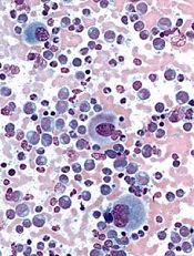
Image from UCSD
The Japanese Pharmaceuticals and Medical Devices Agency (PMDA) has approved 2 uses of the tyrosine kinase inhibitor (TKI) ponatinib (Iclusig®).
The drug is now approved to treat recurrent or refractory Philadelphia chromosome-positive acute lymphoblastic leukemia (Ph+ ALL) and chronic myeloid leukemia (CML) that was resistant to or intolerant of prior treatment.
Ponatinib will be manufactured and sold by Otsuka Pharmaceutical Co., Ltd.
Due to the limited existing treatment options for patients in Japan, Otsuka said it will provide access to ponatinib free of charge as soon as procedures are in place from an ethical standpoint.
This program will be offered at medical institutions where clinical trials of ponatinib were performed and which are amenable to accepting the drug access program until the product is listed on the Japan National Health Insurance price list.
About ponatinib
Ponatinib is a TKI discovered by ARIAD Pharmaceuticals, Inc. The drug has demonstrated activity against native and mutated BCR-ABL and other kinases.
The PMDA’s approval of ponatinib for CML and Ph+ ALL is based on data from a phase 1/2 trial of Japanese patients, a phase 1 trial, and the phase 2 PACE trial.
Extended follow-up data from the PACE trial, collected in 2013, suggested ponatinib can increase the risk of thrombotic events. When these data came to light, officials in the European Union and the US, where ponatinib had already been approved, began to investigate the drug.
Ponatinib was pulled from the US market for a little over 2 months, and trials of the TKI were placed on partial hold while the US Food and Drug Administration evaluated the drug’s safety. Ponatinib went back on the market in January 2014, with new safety measures in place.
Ponatinib was not pulled from the market in the European Union, but the European Medicine’s Agency released recommendations for safer use of the TKI. The Committee for Medicinal Products for Human Use reviewed data on ponatinib and decided its benefits outweigh its risks.
In addition to the European Union and the US, ponatinib has been approved in Australia, Canada, Israel, and Switzerland. ![]()
Study shows RT underused in developing countries

Photo courtesy of ASTRO
BOSTON—A new study suggests that roughly half of cancer patients in developing countries need radiation therapy (RT) to treat their disease, but many of these patients do not have access to it.
Examining 9 developing countries, investigators found that between 18% and 82% of patients who can benefit from RT do not receive the treatment.
These findings were presented at ASTRO’s 58th Annual Meeting (abstract 82).
“Access to radiation therapy remains limited in low-and middle-income countries,” said study investigator Elena Fidarova, MD, of the International Atomic Energy Agency in Vienna, Austria.
“In Ghana and the Philippines, for example, about 8 in 10 cancer patients who need radiation therapy will not receive needed treatment.”
Dr Fidarova and her colleagues conducted this study to assess levels of optimal and actual RT utilization (RTU) and calculate unmet RT need in 9 developing countries—Costa Rica, Ghana, Malaysia, the Philippines, Romania, Serbia, Slovenia, Tunisia, and Uruguay.
The investigators determined the optimal and actual RTU rates for each country. The optimal RTU rate is the proportion of all newly diagnosed cancer patients who have an indication for RT at least once in their lifetime.
An indication for RT was defined as a clinical scenario for which RT is recommended as the treatment of choice because there is evidence of its superiority to alternative modalities and/or no treatment (eg, better survival, local control, or quality of life profiles).
In clinical situations where RT was equivalent to other treatment options, all comparable modalities were included in the model, and a subsequent sensitivity analysis was conducted to determine the proportion of these patients for whom RT was indicated.
Results
The median optimal RTU for all countries was 52%. Optimal RTU rates ranged from a low of 47% for Costa Rica to a high of 56% for Tunisia. Differences in optimal RTU rates are attributable to varying incidence rates of cancer types in each country.
The median actual RTU rate was roughly half of optimal utilization, suggesting that nearly half of cancer patients across these 9 countries combined may not be receiving adequate care for their disease.
The median actual RTU rate was 28%. The lowest rates of utilization were in Ghana (9%) and the Philippines (10.3%), while the highest utilization rates were in Tunisia (46%) and Uruguay (37%).
Actual RTU rates were lower than optimal RTU rates for all 9 countries, with the smallest difference in Tunisia and the widest gap in Ghana—at nearly 43 percentage points.
The median level of unmet need was 47% for all countries combined.
Ghana and the Philippines had the highest levels of unmet need, at 82.3% and 80.5%, respectively. Costa Rica and Tunisia had the lowest levels of unmet need, at 25.5% and 18%, respectively.
The unmet need was especially high in countries with limited resources and a large population. The number of teletherapy machines per 1000 cancer cases ranged from a high of 1.3 in Tunisia to a low of 0.19 in Ghana.
The strong correlation between the actual RTU rates and the number of teletherapy machines per 1000 cancer cases/year in each country confirms that, although other access factors may be at play, the availability of RT machines is an important factor in RT utilization.
“Differences between optimal and actual RTU rates and the high percentage of unmet RT need likely stem from a number of complex reasons, although inadequate capacity for radiation therapy is the most obvious factor,” Dr Fidarova said.
“As obstacles in access to existing RT services—such as inadequate referral patterns, affordability of treatment, and geographical distribution of centers—differ by country, so does the ideal mix of solutions.” ![]()
Photo courtesy of ASTRO
BOSTON—A new study suggests that roughly half of cancer patients in developing countries need radiation therapy (RT) to treat their disease, but many of these patients do not have access to it.
Examining 9 developing countries, investigators found that between 18% and 82% of patients who can benefit from RT do not receive the treatment.
These findings were presented at ASTRO’s 58th Annual Meeting (abstract 82).
“Access to radiation therapy remains limited in low-and middle-income countries,” said study investigator Elena Fidarova, MD, of the International Atomic Energy Agency in Vienna, Austria.
“In Ghana and the Philippines, for example, about 8 in 10 cancer patients who need radiation therapy will not receive needed treatment.”
Dr Fidarova and her colleagues conducted this study to assess levels of optimal and actual RT utilization (RTU) and calculate unmet RT need in 9 developing countries—Costa Rica, Ghana, Malaysia, the Philippines, Romania, Serbia, Slovenia, Tunisia, and Uruguay.
The investigators determined the optimal and actual RTU rates for each country. The optimal RTU rate is the proportion of all newly diagnosed cancer patients who have an indication for RT at least once in their lifetime.
An indication for RT was defined as a clinical scenario for which RT is recommended as the treatment of choice because there is evidence of its superiority to alternative modalities and/or no treatment (eg, better survival, local control, or quality of life profiles).
In clinical situations where RT was equivalent to other treatment options, all comparable modalities were included in the model, and a subsequent sensitivity analysis was conducted to determine the proportion of these patients for whom RT was indicated.
Results
The median optimal RTU for all countries was 52%. Optimal RTU rates ranged from a low of 47% for Costa Rica to a high of 56% for Tunisia. Differences in optimal RTU rates are attributable to varying incidence rates of cancer types in each country.
The median actual RTU rate was roughly half of optimal utilization, suggesting that nearly half of cancer patients across these 9 countries combined may not be receiving adequate care for their disease.
The median actual RTU rate was 28%. The lowest rates of utilization were in Ghana (9%) and the Philippines (10.3%), while the highest utilization rates were in Tunisia (46%) and Uruguay (37%).
Actual RTU rates were lower than optimal RTU rates for all 9 countries, with the smallest difference in Tunisia and the widest gap in Ghana—at nearly 43 percentage points.
The median level of unmet need was 47% for all countries combined.
Ghana and the Philippines had the highest levels of unmet need, at 82.3% and 80.5%, respectively. Costa Rica and Tunisia had the lowest levels of unmet need, at 25.5% and 18%, respectively.
The unmet need was especially high in countries with limited resources and a large population. The number of teletherapy machines per 1000 cancer cases ranged from a high of 1.3 in Tunisia to a low of 0.19 in Ghana.
The strong correlation between the actual RTU rates and the number of teletherapy machines per 1000 cancer cases/year in each country confirms that, although other access factors may be at play, the availability of RT machines is an important factor in RT utilization.
“Differences between optimal and actual RTU rates and the high percentage of unmet RT need likely stem from a number of complex reasons, although inadequate capacity for radiation therapy is the most obvious factor,” Dr Fidarova said.
“As obstacles in access to existing RT services—such as inadequate referral patterns, affordability of treatment, and geographical distribution of centers—differ by country, so does the ideal mix of solutions.”
DNA methylation rises sharply at progression of Ph+ CML
A large uptick in DNA methylation occurs at the time of progression in patients with Philadelphia chromosome–positive chronic myeloid leukemia (CML), according to results from a comparative cohort study. The observation raises the possibility of new therapeutic targets for advanced disease.
Researchers led by Gerwin Heller, PhD, of the Medical University of Vienna’s Clinical Division of Oncology and Comprehensive Cancer Center, Austria, collected peripheral blood mononuclear cells from 23 patients with CML during various disease phases (chronic phase in 17, acute phase in 5, and blast crisis in 9) and from 5 healthy controls.

They isolated DNA and analyzed it with a novel next-generation sequencing approach, known as reduced representation bisulfite sequencing, to identify the presence and extent of methylation.
Compared with the healthy controls, patients in chronic-phase CML had 666 differentially methylated CpG (cytosine-phosphate-guanine) sites, whereas those in blast crisis had 6,912 differentially methylated CpG sites (Leukemia. 2016;30:1861-8). Some 44% of the former but 88% of the latter affected CpG sites showed increased methylation.
Among patients with chronic-phase CML who experienced progression to the accelerated phase or blast crisis, analyses identified between 344 and 897 genes that were methylated at the time of progression but had not been at the time of diagnosis.
RNA sequencing determined that many of these newly methylated genes showed downregulated expression in the samples collected during blast crisis, compared with those collected during chronic phase disease. These genes included some tumor suppressor genes (EPB41L3, PRDX2), regulators of cell proliferation (BCL11B, NDRG2, PID1), and regulators of drug metabolism (CYP1B1), among others.
Treatment of CML cells with the epigenetically active drugs azacitidine (a DNA methyltransferase inhibitor) and trichostatin A (a histone deacetylase inhibitor) led to re-expression of the CYP1B1 gene.
“Together, our results demonstrate that CpG site methylation clearly increases during CML progression and that it may provide a useful basis for revealing new targets of therapy in advanced CML,” wrote Dr. Heller and colleagues, who disclosed that one of them has received research grants from Novartis and Ariad.
“Our findings suggest that investigating the efficacy of [DNA methyltransferase inhibitors] in patients with progressed CML should be considered,” they said.
A large uptick in DNA methylation occurs at the time of progression in patients with Philadelphia chromosome–positive chronic myeloid leukemia (CML), according to results from a comparative cohort study. The observation raises the possibility of new therapeutic targets for advanced disease.
Researchers led by Gerwin Heller, PhD, of the Medical University of Vienna’s Clinical Division of Oncology and Comprehensive Cancer Center, Austria, collected peripheral blood mononuclear cells from 23 patients with CML during various disease phases (chronic phase in 17, acute phase in 5, and blast crisis in 9) and from 5 healthy controls.
They isolated DNA and analyzed it with a novel next-generation sequencing approach, known as reduced representation bisulfite sequencing, to identify the presence and extent of methylation.
Compared with the healthy controls, patients in chronic-phase CML had 666 differentially methylated CpG (cytosine-phosphate-guanine) sites, whereas those in blast crisis had 6,912 differentially methylated CpG sites (Leukemia. 2016;30:1861-8). Some 44% of the former but 88% of the latter affected CpG sites showed increased methylation.
Among patients with chronic-phase CML who experienced progression to the accelerated phase or blast crisis, analyses identified between 344 and 897 genes that were methylated at the time of progression but had not been at the time of diagnosis.
RNA sequencing determined that many of these newly methylated genes showed downregulated expression in the samples collected during blast crisis, compared with those collected during chronic phase disease. These genes included some tumor suppressor genes (EPB41L3, PRDX2), regulators of cell proliferation (BCL11B, NDRG2, PID1), and regulators of drug metabolism (CYP1B1), among others.
Treatment of CML cells with the epigenetically active drugs azacitidine (a DNA methyltransferase inhibitor) and trichostatin A (a histone deacetylase inhibitor) led to re-expression of the CYP1B1 gene.
“Together, our results demonstrate that CpG site methylation clearly increases during CML progression and that it may provide a useful basis for revealing new targets of therapy in advanced CML,” wrote Dr. Heller and colleagues, who disclosed that one of them has received research grants from Novartis and Ariad.
“Our findings suggest that investigating the efficacy of [DNA methyltransferase inhibitors] in patients with progressed CML should be considered,” they said.
FROM LEUKEMIA
Key clinical point: Increases in DNA methylation occur at progression of Ph+ CML; this realization may enable new therapeutic targets.
Major finding: Patients who progressed from chronic phase to accelerated phase or blast crisis had up to 897 genes that were newly methylated since diagnosis, including some tumor-suppressor genes and regulators of cell proliferation and drug metabolism.
Data source: A cohort study of 23 patients with CML and 5 healthy controls.
Disclosures: The investigators disclosed that one of the authors has received research grants from Novartis and Ariad.

Cells might protect cancer patients from infection

receiving chemotherapy
Photo by Rhoda Baer
Researchers say they have discovered a type of macrophage that may protect against lung infections during chemotherapy.
These macrophages, found in the lungs of mice, were able to survive chemotherapy.
The macrophages could remove bacteria when activated by a vaccine, which improved survival in mice with lethal bacterial pneumonia that had received chemotherapy and were therefore depleted of neutrophils.
The researchers said these results suggest the cells—known as vaccine-induced macrophages (ViMs)— might be able to protect cancer patients from life-threatening infections.
“We have identified a new form of housekeeping macrophage in mice that may, in future, be harnessed to protect against lung infections—like bacterial pneumonia—that remain one of the greatest threats to survival of cancer patients during chemotherapy,” said Peter Murray, PhD, of St. Jude Children’s Research Hospital in Memphis, Tennessee.
Dr Murray and his colleagues detailed this discovery in PNAS.
Working in a mouse model that mimics infection in chemotherapy-treated patients, the researchers found that vaccination protected mice from Pseudomonas aeruginosa pneumonia.
The quest to understand how such protection was possible in the absence of neutrophils led the team to discover ViMs.
The researchers found that ViMs were produced in the lungs following vaccination, proliferated locally, and could persist for at least a month.
Analyses suggested ViMs are closely related to alveolar macrophages, which originate in the embryo, reside in the air-exposed surfaces of alveoli, and are self-maintained in adults.
“All lines of cellular and molecular evidence in this study point to alveolar macrophages as the source of ViMs,” Dr Murray said.
However, unlike alveolar macrophages, the population of ViMs remained stable during chemotherapy and exhibited enhanced phagocytic activity.
When ViMs were transferred to unvaccinated mice depleted of neutrophils via chemotherapy, the animals were protected from lethal Pseudomonas infections.
“We now know that increasing the number of ViMs in the tissue can compensate for the immune deficit caused by chemotherapy,” said study author Akinobu Kamei, MD, of St. Jude Children’s Research Hospital.
“In this study, we relied on vaccination prior to chemotherapy. Going forward, we will explore other, more practical methods for use at the bedside to effectively induce tissue-resident macrophages like ViMs.”
The possible approaches include using drugs or cytokines to induce protection in the immune-compromised host.
How anti-CD44 antibodies fight AML

Image by Lance Liotta
New research appears to explain how antibodies that target CD44 fight acute myeloid leukemia (AML).
Previous research showed that anti-CD44 antibodies inhibit proliferation and induce differentiation in AML, but it wasn’t clear how or why this happens.
The new study suggests anti-CD44 antibodies work by inhibiting 2 “major players” of the PI3K/Akt/mTOR pathway—mTORC1 and mTORC2.
Jasmeen Merzaban, PhD, of King Abdullah University of Science and Technology in Thuwal, Saudi Arabia, and her colleagues described this discovery in a letter to Leukemia.
The researchers tested an anti-CD44 antibody known as A3D8 in cell lines representing different AML subtypes (HL60, THP-1, and KG1a) as well as a mouse model of AML.
In these experiments, A3D8 inhibited proliferation and induced differentiation in AML cells. This was accompanied by a decrease in phosphorylation of the mTORC1 and mTORC2 complexes, which was strongly correlated with inhibition of the PI3K/Akt pathway.
The researchers said this finding is important because a complete shutdown of mTOR signaling is probably needed to disrupt the multiple feedback loops that can fuel AML growth, and drugs that only inhibit one of these complexes have, in the past, failed to demonstrate a therapeutic benefit for patients with AML.
“A growing body of evidence suggests that a broader inhibitor would result in a more potent therapeutic effect,” Dr Merzaban said.
She and her colleagues believe an anti-CD44 antibody like A3D8 might just be that type of inhibitor.
They also noted that A3D8 was able to induce differentiation in different subtypes of AML and did not seem to present any toxicity issues.
“We show that the anti-CD44 antibody used for our studies had no effect on normal blood cells,” said Samah Gadhoum, PhD, a research scientist in Dr Merzaban’s lab.
“However, more work is needed to carefully determine the effect of these antibodies on other cells and other cellular functions within the body.”
The researchers are now conducting follow-up experiments, but they believe their results support the use of anti-CD44 antibodies to treat different types of AML.