User login
No link found between vaccinations, IBD
Neither childhood immunizations nor H1N1 influenza vaccination increased the risk of inflammatory bowel disease, according to a meta-analysis of 11 studies in the August issue of Clinical Gastroenterology and Hepatology published online (2015 [doi:10.1016/j.cgh.2015.04.179]).
“Overall, our results did not find any significant increased risk of developing IBD after childhood immunization with BCG [bacille Calmette-Guérin], diphtheria, tetanus, poliomyelitis, smallpox, pertussis, measles, mumps, and rubella-containing vaccines,” said Dr. Guillaume Pineton de Chambrun of Lille (France) University Hospital and his associates. “The results of this meta-analysis are globally reassuring regarding the risk of developing IBD after childhood vaccination. Vaccines that are developed to protect against an infectious disease or its consequences are, for the majority, not a risk for the subsequent development of intestinal inflammatory disease.”

Besides the childhood vaccines, H1N1 vaccination was not linked to IBD in a single large study of adults (risk ratio, 1.13; 95% confidence interval, 0.97-1.32), the investigators said.
Controversies about immunizations and IBD date to at least 1995, when a report by Thompson et al. linked live measles vaccine to the disease (Lancet 1995;345:1071-4), said the investigators. “However, many publications after this report investigating vaccination with measles-containing vaccines did not show any association between immunization and IBD,” they emphasized. “Epidemiologic studies also investigated other vaccines such as BCG, diphtheria, tetanus, poliomyelitis, smallpox, pertussis, rubella, and mumps, reporting conflicting results.”
To further clarify the issue, they compared rates of IBD, Crohn’s disease, and ulcerative colitis among vaccinated and unvaccinated patients from eight case-control and three cohort studies published between 1970 and June 2014. When looking at IBD overall, 95% CIs for RRs all crossed 1.0, indicating no significant associations, they said. A subgroup analysis did link poliomyelitis vaccination with increased risk of developing Crohn’s disease (RR, 2.28; 95% CI, 1.12-4.63) or ulcerative colitis (RR, 3.48; 95% CI, 1.2-9.71), but the studies were dissimilar enough that the results need to be interpreted cautiously, they added.
To find the studies, the researchers searched MEDLINE, EMBASE, and the Cochrane central trials registry for randomized controlled trials, controlled clinical trials, cohort studies, and case-control studies published in any language and that included the terms ulcerative colitis, Crohn’s disease, inflammatory bowel disease, colitis, or ileitis. This approach yielded six population-based and two hospital-based case-control studies, and three population-based cohort studies. The studies of childhood immunizations included 2,399 patients with IBD and 33,747 controls, and the H1N1 study included 14,842 patients with IBD and almost 2 million controls, said the investigators.
The studies were “extremely heterogeneous” in terms of design, sample size, geographic location, and methods used to help patients recall their vaccination history, the investigators noted. “Vaccination protocols varied between countries and evolved through the years, with different types of vaccines and schedules leading to difficulties in risk evaluation,” they said. “Moreover, some vaccines used were live attenuated vaccines such as measles, oral poliomyelitis, or whole-cell pertussis vaccines, and may have a different effect on immune system activation and dysregulation, compared with other inactivated acellular vaccines.”
The Digitscience Foundation supported the research. The researchers declared having no conflicts of interest.
Neither childhood immunizations nor H1N1 influenza vaccination increased the risk of inflammatory bowel disease, according to a meta-analysis of 11 studies in the August issue of Clinical Gastroenterology and Hepatology published online (2015 [doi:10.1016/j.cgh.2015.04.179]).
“Overall, our results did not find any significant increased risk of developing IBD after childhood immunization with BCG [bacille Calmette-Guérin], diphtheria, tetanus, poliomyelitis, smallpox, pertussis, measles, mumps, and rubella-containing vaccines,” said Dr. Guillaume Pineton de Chambrun of Lille (France) University Hospital and his associates. “The results of this meta-analysis are globally reassuring regarding the risk of developing IBD after childhood vaccination. Vaccines that are developed to protect against an infectious disease or its consequences are, for the majority, not a risk for the subsequent development of intestinal inflammatory disease.”
Besides the childhood vaccines, H1N1 vaccination was not linked to IBD in a single large study of adults (risk ratio, 1.13; 95% confidence interval, 0.97-1.32), the investigators said.
Controversies about immunizations and IBD date to at least 1995, when a report by Thompson et al. linked live measles vaccine to the disease (Lancet 1995;345:1071-4), said the investigators. “However, many publications after this report investigating vaccination with measles-containing vaccines did not show any association between immunization and IBD,” they emphasized. “Epidemiologic studies also investigated other vaccines such as BCG, diphtheria, tetanus, poliomyelitis, smallpox, pertussis, rubella, and mumps, reporting conflicting results.”
To further clarify the issue, they compared rates of IBD, Crohn’s disease, and ulcerative colitis among vaccinated and unvaccinated patients from eight case-control and three cohort studies published between 1970 and June 2014. When looking at IBD overall, 95% CIs for RRs all crossed 1.0, indicating no significant associations, they said. A subgroup analysis did link poliomyelitis vaccination with increased risk of developing Crohn’s disease (RR, 2.28; 95% CI, 1.12-4.63) or ulcerative colitis (RR, 3.48; 95% CI, 1.2-9.71), but the studies were dissimilar enough that the results need to be interpreted cautiously, they added.
To find the studies, the researchers searched MEDLINE, EMBASE, and the Cochrane central trials registry for randomized controlled trials, controlled clinical trials, cohort studies, and case-control studies published in any language and that included the terms ulcerative colitis, Crohn’s disease, inflammatory bowel disease, colitis, or ileitis. This approach yielded six population-based and two hospital-based case-control studies, and three population-based cohort studies. The studies of childhood immunizations included 2,399 patients with IBD and 33,747 controls, and the H1N1 study included 14,842 patients with IBD and almost 2 million controls, said the investigators.
The studies were “extremely heterogeneous” in terms of design, sample size, geographic location, and methods used to help patients recall their vaccination history, the investigators noted. “Vaccination protocols varied between countries and evolved through the years, with different types of vaccines and schedules leading to difficulties in risk evaluation,” they said. “Moreover, some vaccines used were live attenuated vaccines such as measles, oral poliomyelitis, or whole-cell pertussis vaccines, and may have a different effect on immune system activation and dysregulation, compared with other inactivated acellular vaccines.”
The Digitscience Foundation supported the research. The researchers declared having no conflicts of interest.
FROM CLINICAL GASTROENTEROLOGY AND HEPATOLOGY
Key clinical point: A meta-analysis found no link between childhood vaccinations or H1N1 immunization in adults and inflammatory bowel disease.
Major finding: Risk ratios showed no significant associations between IBD and vaccines for BCG infection, diphtheria, tetanus, smallpox, poliomyelitis, measles, rubella, mumps, or H1N1 influenza.
Data source: Systematic review and meta-analysis of eight case-control studies and three cohort studies, encompassing 50,988 total patients.
Disclosures: The Digitscience Foundation supported the research. The researchers declared having no conflicts of interest.

Curcumin and mesalamine top mesalamine alone in ulcerative colitis
More than half of patients with mild to moderate ulcerative colitis who had not responded to optimized mesalamine treatment achieved clinical remission with the addition of 3 grams of curcumin daily, according to a multicenter randomized controlled trial.
In contrast, at week 4, none of the placebo-plus-mesalamine control group had remitted (odds ratio, 42; 95% confidence interval, 2.3-760; P = .01), reported Dr. Alon Lang of Tel-Aviv University, Israel and his associates. The study appears in the August issue of Clinical Gastroenterology and Hepatology.
Mesalamine and curcumin might have “different but potentially synergistic mechanisms of action, hence producing a better outcome” than monotherapy, wrote Dr. Lang and his coinvestigators.
Mesalamine agents are the backbone of treatment for mild to moderate ulcerative colitis, and patients who do not respond to optimized oral and topical therapy are usually stepped up to corticosteroids or immunomodulators, which can have significant side effects, Dr. Lang and his associates noted.

Based on in vitro data supporting the anti-inflammatory and antioxidative properties of curcumin (a phytochemical derived from turmeric), the researchers randomized 50 mesalamine nonresponders who had scored at least 5 but less than 12 on the Simple Clinical Colitis Activity Index to either optimized mesalamine and placebo or optimized mesalamine and 3 grams of curcumin per day. The study excluded patients who had received corticosteroids in the past 12 weeks, were currently receiving anti–tumor necrosis factor agents or cyclosporine, had laboratory abnormalities or significant comorbidities, or had a positive stool culture for Clostridium difficile or enteric pathogens (Clin. Gastroenterol. Hepatol. 2015 [doi:10.1016/j.cgh.2015.02.019])
At week 4, 14 patients (53.8%) in the curcumin-mesalamine group scored 2 or less on the SCCAI, and 17 patients (65.3%) experienced a clinical response, defined as a SCCAI score of 3 or less, the researchers reported. But none of the control group remitted and only three (12.5%) responded clinically (OR, 13.2; 95% CI, 3.1 to 56.6; P < .001), they added. “Admittedly, such nil rate of remission in the placebo arm is much lower than that observed in trials of mesalamine therapy for this indication,” they said. “However, in contrast with prior trials, all patients in the present study were already receiving and failing to respond to optimized oral and topical mesalamine treatment ... it is likely that the zero remission rate and the low (12%) rate of clinical improvement in the placebo arm are a result of this design.”
Use of the SCCAI also could have explained the low remission rate in placebo-treated patients, the researchers said. “Indeed, the SCCAI was recently suggested to be more reflective of actual disease activity compared with other clinical scores, and the strict definition of clinical remission requiring an SCCAI was recently shown to correlate with patients’ genuine sense of remission,” they noted.
In the study, 38% of the intervention group achieved endoscopic remission (a partial Mayo score of 1 or less), compared with none of 16 patients evaluated in the placebo group (OR, 20.7; 95% CI, 1.1 to 393; P = .043). Adverse events were rare and similar between the two groups, the researchers noted.
The trial design lacked a dose-finding component, the sample size was “modest,” and the researchers did not perform a power calculation because no previous studies had looked at the efficacy of curcumin in ulcerative colitis, they wrote.
Sheba Medical Center and the Leona M. and Harry B. Helmsley Charitable Trust helped fund the research. Dr. Lang reported no relevant conflicts of interest, and five coauthors reported relationships with numerous pharmaceutical companies.
Curcumin (diferuloylmethane, a polyphenol) is derived from the Curcuma longa plant. Curcumin has been described as “pharmacodynamically fierce” (owing to its interference in multiple inflammatory and cancer pathways) but “pharmacokinetically feeble” (owing to its chemical instability, poor systemic uptake, and extensive biotransformation) (Pharmacol. Rev. 2013;66:222-307). However, the poor bioavailability of curcumin may be an advantageous property in the treatment of inflammatory bowel disease. In one previous study in ulcerative colitis, oral curcumin (2 g/day) was more effective than was placebo in maintaining remission at 6 months (relapse rates of 5% vs. 21%; P = .04) (Clin. Gastroenterol. Hepatol. 2006;4:1502-6). In another study of patients with mild to moderate distal UC, there was no benefit of rectal curcumin (140 mg dissolved in 20 mL of water taken once daily) over placebo for the induction of remission at 8 weeks (J. Crohns Colitis 2014;8:208-14).

|
| Dr. Themistocles Dassopoulos |
Lang and colleagues performed a methodologically sound trial in patients with mild to moderate UC failing optimized, combined, oral, and topical mesalamine therapy. Fifty subjects were treated with curcumin (3 g/day; n = 26) vs. placebo (n = 24) for 4 weeks.
The trial met the primary endpoint of clinical remission at week 4, which was 53.8% in the curcumin arm vs. 0% in the placebo arm (odds ratio, 42; 95% confidence interval, 2.3-760). The trial also met all the secondary endpoints of clinical response, endoscopic remission and endoscopic improvement. In this small study, adverse events were rare and comparable between the two arms. More studies are needed to assess the therapeutic efficacy of oral and rectal curcumin, explore the dose-response curve, and assess safety. If proven effective in UC, curcumin is poised to become an especially attractive treatment option given its low cost, tolerability, and (apparent) safety at pharmacologic doses.
Dr. Themistocles Dassopoulos, director of the Baylor Center for Inflammatory Bowel Diseases, Dallas. He has no conflicts of interest.
Curcumin (diferuloylmethane, a polyphenol) is derived from the Curcuma longa plant. Curcumin has been described as “pharmacodynamically fierce” (owing to its interference in multiple inflammatory and cancer pathways) but “pharmacokinetically feeble” (owing to its chemical instability, poor systemic uptake, and extensive biotransformation) (Pharmacol. Rev. 2013;66:222-307). However, the poor bioavailability of curcumin may be an advantageous property in the treatment of inflammatory bowel disease. In one previous study in ulcerative colitis, oral curcumin (2 g/day) was more effective than was placebo in maintaining remission at 6 months (relapse rates of 5% vs. 21%; P = .04) (Clin. Gastroenterol. Hepatol. 2006;4:1502-6). In another study of patients with mild to moderate distal UC, there was no benefit of rectal curcumin (140 mg dissolved in 20 mL of water taken once daily) over placebo for the induction of remission at 8 weeks (J. Crohns Colitis 2014;8:208-14).
|
|
| Dr. Themistocles Dassopoulos |
Lang and colleagues performed a methodologically sound trial in patients with mild to moderate UC failing optimized, combined, oral, and topical mesalamine therapy. Fifty subjects were treated with curcumin (3 g/day; n = 26) vs. placebo (n = 24) for 4 weeks.
The trial met the primary endpoint of clinical remission at week 4, which was 53.8% in the curcumin arm vs. 0% in the placebo arm (odds ratio, 42; 95% confidence interval, 2.3-760). The trial also met all the secondary endpoints of clinical response, endoscopic remission and endoscopic improvement. In this small study, adverse events were rare and comparable between the two arms. More studies are needed to assess the therapeutic efficacy of oral and rectal curcumin, explore the dose-response curve, and assess safety. If proven effective in UC, curcumin is poised to become an especially attractive treatment option given its low cost, tolerability, and (apparent) safety at pharmacologic doses.
Dr. Themistocles Dassopoulos, director of the Baylor Center for Inflammatory Bowel Diseases, Dallas. He has no conflicts of interest.
More than half of patients with mild to moderate ulcerative colitis who had not responded to optimized mesalamine treatment achieved clinical remission with the addition of 3 grams of curcumin daily, according to a multicenter randomized controlled trial.
In contrast, at week 4, none of the placebo-plus-mesalamine control group had remitted (odds ratio, 42; 95% confidence interval, 2.3-760; P = .01), reported Dr. Alon Lang of Tel-Aviv University, Israel and his associates. The study appears in the August issue of Clinical Gastroenterology and Hepatology.
Mesalamine and curcumin might have “different but potentially synergistic mechanisms of action, hence producing a better outcome” than monotherapy, wrote Dr. Lang and his coinvestigators.
Mesalamine agents are the backbone of treatment for mild to moderate ulcerative colitis, and patients who do not respond to optimized oral and topical therapy are usually stepped up to corticosteroids or immunomodulators, which can have significant side effects, Dr. Lang and his associates noted.
Based on in vitro data supporting the anti-inflammatory and antioxidative properties of curcumin (a phytochemical derived from turmeric), the researchers randomized 50 mesalamine nonresponders who had scored at least 5 but less than 12 on the Simple Clinical Colitis Activity Index to either optimized mesalamine and placebo or optimized mesalamine and 3 grams of curcumin per day. The study excluded patients who had received corticosteroids in the past 12 weeks, were currently receiving anti–tumor necrosis factor agents or cyclosporine, had laboratory abnormalities or significant comorbidities, or had a positive stool culture for Clostridium difficile or enteric pathogens (Clin. Gastroenterol. Hepatol. 2015 [doi:10.1016/j.cgh.2015.02.019])
At week 4, 14 patients (53.8%) in the curcumin-mesalamine group scored 2 or less on the SCCAI, and 17 patients (65.3%) experienced a clinical response, defined as a SCCAI score of 3 or less, the researchers reported. But none of the control group remitted and only three (12.5%) responded clinically (OR, 13.2; 95% CI, 3.1 to 56.6; P < .001), they added. “Admittedly, such nil rate of remission in the placebo arm is much lower than that observed in trials of mesalamine therapy for this indication,” they said. “However, in contrast with prior trials, all patients in the present study were already receiving and failing to respond to optimized oral and topical mesalamine treatment ... it is likely that the zero remission rate and the low (12%) rate of clinical improvement in the placebo arm are a result of this design.”
Use of the SCCAI also could have explained the low remission rate in placebo-treated patients, the researchers said. “Indeed, the SCCAI was recently suggested to be more reflective of actual disease activity compared with other clinical scores, and the strict definition of clinical remission requiring an SCCAI was recently shown to correlate with patients’ genuine sense of remission,” they noted.
In the study, 38% of the intervention group achieved endoscopic remission (a partial Mayo score of 1 or less), compared with none of 16 patients evaluated in the placebo group (OR, 20.7; 95% CI, 1.1 to 393; P = .043). Adverse events were rare and similar between the two groups, the researchers noted.
The trial design lacked a dose-finding component, the sample size was “modest,” and the researchers did not perform a power calculation because no previous studies had looked at the efficacy of curcumin in ulcerative colitis, they wrote.
Sheba Medical Center and the Leona M. and Harry B. Helmsley Charitable Trust helped fund the research. Dr. Lang reported no relevant conflicts of interest, and five coauthors reported relationships with numerous pharmaceutical companies.
FROM CLINICAL GASTROENTEROLOGY AND HEPATOLOGY
Key clinical point: About half of mesalamine nonresponders with active mild to moderate ulcerative colitis remitted after adding curcumin.
Major finding: Week 4 remission rates were 53.8% for the curcumin-mesalamine group and 0% for the placebo-mesalamine group (P = .01).
Data source: Multicenter randomized, placebo-controlled trial of 50 patients with mild to moderate ulcerative colitis that had failed to respond to optimized mesalamine.
Disclosures: Sheba Medical Center and the Leona M. and Harry B. Helmsley Charitable Trust helped fund the research. Dr. Lang reported no relevant conflicts of interest; five coauthors reported relationships with numerous pharmaceutical companies.

Elective colectomy topped medical therapy for advanced ulcerative colitis
For adults with advanced ulcerative colitis (UC) older than 50 years of age, elective colectomy offered a significantly higher survival rate than did medical therapy, according to a retrospective study of 8,371 patients.
Dr. Meenakshi Bewtra of the University of Pennsylvania, Philadelphia, and her coinvestigators matched 830 UC patients seeking elective colectomy for treatment with 7,541 UC patients opting for more traditional medical therapy, all recruited using data from Medicaid and Medicare from 2000 to 2011 (Ann. Intern. Med. July 14, 2015 [doi:10.7326/M14-0960]).
In total, 63 patients who received elective colectomy died, compared with 783 patients in the medical therapy cohort. Mortality rates per cohort were 34 and 54 per 1,000 person-years, respectively. Furthermore, patients were more likely to respond more favorably to elective colectomy than to medical therapy, with an adjusted hazard ratio of 0.67. Additional post hoc analysis revealed higher survival odds with colectomy for patients age 50 years or older (HR, 0.60; P = .032). “These findings warrant discussion with patients when one is weighing the risks and benefits of different medical therapies and total colectomy,” the investigators said.
The authors noted that the study had several limitations, such as potential residual confounding and the possibility of reduced statistical power in subsequent analyses because several databases were used to cull data.
The study was funded by grants from the National Institutes of Health and the Agency for Healthcare Research and Quality. Dr. Bewtra disclosed receiving a grant from NIH and accepting speaking engagements for Imedex and the Crohn’s & Colitis Foundation of America/Robert Michael Educational Institute outside the submitted work.
FROM THE ANNALS OF INTERNAL MEDICINE
HBV, tuberculosis reactivations after anti-TNFs rare in IBD patients on biologic therapy
WASHINGTON – Reactivation of tuberculosis and hepatitis B virus infections after starting treatment with anti–tumor necrosis factor (TNF) therapy was “rare” in a large national cohort study of veterans with inflammatory bowel disease (IBD), Dr. Jason Hou reported at the annual Digestive Disease Week.
The estimated rate of tuberculosis reactivations was 2.8 per 10,000 patient-years of exposure to anti-TNF therapy “in a fairly well-screened cohort of IBD patients,” said Dr. Hou, codirector of the Inflammatory Bowel Disease Center at Baylor College of Medicine, Houston. The estimate for HBV reactivation or acute liver failure in this cohort was less than 1.4 per 10,000 patient-years of exposure, he noted.

Practice guidelines recommend that patients with IBD should be screened for latent tuberculosis and HBV before starting treatment with an anti-TNF drug, but how often reactivation actually occurs in this population is not known, he pointed out. Moreover, recent studies have questioned the cost-effectiveness of universal screening for HBV before starting treatment, and TB reactivation rates in patients with IBD treated with anti-TNF drugs “have not been assessed on a national population level.”
Dr. Hou and his associates used a national Veterans Affairs database to identify patients who had been diagnosed between 2003 and 2011 with Crohn’s disease or ulcerative colitis, based on ICD-9 codes, and had filled at least one prescription for infliximab, adalimumab, or certolizumab pegol; they also looked for codes related to possible tuberculosis reactivation or acute liver failure related to HBV reactivation at the time of or after anti-TNF therapy was started. Cases were then verified by reviewing electronic medical records of patients to confirm the diagnosis of tuberculosis or HBV, exposure to an anti-TNF drug, and completion of pretreatment screening.
They identified 3,357 patients with IBD who had received a prescription for an anti-TNF treatment, representing 7,210 patient-years of anti-TNF treatment; most were men and were white. The mean time on treatment was about 2 years, and they had started treatment at a mean age of 57 years.
Screening rates for tuberculosis were relatively high and remained stable throughout the study period, at 72% overall, but HBV screening rates were low, at 24% overall. HBV screening was not included in practice guidelines until 2006, and the rate significantly increased from under 10% at the beginning of the period studied to 42% at the end, Dr. Hou noted.
They identified 23 patients with ICD-9 codes related to tuberculosis occurring after they started anti-TNF therapy, but “on chart review, only two patients were confirmed to have tuberculosis reactivation” related to anti-TNF therapy, which occurred at 8 and 18 months of treatment, he said. “Very interestingly, both of these patients had latent tuberculosis and documented completed courses of INH [isonicotinylhydrazine] and a negative chest x-ray prior to [anti-]TNF initiation.” Because the only cases of tuberculosis reactivation occurred in patients with a prior history of latent tuberculosis, “we should maintain extra diligence in those patients even if appropriate screening has been performed,” he advised.
They identified 12 patients with codes related to hepatitis B after the treatment started; 2 had clinically relevant outcomes, but the chart review indicated that neither case was HPV reactivation.
There were two cases of patients with positive HBV tests. One of these patients, who developed acute jaundice with serologies consistent with acute HBV infection, reported a possible exposure to HBV and was newly diagnosed with HIV, associated with the same encounter. This patient was on infliximab and continued treatment, and HBV viremia spontaneously cleared without antiviral treatment, Dr. Hou said.
The second “clinically relevant” case was a patient with chronic HBV infection, identified in a presurgical screen, who was asymptomatic and had normal liver function tests. Infliximab was discontinued, and the patient started antiviral therapy; viremia resolved and the patient resumed treatment with infliximab “without complications,” Dr. Hou said.
Another four patients were identified related to codes for acute liver failure, but on chart review, none had liver failure related to hepatitis B.
The study’s limitations include the possibility that patients may receive medical care outside the VA system, which would not be picked up in the analysis, he said, but added, “when we look at our estimate of tuberculosis reactivation, they mirror almost exactly those reported in the rheumatology literature in screened populations.”
Another limitation is that the study depended on the accuracy of ICD-9 codes for the diagnoses, but all outcomes were verified in the chart review, he said. Dr. Hou is also director of Inflammatory Bowel Diseases and an investigator in the clinical epidemiology and outcomes program in the Center for Innovations in Quality Effectiveness and Safety at the Michael E. DeBakey VA Medical Center, Houston.
WASHINGTON – Reactivation of tuberculosis and hepatitis B virus infections after starting treatment with anti–tumor necrosis factor (TNF) therapy was “rare” in a large national cohort study of veterans with inflammatory bowel disease (IBD), Dr. Jason Hou reported at the annual Digestive Disease Week.
The estimated rate of tuberculosis reactivations was 2.8 per 10,000 patient-years of exposure to anti-TNF therapy “in a fairly well-screened cohort of IBD patients,” said Dr. Hou, codirector of the Inflammatory Bowel Disease Center at Baylor College of Medicine, Houston. The estimate for HBV reactivation or acute liver failure in this cohort was less than 1.4 per 10,000 patient-years of exposure, he noted.
Practice guidelines recommend that patients with IBD should be screened for latent tuberculosis and HBV before starting treatment with an anti-TNF drug, but how often reactivation actually occurs in this population is not known, he pointed out. Moreover, recent studies have questioned the cost-effectiveness of universal screening for HBV before starting treatment, and TB reactivation rates in patients with IBD treated with anti-TNF drugs “have not been assessed on a national population level.”
Dr. Hou and his associates used a national Veterans Affairs database to identify patients who had been diagnosed between 2003 and 2011 with Crohn’s disease or ulcerative colitis, based on ICD-9 codes, and had filled at least one prescription for infliximab, adalimumab, or certolizumab pegol; they also looked for codes related to possible tuberculosis reactivation or acute liver failure related to HBV reactivation at the time of or after anti-TNF therapy was started. Cases were then verified by reviewing electronic medical records of patients to confirm the diagnosis of tuberculosis or HBV, exposure to an anti-TNF drug, and completion of pretreatment screening.
They identified 3,357 patients with IBD who had received a prescription for an anti-TNF treatment, representing 7,210 patient-years of anti-TNF treatment; most were men and were white. The mean time on treatment was about 2 years, and they had started treatment at a mean age of 57 years.
Screening rates for tuberculosis were relatively high and remained stable throughout the study period, at 72% overall, but HBV screening rates were low, at 24% overall. HBV screening was not included in practice guidelines until 2006, and the rate significantly increased from under 10% at the beginning of the period studied to 42% at the end, Dr. Hou noted.
They identified 23 patients with ICD-9 codes related to tuberculosis occurring after they started anti-TNF therapy, but “on chart review, only two patients were confirmed to have tuberculosis reactivation” related to anti-TNF therapy, which occurred at 8 and 18 months of treatment, he said. “Very interestingly, both of these patients had latent tuberculosis and documented completed courses of INH [isonicotinylhydrazine] and a negative chest x-ray prior to [anti-]TNF initiation.” Because the only cases of tuberculosis reactivation occurred in patients with a prior history of latent tuberculosis, “we should maintain extra diligence in those patients even if appropriate screening has been performed,” he advised.
They identified 12 patients with codes related to hepatitis B after the treatment started; 2 had clinically relevant outcomes, but the chart review indicated that neither case was HPV reactivation.
There were two cases of patients with positive HBV tests. One of these patients, who developed acute jaundice with serologies consistent with acute HBV infection, reported a possible exposure to HBV and was newly diagnosed with HIV, associated with the same encounter. This patient was on infliximab and continued treatment, and HBV viremia spontaneously cleared without antiviral treatment, Dr. Hou said.
The second “clinically relevant” case was a patient with chronic HBV infection, identified in a presurgical screen, who was asymptomatic and had normal liver function tests. Infliximab was discontinued, and the patient started antiviral therapy; viremia resolved and the patient resumed treatment with infliximab “without complications,” Dr. Hou said.
Another four patients were identified related to codes for acute liver failure, but on chart review, none had liver failure related to hepatitis B.
The study’s limitations include the possibility that patients may receive medical care outside the VA system, which would not be picked up in the analysis, he said, but added, “when we look at our estimate of tuberculosis reactivation, they mirror almost exactly those reported in the rheumatology literature in screened populations.”
Another limitation is that the study depended on the accuracy of ICD-9 codes for the diagnoses, but all outcomes were verified in the chart review, he said. Dr. Hou is also director of Inflammatory Bowel Diseases and an investigator in the clinical epidemiology and outcomes program in the Center for Innovations in Quality Effectiveness and Safety at the Michael E. DeBakey VA Medical Center, Houston.
AT DDW® 2015
Key clinical point: Significant clinical events related to tuberculosis and hepatitis B virus reactivation after initiating anti–tumor necrosis factor therapy in a screened population are rare.
Major finding: Rates of tuberculosis and HBV were estimated at 2.8 and less than 1.4 cases per 10,000 patient-years of exposure to treatment, respectively, in a large, fairly well screened national cohort of veterans with IBD treated with an anti-TNF drug.
Data source: A retrospective cohort study evaluated the rates of tuberculosis and HBV reactivation, identifying cases of IBD treated with anti-TNF therapy in a Veterans Health Administration database during 2003-2011, confirming cases, treatment, and HBV and TB screening status with electronic medical record review.
Disclosures: Dr. Hou’s financial disclosure includes receiving consulting, speaking, teaching, and/or advisory fees from Janssen, Aptalis, and Abbvie.

Top-down Crohn’s treatment holds up long term
WASHINGTON – More intensive treatment with early combined immunosuppression was associated with a longer time to first flare and fewer flares than conventional management during long-term follow-up of patients with Crohn’s disease.
Fewer patients treated with the “top-down” approach required anti–tumor necrosis factor (anti-TNF) agents or corticosteroids during follow-up, Dr. Daniël Hoekman reported during the annual Digestive Disease Week.
Current guidelines advocate an initial “step-up” approach for Crohn’s disease that focuses on controlling symptoms with corticosteroids followed by immunomodulators and then anti-TNF inhibitors to manage the chronic relapsing-remitting course of Crohn’s.
A new strategy has been proposed with the aim of altering the disease course and slow progression by reversing the treatment paradigm from a step-up to a top-down approach. A 2-year, randomized European trial showed that a top-down approach, combining early immunosuppression with infliximab (Remicade) and azathioprine (Imuran in Canada, Azasan in the United States) followed by azathioprine monotherapy and, if necessary, additional infliximab and corticosteroids, was more effective than step-up management for induction of remission and reduction of steroids in the short term (Lancet 2008;371:660-7).
To investigate the long-term outcome of Crohn’s disease, researchers performed a retrospective chart review in 119 of the 133 trial participants followed for 8 years or 16 semesters (mean 14.2 semesters) after the initial 2-year trial period. Management during the study was left to physician discretion. At the start of the extended follow-up, most patients in both the step-up and top-down groups were on immunomodulators (66% vs. 82%) and only a small portion were using infliximab (15% vs. 20%). A total of 164 endoscopy reports were available for 59% of patients.
During follow-up, significantly more patients treated with the step-up approach than the top-down approach required anti-TNF inhibitors (73% vs. 54%; P = .04) and steroids (62% vs. 41%; P = .02), said Dr. Hoekman of the Academic Medical Center, Amsterdam.
There was no difference between the step-up and top-down groups in long-term remission rates (70% vs. 73%; P = .85).
The top-down group, however, had significantly fewer flares than the step-up group (7% vs. 19%; P = .01) and a longer time to first flare (median 9 semesters vs. 5 semesters; P = .02), he said.
There were no differences between groups in rates of hospitalization or surgery for Crohn’s disease, new fistula, or rescue treatment, defined as use of cyclosporine, experimental therapy, or surgery.
A review of the endoscopy reports suggested a trend for fewer large ulcers per patient in the top-down group than the step-up group, but this did not reach statistical significance (14% vs. 24%; P = .11), Dr. Hoekman said. The median proportion of endoscopies with remission also was similar (49% vs. 43%; P = .46).
Rates of adverse events also were similar in the top-down and step-up groups including infusion reactions (14% vs. 10%), serious infection (22% vs. 10%), malignancy (0 vs. 1 event), and dysplastic lesions (0 vs. 2 events), he said.
On Twitter@pwendl
AT DDW® 2015
Key clinical point: Top-down therapy for Crohn’s disease resulted in fewer flares and a longer time to first flare than did conventional management, but no differences in rates of long-term remission.
Major finding: The top-down group had fewer flares than did the step-up group (7% vs. 19%; P = .01).
Data source: Retrospective chart review of 119 trial participants with Crohn’s disease.
Disclosures: Dr. Hoekman reported having no financial disclosures.
GLUTOX: Identifying nonceliac gluten sensitivity
WASHINGTON – A simple dietary challenge may help identify nonceliac gluten sensitivity in patients with gastrointestinal functional disorders, results of the ongoing, randomized GLUTOX trial suggest.
Nonceliac gluten sensitivity is an emergent syndrome that causes mainly gastrointestinal symptoms and has been thought to be present in about 6% of the population. The problem is that there is no established or well-defined diagnostic flow chart to identify these patients, study author Dr. Luca Elli said at the annual Digestive Disease Week.
To determine whether gluten induces symptoms in patients responding positively to a gluten-free diet and identify those potentially affected by nonceliac gluten sensitivity, GLUTOX enrolled 100 adults with functional GI symptoms and placed them on a gluten-free diet for 21 days. Severity of symptoms was measured before and after the diet using a 10-cm visual analogue scale (VAS) and the 36-item Short Form Health Survey (SF-36).
Patients with at least a 3-cm improvement in baseline VAS were then double-blind, randomly assigned to gluten (5.6 g per day) or placebo capsules for 7 days, followed by a 7-day washout period, and then crossed over to another 7-day cycle of gluten or placebo capsules.

At baseline, the mean age was 38 years, 90% of patients were female, 55 had irritable bowel syndrome, 36 functional dyspepsia, and 9 had other unspecified functional nonspecific gastrointestinal symptoms by ROME III criteria.
Patients with celiac disease or a wheat allergy or who were on an ongoing gluten-free diet were excluded.
In all, 81 patients reported a symptomatic improvement from baseline after the 21-day gluten-free-diet (mean VAS 7.5 vs. 3.3; P value = .001), Dr. Elli, from Fondazione IRCCS Ca’ Granda, Ospedale Maggiore Policlinico, Milan reported in a distinguished abstract plenary session.
“All of the symptoms we found were reduced by the gluten-free diet, but especially patient satisfaction about stool consistency, bloating, and the global satisfaction were improved in an important way,” he said.
Symptom improvements were also associated with significant improvements in the SF-36 physical component summary and mental component summary. Most responders were female (88%); 48 had irritable bowel syndrome, 25 dyspepsia, and 8 other.
After the gluten capsule challenge, 25 of the 81 gluten-free diet responders had a severe symptomatic relapse especially in stool consistency satisfaction, bloating, and abdominal pain, Dr. Elli said.
The relapses were also associated with a significant decrease in SF-36 physical and mental component summaries.
No demographic or biochemical parameters were significantly associated with a response to the gluten challenge, Dr. Elli said. Most of those having a response were female (96%); 13 had irritable bowel syndrome, 10 dyspepsia, and 2 other.
The sequence of the gluten and placebo capsules also had no effect on the results.
If the data are confirmed, it’s possible the double-blind challenge could be used to select gluten-free diet responders and inserted into the diagnostic flow chart for patients with gastrointestinal functional disorders, Dr. Elli said.
A very important open issue is also the 56% of enrolled subjects who responded to the gluten-free diet, but did not show symptoms with the gluten double-blind challenge, he added.
During a discussion of the results, attendees questioned whether the study design, particularly the failure to biopsy patients for celiac disease at enrollment and the short 7-day washout period, was sufficient to answer the question of identifying patients with nonceliac gluten sensitivity.
Session comoderator Dr. Bernd Schnabl, from University of California–San Diego, agreed that the study design was not ideal and that the study would have been strengthened by using biopsy to rule out patients with celiac disease. That said, the study represents a start.
“If we can stratify these patients and re-challenge them longer, it could be helpful in identifying a subpopulation of functional patients who might benefit from a gluten-free diet,” he said.
On Twitter @pwendl
WASHINGTON – A simple dietary challenge may help identify nonceliac gluten sensitivity in patients with gastrointestinal functional disorders, results of the ongoing, randomized GLUTOX trial suggest.
Nonceliac gluten sensitivity is an emergent syndrome that causes mainly gastrointestinal symptoms and has been thought to be present in about 6% of the population. The problem is that there is no established or well-defined diagnostic flow chart to identify these patients, study author Dr. Luca Elli said at the annual Digestive Disease Week.
To determine whether gluten induces symptoms in patients responding positively to a gluten-free diet and identify those potentially affected by nonceliac gluten sensitivity, GLUTOX enrolled 100 adults with functional GI symptoms and placed them on a gluten-free diet for 21 days. Severity of symptoms was measured before and after the diet using a 10-cm visual analogue scale (VAS) and the 36-item Short Form Health Survey (SF-36).
Patients with at least a 3-cm improvement in baseline VAS were then double-blind, randomly assigned to gluten (5.6 g per day) or placebo capsules for 7 days, followed by a 7-day washout period, and then crossed over to another 7-day cycle of gluten or placebo capsules.
At baseline, the mean age was 38 years, 90% of patients were female, 55 had irritable bowel syndrome, 36 functional dyspepsia, and 9 had other unspecified functional nonspecific gastrointestinal symptoms by ROME III criteria.
Patients with celiac disease or a wheat allergy or who were on an ongoing gluten-free diet were excluded.
In all, 81 patients reported a symptomatic improvement from baseline after the 21-day gluten-free-diet (mean VAS 7.5 vs. 3.3; P value = .001), Dr. Elli, from Fondazione IRCCS Ca’ Granda, Ospedale Maggiore Policlinico, Milan reported in a distinguished abstract plenary session.
“All of the symptoms we found were reduced by the gluten-free diet, but especially patient satisfaction about stool consistency, bloating, and the global satisfaction were improved in an important way,” he said.
Symptom improvements were also associated with significant improvements in the SF-36 physical component summary and mental component summary. Most responders were female (88%); 48 had irritable bowel syndrome, 25 dyspepsia, and 8 other.
After the gluten capsule challenge, 25 of the 81 gluten-free diet responders had a severe symptomatic relapse especially in stool consistency satisfaction, bloating, and abdominal pain, Dr. Elli said.
The relapses were also associated with a significant decrease in SF-36 physical and mental component summaries.
No demographic or biochemical parameters were significantly associated with a response to the gluten challenge, Dr. Elli said. Most of those having a response were female (96%); 13 had irritable bowel syndrome, 10 dyspepsia, and 2 other.
The sequence of the gluten and placebo capsules also had no effect on the results.
If the data are confirmed, it’s possible the double-blind challenge could be used to select gluten-free diet responders and inserted into the diagnostic flow chart for patients with gastrointestinal functional disorders, Dr. Elli said.
A very important open issue is also the 56% of enrolled subjects who responded to the gluten-free diet, but did not show symptoms with the gluten double-blind challenge, he added.
During a discussion of the results, attendees questioned whether the study design, particularly the failure to biopsy patients for celiac disease at enrollment and the short 7-day washout period, was sufficient to answer the question of identifying patients with nonceliac gluten sensitivity.
Session comoderator Dr. Bernd Schnabl, from University of California–San Diego, agreed that the study design was not ideal and that the study would have been strengthened by using biopsy to rule out patients with celiac disease. That said, the study represents a start.
“If we can stratify these patients and re-challenge them longer, it could be helpful in identifying a subpopulation of functional patients who might benefit from a gluten-free diet,” he said.
On Twitter @pwendl
AT DDW® 2015
Key clinical point: A randomized, double-blind dietary challenge may be useful in identifying nonceliac gluten sensitivity.
Major finding: More than 30% of patients with functional gastrointestinal disorders were classified as possibly having nonceliac gluten sensitivity.
Data source: Randomized, double-blind study in 100 patients with suspected nonceliac gluten sensitivity.
Disclosures: The study was funded by Fondazione IRCCS Ca’ Granda, Ospedale Maggiore Policlinico. Dr. Elli reported consulting fees from and serving on an advisory committee or review panel for the Dr. Schär Institute.

DDW: New scale measures impact of IBD on sexual function in men
WASHINGTON – Almost 40% of men with inflammatory bowel disease said the disease had a negative effect on their libido and almost 30% said that the disease had prevented them from having sex, in a study that used a new scale designed to evaluate sexual dysfunction in men with IBD, Dr. Aoibhlinn O’Toole reported at the annual Digestive Disease Week.
Dr. O’Toole, an IBD fellow at Beth Israel Deaconess Medical Center, Boston, developed the IBD Sexual Dysfunction Scale (IBDSDS) with her associates at Beth Israel Deaconess and Brigham and Women’s Hospital. The results of the study were based on 175 responses to the IBDSDS questionnaire, which were sent to all the adult male patients treated at the IBD clinics at both medical centers. The IBD-specific tool is now being validated.
Because there has been no IBD-specific scale to measure sexual dysfunction in this group of patients, they developed the IBDSDS and conducted the study to evaluate the prevalence of sexual dysfunction in male patients with IBD and identify factors associated with sexual dysfunction. The process of designing the scale included a literature search of generic questionnaires to identify relevant domains of sexual function and the addition of IBD-specific questions related to the effects of symptoms, medications, and surgery on sexual function.
Despite significant issues that can clearly affect sexuality in this group of patients – including perianal disease and the effect of treatment and symptoms on body image, intimacy, and sexual function – the extent and effects of sexual dysfunction in men with IBD is not well known, Dr. O’Toole pointed out.
The 43 questions in the scale include those that pertain to libido, ejaculation, satisfaction, and body image, as well as elements of two validated screening tools, the International Index of Erectile Function (IIEF), the gold standard screening tool for erectile dysfunction; and the Patient Health Questionnaire 9 (PHQ-9), for depression.
The average age of the 175 respondents was 43 years and the average duration of IBD was 14 years; 57% had Crohn’s disease, 33% had had surgery, 6% had chronic pain, 2% used narcotics, 12% had hypertension, and 4% had diabetes. Almost 90% were in a relationship that “could involve sexual activity” and 2% had not been sexually active in the previous year. In addition, 14% said that they felt that IBD had caused a breakup in an existing relationship, and 21% said they had felt hesitant about starting a new relationship because of IBD.
As for the effects of IBD on sexual function, in the last year, 38% felt that IBD had a negative effect on their libido, 27% said IBD prevented them from having sex, and 18% said that IBD caused problems during sex. In addition, 20% said that IBD made them feel guilty about having sex and 30% said they feared having sex; 2% reported having erectile dysfunction.
The use of erectile-enhancing medications were reported by 23% (prescribed mostly by doctors other than the physician treating the IBD) and 5% were taking testosterone to enhance sexual function. While 78% said they were comfortable speaking about sexual function with their gastroenterologist, only 10% said that their gastroenterologist had initiated a discussion about this topic, Dr. O’Toole said.
Based on other analyses of the results, “we found that impaired sexual function was associated with older age, longer duration of disease, active disease, comorbid depression, presence of an ostomy, and diabetes,” she added.
While the thrombotic risks of testosterone are clear, Dr. O’Toole noted that in patients with IBD, the effects of phosphodiesterase-5 (PDE5) inhibitors like sildenafil (Viagra) on the microcirculation within the gastrointestinal tract and whether the effects of these drugs contribute to microscopic ischemia in the GI tract is unclear, she said. Dr. O’Toole had no relevant financial disclosures.
AT DDW 2015
Key clinical point: Once validated, a new scale designed to evaluate sexual dysfunction in men with inflammatory bowel disease could provide a useful tool to evaluate the impact the disease has on sexual function in male patients.
Major finding: The negative effects of IBD on sexual dysfunction in men with the disease, reflected in the responses to the IBD-specific questionnaire, included negative effects on libido in 38% and fear of having sex in 30%.
Data source: Results were based on the responses to the questionnaire from 175 male patients treated at the IBD clinics at two Boston medical centers.
Disclosures: Dr. O’Toole had no relevant financial disclosures.
Antibiotic therapy an option for acute appendicitis
Antibiotic therapy failed to be shown as noninferior to surgery in a study of adults with uncomplicated acute appendicitis, but it did resolve the problem and avert surgery with no adverse effects in 73% of patients, according to a report published online June 15 in JAMA.
Moreover, patients who did require surgery after initial antibiotic treatment did not develop significant complications and arguably were no worse off than were those who were operated on immediately. In fact, the overall complication rate was 2.8% among patients assigned to antibiotics and 7% among those assigned to antibiotics who eventually underwent appendectomy, compared with a 20.5% complication rate among patients assigned to surgery, said Dr. Paulina Salminen of the division of digestive surgery and urology, Turku (Finland) University Hospital, and her associates.

Their findings indicate that acute uncomplicated appendicitis need no longer be considered a surgical emergency, and that delaying the operation while trying a course of antibiotics “has few consequences,” they noted.
The few previous studies that have compared the two approaches had many limitations, including small study populations.
For their study, the Appendicitis Acuta (AAPAC) trial, Dr. Salminen and her associates included 530 patients aged 18-60 years who presented to six Finnish hospitals with suspected appendicitis during a 2-year period and were followed for 1 year. Appendicitis was confirmed via computed tomography (CT). Patients who had an appendicolith, perforation, abscess, or tumor were excluded from the study.
A total of 273 patients were randomly assigned to undergo immediate open appendectomy (with preoperative prophylactic antibiotics) and the remaining 257 were assigned to a 3-day course of IV ertapenem followed by 7 days of oral levofloxacin and metronidazole. If progressive infection, perforation, or peritonitis was suspected, appendectomy was performed.
Previous studies of uncomplicated appendicitis treated with antibiotics found a 70%-80% success rate, so the researchers prespecified criteria for noninferiority to surgery at 75% for the study group.
The primary endpoint for the antibiotic group – resolution of acute appendicitis with no recurrences for a full year – occurred in 73%. The remaining 27% of patients in this group underwent appendectomy during follow-up, at a median of 102 days after initial presentation. None of these patients developed abscesses or serious infections, “suggesting that the decision to delay appendectomy ... can be made with a low likelihood of major complications,” the investigators said (JAMA 2015 June 15 [doi:10.1001/jama.2015.6154]). Nevertheless, antibiotic treatment did not qualify as noninferior to immediate surgery according to the study’s preset definitions, they said.
This study was supported by a government research grant from the EVO Foundation to Turku University Hospital. Dr. Salminen reported receiving lecture fees from Merck and Roche; there were no other financial disclosures.
This trial technically had a negative result because antibiotic therapy failed to meet the boundary for noninferiority, but in practical terms the findings fully justify a new approach to uncomplicated acute appendicitis.
The study findings dispel the notion that appendectomy is always an emergency and suggest instead that, given our current precise diagnostic capabilities and effective wide-spectrum antibiotics, a trial of antibiotic therapy is reasonable. However, it’s important to note that children, adolescents, pregnant women, and patients with complications were excluded from this trial so the findings do not apply to those patient groups.
Edward Livingston, M.D., is deputy editor of JAMA. Corrine Vons, M.D., Ph.D., is in the digestive surgery department at Jean-Verdier Hospital, Bondy, France. They reported having no financial conflicts of interest. Dr. Livingston and Dr. Vons made these remarks in an editorial accompanying Dr. Salminen’s report (JAMA 2015;313:2327-8).
Antibiotic therapy failed to be shown as noninferior to surgery in a study of adults with uncomplicated acute appendicitis, but it did resolve the problem and avert surgery with no adverse effects in 73% of patients, according to a report published online June 15 in JAMA.
Moreover, patients who did require surgery after initial antibiotic treatment did not develop significant complications and arguably were no worse off than were those who were operated on immediately. In fact, the overall complication rate was 2.8% among patients assigned to antibiotics and 7% among those assigned to antibiotics who eventually underwent appendectomy, compared with a 20.5% complication rate among patients assigned to surgery, said Dr. Paulina Salminen of the division of digestive surgery and urology, Turku (Finland) University Hospital, and her associates.
Their findings indicate that acute uncomplicated appendicitis need no longer be considered a surgical emergency, and that delaying the operation while trying a course of antibiotics “has few consequences,” they noted.
The few previous studies that have compared the two approaches had many limitations, including small study populations.
For their study, the Appendicitis Acuta (AAPAC) trial, Dr. Salminen and her associates included 530 patients aged 18-60 years who presented to six Finnish hospitals with suspected appendicitis during a 2-year period and were followed for 1 year. Appendicitis was confirmed via computed tomography (CT). Patients who had an appendicolith, perforation, abscess, or tumor were excluded from the study.
A total of 273 patients were randomly assigned to undergo immediate open appendectomy (with preoperative prophylactic antibiotics) and the remaining 257 were assigned to a 3-day course of IV ertapenem followed by 7 days of oral levofloxacin and metronidazole. If progressive infection, perforation, or peritonitis was suspected, appendectomy was performed.
Previous studies of uncomplicated appendicitis treated with antibiotics found a 70%-80% success rate, so the researchers prespecified criteria for noninferiority to surgery at 75% for the study group.
The primary endpoint for the antibiotic group – resolution of acute appendicitis with no recurrences for a full year – occurred in 73%. The remaining 27% of patients in this group underwent appendectomy during follow-up, at a median of 102 days after initial presentation. None of these patients developed abscesses or serious infections, “suggesting that the decision to delay appendectomy ... can be made with a low likelihood of major complications,” the investigators said (JAMA 2015 June 15 [doi:10.1001/jama.2015.6154]). Nevertheless, antibiotic treatment did not qualify as noninferior to immediate surgery according to the study’s preset definitions, they said.
This study was supported by a government research grant from the EVO Foundation to Turku University Hospital. Dr. Salminen reported receiving lecture fees from Merck and Roche; there were no other financial disclosures.
FROM JAMA
Key clinical point: Antibiotic therapy was not noninferior to surgery for uncomplicated acute appendicitis, but it averted surgery in a clear majority without any adverse effects.
Major finding: The primary endpoint for the antibiotic group – resolution of acute appendicitis with no recurrences for a full year – occurred in 73% of patients.
Data source: An open-label randomized noninferiority trial involving 530 adults treated at six Finnish hospitals during a 2-year period.
Disclosures: This study was supported by a government research grant from the EVO Foundation to Turku University Hospital. Dr. Salminen reported receiving lecture fees from Merck and Roche; there were no other financial disclosures.
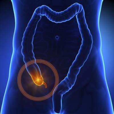
Rotavirus vaccine slashed hospitalizations for acute gastroenteritis
Hospitalizations for acute gastroenteritis in children aged 5 years and younger plummeted after routine rotavirus vaccination was implemented in 2006, with rotavirus-specific hospitalizations plunging 94% and all-cause gastroenteritis hospitalizations dropping 55% in the most recent year for which data are available, according to a report published online June 9 in JAMA.
Researchers assessed the vaccine’s effect by analyzing information in a Healthcare Cost and Utilization Project database covering community and academic hospitals. They focused on 1,201,458 hospitalizations coded for all-cause acute gastroenteritis, including 199,812 (17%) that were coded specifically for rotavirus, which occurred in 26 states during 2000 through 2012 among children under age 5 years.

The mean annual hospitalization rate for all-cause acute gastroenteritis was 76 per 10,000 before the vaccine was introduced, which declined by 31% in 2008, 33% in 2009, 48% in 2010, 47% in 2011, and 55% in 2012. The marked decrease occurred in both boys and girls, across all race/ethnicity categories, and at all ages. The reduction was greatest among children aged 6-23 months, Dr. Eyal Leshem of the Division of Viral Diseases, Centers for Disease Control and Prevention, Atlanta, and his associates said in a research letter.
The mean annual hospitalization rate for rotavirus-specific gastroenteritis dropped even more dramatically, from 16 per 10,000 before the vaccine was introduced by 70% in 2008, 63% in 2009, 90% in 2010, 79% in 2011, and 94% in 2012, the investigators said (JAMA 2015;313:2282-4).
The nonlinear pattern of both declines “might be explained by accumulation of susceptible, nonimmune population during seasons of markedly decreased transmission,” Dr. Leshem and his associates noted.
“Our findings support continued efforts to increase rotavirus vaccine coverage,” they added.
This study was supported by the Centers for Disease Control and Prevention and the Agency for Healthcare Research and Quality. Dr. Leshem and his associates reported having no relevant financial conflicts of interest.
Hospitalizations for acute gastroenteritis in children aged 5 years and younger plummeted after routine rotavirus vaccination was implemented in 2006, with rotavirus-specific hospitalizations plunging 94% and all-cause gastroenteritis hospitalizations dropping 55% in the most recent year for which data are available, according to a report published online June 9 in JAMA.
Researchers assessed the vaccine’s effect by analyzing information in a Healthcare Cost and Utilization Project database covering community and academic hospitals. They focused on 1,201,458 hospitalizations coded for all-cause acute gastroenteritis, including 199,812 (17%) that were coded specifically for rotavirus, which occurred in 26 states during 2000 through 2012 among children under age 5 years.
The mean annual hospitalization rate for all-cause acute gastroenteritis was 76 per 10,000 before the vaccine was introduced, which declined by 31% in 2008, 33% in 2009, 48% in 2010, 47% in 2011, and 55% in 2012. The marked decrease occurred in both boys and girls, across all race/ethnicity categories, and at all ages. The reduction was greatest among children aged 6-23 months, Dr. Eyal Leshem of the Division of Viral Diseases, Centers for Disease Control and Prevention, Atlanta, and his associates said in a research letter.
The mean annual hospitalization rate for rotavirus-specific gastroenteritis dropped even more dramatically, from 16 per 10,000 before the vaccine was introduced by 70% in 2008, 63% in 2009, 90% in 2010, 79% in 2011, and 94% in 2012, the investigators said (JAMA 2015;313:2282-4).
The nonlinear pattern of both declines “might be explained by accumulation of susceptible, nonimmune population during seasons of markedly decreased transmission,” Dr. Leshem and his associates noted.
“Our findings support continued efforts to increase rotavirus vaccine coverage,” they added.
This study was supported by the Centers for Disease Control and Prevention and the Agency for Healthcare Research and Quality. Dr. Leshem and his associates reported having no relevant financial conflicts of interest.
FROM JAMA
Key clinical point: All hospitalizations for acute gastroenteritis in children aged 5 years and younger plummeted after routine rotavirus vaccination was implemented in 2006.
Major finding: The mean annual hospitalization rate for all-cause acute gastroenteritis was 76 per 10,000 before the vaccine was introduced, which declined by 31% in 2008, 33% in 2009, 48% in 2010, 47% in 2011, and 55% in 2012.
Data source: An analysis of information in a Healthcare Cost and Utilization Project database concerning 1,201,458 hospitalizations for acute gastroenteritis in 26 states from 2000 through 2012.
Disclosures: This study was supported by the Centers for Disease Control and Prevention and the Agency for Healthcare Research and Quality. Dr. Leshem and his associates reported having no relevant financial conflicts of interest.

DDW: Uveitis significantly more common in black patients with IBD
WASHINGTON – Black patients with inflammatory bowel disease were significantly more likely to have at least one extraintestinal manifestation of the disease than were white patients, particularly uveitis, in a study of more than 500 people with Crohn’s disease or ulcerative colitis, Dr. Jemilat Badamas reported at the annual Digestive Disease Week.
Black patients were about twice as likely to have uveitis than were white patients in the study, after researchers controlled for other risk factors for extraintestinal manifestations of IBD, said Dr. Badamas, a gastroenterology fellow at Johns Hopkins University, Baltimore. Based on these results, and the increased rate of uveitis among black IBD patients seen in other studies, “practitioners should consider proactive referral of African-Americans [with IBD] to be screened for uveitis,” she said.
The study compared the incidence of various extraintestinal manifestations of IBD among 196 black patients and 342 white patients with a confirmed diagnosis of Crohn’s disease (CD) or ulcerative colitis (UC), which included uveitis, small and large joint disease, sacroiliitis, ankylosing spondylitis, episcleritis, pyoderma gangrenosum (PG), erythema nodosum (EN), and progressive sclerosing cholangitis, confirmed in the medical records. All the patients had been treated at outpatient gastroenterology clinics and infliximab infusion clinics at Johns Hopkins Hospital, including patients who were hospitalized and were subsequently followed up at the GI clinics. Cases with no information on extraintestinal manifestations were excluded.
Enrollment of all patients at one medical center overcame one of the limitations of previous studies evaluating racial differences in extraintestinal manifestations of IBD, where most of the black and white patients were treated in different centers, so researchers could not account for differences in practices at different institutions or access to subspecialty care, she pointed out.
In the Johns Hopkins study, patients in both groups were diagnosed with IBD at a mean age of 28-29 years. Among the black patients, 62% were female and they were enrolled in the study at a mean age of 38 years. Among the white patients, half were female and they were enrolled at a mean age of 41 years. The data on these patients are being collected as part of the Multicenter African American Inflammatory Bowel Disease Study (MAAIS).
More black patients (41%) had at least one extraintestinal manifestation of IBD compared with white patients (33%). The most common was large joint disease, affecting 25.5% of black patients and 19% of white patients, Dr. Badamas said. Uveitis was more common in black patients (6.9% vs. 2.7%), a difference that was statistically significant (P = 0.021). Pyoderma gangrenosum was more common in black patients (4.8% vs. 1.8%), a difference of borderline significance (P = 0.051).
After the researchers controlled for gender, disease location, and extent of disease, which are factors known to be associated with extraintestinal manifestations of IBD, there were no significant differences between the two groups for most of the manifestations, she said. But black patients were about two times more likely to have uveitis, compared with white patients (odds ratio, 2.3), and were 1.5 times more likely to have at least one extraintestinal manifestation (OR, 1.5), she added.
The study did not include a temporal analysis to determine if any of these manifestations occurred before the diagnosis of IBD was made, but Dr. Badamas said that most were diagnosed after the diagnosis of IBD.
Dr. Badamas had no relevant disclosures.
AT DDW 2015
Key clinical point: The finding that uveitis was more common among black patients with inflammatory bowel disease (IBD) may justify screening this group of patients for uveitis.
Major finding: Black patients with IBD were 1.5 times more likely to have at least one extraintestinal manifestation of IBD and about twice as likely to have uveitis than were white patients – statistically significant differences in a study of more than 500 patients with IBD.
Data source: Study of extraintestinal manifestations of IBD in 196 black patients and 342 white patients with Crohn’s disease or ulcerative colitis, treated at Johns Hopkins clinics, who are part of the Multicenter African-American Inflammatory Bowel Disease Study (MAAIS).
Disclosures: Dr. Badamas had no relevant disclosures.