User login
Research focus on drug-induced liver injury on the rise
PHILADELPHIA – Drug-induced liver injury (DILI) continues to be a hot topic, but new advances have helped increase the amount of research done and improve the quality of treatment for patients suffering from the condition.
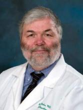
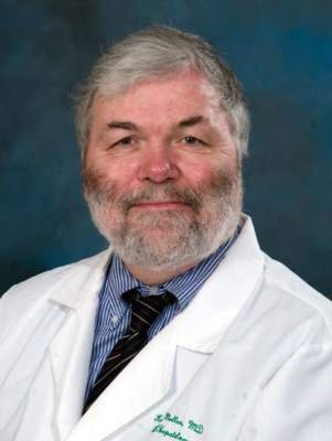
“It’s been 10 years since the DILI Network was started at [the National Institutes of Health], and it’s giving us a lot of important information about what’s happening with [DILI] in the United States,” explained Dr. James H. Lewis of Georgetown University in Washington, D.C. “However, we are still playing catch up with many nations around the world, who have had similar registries for a longer time.”
While incidents of DILI and, more important, severe DILI, continue to be low in the United States, DILI is the leading cause of acute liver failure in Americans, and a major cause of emergency liver transplant. Further research into new drugs is stymied by hepatoxicity, which is “the main reason why many drugs are pulled from development.”
“When it comes to drugs that are causing injury, these are fairly old friends,” said Dr. Lewis. “They’ve been around for a long time and they don’t change a whole lot, Very interesting [is] that the TB drugs lead the list, especially INH [isoniazid] and pyrazinamide, as well as antibiotics, especially augmentin.”
Nevertheless, activity continues to increase in the DILI field. Looking at PubMed, said Dr. Lewis, there are already over 300 citations involving DILI from January through April of this year, over 1,500 in all of 2014, and over 6,000 from 2010 to 2014. Compared to that, there were around 7,000 in total from 2000 to 2009.
The relationship between dosage and likelihood of liver injury still needs examination, declared Dr. Lewis, citing separate works done by Dr. Hy Zimmerman and Dr. Jack Uetretcht. The former determined that there were certain drugs for which the likelihood of liver injury could be predicted, such as acetaminophen, and those for which such risk was idiosyncratic.
Dr. Uetrecht determined that 10-mg doses were the cutoff at which liver injury became possible. In 2008, work by Lammert et al., published in Hepatology, revealed that doses of 10 mg or less resulted in a 9% risk of DILI, compared with 14.2% for doses of 11-49 mg, and 77% for doses at or above 50 mg. “There are over 200 drugs that have been found to cause DILI, but the top 10 alone account for 50% of all DILI, and the top 25 drugs account for [67%],” said Dr. Lewis. “This means that the other 175 drugs have maybe one case of DILI in the [DILI Network] entire database.”
What this means, concluded Dr. Lewis, is that DILI does not occur often with most drugs, and the ones that cause it are those that are already known.
Dr. Lewis disclosed receiving grant/research support from Ocera Therapeutics, Inc.; consultant relationships with GlaxoSmithKline, Otsuka Pharmaceutical Co. Ltd., Takeda Pharmaceuticals U.S.A. Inc., AstraZeneca, and Lundbeck Inc.; and being a member of the speaking bureau for Gilead Science, Inc.
AT THE DIGESTIVE DISEASES MEETING
Confusion surrounding hepatic encephalopathy persists
PHILADELPHIA – The treatment of patients with hepatic encephalopathy is still plagued by confusion, misdiagnosis, and poor treatment, and requires new protocols to improve overall patient care.
“It’s surprisingly difficult to define hepatic encephalopathy [HE],” said Dr. Kevin D. Mullen, from the Metrohealth Medical Center in Cleveland, Ohio, who spoke at the second annual Digestive Diseases: New Advances meeting on Saturday. “It can range from subtle, minimal changes all the way to full-blown coma, but the only way to confirm [HE] is through psychometric testing.”
This inability to properly define HE can lead to problems for patients, Dr. Mullen explained. Many physicians tend to use too much lactulose in HE patients, which can lead to dehydration and hypernatremia, especially in patients in an hepatic coma. Hypernatremia is often treated with furosemide, which can lead to deafness in patients and delay recovery.
Additionally, improper use of rifaximin and metronidazole are common issues that need to be addressed by hepatologists, as well as the beliefs that precipitating factors do not need to be looked at very carefully when diagnosing and treating the condition, as well as the notion that concomitant disease – especially hypothyroidism – rarely, if ever, causes coma.
Management of HE, therefore, should always include identification and treatment of precipitating factors, physicians should rule out other potential causes of encephalopathy, unconscious patients should be given supportive care as soon as possible, and specific additional measures should begin right away based on the precipitating factors, which can also include hypo/hyperglycemia, CNS or systemic sepsis, and post-ictal confusion.
Dr. Mullen also talked about covert hepatic encephalopathy, a relatively new term that describes milder HEs that often are undiagnosed but can still cause severe consequences. Covert HE is often found in patients with cirrhosis, and can lead to cognitive deficiencies in attention, information processing, motor abilities, and can impair social, physical, and job-related interactions.
“The terms ‘subclinical’ and ‘minimal’ keep popping up, but we’re trying very hard to get people to use ‘covert,’” explained Dr. Mullen. “These are patients who have a normal neurological examination [but] subnormal performance in two or more psychometric tests, such as number connection tests A and B, line drawing, serial dotting, and digit symbol.”
Dr. Mullen disclosed being on the speaker’s bureau for Salix Pharmaceuticals, Inc.
FROM THE DIGESTIVE DISEASES MEETING
Treament of IBS should focus on head as well as gut
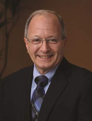
PHILADELPHIA – The pathophysiology of irritable bowel syndrome (IBS) was the focus of a presentation given by Dr. Anthony J. Lembo at the 2nd annual Digestive Diseases meeting in Philadelphia on Friday.
Specifically, Dr. Lembo discussed the debate about whether diagnosing the condition should focus on a patient’s gut or head; that is, if enhanced perception of a bowel disorder can lead to visceral hypersensitivity and altered motility, which results in a diagnosis of IBS when, in fact, the issue is psychosomatic. Dr. Lembo, a professor at Harvard Medical School and Beth Israel Deaconess Medical Center, explained that there is a growing shift toward diagnosing IBS as a brain-to-gut interaction rather than as something based purely on limited motility. Patients presenting with comorbid conditions such as depression, migraine, anxiety, neuralgia, chronic fatigue or pain, or fibromytosis are all more likely to not only have IBS, but to have more severe symptoms and lower quality of life.
“When I see a patient who has a lot of these symptoms together, I realize that I need to be very aggressive and use a multimodal approach,” said Dr. Lembo. “I talk to their other physicians, and do more of the brain treatment, psychological treatment, CNS drugs, as well as some peripheral ones.”
For these reasons, Dr. Lembo said that the current Rome III criteria for treatment of IBS are not sufficient to be used on their own in diagnosing and treating IBS, saying “IBS is a heterogeneous disorder with both peripheral and central mechanisms” that must be treated as such. In treating the central nervous system, Dr. Lembo discussed 5-HT3 antagonists, serotonin modulators, antidepressants, and placebos. Peripheral treatments can come in the form of either diets, fiber regimens, antibiotics, GC-C agonists*, 5-HT3 antagonists, and probiotics/prebiotics, although efficacies and approvals vary.
Dr. Lembo disclosed numerous potential conflicts of interest: consultantships with Ironwood/Forest, Astra-Zeneca, Furiex, Salix, Prometheus, Astellas, and GlaxoSmithKlinel; honoraria from Ironwood/Forest, Astra-Zeneca, Furiex, Salix, Prometheus, and GlaxoSmithKline; and research grants/contracts from Prometheus and Furiex.
*Content was corrected on 7/1/2015.
FROM THE ANNUAL DIGESTIVE DISEASES MEETING
Celiac disease remains underdiagnosed
Celiac disease is not uncommon, affecting about 1% of the population in the United States and around the world, but the biggest barrier to patients receiving treatment is practitioners’ failure to recognize the condition, said Dr. Peter H.R. Green, director of the celiac disease center at Columbia University, New York.
There’s a very low rate of diagnosis in the United States, compared with other countries, according to Dr. Green. Only about 18% of U.S. patients with celiac are properly diagnosed. Physicians need to be more aware of the condition as it is on the rise. The incidence of celiac disease has increased four- to fivefold over the past 50 years, he said.
“Patients, when they get diagnosed, have often had a long history” of symptoms, Dr. Green said at Digestive Diseases: New Advances. “They say, ‘Why didn’t my doctor think of this? I’ve been complaining so long,’ etc.”
The main symptoms of celiac disease are diarrhea or bloating, iron-deficiency anemia, osteoporosis, fatigue, hypothyroidism, and/or weight loss. “The host of presentations is very great, which is one of the factors that may be contributing to the low rate of diagnosis. Almost anyone you could think of could have celiac disease based on these symptoms,” he said.
Once a physician considers celiac disease as a possible diagnosis, the rest is easy, he said. The most definitive blood test is an anti-tissue transglutaminase IgA, which is available through most laboratories. If the test results show antibodies toward gluten, or if it’s inconclusive, it should be followed up by an intestinal biopsy to confirm the diagnosis. From there, patients could start a gluten-free diet.
Although biopsy rates have been increasing, as recently as 2009 only 51% of individuals undergoing upper endoscopy with signs and symptoms of celiac disease had had a small-bowel biopsy, judging from findings from one of his recent studies, Dr. Green reported. Patients who are less likely to have a biopsy are nonwhite or male or have weight loss as the presenting symptom. More recent studies indicate that several biopsies should be taken, including four to six from the descending duodenum and two from the duodenal bulb, the portion closest to the stomach.
Once diagnosed, patients should be managed in a center that specializes in celiac disease and should be referred to a knowledgeable dietitian to learn what to avoid and what to eat. “People will just eat the same stuff because they think it’s safe,” Dr. Green said.
Antibody levels should be checked again 6 and 12 months after starting a gluten-free diet to ensure that antibodies normalize. Levels of iron, folate, and B12 also should be checked, he said, as well as bone density and thyroid function. In addition, patients should be encouraged to follow up with their physician at least annually. Whether patients need a follow-up biopsy remains controversial. “We think they do, but maybe in 2-3 years, when you can assess the effects of the diet,” he said.
Some patients have true celiac, while others may have a wheat allergy or just a gluten sensitivity. There has been an increased interest in gluten-free diets not just among those affected with celiac disease but by people who think it will help with weight loss or by those who think it’s healthy. “In fact, a gluten-free diet is not healthy,” Dr. Green said. “It’s low in fiber, low in B vitamins, high in heavy metals, and there is some concern about toxins in corn.” Patients should make sure to have a blood test for celiac disease before embarking on a gluten-free diet. Some patients studied who did not have celiac had diagnoses such as bacterial overgrowth in the small intestine; intolerance to fructose, lactose, or other foods; and microscopic colitis.
Risk factors for developing celiac disease include being born via cesarean section, taking proton pump inhibitors or antibiotics, or having a history of GI infections such as rotavirus or campylobacter, although it’s unclear why the disorder can occur at any age, he said at the meeting, which was held by Global Academy for Medical Education and Rutgers, the State University of New Jersey. Global Academy and this news organization are owned by the same company.
Dr. Green reported no relevant financial disclosures.
Barrett’s esophagus increasing in frequency
Patients with Barrett’s esophagus, a condition in which cells lining the esophagus are replaced by the type of cells that normally line the intestine, need to be monitored carefully to prevent the development of esophageal cancer, a gastroenterologist expert in the condition said here at Digestive Diseases: New Advances. The condition can occur following exposure to stomach acids and bile from gastroesophageal reflux disease (GERD).
“The reason we worry about Barrett’s is because it’s a major risk factor for developing esophageal adenocarcinoma, and that’s a tumor that has been experiencing an enormous increase in frequency over the past few decades,” said Dr. Stuart Spechler, chief of gastroenterology for the VA North Texas Healthcare System and co-director of the Esophageal Diseases Center at the University of Texas Southwestern Medical Center in Dallas.
The incidence of esophageal adenocarcinoma increased more than sevenfold over 33 years, going from 3.6 per million in 1973 to 25.6 per million in 2006. Barrett’s esophagus affects about 5.6% of U.S. adults, he noted.
Endoscopic screening for Barrett’s esophagus is recommended for patients with multiple risk factors for esophageal cancer, including chronic GERD (having GERD for more than 5 years), hiatal hernia, age of at least 50 years, male gender, white race, and an elevated body mass index, especially if there’s an intra-abdominal body fat distribution, Dr. Spechler said.
Barrett’s esophagus traditionally is treated with proton pump inhibitors because of their ability to decrease gastric acid production and acid reflux and heal reflux esophagitis. “We also recommend that these patients have regular endoscopic surveillance to look for precancerous changes,” most notably dysplasia, he said. If no dysplasia is found, patients should receive a repeat endoscopy every 3-5 years. Patients with high-grade dysplasia should be treated with endoscopic eradication therapy. The risk of developing cancer in patients with high-grade dysplasia is about 6% per year, he said, which is “sufficient to warrant intervention.”
For patients with visible mucosal irregularities, endoscopic mucosal resection can determine how far the neoplasms have spread, Dr. Spechler said.
For those neoplasms confined to the mucosa, like high-grade dysplasia, radiofrequency ablation can be used to burn the abnormal mucosa away. As the esophagus heals, normal cells grow back. The procedure is safe and effective and has been shown to prevent neoplastic progression at 1 year, he said – the most serious complication is esophageal stricture (N. Engl. J. Med. 2009;360:2277-88). Radiofrequency ablation also can be considered in patients with low-grade dysplasia, and even for highly selected patients with nondysplastic Barrett’s esophagus deemed to be at increased risk for progression to high-grade dysplasia or cancer.
“We do worry about Barrett’s esophagus recurring after endoscopic eradication, and there is a rate of recurrence, but this newer treatment seems to be very effective, at least in the short term, for preventing progression to cancer,” he said.
More advanced cancers can be treated by surgery and chemotherapy or radiation therapy.
Overall, “this is a condition that’s increasing in frequency, and we do worry about it, but as long as you are treating patients, screening for Barrett’s esophagus, and following them regularly with surveillance, the prognosis for any individual patient is really excellent,” he said at the meeting, which was held by Global Academy for Medical Education and Rutgers, the State University of New Jersey. Global Academy and this news organization are owned by the same company.
Dr. Spechler has served a consultant for Takeda Pharmaceuticals and is currently a consultant for Interpace Diagnostics.


