User login
Manometry, esophagram combo detects achalasia posttreatment problems
High-resolution manometry with esophageal pressure topography is known to improve the accuracy of manometry for detecting achalasia and defined clinically relevant subtypes prior to treatment, but new data demonstrate that the procedure also is useful for evaluating treatment efficacy.
In a study of 31 men and 19 women aged 20-79 years, resolution of the achalasia pattern on high-resolution manometry with esophageal pressure topography (HRM-EPT) after treatment was associated with symptom improvement and reduced bolus retention, reported Dr. Frédéric Nicodème of Northwestern University, Chicago, and his colleagues.
Video source: American Gastroenterological Association's YouTube Channel
Study participants included one cohort of 25 patients who underwent endoscopy, HRM, timed barium esophagram (TBE) following a 200-mL barium swallow, and symptom assessment before treatment, and a second cohort of 25 treated patients who had pretreatment type 1 or 2 achalasia and who were undergoing a posttreatment study protocol including HRM, TBE, endoscopy, and symptom assessment. The investigators assessed dysphagia, regurgitation, retrosternal pain, and weight loss and used them to calculate an Eckardt score (ES) for both cohorts.
In the untreated cohort, which included 10 patients with type 1 achalasia and 15 with type 2 achalasia, the key EPT metric of integrated relaxation pressure (IRP) was significantly greater in the type 2 patients, as was the nadir-relaxation pressure. No differences were seen based on disease type with respect to resting esophagogastric junction (EGJ) pressure, barium column height, barium column width, or ES (Clin. Gastroenterol. Hepatol. 2012 Oct. 12 [doi: 10.1016/j.cgh.2012.10.015]).
No correlation was found between TBE column height at 5 minutes and IRP, resting EGJ pressure, nadir-EGJ relaxation pressure, or ES, and no correlation was seen between ES and IRP, resting EGJ pressure, or nadir-EGJ relaxation pressure.
In the posttreatment cohort, 10 patients, including 6 with type 1 disease and 4 with type 2 disease, had EPT findings of persistent achalasia pattern, and 15 had resolution of the achalasia pattern. Of these 15 patients, 8 converted to absent peristalsis and 7 to weak peristalsis.
"The IRP, resting EGJ pressure, and nadir-EGJ were all significantly correlated with posttreatment ES," the investigators noted.
Furthermore, TBE column height at 5 minutes, IRP, and ES were significantly lower in patients with resolved achalasia patterns on HRM than in those with a persistent achalasia pattern; the subgroup of 7 patients with weak peristalsis appeared to have the best outcome.
"The median TBE column height at 5 minutes was significantly lower [in those with weak peristalsis] than the three other groups, and ES showed a trend toward a lower value compared to the other 3 groups," they explained.
No significant correlations between TBE column height at 5 minutes and resting EGJ pressure or nadir-EGJ relaxation pressure were noted in the posttreatment patients. Only the IRP showed a weak correlation.
"The correlation between TBE column height at 5 minutes and ES after treatment was also not significant. However, the median TBE column height at 5 minutes for patients with an ES of 3 or greater was significantly greater than that for patients with an ES less than 3," they wrote.
"Our findings suggest that resolution of the achalasia pattern on EPT after treatment was associated with an improvement in symptoms and reduced bolus retention. Although the IRP was not strongly linearly correlated with symptom severity, when analyzed dichotomously patients with an IRP greater than or equal to 15 mm Hg had worse symptom scores and greater bolus retention on TBE compared to those with normal IRP values," the investigators wrote.
In contrast, no EPT pattern or metric, and no TBE variable predicted symptom severity in the untreated cohort, and barium height on TBE did not distinguish achalasia EPT subtypes, they noted.
"These findings suggest that in addition to its proven utility in detecting pretreatment achalasia, EPT also has utility in the management of posttreatment achalasia that can complement TBE," they said.
Although TBE did not distinguish disease subtypes, it does complement EPT, especially when the disease has progressed to an anatomic-dominant disorder, they said. Additional studies in larger series of patients are needed before and after therapy to confidently establish the relative merits of each evaluation with respect to the prediction of long-term outcomes and cost-effectiveness, they said.
This study was supported by the Public Health Service. The authors reported having no disclosures.
Most patients with achalasia do well in the postoperative period after either pneumatic dilation (PD) or laparoscopic surgical myotomy, with an initial success rate as high as 90% after 2 years. However, predicting failure based on symptom recurrence is problematic, as symptoms are often underreported or absent due to esophageal dilation or impaired esophageal sensation. Thus, a strategy of avoiding esophageal decompensation due to delayed diagnosis cannot solely rely on patient report of symptom recurrence. Objective testing by barium esophagram or esophageal manometry may be needed.

|
|
This study adds new knowledge regarding the benefit of high-resolution manometry (HRM) in the management of posttreatment achalasia. The novelty of the study is in its use of timed barium esophagram (TBE), HRM, and symptoms in a group of patients with achalasia pre- and posttherapy. TBE was previously advocated as the objective test of choice; however, this study suggests that TBE and HRM may be complementary in this regard.
What is clear from this report as well as others regarding objective testing in achalasia is that patients’ symptoms alone are a poor marker of esophageal emptying. Posttherapy testing with HRM and/or TBE with special emphasis on bolus emptying should be employed in order to avoid long-term complications with achalasia. The choice between the tests may depend on the expertise and availability of each of the proposed test modalities.
Dr. Vaezi is clinical director of the division of gastroenterology, hepatology, and nutrition, and director of the Center for Swallowing and Esophageal Disorders and Clinical Research at Vanderbilt University Medical Center, Nashville, Tenn. He said he had no conflicts of interest to disclose.
Most patients with achalasia do well in the postoperative period after either pneumatic dilation (PD) or laparoscopic surgical myotomy, with an initial success rate as high as 90% after 2 years. However, predicting failure based on symptom recurrence is problematic, as symptoms are often underreported or absent due to esophageal dilation or impaired esophageal sensation. Thus, a strategy of avoiding esophageal decompensation due to delayed diagnosis cannot solely rely on patient report of symptom recurrence. Objective testing by barium esophagram or esophageal manometry may be needed.
|
|
|
This study adds new knowledge regarding the benefit of high-resolution manometry (HRM) in the management of posttreatment achalasia. The novelty of the study is in its use of timed barium esophagram (TBE), HRM, and symptoms in a group of patients with achalasia pre- and posttherapy. TBE was previously advocated as the objective test of choice; however, this study suggests that TBE and HRM may be complementary in this regard.
What is clear from this report as well as others regarding objective testing in achalasia is that patients’ symptoms alone are a poor marker of esophageal emptying. Posttherapy testing with HRM and/or TBE with special emphasis on bolus emptying should be employed in order to avoid long-term complications with achalasia. The choice between the tests may depend on the expertise and availability of each of the proposed test modalities.
Dr. Vaezi is clinical director of the division of gastroenterology, hepatology, and nutrition, and director of the Center for Swallowing and Esophageal Disorders and Clinical Research at Vanderbilt University Medical Center, Nashville, Tenn. He said he had no conflicts of interest to disclose.
High-resolution manometry with esophageal pressure topography is known to improve the accuracy of manometry for detecting achalasia and defined clinically relevant subtypes prior to treatment, but new data demonstrate that the procedure also is useful for evaluating treatment efficacy.
In a study of 31 men and 19 women aged 20-79 years, resolution of the achalasia pattern on high-resolution manometry with esophageal pressure topography (HRM-EPT) after treatment was associated with symptom improvement and reduced bolus retention, reported Dr. Frédéric Nicodème of Northwestern University, Chicago, and his colleagues.
Video source: American Gastroenterological Association's YouTube Channel
Study participants included one cohort of 25 patients who underwent endoscopy, HRM, timed barium esophagram (TBE) following a 200-mL barium swallow, and symptom assessment before treatment, and a second cohort of 25 treated patients who had pretreatment type 1 or 2 achalasia and who were undergoing a posttreatment study protocol including HRM, TBE, endoscopy, and symptom assessment. The investigators assessed dysphagia, regurgitation, retrosternal pain, and weight loss and used them to calculate an Eckardt score (ES) for both cohorts.
In the untreated cohort, which included 10 patients with type 1 achalasia and 15 with type 2 achalasia, the key EPT metric of integrated relaxation pressure (IRP) was significantly greater in the type 2 patients, as was the nadir-relaxation pressure. No differences were seen based on disease type with respect to resting esophagogastric junction (EGJ) pressure, barium column height, barium column width, or ES (Clin. Gastroenterol. Hepatol. 2012 Oct. 12 [doi: 10.1016/j.cgh.2012.10.015]).
No correlation was found between TBE column height at 5 minutes and IRP, resting EGJ pressure, nadir-EGJ relaxation pressure, or ES, and no correlation was seen between ES and IRP, resting EGJ pressure, or nadir-EGJ relaxation pressure.
In the posttreatment cohort, 10 patients, including 6 with type 1 disease and 4 with type 2 disease, had EPT findings of persistent achalasia pattern, and 15 had resolution of the achalasia pattern. Of these 15 patients, 8 converted to absent peristalsis and 7 to weak peristalsis.
"The IRP, resting EGJ pressure, and nadir-EGJ were all significantly correlated with posttreatment ES," the investigators noted.
Furthermore, TBE column height at 5 minutes, IRP, and ES were significantly lower in patients with resolved achalasia patterns on HRM than in those with a persistent achalasia pattern; the subgroup of 7 patients with weak peristalsis appeared to have the best outcome.
"The median TBE column height at 5 minutes was significantly lower [in those with weak peristalsis] than the three other groups, and ES showed a trend toward a lower value compared to the other 3 groups," they explained.
No significant correlations between TBE column height at 5 minutes and resting EGJ pressure or nadir-EGJ relaxation pressure were noted in the posttreatment patients. Only the IRP showed a weak correlation.
"The correlation between TBE column height at 5 minutes and ES after treatment was also not significant. However, the median TBE column height at 5 minutes for patients with an ES of 3 or greater was significantly greater than that for patients with an ES less than 3," they wrote.
"Our findings suggest that resolution of the achalasia pattern on EPT after treatment was associated with an improvement in symptoms and reduced bolus retention. Although the IRP was not strongly linearly correlated with symptom severity, when analyzed dichotomously patients with an IRP greater than or equal to 15 mm Hg had worse symptom scores and greater bolus retention on TBE compared to those with normal IRP values," the investigators wrote.
In contrast, no EPT pattern or metric, and no TBE variable predicted symptom severity in the untreated cohort, and barium height on TBE did not distinguish achalasia EPT subtypes, they noted.
"These findings suggest that in addition to its proven utility in detecting pretreatment achalasia, EPT also has utility in the management of posttreatment achalasia that can complement TBE," they said.
Although TBE did not distinguish disease subtypes, it does complement EPT, especially when the disease has progressed to an anatomic-dominant disorder, they said. Additional studies in larger series of patients are needed before and after therapy to confidently establish the relative merits of each evaluation with respect to the prediction of long-term outcomes and cost-effectiveness, they said.
This study was supported by the Public Health Service. The authors reported having no disclosures.
FROM CLINICAL GASTROENTEROLOGY AND HEPATOLOGY
Major Finding: In a study of 31 men and 19 women aged 20-79 years, resolution of the achalasia pattern on HRM-EPT after treatment was associated with symptom improvement and reduced bolus retention.
Data Source: A prospective study of 50 achalasia patients.
Disclosures: This study was supported by the Public Health Service. The authors reported having no disclosures.

Scoring tool targets hospitalized patients needing acid suppressants
A scoring system based on demographics and comorbidities predicted which hospitalized patients were at risk for nosocomial gastrointestinal bleeding.
Used as a guideline for administering acid suppressors, the system found that treating fewer than 100 high-risk patients would prevent one case of bleeding, Dr. Shoshana Herzig and her colleagues wrote in the January online issue of General Internal Medicine (2013 [doi:10.1007/s11606-012-2296-x]).
"This scoring system allows identification of subsets of patients in whom the risk of nosocomial gastrointestinal bleeding may be higher enough to warrant the use of prophylactic acid-suppressive medication, in the absence of other indicators for use," said Dr. Herzig of Beth Israel Deaconess Medical Center and Harvard Medical School, Boston, and her coauthors.

The model "allows for risk stratification of patients using readily available information, and can be used to guide more selective use of acid-suppressive medication in patients outside of the ICU," the investigators wrote. The investigators reviewed about 76,000 records of noncritically ill patients hospitalized for more than 3 days, from 2004 through 2007. Nosocomial gastrointestinal bleeding occurred in about 3% of the group.
The authors constructed the risk score by identifying several factors that could potentially affect bleeding, including age, sex, comorbid conditions, admission service, and the receipt of certain medications during hospitalization.
A multivariate analysis identified those factors that significantly correlated with gastrointestinal bleeding. These included:
• Age more than 60 years (odds ratio, 2.2).
• Male sex (OR, 1.6).
• Liver disease (OR, 2.1).
• Acute renal failure (OR, 1.9).
• Admission to a medical service (OR, 2.7).
• Prophylactic anticoagulation (OR, 1.7).
• Coagulotherapy without antiplatelet medication (OR, 2.6).
• Coagulotherapy with one antiplatelet agent (OR, 3.2).
• Coagulotherapy with dual antiplatelet agents (OR, 3.3).
Based on these factors, they constructed a points-based risk scoring system that significantly correlated with the incidence of gastrointestinal bleeding. Patients in the lowest-score quartile were at the least risk of bleeding (less than 0.3%), while those in the highest quartile had the highest risk (1.5%).
The number needed to treat (NNT) to prevent one bleed increased as the risk score increased. The NNT was 500 in patients whose score was at least 6, 179 in those whose score was at least 8, 95 in those whose score was at least 10 (moderate risk), and 48 in those whose score was at least 12 (high risk).
The patients’ mean age was 56 years, although the range was very wide (18-107 years); 40% were male. Most patients received an acid-suppressing medication (58%). Proton pump inhibitors were most commonly used (81%); 29% received a histamine-2 receptor antagonist.
"With further validation at other medical centers, this scoring system may help clinicians individualize the decision to prescribe acid-suppressive medication as prophylaxis," the authors said.
The study was funded by grants from the National Institute on Aging and the National Center for Research Resources. None of the authors reported any financial conflicts.
A scoring system based on demographics and comorbidities predicted which hospitalized patients were at risk for nosocomial gastrointestinal bleeding.
Used as a guideline for administering acid suppressors, the system found that treating fewer than 100 high-risk patients would prevent one case of bleeding, Dr. Shoshana Herzig and her colleagues wrote in the January online issue of General Internal Medicine (2013 [doi:10.1007/s11606-012-2296-x]).
"This scoring system allows identification of subsets of patients in whom the risk of nosocomial gastrointestinal bleeding may be higher enough to warrant the use of prophylactic acid-suppressive medication, in the absence of other indicators for use," said Dr. Herzig of Beth Israel Deaconess Medical Center and Harvard Medical School, Boston, and her coauthors.
The model "allows for risk stratification of patients using readily available information, and can be used to guide more selective use of acid-suppressive medication in patients outside of the ICU," the investigators wrote. The investigators reviewed about 76,000 records of noncritically ill patients hospitalized for more than 3 days, from 2004 through 2007. Nosocomial gastrointestinal bleeding occurred in about 3% of the group.
The authors constructed the risk score by identifying several factors that could potentially affect bleeding, including age, sex, comorbid conditions, admission service, and the receipt of certain medications during hospitalization.
A multivariate analysis identified those factors that significantly correlated with gastrointestinal bleeding. These included:
• Age more than 60 years (odds ratio, 2.2).
• Male sex (OR, 1.6).
• Liver disease (OR, 2.1).
• Acute renal failure (OR, 1.9).
• Admission to a medical service (OR, 2.7).
• Prophylactic anticoagulation (OR, 1.7).
• Coagulotherapy without antiplatelet medication (OR, 2.6).
• Coagulotherapy with one antiplatelet agent (OR, 3.2).
• Coagulotherapy with dual antiplatelet agents (OR, 3.3).
Based on these factors, they constructed a points-based risk scoring system that significantly correlated with the incidence of gastrointestinal bleeding. Patients in the lowest-score quartile were at the least risk of bleeding (less than 0.3%), while those in the highest quartile had the highest risk (1.5%).
The number needed to treat (NNT) to prevent one bleed increased as the risk score increased. The NNT was 500 in patients whose score was at least 6, 179 in those whose score was at least 8, 95 in those whose score was at least 10 (moderate risk), and 48 in those whose score was at least 12 (high risk).
The patients’ mean age was 56 years, although the range was very wide (18-107 years); 40% were male. Most patients received an acid-suppressing medication (58%). Proton pump inhibitors were most commonly used (81%); 29% received a histamine-2 receptor antagonist.
"With further validation at other medical centers, this scoring system may help clinicians individualize the decision to prescribe acid-suppressive medication as prophylaxis," the authors said.
The study was funded by grants from the National Institute on Aging and the National Center for Research Resources. None of the authors reported any financial conflicts.
FROM GENERAL INTERNAL MEDICINE
Major Finding: Acid suppressants would prevent one nosocomial gastrointestinal bleed for every 48 high-risk patients and every 95 moderate-risk patients treated, a risk-scoring system showed.
Data Source: A retrospective chart study of more than 76,000 patients.
Disclosures: The study was funded by grants from the National Institute on Aging and the National Center for Research Resources. None of the authors reported having any financial conflicts.

Restrictive transfusion strategy superior for severe acute GI bleeding
For patients with severe acute GI bleeding, outcomes are significantly better when a restrictive transfusion strategy is used – limiting the hemoglobin threshold to 7 g/dL – rather than a liberal transfusion strategy allowing a 9 g/dL threshold, according to a report published online Jan. 2 in the New England Journal of Medicine.
In a single-center randomized controlled trial involving 889 patients, the restrictive transfusion strategy resulted in significantly lower mortality, lower rates of rebleeding, less frequent need for rescue therapy, fewer complications, and shorter hospitalizations than did the liberal transfusion strategy. "Our results suggest that in patients with acute GI bleeding, a strategy of not performing transfusion until the hemoglobin concentration falls below 7 g/dL is a safe and effective approach," said Dr. Càndid Villanueva of Hospital de Sant Pau, Barcelona, and his associates.
"Current international guidelines recommend decreasing the hemoglobin threshold level for transfusion ... from 10g/dL to 7 g/dL" in such patients, but these recommendations are based on trials involving critically ill patients with normovolemic anemia that did not include subjects with acute bleeding. "Transfusion requirements may be different for patients with acute hemorrhage due to factors such as hemodynamic instability or rapid onset of anemia" resulting from extremely low hemoglobin levels.
In particular, results of animal studies suggest that transfusion can be especially harmful in patients with bleeding from portal hypertension sources, "since restitution of blood volume after hemorrhage can lead to a rebound increase in portal pressure, which is associated with a risk of rebleeding," the investigators noted.
To examine the effects of different transfusion strategies in this setting, Dr. Villanueva and his colleagues enrolled adults who presented with hematemesis, melena, or both, randomly assigning 444 to receive restrictive transfusion (with a target range for the posttransfusion hemoglobin level of 7-9 g/dL) and 445 to receive liberal transfusion (with a target range of 9-11 g/dL).
The study protocol permitted transfusions to be administered at the discretion of the attending physician any time symptoms or signs of anemia developed, massive bleeding occurred, or surgical intervention was needed, as well as when hemoglobin levels dipped below the assigned threshold.
All the study subjects underwent emergency gastroscopy within 6 hours of presentation, with appropriate treatment when the source of the bleeding was identified. Diagnoses included peptic ulcer, esophageal varices, cirrhosis, portal hypertension, and nonvariceal lesions.
The primary outcome measure, mortality from any cause at 45 days, was significantly lower in the restrictive-strategy group (5%) than the liberal-strategy group (9%). Death resulted from uncontrolled bleeding in 0.7% vs 3.1% of the 2 groups, respectively, the researchers said (N. Engl. J. Med. 2013 Jan. 2 [doi:10.1056/NEJMoa1211801]).
The rate of rebleeding also was significantly lower with the restrictive strategy (10% vs. 16%), and length of hospital stay was significantly shorter. In addition, rescue therapy for esophageal varices with balloon tamponade or a transjugular intrahepatic portosystemic shunt was required less often in the restrictive-strategy group than in the liberal-strategy group, as was emergency surgery to control further bleeding from peptic ulcer.
The rate of overall complications was significantly lower with the restrictive strategy (40%) than with the liberal strategy (48%), as was the rate of serious adverse events. In addition, transfusion reactions and cardiac events such as pulmonary edema were more frequent with the liberal strategy.
"Our results are consistent with those from previous observational studies and randomized trials performed in other settings, which have shown that a restrictive transfusion strategy did not increase, and even decreased, the mortality observed with a liberal transfusion strategy," Dr. Villanueva and his associates said.
This important study "provides long-awaited evidence to guide practice and justify current recommendations for the management of upper GI bleeding," said Dr. Loren Laine.
A restrictive transfusion strategy reduced the relative risk of mortality at 45 days by 45%. Extrapolating the study findings, only 25 patients would have to be treated with a restrictive rather than a liberal transfusion strategy to avert one additional death.
Dr. Laine is at Yale University, New Haven, Conn., and the Veterans Affairs Connecticut Healthcare System, West Haven. He reported no financial conflicts of interest. These remarks were taken from his editorial accompanying Dr. Villanueva’s report (N. Engl. J. Med. 2013 Jan. 2 [doi:10.1056/NEJMe1212009]).
For patients with severe acute GI bleeding, outcomes are significantly better when a restrictive transfusion strategy is used – limiting the hemoglobin threshold to 7 g/dL – rather than a liberal transfusion strategy allowing a 9 g/dL threshold, according to a report published online Jan. 2 in the New England Journal of Medicine.
In a single-center randomized controlled trial involving 889 patients, the restrictive transfusion strategy resulted in significantly lower mortality, lower rates of rebleeding, less frequent need for rescue therapy, fewer complications, and shorter hospitalizations than did the liberal transfusion strategy. "Our results suggest that in patients with acute GI bleeding, a strategy of not performing transfusion until the hemoglobin concentration falls below 7 g/dL is a safe and effective approach," said Dr. Càndid Villanueva of Hospital de Sant Pau, Barcelona, and his associates.
"Current international guidelines recommend decreasing the hemoglobin threshold level for transfusion ... from 10g/dL to 7 g/dL" in such patients, but these recommendations are based on trials involving critically ill patients with normovolemic anemia that did not include subjects with acute bleeding. "Transfusion requirements may be different for patients with acute hemorrhage due to factors such as hemodynamic instability or rapid onset of anemia" resulting from extremely low hemoglobin levels.
In particular, results of animal studies suggest that transfusion can be especially harmful in patients with bleeding from portal hypertension sources, "since restitution of blood volume after hemorrhage can lead to a rebound increase in portal pressure, which is associated with a risk of rebleeding," the investigators noted.
To examine the effects of different transfusion strategies in this setting, Dr. Villanueva and his colleagues enrolled adults who presented with hematemesis, melena, or both, randomly assigning 444 to receive restrictive transfusion (with a target range for the posttransfusion hemoglobin level of 7-9 g/dL) and 445 to receive liberal transfusion (with a target range of 9-11 g/dL).
The study protocol permitted transfusions to be administered at the discretion of the attending physician any time symptoms or signs of anemia developed, massive bleeding occurred, or surgical intervention was needed, as well as when hemoglobin levels dipped below the assigned threshold.
All the study subjects underwent emergency gastroscopy within 6 hours of presentation, with appropriate treatment when the source of the bleeding was identified. Diagnoses included peptic ulcer, esophageal varices, cirrhosis, portal hypertension, and nonvariceal lesions.
The primary outcome measure, mortality from any cause at 45 days, was significantly lower in the restrictive-strategy group (5%) than the liberal-strategy group (9%). Death resulted from uncontrolled bleeding in 0.7% vs 3.1% of the 2 groups, respectively, the researchers said (N. Engl. J. Med. 2013 Jan. 2 [doi:10.1056/NEJMoa1211801]).
The rate of rebleeding also was significantly lower with the restrictive strategy (10% vs. 16%), and length of hospital stay was significantly shorter. In addition, rescue therapy for esophageal varices with balloon tamponade or a transjugular intrahepatic portosystemic shunt was required less often in the restrictive-strategy group than in the liberal-strategy group, as was emergency surgery to control further bleeding from peptic ulcer.
The rate of overall complications was significantly lower with the restrictive strategy (40%) than with the liberal strategy (48%), as was the rate of serious adverse events. In addition, transfusion reactions and cardiac events such as pulmonary edema were more frequent with the liberal strategy.
"Our results are consistent with those from previous observational studies and randomized trials performed in other settings, which have shown that a restrictive transfusion strategy did not increase, and even decreased, the mortality observed with a liberal transfusion strategy," Dr. Villanueva and his associates said.
FROM THE NEW ENGLAND JOURNAL OF MEDICINE
Major Finding: Mortality from any cause at 45 days was significantly lower in the restrictive-strategy group (5%) than in the liberal-strategy group (9%).
Data Source: A single-center randomized controlled trial of 889 patients.
Disclosures: This study was supported in part by the Fundacio Investigacio Sant Pau. Dr. Villanueva reported no financial conflicts of interest; one of his associates reported ties to Sequana Medical.
Lower esophageal electrical stimulation therapy targets GERD
Electrical stimulation of the lower esophageal sphincter using an implanted device shows promise for the treatment of gastroesophageal reflux, according to findings from three recent studies.
In one open-label pilot extension study, 77% of 23 patients treated with EndoStim’s Lower Esophageal Sphincter (LES) stimulation system reported normalization of (or at least a 50% reduction in) distal esophageal acid exposure at 12 months’ follow-up, Dr. Edy Soffer reported in October at the annual meeting of the American College of Gastroenterology.

The patients also experienced significant improvement in their median GERD-Health Related Quality of Life (GERD-HRQL) score while implanted with the LES stimulation system (LES-EST), compared with their score while on proton pump inhibitor (PPI) therapy (increase of 9 vs. 2 points) and while off PPI therapy (increase of 23.5 vs. 2), said Dr. Soffer, professor of clinical medicine and director of the GI motility program at the University of Southern California, Los Angeles.
All but one patient reported cessation of regular PPI use, and no implantation- or stimulation-related unanticipated adverse events or "untoward sensation" due to stimulation occurred during follow-up; nor was swallowing function as assessed by manometry affected.
Patients included in the study were GERD patients with a mean age of 53 years who were at least partially responsive to PPI therapy, and who had off-PPI GERD-HRQL scores of at least 20, as well as hiatal hernia.
The findings indicate that LES-EST, which uses low-energy electrical pulses to strengthen a weak or dysfunctional lower esophageal sphincter, is safe and effective for long-term use, he said.
In an interview, Dr. Soffer said that the findings have held up at 18 months of follow-up.
"The results are comparable to those observed at 12 months with regard to control of symptoms, and with near elimination of the use of PPIs," he said, noting that the safety profile remains excellent, with no new adverse event reported beyond those seen at 12 months’ follow-up.
Physiological studies such as esophageal pH will be conducted at 24 months, he said.
In a related study presented in a poster at the meeting, Michael Crowell, Ph.D., looked more closely at the effects of LES-EST on both distal and proximal esophageal acid exposure in a post hoc analysis.
In 19 patients with a median age of 54 years, LES-EST was associated with normalization of total and upright proximal esophageal acid exposure, which improved from 0.4% and 0.6%, respectively, at baseline to 0% at 12 months’ follow-up. Supine esophageal acid exposure was unchanged from 0% at baseline, said Dr. Crowell of the Mayo Clinic, Scottsdale, Ariz.
This was true even among seven patients with abnormal proximal esophageal pH, whose total, upright, and supine proximal esophageal acid exposure values at baseline were 1.7%, 2.9%, and 0.3%, respectively. Distal esophageal pH improved from 10.2% to 3.6% for the entire cohort, and from 9.3% to 3.4% in those seven patients.
Patients in this study had GERD that was at least partially responsive to PPIs, a hiatal hernia of less than 3 cm, and esophagitis of less than Los Angeles Classification grade D. Electrical stimulation was administered for 220 microseconds at 20 Hz and 5-8 mA in 6-12 30-minute sessions starting on day 1 after implantation.
No gastrointestinal side effects occurred in the patients, nor were there any device- or procedure-related adverse events.
LES-EST may be effective in treating proximal GERD, Dr. Crowell concluded.
In an interview, he added that the findings are important, particularly for the one-third of patients who remain symptomatic on PPIs.
"LES dysfunction is the root cause of GERD. Medications such as PPIs block stomach acid production, but do not address the pathophysiology of GERD. Hence, more than a third of patients continue to suffer from symptoms despite maximal medical therapy," he said.
Additionally, there are significant safety concerns with long-term acid suppression, he noted.
LES-EST, on the other hand, addresses the root cause of GERD by improving LES pressure and function, thereby restoring the LES physiology and its barrier function, preventing reflux of gastric acid into the esophagus.
"LES stimulation does not affect normal LES relaxation, allowing for the patient to swallow normally, and has no side effects," he said.
In a third, unrelated study also reported in a poster at the meeting, LES-EST was similarly effective.
Of 11 patients treated with EndoStim’s LES stimulation system as part of an international multicenter study, 10 were able to discontinue PPI therapy at 3-6 months of follow-up, according to Dr. Arjan J. Bredenoord of Academic Medical Center Utrecht, Rotterdam.
Those 10 patients experienced significant improvements in median GERD-HRQL scores from 32 (off PPIs) and 22 (on PPIs) to 9 (on LES-EST) at 3 months, and the scores remained stable at 9 points in 3 patients who were followed for 6 months.
Median esophageal acid exposure improved from 11.8% at baseline to 7.8% at 3 months and 7.3% at 6 months, Dr. Bredenoord said.
The remaining patient suffered a small bowl trocar perforation during implantation of the device and underwent successful repair. An additional 13 adverse events, including 1 serious adverse event, were reported in 4 patients. Nine of these were related to the device or procedure, including seven complaints of pain at the implant site and one case of postoperative nausea.
The treatment was safe, with no GI or cardiac side effects, Dr. Bredenoord concluded, noting that long-term safety and efficacy results in a larger group of patients are currently being analyzed.
In an interview, Dr. Bredenoord noted that the findings are among the first in patients outside South America, where the earliest trials of the EndoStim LES stimulation system were conducted.
"The data confirm the favorable outcome of the patients in Chile, and show that both symptoms and reflux are reduced in European patients as well," he said.
However, the experience remains limited, and additional study comparing the device with other treatments and/or with placebo is needed, as are longer-term outcomes data.
"I think that although these results suggest this is a promising treatment, it would be best to treat patients only in trials" at this point, he said. Because trial participants have been carefully selected, it remains unclear whether this treatment is suitable for the entire GERD population, he added.
Dr. Soffer agreed, and said that if the results are reproduced in additional studies with a larger number of patients, the intervention will provide an alternative therapy for GERD patients who are not satisfied with PPIs or who have concerns about side effects and the chronic use of treatment, and who also have concerns about the established surgical treatment for GERD.
"The importance of this intervention is in providing a bridge between the currently established drug therapy for GERD [PPIs] and the established surgical therapy [fundoplication]. While PPIs are effective in a substantial number of GERD patients, they do not correct the underlying pathophysiology of GERD, resulting in a substantial number of patients who remain symptomatic in spite of therapy," he said.
These patients can be offered fundoplication, which is effective in expert hands, but the surgery has side effects and is performed less and less often, he added.
"Consequently, there is a search for a bridge intervention, endoscopic or surgical, for the treatment of GERD," he said.
LES-EST appears to have the potential to be such a bridge intervention.
"The procedure is simple and nondisruptive, and addresses pathophysiology. The safety profile thus far is excellent, and the effect on esophageal acid exposure, symptoms, and PPI use is sustained over a period of 12-18 moths. Furthermore, the treatment can be optimized to individual needs by adjusting delivery of stimulation to coincide with periods when symptoms and acid reflux are detected," he said.
All three of these studies presented at the meeting were supported by EndoStim BV. Dr. Soffer disclosed that he has stockholder/ownership interest in the company. Dr. Crowell disclosed that he has served as a consultant to the company. Dr. Bredenoord reported receiving grant and/or research support from the company.
Gastroesophageal reflux disease is one of the most common chronic gastrointestinal disorders. The mainstay of medical therapy is to suppress gastric acid secretion with medications such as PPIs. An alternative approach to acid suppression is modulation of the antireflux barrier, composed of the lower esophageal sphincter (LES) and the crural diaphragm. Surgical fundoplication is a well-established method to augment the function of the antireflux barrier. Fundoplication has proven effective in high-quality trials, but it also has well-documented side effects such as dysphagia, bloating, and diarrhea in a subset of patients.
Additional means of enhancing the function of the antireflux barrier include pharmacological inhibition of transient LES relaxations (TLESR) with various agents and several endoscopic antireflux procedures. Unfortunately, the use of TLESR inhibitors and endoscopic procedures for GERD is very limited due to side effects and adverse events, along with insufficient efficacy.
Electrical stimulation of the LES has been previously shown to increase LES resting tone in an animal model (Am. J. Physiol. Gastrointest. Liver Physiol. 2008;295:G389-94). More recently, in a 6-month, open-label trial, electrical stimulation of the LES was found to reduce or eliminate PPI use and to improve symptoms and 24-hour esophageal pH parameters in GERD patients who were at least partially responsive to PPIs (Surg. Endosc. 2012 Oct. 17 [doi: 10.1007/s00464-012-2561-4]).
During the recent American College of Gastroenterology meeting, 1-year data for the open-label trial mentioned above was presented by Dr. Soffer, Dr. Crowell, and their collaborators, showing that improvement in symptoms, PPI use, and distal esophageal acid exposure persisted at 12 months with minimal side effects or adverse events.
Preliminary results of an international multicenter trial of LES electrical stimulation for refractory GERD were also presented at the meeting by Dr. Bredenoord and his colleagues. They found that LES electrical stimulation improved symptoms, PPI use, and esophageal acid exposure in this group of patients, but the preliminary number of patients was small and follow-up was mostly limited to 3 months. Importantly, they did report some adverse events.
These studies provide additional information to suggest that electrical stimulation of the LES may be an effective treatment for GERD. While the available data is compelling, more work will be required to evaluate the durability of the procedure, assess long-term safety and side effects, study the effects of stimulation on esophageal sensation, and define which GERD patients are best suited for this treatment. This will be ideally accomplished by randomized controlled trials that will compare electrical stimulation of the LES to the well-established pharmacological and surgical approaches currently used to treat GERD.
Marcelo F. Vela, M.D., is an associate professor of medicine and director of gastrointestinal motility at Baylor College of Medicine and the Michael E. DeBakey VA Medical Center, both in Houston. He is a member of an advisory panel for Given Imaging.
Electrical stimulation of the lower esophageal sphincter using an implanted device shows promise for the treatment of gastroesophageal reflux, according to findings from three recent studies.
In one open-label pilot extension study, 77% of 23 patients treated with EndoStim’s Lower Esophageal Sphincter (LES) stimulation system reported normalization of (or at least a 50% reduction in) distal esophageal acid exposure at 12 months’ follow-up, Dr. Edy Soffer reported in October at the annual meeting of the American College of Gastroenterology.
The patients also experienced significant improvement in their median GERD-Health Related Quality of Life (GERD-HRQL) score while implanted with the LES stimulation system (LES-EST), compared with their score while on proton pump inhibitor (PPI) therapy (increase of 9 vs. 2 points) and while off PPI therapy (increase of 23.5 vs. 2), said Dr. Soffer, professor of clinical medicine and director of the GI motility program at the University of Southern California, Los Angeles.
All but one patient reported cessation of regular PPI use, and no implantation- or stimulation-related unanticipated adverse events or "untoward sensation" due to stimulation occurred during follow-up; nor was swallowing function as assessed by manometry affected.
Patients included in the study were GERD patients with a mean age of 53 years who were at least partially responsive to PPI therapy, and who had off-PPI GERD-HRQL scores of at least 20, as well as hiatal hernia.
The findings indicate that LES-EST, which uses low-energy electrical pulses to strengthen a weak or dysfunctional lower esophageal sphincter, is safe and effective for long-term use, he said.
In an interview, Dr. Soffer said that the findings have held up at 18 months of follow-up.
"The results are comparable to those observed at 12 months with regard to control of symptoms, and with near elimination of the use of PPIs," he said, noting that the safety profile remains excellent, with no new adverse event reported beyond those seen at 12 months’ follow-up.
Physiological studies such as esophageal pH will be conducted at 24 months, he said.
In a related study presented in a poster at the meeting, Michael Crowell, Ph.D., looked more closely at the effects of LES-EST on both distal and proximal esophageal acid exposure in a post hoc analysis.
In 19 patients with a median age of 54 years, LES-EST was associated with normalization of total and upright proximal esophageal acid exposure, which improved from 0.4% and 0.6%, respectively, at baseline to 0% at 12 months’ follow-up. Supine esophageal acid exposure was unchanged from 0% at baseline, said Dr. Crowell of the Mayo Clinic, Scottsdale, Ariz.
This was true even among seven patients with abnormal proximal esophageal pH, whose total, upright, and supine proximal esophageal acid exposure values at baseline were 1.7%, 2.9%, and 0.3%, respectively. Distal esophageal pH improved from 10.2% to 3.6% for the entire cohort, and from 9.3% to 3.4% in those seven patients.
Patients in this study had GERD that was at least partially responsive to PPIs, a hiatal hernia of less than 3 cm, and esophagitis of less than Los Angeles Classification grade D. Electrical stimulation was administered for 220 microseconds at 20 Hz and 5-8 mA in 6-12 30-minute sessions starting on day 1 after implantation.
No gastrointestinal side effects occurred in the patients, nor were there any device- or procedure-related adverse events.
LES-EST may be effective in treating proximal GERD, Dr. Crowell concluded.
In an interview, he added that the findings are important, particularly for the one-third of patients who remain symptomatic on PPIs.
"LES dysfunction is the root cause of GERD. Medications such as PPIs block stomach acid production, but do not address the pathophysiology of GERD. Hence, more than a third of patients continue to suffer from symptoms despite maximal medical therapy," he said.
Additionally, there are significant safety concerns with long-term acid suppression, he noted.
LES-EST, on the other hand, addresses the root cause of GERD by improving LES pressure and function, thereby restoring the LES physiology and its barrier function, preventing reflux of gastric acid into the esophagus.
"LES stimulation does not affect normal LES relaxation, allowing for the patient to swallow normally, and has no side effects," he said.
In a third, unrelated study also reported in a poster at the meeting, LES-EST was similarly effective.
Of 11 patients treated with EndoStim’s LES stimulation system as part of an international multicenter study, 10 were able to discontinue PPI therapy at 3-6 months of follow-up, according to Dr. Arjan J. Bredenoord of Academic Medical Center Utrecht, Rotterdam.
Those 10 patients experienced significant improvements in median GERD-HRQL scores from 32 (off PPIs) and 22 (on PPIs) to 9 (on LES-EST) at 3 months, and the scores remained stable at 9 points in 3 patients who were followed for 6 months.
Median esophageal acid exposure improved from 11.8% at baseline to 7.8% at 3 months and 7.3% at 6 months, Dr. Bredenoord said.
The remaining patient suffered a small bowl trocar perforation during implantation of the device and underwent successful repair. An additional 13 adverse events, including 1 serious adverse event, were reported in 4 patients. Nine of these were related to the device or procedure, including seven complaints of pain at the implant site and one case of postoperative nausea.
The treatment was safe, with no GI or cardiac side effects, Dr. Bredenoord concluded, noting that long-term safety and efficacy results in a larger group of patients are currently being analyzed.
In an interview, Dr. Bredenoord noted that the findings are among the first in patients outside South America, where the earliest trials of the EndoStim LES stimulation system were conducted.
"The data confirm the favorable outcome of the patients in Chile, and show that both symptoms and reflux are reduced in European patients as well," he said.
However, the experience remains limited, and additional study comparing the device with other treatments and/or with placebo is needed, as are longer-term outcomes data.
"I think that although these results suggest this is a promising treatment, it would be best to treat patients only in trials" at this point, he said. Because trial participants have been carefully selected, it remains unclear whether this treatment is suitable for the entire GERD population, he added.
Dr. Soffer agreed, and said that if the results are reproduced in additional studies with a larger number of patients, the intervention will provide an alternative therapy for GERD patients who are not satisfied with PPIs or who have concerns about side effects and the chronic use of treatment, and who also have concerns about the established surgical treatment for GERD.
"The importance of this intervention is in providing a bridge between the currently established drug therapy for GERD [PPIs] and the established surgical therapy [fundoplication]. While PPIs are effective in a substantial number of GERD patients, they do not correct the underlying pathophysiology of GERD, resulting in a substantial number of patients who remain symptomatic in spite of therapy," he said.
These patients can be offered fundoplication, which is effective in expert hands, but the surgery has side effects and is performed less and less often, he added.
"Consequently, there is a search for a bridge intervention, endoscopic or surgical, for the treatment of GERD," he said.
LES-EST appears to have the potential to be such a bridge intervention.
"The procedure is simple and nondisruptive, and addresses pathophysiology. The safety profile thus far is excellent, and the effect on esophageal acid exposure, symptoms, and PPI use is sustained over a period of 12-18 moths. Furthermore, the treatment can be optimized to individual needs by adjusting delivery of stimulation to coincide with periods when symptoms and acid reflux are detected," he said.
All three of these studies presented at the meeting were supported by EndoStim BV. Dr. Soffer disclosed that he has stockholder/ownership interest in the company. Dr. Crowell disclosed that he has served as a consultant to the company. Dr. Bredenoord reported receiving grant and/or research support from the company.
FROM THE ANNUAL MEETING OF THE AMERICAN COLLEGE OF GASTROENTEROLOGY
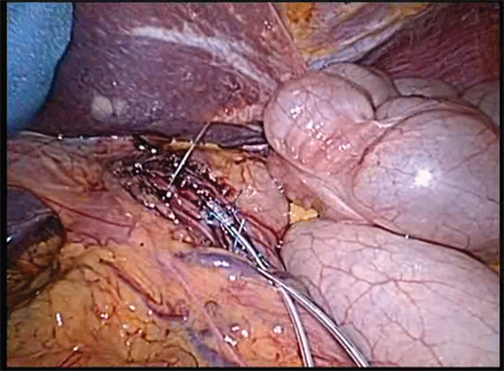
Study connects aspirin 'resistance' to enteric coating
In a study of 400 healthy adults, measurements indicating the presence of what has been thought to be aspirin resistance were evident only with enteric-coated aspirin, not immediate-release aspirin, which the authors have described as aspirin "pseudoresistance."
The researchers used several measurements to evaluate responses reflecting the activity of cyclooxygenase-1 (COX-1), the molecular target of aspirin, including platelet aggregation and serum thromboxane formation, which could indicate a genetic cause of aspirin resistance. Still, "we failed to find a single person who satisfied" the criteria for true aspirin resistance, reported Dr. Tilo Grosser, of the Institute for Translational Medicine and Therapeutics, Perelman School of Medicine in Philadelphia, and his associates. "By contrast, pseudoresistance, due to delayed and reduced drug absorption, was common after ingestion of enteric-coated aspirin," they said.
"These observations question the value of seeking to diagnose aspirin resistance with single point-of-care diagnostic approaches and support the finding of inconsistent platelet inhibition following enteric-coated preparations of aspirin," they concluded. The study appeared in Circulation online Dec. 4 (2012 [doi: 10.1161/CIRCULATIONAHA.112.117283]).
The concept of aspirin resistance has been used to explain treatment failures occurring in patients, with estimates of incidence ranging from 5% to 20% in studies, they noted.
The study had several phases, which included screening the study participants, aged 18-55 years, for their response to a single 325-mg dose of immediate-release or enteric-coated aspirin, repeating testing among those who appeared to be resistant to aspirin, and comparing responses in responders and nonresponders.
Among the specific findings: All of the individuals who received a dose of immediate-release aspirin were responders, but up to 49% of those who received enteric aspirin were considered nonresponders. With repeated exposure, individuals categorized as nonresponders when given enteric-coated aspirin showed evidence of responses when administered immediate-release aspirin.
"What they have shown in an extremely elegant way is that aspirin resistance, at least in healthy volunteers, doesn’t exist," Dr. Deepak Bhatt said in an interview.
Of great interest to physicians and patients is the finding that in the individuals who appeared to be aspirin resistant, enteric coating on aspirin was delaying and reducing absorption, which is consistent with old data that indicate enteric coating can interfere with aspirin absorption, he said. While this may have been suspected as an explanation for so-called aspirin resistance, "this is the first time someone has really nailed it down in a scientifically unassailable way," said Dr. Bhatt, professor of medicine at Harvard University, Boston, and chief of cardiology at VA Boston Healthcare System.
Although the results need to be replicated in patients with actual disease, such as diabetes or atherosclerosis, he said the results will potentially impact practice, although immediate-release aspirin is recommended for patients who are being stented or are having a myocardial infarction, for the rapid pharmacologic effect.
But the results raise further questions about more chronic administration of aspirin in patients who are not acutely ill and how many of these patients may not be fully absorbing aspirin if they are taking an enteric-coated formulation. "If there is a proportion of patients who are not getting the full anticlotting effect of aspirin ... that means potentially millions of patients are taking enteric-coated aspirin and not getting the protection they or their doctors are assuming they are getting," he said.
Another consideration, which he said is not widely appreciated, is that evidence that enteric coating reduces stomach bleeding "is quite scant, some might even say nonexistent." While enteric coated aspirin does reduce stomach upset, it is used for GI bleeding protection, "but if you’re not getting the GI benefit, and now potentially there is this issue of pseudoresistance these authors are raising, you’ve got to think twice about whether enteric coating is doing anything useful or good."
Dr. Bhatt’s recommendation to clinicians, which predates the publication of this study, is to make sure that patients who are prescribed the immediate-release aspirin get the correct formulation, and not end up with an enteric-coated formulation unintentionally.
Dr. Sanjay Kaul, director of the vascular physiology and thrombosis research laboratory at the Burns and Allen Research Institute, Cedars-Sinai Medical Center, Los Angeles, also advised caution about extrapolating the results of a study of healthy volunteers to patients with heart disease or chronic illnesses that may affect how aspirin works in the body. The study was not designed to address comparative clinical efficacy or safety between the two formulations, and he views it as "a mechanistic study demonstrating a pharmacokinetic reason for variability in aspirin responsiveness."
In an interview, he added that testing for aspirin resistance is "of dubious value," and the concept, like resistance to clopidogrel, "is driven to a large extent by marketing considerations," and the development of tests to assess aspirin responsiveness.
Dr. Kaul said he will continue to recommend immediate-release formulation for rapid onset of action and switch to enteric-coated aspirin for chronic use, especially in patients with a history of gastric intolerance or side effects. Although "unequivocal evidence" of safety advantages over immediate-release aspirin, such as GI bleeding, is lacking, data have shown reduced rates of gastric erosions on endoscopy associated with enteric-coated aspirin, although the clinical relevance of the erosions is unclear, he said.
The study was funded by the National Heart, Lung, and Blood Institute; the National Center for Research Resources; the American Heart Association; and Bayer HealthCare. Coauthor Dr. Garret FitzGerald of the University of Pennsylvania, Philadelphia, received research funding from Bayer HealthCare to support partial funding of this study, and Dr. Grosser received consultancy fees from PLx Pharma. The four remaining coauthors had no potential conflicts of interest to disclose.
Dr. Bhatt said his disclosures include being an unpaid consultant to PLx Pharma. Dr. Kaul said he had no relevant disclosures.
FROM CIRCULATION
Major Finding: "Resistance" to a 325-mg dose of enteric-coated aspirin was as high as 49%, compared with 0% after the same patients took immediate-release aspirin.
Data Source: A study of platelet aggregation and other responses in 400 healthy, nonsmoking adults.
Disclosures: The study was funded by the National Heart, Lung, and Blood Institute; the National Center for Research Resources; the American Heart Association; and Bayer HealthCare. Coauthor Dr. Garret FitzGerald of the University of Pennsylvania, Philadelphia, received research funding from Bayer HealthCare to support partial funding of this study, and Dr. Grosser received consultancy fees from PLx Pharma. The four remaining coauthors had no potential conflicts of interest to disclose. Dr. Bhatt said his disclosures include being an unpaid consultant to PLx Pharma. Dr. Kaul said he had no relevant disclosures.
Arrhythmia risk prompts 32-mg IV ondansetron withdrawal
The 32-mg intravenous dose of the antinausea drug ondansetron will no longer be marketed because it has been linked to a risk of QT prolongation, the Food and Drug Administration announced.
A statement on the agency’s MedWatch website said that the withdrawal of the 32-mg dose, available in premixed solutions of dextrose or sodium chloride, is expected to last through early 2013. Since this dose accounts for a "very small percentage" of the market, the voluntary withdrawal is not expected to contribute to a shortage of intravenous ondansetron, the Dec. 4 statement said.
The drug is marketed as Zofran and is available in generic formulations for preventing chemotherapy-induced nausea and vomiting. The FDA is recommending three doses of IV ondansetron at a dose of 0.15 mg/kg administered every 4 hours for three doses to prevent chemotherapy-induced nausea and vomiting but added: "If the calculated weight-based dose were to exceed 16 mg, the potential for prolonged QT interval would be greater; therefore, no single intravenous dose should exceed 16 mg." QT prolongation can result in the potentially fatal arrhythmia, torsades de pointes.
The FDA also stated that, currently, there is not enough information to recommend another single-dose intravenous regimen and that oral ondansetron "remains effective for the prevention of chemotherapy-induced nausea and vomiting."
Ondansetron also is approved for postoperative nausea and vomiting.
The announcement follows a previous FDA notice in June 2012 that the 32-mg single-IV dose of ondansetron should be avoided, citing preliminary results from a study suggesting that the 32-mg dose may cause QT prolongation. The agency first announced that it was conducting a safety review of ondansetron in September 2011.
At the time of the June 2012 statement, GlaxoSmithKline, the manufacturer of Zofran, which conducted the QT study, announced that it was removing the 32-mg single-IV dose from the drug’s label. In November, the labeling was revised and no longer includes the 32-mg IV dose; it includes information about QT prolongation and dosing information that no single-IV weight-based dose should exceed 16 mg.
Manufacturers of ondansetron intravenous products are Baxter Healthcare, Hospira, Teva, Bedford Labs, and Claris Lifesciences.
Serious adverse events associated with ondansetron should be reported to the FDA at 800-332-1088 or http://www.fda.gov/Safety/MedWatch/default.htm.
Study Finds 'PPI Test' a Poor Predictor of GERD
Empiric acid suppression with the "PPI test," widely used to assess whether upper-GI symptoms are due to gastroesophageal reflux disease, actually performs poorly as a predictor of GERD, Dr. Peter Bytzer and his colleagues wrote in the December issue of Clinical Gastroenterology and Hepatology.
Typically, clinicians perform a PPI test to diagnose GERD without the need for endoscopy. They give patients a 2-week course of a proton pump inhibitor and then determine whether the brief treatment ameliorated upper GI symptoms. If so, the symptoms are assumed to be acid related, said Dr. Bytzer of Copenhagen University and his associates.
But in their study, such a PPI test "showed a disappointingly low validity" for diagnosing GERD, and couldn’t reliably distinguish between reflux esophagitis and nonerosive reflux disease. Even when combined with a clinical diagnosis made by a primary care physician, a clinical diagnosis made by a gastroenterologist, or the results of a detailed diagnostic questionnaire, the outcome of a PPI test didn’t add reliable information allowing clinicians to separate patients who had GERD from those who did not, the researchers said (Clin. Gastroenterol. Hepatol. 2012 [doi:10.1016/j.cgh.2012.06.030]).
Dr. Bytzer and his colleagues analyzed data originally collected in the DIAMOND clinical trial, an international study that found the Reflux Disease Questionnaire to be useful in diagnosing GERD among patients who consulted primary care physicians because of frequent upper GI symptoms. The DIAMOND study results indicated that unlike the Reflux Disease Questionnaire, PPI testing had very limited diagnostic value.
Dr. Bytzer and his associates performed more detailed analyses of the data, using different combinations of three main symptoms (heartburn, central chest pain, and dysphagia or regurgitation); different definitions of "PPI response"; and assessments of various subgroups of patients, all in the hope of improving the diagnostic yield of PPI testing. They compared the diagnostic usefulness of PPI testing against state-of-the-art comprehensive diagnostic work-ups for GERD in 299 of the DIAMOND study subjects.
"This large database provides the best platform so far to test whether there are better approaches for scoring a test of PPI therapy," the investigators noted.
All of the subjects underwent endoscopy with concomitant wireless esophageal pH monitoring and symptom assessment. They then completed the detailed Reflux Disease Questionnaire, and participated in a 2-week PPI test using esomeprazole (Nexium), during which they recorded GI symptoms in a diary.
A total of 69% of the patients who did have GERD showed a positive response to the PPI test. This means that 31% of the study participants with GERD were not identified by PPI testing.
Moreover, 51% of the patients who did not have GERD also showed a positive response to the PPI test, the investigators said.
Both patients who had GERD and patients who did not reported that their symptom response to acid suppression increased over the course of the first 5-6 days of treatment and then leveled off for the remaining 8-9 days.
The PPI test performed only marginally better across various subgroups of patients, such as those with GERD plus reflux esophagitis (as compared with patients who had GERD but no reflux esophagitis) or those whose physicians were certain of their GERD diagnosis (compared with patients whose physicians were uncertain about their GERD diagnosis).
For example, 48% of the study participants whose physicians predicted that they had GERD showed a positive response on PPI testing – a rate that was nearly identical to the 47% of study subjects whose physicians predicted that they had some disorder other than GERD.
Similarly, the PPI test performed only marginally better when different symptom combinations and different definitions of "PPI response" were assessed.
"In line with other researchers, we found that ... the clinical value of [the PPI test] is very limited," Dr. Bytzer and his colleagues said.
This study was funded by AstraZeneca. Dr. Bytzer reported ties to AstraZeneca, Boehringer Ingelheim, Eisai, Nycomed, Reckitt Benckiser, Takeda, and Wyeth, and his associates reported ties to numerous industry sources, including employment by AstraZeneca.
FROM CLINICAL GASTROENTEROLOGY AND HEPATOLOGY
Major Finding: Thirty-one percent of study subjects who had GERD showed a negative response to the PPI test, indicating that they did not have the disorder, while 51% of those who did not have GERD showed a false-positive response.
Data Source: Investigators reported a secondary analysis of data from the international DIAMOND study, comparing the results of PPI testing to the actual diagnosis of GERD or other disorders.
Disclosures: This study was funded by AstraZeneca. Dr. Bytzer reported ties to AstraZeneca, Boehringer Ingelheim, Eisai, Nycomed, Reckitt Benckiser, Takeda, and Wyeth, and his associates reported ties to numerous industry sources, including employment by AstraZeneca.
Bariatric Surgery Safety Has Increased With Medicare Coverage
CHICAGO – Complication rates for bariatric surgery patients on Medicare declined after Medicare began to cover the procedure in 2006, an analysis of state inpatient data from 12 states has found.
Data from previous studies suggest that bariatric surgery outcomes for Medicare patients improved after the implementation of Medicare’s National Coverage Determination in 2006, said Dr. Justin B. Dimick of the University of Michigan, Ann Arbor. But specific safety data on Medicare patients who have undergone bariatric surgery since the time of the decision are limited, Dr. Dimick said. The decision allowed for Medicare coverage of patients who sought care at facilities certified by the American College of Surgeons or the American Society for Metabolic and Bariatric Surgery.

To assess the impact of the National Coverage Determination on the safety of bariatric surgery, Dr. Dimick and his colleagues reviewed state inpatient data from 12 large, geographically dispersed states. Patients were identified on the basis of ICD-9 codes and diagnosis-related groups for weight loss surgery.
The outcomes were categorized according to any complication, a serious complication, or a reoperation.
The percentage of Medicare patients with any complications dropped from 12% before the determination to 8% afterward. Similarly, the percentage of non-Medicare patients with any complications dropped from 7% before to 5% after the determination.
In a multivariate analysis, the factors contributing to improved outcomes for bariatric surgery in Medicare patients were an increase in the use of laparoscopic gastric banding (lap band surgery), the transfer of patients to better hospitals, and quality improvement within individual hospitals, Dr. Dimick said.
Improvements in the safety of bariatric surgery did not, however, result from patients being redirected to safer hospitals, said Dr. Dimick. In fact, complication rates for procedures performed at Centers of Excellence versus non–Centers of Excellence were not significantly different (odds ratio for any complications, 0.97), he noted.
"CMS should consider dropping the COE [Centers of Excellence] aspect of the coverage decision [that] limits patient access without a beneficial improvement in outcomes," Dr. Dimick said. "Alternatively, CMS could revise the national coverage decision to further encourage participation in a quality improvement registry." Such a registry would need to include measures of long-term effectiveness to identify any unintended consequences of the increase in lap band surgery, he added.
Dr. Dimick is an equity owner and cofounder of ArborMetrix, a health care analytics and software firm.
CHICAGO – Complication rates for bariatric surgery patients on Medicare declined after Medicare began to cover the procedure in 2006, an analysis of state inpatient data from 12 states has found.
Data from previous studies suggest that bariatric surgery outcomes for Medicare patients improved after the implementation of Medicare’s National Coverage Determination in 2006, said Dr. Justin B. Dimick of the University of Michigan, Ann Arbor. But specific safety data on Medicare patients who have undergone bariatric surgery since the time of the decision are limited, Dr. Dimick said. The decision allowed for Medicare coverage of patients who sought care at facilities certified by the American College of Surgeons or the American Society for Metabolic and Bariatric Surgery.
To assess the impact of the National Coverage Determination on the safety of bariatric surgery, Dr. Dimick and his colleagues reviewed state inpatient data from 12 large, geographically dispersed states. Patients were identified on the basis of ICD-9 codes and diagnosis-related groups for weight loss surgery.
The outcomes were categorized according to any complication, a serious complication, or a reoperation.
The percentage of Medicare patients with any complications dropped from 12% before the determination to 8% afterward. Similarly, the percentage of non-Medicare patients with any complications dropped from 7% before to 5% after the determination.
In a multivariate analysis, the factors contributing to improved outcomes for bariatric surgery in Medicare patients were an increase in the use of laparoscopic gastric banding (lap band surgery), the transfer of patients to better hospitals, and quality improvement within individual hospitals, Dr. Dimick said.
Improvements in the safety of bariatric surgery did not, however, result from patients being redirected to safer hospitals, said Dr. Dimick. In fact, complication rates for procedures performed at Centers of Excellence versus non–Centers of Excellence were not significantly different (odds ratio for any complications, 0.97), he noted.
"CMS should consider dropping the COE [Centers of Excellence] aspect of the coverage decision [that] limits patient access without a beneficial improvement in outcomes," Dr. Dimick said. "Alternatively, CMS could revise the national coverage decision to further encourage participation in a quality improvement registry." Such a registry would need to include measures of long-term effectiveness to identify any unintended consequences of the increase in lap band surgery, he added.
Dr. Dimick is an equity owner and cofounder of ArborMetrix, a health care analytics and software firm.
AT THE ANNUAL CLINICAL CONGRESS OF THE AMERICAN COLLEGE OF SURGEONS
Major Finding: The percentage of Medicare patients with any complications dropped from 12% before the National Coverage Determination to 8% afterward.
Data Source: The data come from state inpatient data from 12 states.
Disclosures: Dr. Dimick is an equity owner and cofounder of ArborMetrix, a health care analytics and software firm.

Two Common Genetic Variants Linked to Barrett's Esophagus
Two common genetic variants on chromosomes 6p21 and 16q24 were found to be associated with Barrett’s esophagus in the first genome-wide association study of susceptibility to the disorder, according to a letter to the editor published online Sept. 9 in Nature Genetics.
Genetic factors have long been suspected to play a role in the development of Barrett’s esophagus since relative risks for the disorder, as well as for gastroesophageal reflux disease and esophageal adenocarcinoma, are increased two- to fourfold in first-degree relatives of affected patients.
However, "extensive candidate gene and linkage searches have to date been unsuccessful in identifying genetic variants that are associated with risk of Barrett’s esophagus," wrote Dr. Zhan Su of the Wellcome Trust Centre for Human Genetics in Oxford, England, and his associates.
Their findings provide the first direct evidence that the etiology of Barrett’s esophagus has a genetic component, the researchers noted.
Dr. Su and his colleagues performed the genome-wide association study as part of a Wellcome Trust consortium study of the genetic components of 15 common disorders and traits. They identified the two genetic variants in a discovery analysis and confirmed the findings in another five replication cohorts.
The discovery analysis involved genotyping of 1,852 adults with histologically confirmed Barrett’s esophagus who were recruited from sites across the United Kingdom. A representative sample of 5,172 control subjects also was analyzed. A total of 521,744 single-nucleotide polymorphisms (SNPs) were typed.
The 100 SNPs that were associated to some degree with Barrett’s esophagus were then analyzed in a separate cohort of 1,105 cases from two clinical trials, 4,421 controls from another U.K. sample, and 2,578 controls from yet another U.K .sample.
The top 16 SNPs from these analyses were then analyzed in a second replication cohort of 473 cases and 1,780 controls from a Dutch genotyping database. Two of the SNPs were found to be significantly associated with Barrett\'’s esophagus: rs9257809 on chromosome 6p21 and rs9936833 on chromosome 16q24.
It is notable that the closest coding gene to the rs9936833 SNP on chromosome 16q24 is a gene that encodes a protein involved in the development of the gastrointestinal tract. It has been reported that, when this gene is inactivated, structural alterations in the esophagus, especially atresia, occur. The rs9257809 SNP on 6p21 is near the telomeric edge of the major histocompatibility complex region.
The two SNPs were then examined further in an Irish cohort of 245 cases and 473 controls, a U.K. cohort of 1,765 cases and 1,586 controls, and a cohort of 2,398 cases and 2,167 controls from Europe, Australia, and the United States.
Overall, the two SNPs "showed compelling evidence for association," with Barrett’s esophagus, reflected in the significance of the combined P values for rs9257809 (odds ratio, 1.21) and rs9936833 (odds ratio, 1.14), the investigators reported (Nat. Genet. 2012 Sept. 9 [doi:10.1038/ng.2408]).
In a subgroup analysis involving only patients who had histologic evidence of intestinal metaplasia, the same two SNPs were again strongly associated with Barrett’s esophagus. Neither SNP, however, was associated with either the circumferential extent or the maximal length of the affected segment of esophagus.
Given that men are known to be more susceptible than women to Barrett’s esophagus, the investigators also performed a sex-stratified analysis of the data and found that the association between both SNPs and the disorder was stronger in men than in women. "This finding warrants further investigation," they noted.
Similarly, given the known association between obesity and Barrett’s esophagus, the researchers analyzed the 40 SNPs that previously have been linked with either body mass index or waist-to-hip ratio. These latter SNPs were "more likely than expected by chance to show effects in the same direction in association with Barrett’s esophagus, suggesting that genetic effects may in part underpin the epidemiologic observation that BMI is a risk factor for Barrett’s esophagus," Dr. Su and his associates said.
"Given that Barrett’s esophagus has an accepted status as a precursor lesion, the SNPs that we have identified could also essentially be risk factors for esophageal adenocarcinoma and may give clues as to the biology of both of these important phenotypes," they added.
The financial disclosures of Dr. Su and his 143 coauthors are listed in the online version of this article.
FROM NATURE GENETICS
Major Finding: The rs9257809 SNP on chromosome 6p21 and the rs9936833 SNP on chromosome 16q24 both were strongly associated with an increased risk of developing Barrett's esophagus.
Data Source: The investigators conducted a genome-wide association study of susceptibility to Barrett’s esophagus, analyzing the genotypes of 7,838 patients and 15,599 control subjects in several countries.
Disclosures: The financial disclosures of Dr. Su and his 143 coauthors are listed in the online version of this article.
Ablation Cost Effective for High-Grade Dysplasia in Barrett's Esophagus
Radiofrequency ablation for high-grade dysplasia in the setting of Barrett’s esophagus is more cost effective than endoscopic surveillance until progression to cancer, reported Dr. Chin Hur and his colleagues in the September issue of Gastroenterology.
Moreover, the use of radiofrequency ablation (RFA) for stable, confirmed low-grade dysplasia can also be cost effective, said the investigators.
Dr. Hur of Harvard University and Massachusetts General Hospital, both in Boston, and his colleagues conducted several analyses comparing three treatment strategies for each of three disease states: Barrett’s esophagus with high-grade dysplasia, Barrett’s esophagus with low-grade dysplasia, and Barrett’s esophagus with no dysplasia.
The treatment strategies consisted of surveillance followed by esophagectomy upon disease progression to cancer, RFA upon disease progression to a higher-grade dysplasia or cancer, or initial RFA before disease progression (Gastroenterology 2012 [doi: 10.1053/j.gastro.2012.05.010]).
Cost calculations were based on Medicare reimbursement rates for 2011. The cost estimate for RFA, for example, was $6,400, and the cost of esophagectomy was $25,882. Based on these values, the authors then conducted a base-case analysis.
They found that in high-grade dysplasia, the strategy of initial RFA resulted in 0.704 more quality-adjusted life-years (QALYs) and cost $25,609 less than the strategy of surveillance without RFA followed by esophagectomy upon disease progression to cancer, assuming a 1% disease progression rate.
Similarly, in low-grade dysplasia, RFA upon disease progression to high-grade dysplasia also bested the strategy of surveillance until cancer and esophagectomy, resulting in 0.17 more QALYs and costing $7,446 less, assuming a 0.5% progression rate.
However, in low-grade dysplasia, when comparing initial RFA versus surveillance until progression to high-grade dysplasia and then RFA, the authors found that the latter approach cost only $1,969 less than the former, and that the former was associated with a 0.108 gain in QALYs.
That amounted to an incremental cost-effectiveness ratio of $18,231 per QALY – "below our willingness-to-pay threshold of $100,000/QALY, making it the most plausible strategy in terms of cost-effectiveness."
Finally, in scenarios involving Barrett’s esophagus but no dysplasia, RFA upon progression of disease to high-grade dysplasia was still the most cost-effective strategy: It was associated with a savings of $7,709 as well as 0.194 additional QALYs, compared with esophagectomy upon disease progression to cancer, assuming a 0.33% progression rate.
The authors noted that their model relies on a stringent definition of low-grade dysplasia that assumes "review and agreement between more than one expert pathologist" as well as a consistent level of dysplasia found on more than one endoscopy spaced at least 6 months apart. In addition, the progression rates used in this analysis were based on the current literature, but were still estimates involving some uncertainty.
"We believe that a multicenter, randomized, controlled trial for initial RF ablation versus surveillance in patients with BE without dysplasia is needed to confirm our model results and to inform clinical decision making," they added.
Long-term follow-up data from such a study could "provide much needed data regarding cancer progression and the need for surveillance, which significantly impacts the cost-effectiveness and patients’ preferences for RFA."
The authors stated that they had no disclosures relevant to this study. The study was supported by grants from the National Institutes of Health.
FROM GASTROENTEROLOGY