User login
Plasma Tau Level Is Chronically Elevated in TBI
Peripheral plasma levels of the CNS protein tau are chronically elevated after traumatic brain injury (TBI) and appear to correlate with the severity of postconcussive symptoms, according to a report published online ahead of print August 3 in JAMA Neurology.
If these findings are confirmed, tau may be the first biomarker that is sensitive and specific to persistent TBI-related symptoms. The results also suggest that “months to years after the primary brain injury, there may be a continuation of secondary injuries with residual axonal degeneration and blood–brain barrier disruptions in this population that may contribute to the maintenance of postconcussive disorder symptoms and affect symptom severity,” said Anlys Olivera, PhD, of the National Institute of Nursing Research in Bethesda, Maryland, and her associates.
Tau stabilizes the structure of the axonal cytoskeleton. It is elevated in the CSF and the peripheral blood (albeit in extremely low concentrations) of patients with severe TBI, professional boxers, and athletes who sustain concussions. The extremely low levels of tau in the peripheral blood have been difficult to measure until the recent development of an ultrahigh-sensitivity immunoassay technology. Using this innovation, the researchers were able to examine for the first time the associations between plasma tau levels and the frequency and severity of deployment-related TBIs.
Over a two-year period, Dr. Olivera and her associates assessed tau levels in 70 members of the military who self-reported one or more TBIs and 28 military control subjects without TBI who were matched for age, sex, race, time since deployment, and number of deployments. Almost all participants in the TBI group had been injured at least 18 months previously. The most common causes of TBI were blows to the head, exposure to blasts, vehicular crashes, and sports-related concussions.
Total tau was significantly increased in the TBI group (mean level, 1.13 pg/mL), compared with the control group (0.63 pg/mL). Total tau also increased with increasing severity of the initial brain injury, with increasing number of TBIs, and with increasing severity of present-day postconcussive symptoms. These associations, moreover, were independent of symptoms of post-traumatic stress disorder and depression, which were prevalent in the TBI group, the investigators said.
Tau is not only a marker of brain injury, it also can contribute to secondary injury processes such as inflammation, which makes it a potential target for therapy. If the findings of this study are confirmed and extended to demonstrate a direct mechanistic relationship between TBI and tau aggregation, treatments such as the direct delivery of proteasomes “would be invaluable, considering the dearth of treatments for TBIs and chronic [postconcussive disorder] symptoms,” Dr. Olivera and her associates said.
Among the study limitations cited by the investigators are lack of neuroimaging and neuropsychologic data.
—Mary Ann Moon
Suggested Reading
Olivera A, Lejbman N, Jeromin A, et al. Peripheral total tau in military personnel who sustain traumatic brain injuries during deployment. JAMA Neurol. 2015 Aug 3 [Epub ahead of print].
Peripheral plasma levels of the CNS protein tau are chronically elevated after traumatic brain injury (TBI) and appear to correlate with the severity of postconcussive symptoms, according to a report published online ahead of print August 3 in JAMA Neurology.
If these findings are confirmed, tau may be the first biomarker that is sensitive and specific to persistent TBI-related symptoms. The results also suggest that “months to years after the primary brain injury, there may be a continuation of secondary injuries with residual axonal degeneration and blood–brain barrier disruptions in this population that may contribute to the maintenance of postconcussive disorder symptoms and affect symptom severity,” said Anlys Olivera, PhD, of the National Institute of Nursing Research in Bethesda, Maryland, and her associates.
Tau stabilizes the structure of the axonal cytoskeleton. It is elevated in the CSF and the peripheral blood (albeit in extremely low concentrations) of patients with severe TBI, professional boxers, and athletes who sustain concussions. The extremely low levels of tau in the peripheral blood have been difficult to measure until the recent development of an ultrahigh-sensitivity immunoassay technology. Using this innovation, the researchers were able to examine for the first time the associations between plasma tau levels and the frequency and severity of deployment-related TBIs.
Over a two-year period, Dr. Olivera and her associates assessed tau levels in 70 members of the military who self-reported one or more TBIs and 28 military control subjects without TBI who were matched for age, sex, race, time since deployment, and number of deployments. Almost all participants in the TBI group had been injured at least 18 months previously. The most common causes of TBI were blows to the head, exposure to blasts, vehicular crashes, and sports-related concussions.
Total tau was significantly increased in the TBI group (mean level, 1.13 pg/mL), compared with the control group (0.63 pg/mL). Total tau also increased with increasing severity of the initial brain injury, with increasing number of TBIs, and with increasing severity of present-day postconcussive symptoms. These associations, moreover, were independent of symptoms of post-traumatic stress disorder and depression, which were prevalent in the TBI group, the investigators said.
Tau is not only a marker of brain injury, it also can contribute to secondary injury processes such as inflammation, which makes it a potential target for therapy. If the findings of this study are confirmed and extended to demonstrate a direct mechanistic relationship between TBI and tau aggregation, treatments such as the direct delivery of proteasomes “would be invaluable, considering the dearth of treatments for TBIs and chronic [postconcussive disorder] symptoms,” Dr. Olivera and her associates said.
Among the study limitations cited by the investigators are lack of neuroimaging and neuropsychologic data.
—Mary Ann Moon
Suggested Reading
Olivera A, Lejbman N, Jeromin A, et al. Peripheral total tau in military personnel who sustain traumatic brain injuries during deployment. JAMA Neurol. 2015 Aug 3 [Epub ahead of print].
Suggested Reading
Olivera A, Lejbman N, Jeromin A, et al. Peripheral total tau in military personnel who sustain traumatic brain injuries during deployment. JAMA Neurol. 2015 Aug 3 [Epub ahead of print].
The VA/DoD Chronic Effects of Neurotrauma Consortium: An Overview at Year 1
The Chronic Effects of Neuro-trauma Consortium (CENC) is a federally funded research project devised to address the long-term effects of mild traumatic brain injury (mTBI) in military service members (SMs) and veterans. Announced by President Barack Obama on August 20, 2013, the CENC is one of 2 major initiatives developed in response to injuries incurred by U.S. service personnel during Operation Enduring Freedom (OEF) and Operation Iraqi Freedom (OIF) as part of the National Research Action Plan. The CENC is jointly funded by the DoD and the VA, with a budget of $62.175 million over 5 years.
The consortium funds basic science, clinical, and translational research efforts with a closely integrated supportive infrastructure, including administrative services, regulatory guidance, study design, biostatistical consultation, data management, common data element application, and interdisciplinary communication. In addition, the consortium facilitates and integrates the activities of a diverse group of skilled specialty research teams, allowing them to fully focus their efforts on understanding and clarifying the relationship between combat-related mTBI and chronic neurotrauma effects, including neurodegeneration.
Background
Nearly 20% of the more than 2.6 million U.S. SMs deployed since 2003 to OEF and OIF have sustained at least 1 TBI, predominantly mTBI. Almost 8% of all OEF/OIF veterans demonstrate persistent post-TBI symptoms more than 6 months postinjury. Acute mTBI effects are typically transient, with headache, cognitive, behavioral, balance, and sleep symptoms most often seen, but symptoms may persist and even lead to lifelong disability. In these individuals, additional chronic effects, such as neuroendocrinologic abnormalities, seizures and seizurelike disorders, fatigue, vision and hearing abnormalities, and numerous other somatic symptoms are more common over time. The long-term effects from single or repeated mTBIs on the persistence of these symptoms, on combat and trauma-related comorbidities, and on long-term brain functioning are unknown.
Increasing evidence supports the link between both concussions and combat-related trauma with chronic traumatic encephalopathy (CTE), which results in progressive cognitive and behavioral decline in subpopulations 5 to 50 years out from repeated or cumulative mTBI exposures. The possibility of a link between mTBI, persistent symptoms, and early dementia has widespread implications for SMs and veterans; however, these chronic and late-life effects of mTBI are poorly understood.
Traumatic brain injuries of mixed severity have been linked to a higher incidence of Alzheimer disease (AD) and other dementias and an earlier onset of AD, although negative findings have also been reported. Chronic traumatic encephalopathy has been reported to occur in retired boxers at higher rates and at younger ages compared with dementia in the general population. More recently, brain autopsies of athletes from a variety of sports with confirmed CTE have demonstrated elevated tau proteins, tau-immunoreactive neurofibrillary tangles, and neuropil threads, suggesting that pathologic processes similar to those occurring in AD may be involved. Longitudinal research bridging SMs, veterans, and athletes with neurotrauma has been fragmented and incompletely focused on the strategic needs (eg, troop readiness) and vision of the DoD and VA.
Critical gaps exist in the literature with few prospective, well-controlled, longitudinal studies on late-life outcomes and neurodegeneration after mTBI, as well as in related basic science research. These research gaps are particularly prominent in the potentially unique injuries and difficulties seen in combat-exposed populations. The existing research, although suggestive, is not rigorous or robust enough to allow for a clear understanding of the relationships, risks, and potential effective interventions for mTBI, chronic symptoms, and neurodegeneration.
The CENC was developed to create a road map of existing knowledge gaps, to recruit the top relevant subject matter experts in the country, to develop and establish a cohesive set of rigorously designed studies to address these knowledge voids, and to leverage core consortium resources both efficiently and effectively.
Related: The Right Care at the Right Time and in the Right Place: The Role of Technology in the VHA
Given these gaps in scientific research and knowledge, the DoD and VA jointly issued a request for proposals to fund a project to address these concerns. After a competitive application process, an integrated proposal, led by researchers at Virginia Commonwealth University (VCU) was announced as the recipient of the Presidential award.
Consortium Structure
The CENC, serving as the comprehensive research network for DoD and VA, focuses on (1) identifying and characterizing the anatomic, molecular, and physiologic mechanisms of chronic injury from mTBI and potential neurodegeneration; (2) investigating the relationship of comorbidities (psychological, neurologic, sensory, motor, pain, cognitive, and neuroendocrine) of trauma and combat exposure to TBI with neurodegeneration; and (3) assessing the efficacy of existing and novel treatment and rehabilitation strategies for chronic effects and neurodegeneration following TBI.
The consortium is a collaboration among more than 30 universities, nonprofit research organizations, VAMCs, and military medical centers made up of a leadership core, 5 research infrastructure cores, 8 active studies, a data safety monitoring committee, a consumer advisory board, a scientific advisory board, and an independent granting mechanism to foster additional research in chronic effects after mTBI.
Leadership Core
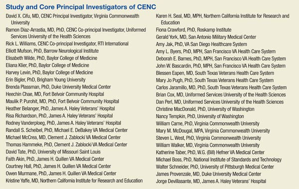
The principal investigator for CENC is David X. Cifu, MD, chairman and professor of the VCU Department of Physical Medicine and Rehabilitation in Richmond, Virginia. The consortium co-principal investigators are Ramon Diaz-Arrastia, MD, PhD, professor of neurology, Uniformed Services University of the Health Sciences (USUHS) and director of the clinical research at the Center for Neuroscience and Regenerative Medicine in Bethesda, Maryland, and Rick L. Williams, PhD, co-principal investigator for CENC and senior statistician at RTI International in Raleigh, North Carolina.
Research Cores
The CENC operates 5 research infrastructure cores. The Biorepository Core, led by Dr. Diaz-Arrastia at USUHS, manages the storage and processing of biologic (blood and saliva) samples collected through all CENC protocols. The Biostatistics Core, led by Dr. Williams; Nancy Temkin, PhD; and Heather Belanger, PhD at RTI, provides study design guidance and biostatistical analysis to facilitate knowledge translation and dissemination.
The Data and Study Management Core is led by Dr. Williams at RTI. It centrally and securely maintains all collected data; oversees the clinical monitoring of research sites; provides a consortium research manager for each study who interacts with the study leadership, study site leaders, and staff; expedites and guides clinical protocols through regulatory approval processes; coordinates patient accrual and study activities across sites; develops and monitors data acquisition compliance; and facilitates exportation of all data collection to the Federal Interagency Traumatic Brain Injury Research informatics system.
The Neuroimaging Core is led by Elisabeth Wilde, PhD, at Baylor College of Medicine and the Michael E. DeBakey VAMC in Houston, Texas. This core facilitates sequence development and pulse programming; provides training and supervision of technologists and support personnel; ensures acquisition, transfer, and storage of imaging data; oversees quality assurance; performs conventional and advanced imaging analysis; and interprets neuroimaging data.
The Neuropathology Core is led by Dr. Dan Perl and colocated at USUHS and Edith Norse Rogers Memorial Veterans Hospital/VA Boston Healthcare System. Dr. Perl manages the collection of brain specimens from the participants, using an existing national network of dieners and neuropathologists, catalogs and stores tissues, and administers requests for use of these tissues.
Active Research Studies
The Longitudinal Cohort Study addresses a critical research gap by identifying and characterizing the late effects of mTBI and assessing the influence and interaction of the many potential risk factors for early dementia. The study uses a wide array of self-report, laboratory, biophysical, neuropsychologic, and imaging assessment tools to evaluate a cohort (n = 880) of U.S. OEF/OIF combatants who have had at least 1 mTBI and a control group of participants (n = 220) who have experienced combat but have not had a mTBI, and then re-assesses them annually (in person or via telephone), with the goal of following the cohort for as long as resources are available.
Collaborating sites for this study include Hunter Holmes McGuire VAMC in Richmond, Virginia; James A. Haley Veterans’ Hospital in Tampa, Florida; Michael E. DeBakey VAMC in Houston, Texas; Audie L. Murphy Memorial Veterans Hospital in San Antonio, Texas; VA Boston Healthcare System; Minneapolis VA Health Care System in Minnesota; and Fort Belvoir in Virginia. Dr. Cifu and Dr. William Walker lead this study.
Epidemiology of mTBI and Neurosensory Outcomes
This project integrates and analyzes several VA, DoD, and Centers for Medicare and Medicaid Services health care system data sets to study the chronic effects of mTBI on neurodegenerative disease and other comorbidities. The primary aims of the project include evaluating the association between mTBI and short-term clinical outcomes, including factors associated with resilience and effects of treatment; investigating long-term clinical outcomes, including neurosensory disorders and mortality; and identifying factors associated with low- and high-distress trajectories of comorbid burden after mTBI. Dr. Kristine Yaffe, Dr. Mary Jo Pugh, and Dr. Michael McCrea, are the leads of this study.
Tau Modification and Aggregation in TBI
This study aims to develop an animal model of repetitive-mTBI, which will allow the tracking of progressive intraneuronal tau alterations that can be correlated with behavioral dysfunction, neuronal protein, and gene expression signatures that can be used to assess the effects of interventions. The observations made in the animal model will be compared with findings generated from tissue obtained at autopsy from deceased SMs and veterans who sustained repetitive-mTBI. Dr. Fiona Crawford and Dr. Elliott Mufson lead this study.
Otolith Dysfunction
This study is examining the effect of inner ear dysfunction on balance, gait, and quality of life (QOL). Recent evidence suggests that otolith organ dysfunction can occur in patients with mTBI or blast exposure. If the dizziness and imbalance symptoms that occur following head injury or blast exposure are related to injury to the otolith organs rather than to the horizontal semicircular canal, then new treatment approaches may be necessary to focus on otolith organ pathway recovery. Performance on balance tasks while standing and walking and questionnaires on the impact on QOL will be compared in 4 groups of individuals (n = 120) with and without head injury/blast exposure (otolith organ dysfunction, horizontal canal dysfunction, both otolith and horizontal canal dysfunction, and healthy individuals). Dr. Faith Akin leads this study.
ADAPT
The ADAPT study (Assessment and Long-term Outcome and Disability in Active Duty Military Prospectively Examined following Concussive TBI) is investigating the association of early clinical and imaging measures with late (5 year) clinical outcome after blast-related mTBI from combat. The study (n = 100) will use 5-year follow-up advanced magnetic resonance imaging (MRI) and clinical outcome measures of combat mTBI, as a continuation of previous longitudinal research efforts (n = 575). Two groups of subjects will be studied: subjects who sustained a mTBI from blast during deployment and subjects without history of blast exposure and no diagnosis of deployment mTBI. Dr. Christine MacDonald leads this study.
Diffusion Tensor Imaging Phantom Study
This study involves the development and testing of a novel phantom that would be used to enhance accuracy, consistency, and reliability in both isotropic and anisotropic measurements derived from diffusion imaging, as well as other MRI-based measurements, using universal fluid disk chambers in a single phantom. Currently, the acquisition of diffusion data in large studies and clinical trials lacks standardization, and important differences exist in how data are acquired on scanners of different manufacturers, using different hardware or software, or when different acquisition parameters are used. As a result, development of large pools of data and the creation of normative data are hampered by inhomogeneity in the data set, which is difficult to analyze. The study team will perform detailed testing of the phantom materials and phantoms themselves, as well as examine diffusion imaging on 1 to 2 human volunteers at each of the 4 sites. Intra- and interscanner differences will be measured, and based on these findings, a more standardized imaging protocol that will provide optimal uniformity of diffusion imaging will be designed. Dr. Elisabeth Wilde leads this study.
Novel White Matter Imaging to Improve mTBI Diagnosis
This study will use myelin-sensitive novel imaging techniques (McDespot [multi-component driven equilibrium single pulse observation of T1/T2]) to improve correspondence with diagnostic groups after trauma exposure and correlation with cognitive deficits in mTBI. The study will recruit individuals (n = 82) from 4 groups, comorbid mTBI and posttraumatic stress disorder (PTSD), only mTBI, only PTSD, and controls who will be prospectively comprehensively assessed clinically (clinical interview, physical exam, neuropsychological assessment) and with advanced imaging (including McDespot, diffusion tensor imaging, and other forms of imaging). Dr. Amy Jak leads this study.
Peer Review Program
The CENC has an integrated grant program to identify scientifically valid and strategically important research projects. To date, 2 rounds of proposal requests and project support have been completed. Scientific review is conducted under the CENC Peer Review Program. Scientifically meritorious studies are identified by independent peer review and then undergo a Programmatic Review by CENC leadership before being recommended for funding to the Government Steering Committee (GSC). Studies that are recommended must address road map gaps, develop innovative approaches, or provide an avenue for new researchers and novel research approaches to contribute to the consortium mission to advance the science of brain injury treatment and prevention. The CENC grant program is administered by Dr. Steven L. West.
Consumer Advisory Board
The Consumer Advisory Board (CAB) advises and makes nonbinding recommendations to CENC. The responsibilities of the committee members include (1) providing information that helps CENC leadership better appreciate and understand the issues and needs of TBI survivors and their support networks so appropriate research can be designed and implemented; (2) evaluating existing research and making recommendations for additions and/or modifications to project procedures; (3) providing input for the road map for future research based on members’ personal experiences and knowledge; and (4) providing linkages to targeted communities for direct feedback and to assist in forming collaborative partnerships.
The CAB is composed of survivors of TBI, family members of survivors of TBI, providers of TBI services, service organizations with specific ties to SMs and veterans, and clinical and corporate representatives of transportation services for the disabled, the independent living movement, and assistive technology. Persons who are heavily engaged in political activity or who actively endorse a specific device or product are not eligible for membership on the CAB. Membership is composed of persons nominated by CENC leadership and approved by the GSC. The CAB is co-chaired by Charles Gatlin, MS, and General (Ret.) Peter Chiarelli.
Scientific Advisory Board
The members of the Scientific Advisory Board (SAB) advise and make nonbinding recommendations to CENC. Responsibilities of the committee members include (1) providing information that may help the consortium leadership better understand the issues related to TBI; (2) evaluating existing research; (3) recommending additions and/or modifications to project procedures; and (4) assisting CENC by helping leverage relationships with other researchers. The SAB is composed of members of the research community on TBI who are not part of CENC. Persons who may be considered to have positions of authority, such as active or retired flag officers or chief executive officers, may be eligible for general SAB membership but are not be eligible for chair positions. Membership is composed of persons nominated by CENC leadership and approved by the GSC. Col. Jamie Grimes, MD, and Henry Lew, MD, PhD, co-chair the SAB.
Federal Oversight
The GSC oversees CENC. Members of the GSC are DoD and VA appointed and represent both government agencies and nongovernment subject matter experts. The GSC approves all studies to be conducted, recommends new studies, and identifies existing and new requirements. The GSC is the overall main governing and management committee for the project and the committee through which the DoD and VA interact and collaborate with the CENC. The GSC determines all major scientific decisions, and clinical studies proposed by the CENC committee proceed to the implementation stage only with the approval of the GSC.
Acknowledgements
This research is supported by grants 1-I01-RX-001135-01-A2 (PI: F. Aiken), 1-I01-RX-001774-01 (PI: F. Crawford), 1-I01-RX-001880-01 (PI: E. Wilde), 1-I01-CX-001135-01 (PI: S. Cifu), and 1-I01-CX-001246-01 (PI: K. Yaffe) from the U.S. Department of Veterans Affairs and by grant W81XWH-13-2-0095 (PI: D. Cifu) from the U.S. Department of Defense, Congressionally Directed Medical Research Programs. The ideas and opinions expressed in this paper do not necessarily represent the views of the Department of Veterans Affairs, the Department of Defense, or the U.S. Government.
Author disclosures
The authors report no actual or potential conflicts of interest with regard to this article.
Disclaimer
The opinions expressed herein are those of the authors and do not necessarily reflect those of Federal Practitioner, Frontline Medical Communications Inc., the U.S. Government, or any of its agencies.
The Chronic Effects of Neuro-trauma Consortium (CENC) is a federally funded research project devised to address the long-term effects of mild traumatic brain injury (mTBI) in military service members (SMs) and veterans. Announced by President Barack Obama on August 20, 2013, the CENC is one of 2 major initiatives developed in response to injuries incurred by U.S. service personnel during Operation Enduring Freedom (OEF) and Operation Iraqi Freedom (OIF) as part of the National Research Action Plan. The CENC is jointly funded by the DoD and the VA, with a budget of $62.175 million over 5 years.
The consortium funds basic science, clinical, and translational research efforts with a closely integrated supportive infrastructure, including administrative services, regulatory guidance, study design, biostatistical consultation, data management, common data element application, and interdisciplinary communication. In addition, the consortium facilitates and integrates the activities of a diverse group of skilled specialty research teams, allowing them to fully focus their efforts on understanding and clarifying the relationship between combat-related mTBI and chronic neurotrauma effects, including neurodegeneration.
Background
Nearly 20% of the more than 2.6 million U.S. SMs deployed since 2003 to OEF and OIF have sustained at least 1 TBI, predominantly mTBI. Almost 8% of all OEF/OIF veterans demonstrate persistent post-TBI symptoms more than 6 months postinjury. Acute mTBI effects are typically transient, with headache, cognitive, behavioral, balance, and sleep symptoms most often seen, but symptoms may persist and even lead to lifelong disability. In these individuals, additional chronic effects, such as neuroendocrinologic abnormalities, seizures and seizurelike disorders, fatigue, vision and hearing abnormalities, and numerous other somatic symptoms are more common over time. The long-term effects from single or repeated mTBIs on the persistence of these symptoms, on combat and trauma-related comorbidities, and on long-term brain functioning are unknown.
Increasing evidence supports the link between both concussions and combat-related trauma with chronic traumatic encephalopathy (CTE), which results in progressive cognitive and behavioral decline in subpopulations 5 to 50 years out from repeated or cumulative mTBI exposures. The possibility of a link between mTBI, persistent symptoms, and early dementia has widespread implications for SMs and veterans; however, these chronic and late-life effects of mTBI are poorly understood.
Traumatic brain injuries of mixed severity have been linked to a higher incidence of Alzheimer disease (AD) and other dementias and an earlier onset of AD, although negative findings have also been reported. Chronic traumatic encephalopathy has been reported to occur in retired boxers at higher rates and at younger ages compared with dementia in the general population. More recently, brain autopsies of athletes from a variety of sports with confirmed CTE have demonstrated elevated tau proteins, tau-immunoreactive neurofibrillary tangles, and neuropil threads, suggesting that pathologic processes similar to those occurring in AD may be involved. Longitudinal research bridging SMs, veterans, and athletes with neurotrauma has been fragmented and incompletely focused on the strategic needs (eg, troop readiness) and vision of the DoD and VA.
Critical gaps exist in the literature with few prospective, well-controlled, longitudinal studies on late-life outcomes and neurodegeneration after mTBI, as well as in related basic science research. These research gaps are particularly prominent in the potentially unique injuries and difficulties seen in combat-exposed populations. The existing research, although suggestive, is not rigorous or robust enough to allow for a clear understanding of the relationships, risks, and potential effective interventions for mTBI, chronic symptoms, and neurodegeneration.
The CENC was developed to create a road map of existing knowledge gaps, to recruit the top relevant subject matter experts in the country, to develop and establish a cohesive set of rigorously designed studies to address these knowledge voids, and to leverage core consortium resources both efficiently and effectively.
Related: The Right Care at the Right Time and in the Right Place: The Role of Technology in the VHA
Given these gaps in scientific research and knowledge, the DoD and VA jointly issued a request for proposals to fund a project to address these concerns. After a competitive application process, an integrated proposal, led by researchers at Virginia Commonwealth University (VCU) was announced as the recipient of the Presidential award.
Consortium Structure
The CENC, serving as the comprehensive research network for DoD and VA, focuses on (1) identifying and characterizing the anatomic, molecular, and physiologic mechanisms of chronic injury from mTBI and potential neurodegeneration; (2) investigating the relationship of comorbidities (psychological, neurologic, sensory, motor, pain, cognitive, and neuroendocrine) of trauma and combat exposure to TBI with neurodegeneration; and (3) assessing the efficacy of existing and novel treatment and rehabilitation strategies for chronic effects and neurodegeneration following TBI.
The consortium is a collaboration among more than 30 universities, nonprofit research organizations, VAMCs, and military medical centers made up of a leadership core, 5 research infrastructure cores, 8 active studies, a data safety monitoring committee, a consumer advisory board, a scientific advisory board, and an independent granting mechanism to foster additional research in chronic effects after mTBI.
Leadership Core
The principal investigator for CENC is David X. Cifu, MD, chairman and professor of the VCU Department of Physical Medicine and Rehabilitation in Richmond, Virginia. The consortium co-principal investigators are Ramon Diaz-Arrastia, MD, PhD, professor of neurology, Uniformed Services University of the Health Sciences (USUHS) and director of the clinical research at the Center for Neuroscience and Regenerative Medicine in Bethesda, Maryland, and Rick L. Williams, PhD, co-principal investigator for CENC and senior statistician at RTI International in Raleigh, North Carolina.
Research Cores
The CENC operates 5 research infrastructure cores. The Biorepository Core, led by Dr. Diaz-Arrastia at USUHS, manages the storage and processing of biologic (blood and saliva) samples collected through all CENC protocols. The Biostatistics Core, led by Dr. Williams; Nancy Temkin, PhD; and Heather Belanger, PhD at RTI, provides study design guidance and biostatistical analysis to facilitate knowledge translation and dissemination.
The Data and Study Management Core is led by Dr. Williams at RTI. It centrally and securely maintains all collected data; oversees the clinical monitoring of research sites; provides a consortium research manager for each study who interacts with the study leadership, study site leaders, and staff; expedites and guides clinical protocols through regulatory approval processes; coordinates patient accrual and study activities across sites; develops and monitors data acquisition compliance; and facilitates exportation of all data collection to the Federal Interagency Traumatic Brain Injury Research informatics system.
The Neuroimaging Core is led by Elisabeth Wilde, PhD, at Baylor College of Medicine and the Michael E. DeBakey VAMC in Houston, Texas. This core facilitates sequence development and pulse programming; provides training and supervision of technologists and support personnel; ensures acquisition, transfer, and storage of imaging data; oversees quality assurance; performs conventional and advanced imaging analysis; and interprets neuroimaging data.
The Neuropathology Core is led by Dr. Dan Perl and colocated at USUHS and Edith Norse Rogers Memorial Veterans Hospital/VA Boston Healthcare System. Dr. Perl manages the collection of brain specimens from the participants, using an existing national network of dieners and neuropathologists, catalogs and stores tissues, and administers requests for use of these tissues.
Active Research Studies
The Longitudinal Cohort Study addresses a critical research gap by identifying and characterizing the late effects of mTBI and assessing the influence and interaction of the many potential risk factors for early dementia. The study uses a wide array of self-report, laboratory, biophysical, neuropsychologic, and imaging assessment tools to evaluate a cohort (n = 880) of U.S. OEF/OIF combatants who have had at least 1 mTBI and a control group of participants (n = 220) who have experienced combat but have not had a mTBI, and then re-assesses them annually (in person or via telephone), with the goal of following the cohort for as long as resources are available.
Collaborating sites for this study include Hunter Holmes McGuire VAMC in Richmond, Virginia; James A. Haley Veterans’ Hospital in Tampa, Florida; Michael E. DeBakey VAMC in Houston, Texas; Audie L. Murphy Memorial Veterans Hospital in San Antonio, Texas; VA Boston Healthcare System; Minneapolis VA Health Care System in Minnesota; and Fort Belvoir in Virginia. Dr. Cifu and Dr. William Walker lead this study.
Epidemiology of mTBI and Neurosensory Outcomes
This project integrates and analyzes several VA, DoD, and Centers for Medicare and Medicaid Services health care system data sets to study the chronic effects of mTBI on neurodegenerative disease and other comorbidities. The primary aims of the project include evaluating the association between mTBI and short-term clinical outcomes, including factors associated with resilience and effects of treatment; investigating long-term clinical outcomes, including neurosensory disorders and mortality; and identifying factors associated with low- and high-distress trajectories of comorbid burden after mTBI. Dr. Kristine Yaffe, Dr. Mary Jo Pugh, and Dr. Michael McCrea, are the leads of this study.
Tau Modification and Aggregation in TBI
This study aims to develop an animal model of repetitive-mTBI, which will allow the tracking of progressive intraneuronal tau alterations that can be correlated with behavioral dysfunction, neuronal protein, and gene expression signatures that can be used to assess the effects of interventions. The observations made in the animal model will be compared with findings generated from tissue obtained at autopsy from deceased SMs and veterans who sustained repetitive-mTBI. Dr. Fiona Crawford and Dr. Elliott Mufson lead this study.
Otolith Dysfunction
This study is examining the effect of inner ear dysfunction on balance, gait, and quality of life (QOL). Recent evidence suggests that otolith organ dysfunction can occur in patients with mTBI or blast exposure. If the dizziness and imbalance symptoms that occur following head injury or blast exposure are related to injury to the otolith organs rather than to the horizontal semicircular canal, then new treatment approaches may be necessary to focus on otolith organ pathway recovery. Performance on balance tasks while standing and walking and questionnaires on the impact on QOL will be compared in 4 groups of individuals (n = 120) with and without head injury/blast exposure (otolith organ dysfunction, horizontal canal dysfunction, both otolith and horizontal canal dysfunction, and healthy individuals). Dr. Faith Akin leads this study.
ADAPT
The ADAPT study (Assessment and Long-term Outcome and Disability in Active Duty Military Prospectively Examined following Concussive TBI) is investigating the association of early clinical and imaging measures with late (5 year) clinical outcome after blast-related mTBI from combat. The study (n = 100) will use 5-year follow-up advanced magnetic resonance imaging (MRI) and clinical outcome measures of combat mTBI, as a continuation of previous longitudinal research efforts (n = 575). Two groups of subjects will be studied: subjects who sustained a mTBI from blast during deployment and subjects without history of blast exposure and no diagnosis of deployment mTBI. Dr. Christine MacDonald leads this study.
Diffusion Tensor Imaging Phantom Study
This study involves the development and testing of a novel phantom that would be used to enhance accuracy, consistency, and reliability in both isotropic and anisotropic measurements derived from diffusion imaging, as well as other MRI-based measurements, using universal fluid disk chambers in a single phantom. Currently, the acquisition of diffusion data in large studies and clinical trials lacks standardization, and important differences exist in how data are acquired on scanners of different manufacturers, using different hardware or software, or when different acquisition parameters are used. As a result, development of large pools of data and the creation of normative data are hampered by inhomogeneity in the data set, which is difficult to analyze. The study team will perform detailed testing of the phantom materials and phantoms themselves, as well as examine diffusion imaging on 1 to 2 human volunteers at each of the 4 sites. Intra- and interscanner differences will be measured, and based on these findings, a more standardized imaging protocol that will provide optimal uniformity of diffusion imaging will be designed. Dr. Elisabeth Wilde leads this study.
Novel White Matter Imaging to Improve mTBI Diagnosis
This study will use myelin-sensitive novel imaging techniques (McDespot [multi-component driven equilibrium single pulse observation of T1/T2]) to improve correspondence with diagnostic groups after trauma exposure and correlation with cognitive deficits in mTBI. The study will recruit individuals (n = 82) from 4 groups, comorbid mTBI and posttraumatic stress disorder (PTSD), only mTBI, only PTSD, and controls who will be prospectively comprehensively assessed clinically (clinical interview, physical exam, neuropsychological assessment) and with advanced imaging (including McDespot, diffusion tensor imaging, and other forms of imaging). Dr. Amy Jak leads this study.
Peer Review Program
The CENC has an integrated grant program to identify scientifically valid and strategically important research projects. To date, 2 rounds of proposal requests and project support have been completed. Scientific review is conducted under the CENC Peer Review Program. Scientifically meritorious studies are identified by independent peer review and then undergo a Programmatic Review by CENC leadership before being recommended for funding to the Government Steering Committee (GSC). Studies that are recommended must address road map gaps, develop innovative approaches, or provide an avenue for new researchers and novel research approaches to contribute to the consortium mission to advance the science of brain injury treatment and prevention. The CENC grant program is administered by Dr. Steven L. West.
Consumer Advisory Board
The Consumer Advisory Board (CAB) advises and makes nonbinding recommendations to CENC. The responsibilities of the committee members include (1) providing information that helps CENC leadership better appreciate and understand the issues and needs of TBI survivors and their support networks so appropriate research can be designed and implemented; (2) evaluating existing research and making recommendations for additions and/or modifications to project procedures; (3) providing input for the road map for future research based on members’ personal experiences and knowledge; and (4) providing linkages to targeted communities for direct feedback and to assist in forming collaborative partnerships.
The CAB is composed of survivors of TBI, family members of survivors of TBI, providers of TBI services, service organizations with specific ties to SMs and veterans, and clinical and corporate representatives of transportation services for the disabled, the independent living movement, and assistive technology. Persons who are heavily engaged in political activity or who actively endorse a specific device or product are not eligible for membership on the CAB. Membership is composed of persons nominated by CENC leadership and approved by the GSC. The CAB is co-chaired by Charles Gatlin, MS, and General (Ret.) Peter Chiarelli.
Scientific Advisory Board
The members of the Scientific Advisory Board (SAB) advise and make nonbinding recommendations to CENC. Responsibilities of the committee members include (1) providing information that may help the consortium leadership better understand the issues related to TBI; (2) evaluating existing research; (3) recommending additions and/or modifications to project procedures; and (4) assisting CENC by helping leverage relationships with other researchers. The SAB is composed of members of the research community on TBI who are not part of CENC. Persons who may be considered to have positions of authority, such as active or retired flag officers or chief executive officers, may be eligible for general SAB membership but are not be eligible for chair positions. Membership is composed of persons nominated by CENC leadership and approved by the GSC. Col. Jamie Grimes, MD, and Henry Lew, MD, PhD, co-chair the SAB.
Federal Oversight
The GSC oversees CENC. Members of the GSC are DoD and VA appointed and represent both government agencies and nongovernment subject matter experts. The GSC approves all studies to be conducted, recommends new studies, and identifies existing and new requirements. The GSC is the overall main governing and management committee for the project and the committee through which the DoD and VA interact and collaborate with the CENC. The GSC determines all major scientific decisions, and clinical studies proposed by the CENC committee proceed to the implementation stage only with the approval of the GSC.
Acknowledgements
This research is supported by grants 1-I01-RX-001135-01-A2 (PI: F. Aiken), 1-I01-RX-001774-01 (PI: F. Crawford), 1-I01-RX-001880-01 (PI: E. Wilde), 1-I01-CX-001135-01 (PI: S. Cifu), and 1-I01-CX-001246-01 (PI: K. Yaffe) from the U.S. Department of Veterans Affairs and by grant W81XWH-13-2-0095 (PI: D. Cifu) from the U.S. Department of Defense, Congressionally Directed Medical Research Programs. The ideas and opinions expressed in this paper do not necessarily represent the views of the Department of Veterans Affairs, the Department of Defense, or the U.S. Government.
Author disclosures
The authors report no actual or potential conflicts of interest with regard to this article.
Disclaimer
The opinions expressed herein are those of the authors and do not necessarily reflect those of Federal Practitioner, Frontline Medical Communications Inc., the U.S. Government, or any of its agencies.
Player-to-Player Contact Is the Main Source of High School Soccer Concussions
Head contact with other players, not with the ball, is the main source of concussions among high school soccer players, according to research published online ahead of print July 13 in JAMA Pediatrics.
Several studies have shown that heading the ball is responsible for many soccer-related concussions. Some people have called for banning heading, especially among children and adolescents, to make the sport safer. No large study, however, had examined the exact mechanism of head injuries among school-aged soccer players, so such prevention efforts could not be considered evidence-based, said R. Dawn Comstock, PhD, an epidemiologist at the University of Colorado Denver in Aurora.
Dr. Comstock and colleagues performed a retrospective analysis of data from a large, Internet-based sports injury surveillance study, focusing on concussions sustained during soccer practices or games that required medical attention and restricted the athlete’s participation for one or more days. The investigators assessed nationally representative samples of 100 high schools every year for nine years. There were 627 concussions during 1,393,753 athlete exposures among girls (4.50 per 10,000 exposures) and 442 concussions during 1,592,238 athlete exposures among boys (2.78 per 10,000 exposures).
The most common mechanism of concussion was player-to-player contact among boys (68.8%) and girls (51.3%). Contact with the ball accounted for 17% of concussions among boys and 29% among girls.
The number and types of concussion symptoms were the same, regardless of whether the concussion was caused by player-to-player contact or player-to-ball contact. However, symptom resolution time was slightly but significantly longer for both boys and girls when the concussion was caused by collision with a ball or goal post.
“We postulate that banning heading from soccer will have limited effectiveness as a primary prevention mechanism unless such a ban is combined with concurrent efforts to reduce athlete–athlete contact throughout the game,” Dr. Comstock and her associates said.
“It may be culturally more tolerable to the soccer community to attempt to reduce athlete–athlete contact across all phases of play through better enforcement of existing rules, enhanced education of athletes on the rules of the game, and improved coaching of activities such as heading,” rather than simply banning heading, said the researchers.
—Mary Ann Moon
Brain Training for TBI Patients
Making the brain work strategically did more good for patients with brain injury than did education about how the brain works, evidenced through Strategic Memory Advanced Reasoning Training (SMART), a DoD-funded study conducted by researchers from The University of Texas at Dallas. Sixty participants aged 19 to 65 years were tested on complex abstraction and innovation or participated in an educational program. Both groups had 18 hours of training in 12 group sessions over 8 weeks.
Related: Stopping TBI-Related Brain Degeneration
The SMART group improved complex abstraction scores by > 20% and memory scores by > 30%; they also reported a 60% reduction in depressive symptoms and a 40% reduction in symptoms related to posttraumatic stress disorder (PTSD).
Related: Protecting Sensory Health
Notably, blood flow to the frontal lobe, anterior cingulate, and precuneus increased significantly in the study group, compared with the education-only patients. According to a study investigator, the increased blood flow implies that the brain is undergoing changes suggestive of improved neural health. Reduction in blood flow to the precuneus has been linked to the emotional regulation of stress and to the severity of traumatic brain injury and PTSD symptoms; the frontal region is associated with increased abstract thinking, and the anterior cingulate is associated with superior cognitive performance. The researchers suggest that improved abstract thinking and executive functioning could help the injured brain to down-regulate emotional reactions.
Related: Attention Deficit/Hyperactivity Disorder in a VA Polytrauma Clinic
The study patients realized cognitive, psychological, and other benefits for 3 to 4 months after training, which the researchers say may mean they continued to improve after the training ended. One investigator says that the findings suggest that brain injuries should be treated more like a chronic health condition than a single short-term event.
SourceVas A, Chapman S, Aslan S, et al. Neuropsychol Rehabil. 2015:1-30. [Online ahead of print.]
Ideas for Helping TBI Patients
Can crowdsourcing produce innovative and practical ways to fill health care gaps? The Defense Centers of Excellence for Psychological Health and Traumatic Brain Injury (DCoE) hopes so.
Everyone—military, civilians, caregivers, and clinicians, people living with posttraumatic stress disorder or traumatic brain injury (TBI)—is eligible to share ideas. The site posts contributions, such as one from a woman whose husband could not attend open-casket funerals because he “could smell the dead body.” She suggests collecting anecdotal behaviors from spouses or caregivers and sharing the information to help people understand why veterans who have survived combat have different social cues. Another entry advocates for a mobile application to help people deal with ongoing fatigue. A third promotes emotional freedom techniques (“tapping” on acupressure points) for relieving symptoms of long-standing trauma.
Ideas can address prevention of TBI, a product or service that helps caregivers, or anything related to improving care. Winners will be announced at the DCoE Challenge Community website.
Stopping TBI-Related Brain Degeneration
A drug that blocks overproduction of molecules that cause brain inflammation after traumatic brain injury (TBI) may offer hope to those who have experienced severe head trauma. Researchers from the University of Kentucky’s Sanders-Brown Center on Aging in Lexington conducted a study in mice that suggests that treatment with the drug may interrupt the process that links head injury with later development of degenerative brain diseases, such as Alzheimer disease.
Related: TBI Assisted Living Program Extended
The drug, known as MW151, was given to mice 1 week after TBI. After 3 weeks of treatment, those mice no longer showed learning and memory problems, unlike the mice that did not receive MW151, the researchers said.
Related: Resilience and Reintegration
More than a million people in the U.S. seek treatment for TBI each year, and the impact of earlier onset of dementia in such a large number of people is “simply unthinkable,” according to Linda Van Eldik, PhD, director of the Sanders-Brown Center and the developer of the drug. The study findings, she says, “could have a large impact both socially and economically.”
Sources
Webster SJ, Van Eldik LJ, Watterson DM, Bachstetter AD. J Neurosci. 2015;35(16):6554-6569.
doi: 10.1523/JNEUROSCI.0291-15.2015.
Dawahare L. Researchers see promise in treatment to reduce incidence of dementia after traumatic brain injury. UKNOW, University of Kentucky News. April 23, 2015. http://uknow.uky.edu/content/researchers-see-promise-treatment-reduce-incidence-dementia-after-traumatic-brain-injury. Accessed May 19, 2015.
TBI Assisted Living Program Extended
The Assisted Living Pilot Program for Veterans With Traumatic Brain Injury (AL-TBI), originally slated to end in 2014, has been extended until October 6, 2017.
Related: New Guidelines on Concussion and Sleep Disturbance
Under the program, eligible veterans are placed in private sector TBI residential care facilities that specialize in neurobehavioral rehabilitation. The program offers team-based care and assistance in speech, memory, and mobility.
Related: Depression and Substance Abuse Intensify Suicide Risk
More than 200 veterans have participated in the pilot program at 47 facilities in 22 states; 101 are currently enrolled. The VA continues to accept eligible veterans into the program (www.polytrauma.va.gov).
To participate, veterans need to be enrolled in VA care, have received hospital care or medical services provided by VA for moderate-to-severe TBI, and be unable to manage 2 or more routine or instrumental activities of daily living without supervision and assistance.
Olfactory Impairment May Indicate TBI Among Blast-Injured Troops
Decreased ability to identify odors may be a marker of acute structural neuropathology resulting from trauma, according to research published online ahead of print March 18 in Neurology. Quantitative identification olfactometry has limited sensitivity but high specificity in detecting this pathology and could inform decisions about whether advanced neuroimaging is required, said the authors.
Michael S. Xydakis, MD, a colonel in the US Air Force and Associate Professor of Surgery at the Uniformed Services University of the Health Sciences in Bethesda, Maryland, and colleagues enrolled 231 consecutive patients with polytrauma in a study to determine whether a quantitative assessment of differential olfactory performance could serve as a reliable antecedent marker for the preclinical detection of intracranial neurotrauma. Participants had been acutely injured from explosions during combat operations in Afghanistan or Iraq, required immediate stateside evacuation, and were enrolled prospectively during two and a half years.
All patients underwent evaluation for possible traumatic brain injury (TBI). The investigators stratified the patients into groups according to severity of TBI and neuroimaging results. Blast-injured troops without TBI who had comparable demographic features and severity of polytrauma formed the comparison control group. An otorhinolaryngologist administered the University of Pennsylvania Smell Identification Test to each patient. Patients were described as having normal, decreased, or absent olfactory function, and the latter two categories were considered to represent olfactory impairment.
Impairment Associated With Frontal and Temporal Lobe Injuries
Approximately 6% of participants had impaired olfactory function. All patients in the mild TBI group and the blast-injured control group had normal olfactory function. Median olfactometric scores did not differ significantly between these two groups. All participants with normal neuroimaging, including 127 patients with mild TBI and 47 controls, had normal olfactory function.
Among the 40 patients with abnormal imaging, 35% had olfactory impairment. Data analysis indicated that olfactometric score predicted abnormal neuroimaging significantly better than chance alone. Olfactory testing was administered to 18 of the patients with abnormal imaging within 14 days after injury. Nine of these patients had impaired function. The remaining 22 soldiers with abnormal imaging underwent testing 15 or more days after injury, and five of them had impaired function. “These results suggest that it is worth testing the hypothesis that sensitivity of olfactory testing to identify patients with structural brain injury may be higher if testing is performed closer to the time of injury,” said Dr. Xydakis.
Approximately 79% of patients with olfactory impairment had injury to the frontal lobe, compared with 42% of patients with normal olfactory function. About 86% of troops with olfactory impairment had either frontal or temporal involvement, compared with 50% of patients with normal function. Approximately 36% of troops with olfactory impairment had both frontal and temporal involvement, compared with 12% of patients with normal function.
Test May Detect Injury Preclinically
“The radiographic findings support a higher-order CNS etiology for the observed impairment,” said Dr. Xydakis. The inclusion of the blast-injured control group with normal olfactometric scores may mitigate the concern that observed impairments resulted from peripheral trauma at the intranasal receptor level.
The finding that only troops with concurrent acute traumatic radiographic abnormalities had olfactory impairment “supports the assertion that impaired olfactory identification is only present in the context of significant intracranial neurotrauma,” he added. “Ultimately, it is the radiographic presence and the radiographic locations of the structural brain injuries that define the probability of subsequent olfactory performance degradation, and not simply the abstract and unquantifiable risk factor of a ‘blow or hit to the head region.’
“The presence of measurable abnormalities with central olfactory dysfunction provides added value to the practicing physician for preclinical detection of intracranial injury and, accordingly, subsequent disease-modifying early interventions,” Dr. Xydakis continued. “While the level of sensitivity for screening purposes is insufficient to exclude all types of post-traumatic neuropathology, the absolute specificity and the association with frontal or temporal lobe injury enhance its value in clinical practice.”
—Erik Greb
Suggested Reading
Xydakis MS, Mulligan LP, Smith AB, et al. Olfactory impairment and traumatic brain injury in blast-injured combat troops: A cohort study. Neurology. 2015 Mar 18 [Epub ahead of print].
Decreased ability to identify odors may be a marker of acute structural neuropathology resulting from trauma, according to research published online ahead of print March 18 in Neurology. Quantitative identification olfactometry has limited sensitivity but high specificity in detecting this pathology and could inform decisions about whether advanced neuroimaging is required, said the authors.
Michael S. Xydakis, MD, a colonel in the US Air Force and Associate Professor of Surgery at the Uniformed Services University of the Health Sciences in Bethesda, Maryland, and colleagues enrolled 231 consecutive patients with polytrauma in a study to determine whether a quantitative assessment of differential olfactory performance could serve as a reliable antecedent marker for the preclinical detection of intracranial neurotrauma. Participants had been acutely injured from explosions during combat operations in Afghanistan or Iraq, required immediate stateside evacuation, and were enrolled prospectively during two and a half years.
All patients underwent evaluation for possible traumatic brain injury (TBI). The investigators stratified the patients into groups according to severity of TBI and neuroimaging results. Blast-injured troops without TBI who had comparable demographic features and severity of polytrauma formed the comparison control group. An otorhinolaryngologist administered the University of Pennsylvania Smell Identification Test to each patient. Patients were described as having normal, decreased, or absent olfactory function, and the latter two categories were considered to represent olfactory impairment.
Impairment Associated With Frontal and Temporal Lobe Injuries
Approximately 6% of participants had impaired olfactory function. All patients in the mild TBI group and the blast-injured control group had normal olfactory function. Median olfactometric scores did not differ significantly between these two groups. All participants with normal neuroimaging, including 127 patients with mild TBI and 47 controls, had normal olfactory function.
Among the 40 patients with abnormal imaging, 35% had olfactory impairment. Data analysis indicated that olfactometric score predicted abnormal neuroimaging significantly better than chance alone. Olfactory testing was administered to 18 of the patients with abnormal imaging within 14 days after injury. Nine of these patients had impaired function. The remaining 22 soldiers with abnormal imaging underwent testing 15 or more days after injury, and five of them had impaired function. “These results suggest that it is worth testing the hypothesis that sensitivity of olfactory testing to identify patients with structural brain injury may be higher if testing is performed closer to the time of injury,” said Dr. Xydakis.
Approximately 79% of patients with olfactory impairment had injury to the frontal lobe, compared with 42% of patients with normal olfactory function. About 86% of troops with olfactory impairment had either frontal or temporal involvement, compared with 50% of patients with normal function. Approximately 36% of troops with olfactory impairment had both frontal and temporal involvement, compared with 12% of patients with normal function.
Test May Detect Injury Preclinically
“The radiographic findings support a higher-order CNS etiology for the observed impairment,” said Dr. Xydakis. The inclusion of the blast-injured control group with normal olfactometric scores may mitigate the concern that observed impairments resulted from peripheral trauma at the intranasal receptor level.
The finding that only troops with concurrent acute traumatic radiographic abnormalities had olfactory impairment “supports the assertion that impaired olfactory identification is only present in the context of significant intracranial neurotrauma,” he added. “Ultimately, it is the radiographic presence and the radiographic locations of the structural brain injuries that define the probability of subsequent olfactory performance degradation, and not simply the abstract and unquantifiable risk factor of a ‘blow or hit to the head region.’
“The presence of measurable abnormalities with central olfactory dysfunction provides added value to the practicing physician for preclinical detection of intracranial injury and, accordingly, subsequent disease-modifying early interventions,” Dr. Xydakis continued. “While the level of sensitivity for screening purposes is insufficient to exclude all types of post-traumatic neuropathology, the absolute specificity and the association with frontal or temporal lobe injury enhance its value in clinical practice.”
—Erik Greb
Suggested Reading
Xydakis MS, Mulligan LP, Smith AB, et al. Olfactory impairment and traumatic brain injury in blast-injured combat troops: A cohort study. Neurology. 2015 Mar 18 [Epub ahead of print].
Suggested Reading
Xydakis MS, Mulligan LP, Smith AB, et al. Olfactory impairment and traumatic brain injury in blast-injured combat troops: A cohort study. Neurology. 2015 Mar 18 [Epub ahead of print].
Does TBI in Later Life Increase the Risk for Parkinson’s Disease?
Patients 55 and older who present to inpatient and emergency department settings with a traumatic brain injury (TBI) have a 44% increased risk of developing Parkinson’s disease over five to seven years, compared with patients in the same age group who present with non-TBI trauma (NTT), according to research published online ahead of print February 27 in Annals of Neurology. In addition, the risk of developing Parkinson’s disease doubles following more severe or more frequent TBI, compared with mild or single TBI. This finding supports a causal association between TBI and Parkinson’s disease.
Raquel C. Gardner, MD, Clinical Instructor and Behavioral Neurology Fellow at the University of California, San Francisco, and colleagues analyzed International Classification of Diseases, Ninth Revision code data collected at California hospitals from 2005 to 2006 to evaluate the risk of developing Parkinson’s disease after TBI in older adulthood. Because of the theoretical possibility that patients with incipient Parkinson’s disease are more likely to fall and sustain a TBI than healthy controls, the researchers examined patients with NTT—defined as fracture, excluding fractures of the head and neck—to reduce possible confounding and reverse causation. To reduce the chance of reverse causation further, researchers excluded cases in which Parkinson’s disease was diagnosed less than a year after the injury.
Researchers identified 52,393 patients with TBI and 113,406 patients with NTT who survived hospitalization and did not have Parkinson’s disease or dementia at baseline. Using Kaplan–Meier estimates and Cox proportional hazards models adjusted for age, sex, race or ethnicity, income, comorbidities, health care use, and trauma severity, they estimated the risk of Parkinson’s disease after TBI during follow-up ending in 2011.
Patients With TBI Were Diagnosed Sooner
Patients with TBI were significantly more likely to be diagnosed with Parkinson’s disease, compared with NTT patients (1.7% of patients vs 1.1% of patients), and patients with TBI were diagnosed with Parkinson’s disease slightly sooner than those with NTT (at 3.1 years, compared with 3.3 years). Researchers found that risk of Parkinson’s disease was similar for TBI sustained via falls and for TBI sustained through other mechanisms.
Researchers also assessed the effect of TBI severity and TBI frequency and found a significant dose response. Patients with mild TBI were 24% more likely to develop Parkinson’s disease, and patients with moderate to severe TBI were 50% more likely to develop Parkinson’s disease, compared with those with NTT. “The evidence for a dose response for increasing TBI severity and TBI frequency, and our persistently significant results despite multiple additional analyses, all enhance causal inference,” the authors said.
A causal association between TBI and Parkinson’s disease may be explained by several possible mechanisms, the researchers said. TBI may reduce motor reserve, thus leading to an earlier diagnosis of Parkinson’s disease in susceptible patients. TBI also may accelerate or augment a pre-existing neurodegenerative cascade or trigger a de novo neurodegenerative cascade. The question of whether typical Parkinson’s disease neuropathologies or unique TBI-specific neuropathology causes post-TBI syndromes deserves further study, they said.
Studies using animal models support a causal mechanism for post-TBI Parkinson’s disease. For example, a progressive loss of dopaminergic neurons and abnormal accumulation of α-synuclein in the substantia nigra have been found in rats after experimentally induced TBI. Other research has begun to replicate these findings in humans.
Information About Patients Was Limited
The study’s limitations include the use of administrative diagnostic codes, which may be poorly sensitive or specific to Parkinson’s disease diagnoses. The researchers lacked information regarding patients’ medical histories and other data about patients’ treatments and outcomes. Also, post-traumatic motor or behavioral abnormalities may complicate the diagnosis of Parkinson’s disease, and diagnoses were not verified by expert review. In addition, the use of a trauma control group essentially controlled for any additional harmful effects of trauma on the nervous system that potentially could increase risk of Parkinson’s disease independently. It is important for large-scale prospective studies, ideally with autopsy confirmation, to confirm these findings, the investigators said.
The results are in line with a 2013 meta-analysis of 22 studies that reported a pooled odds ratio of 1.57 for the association between Parkinson’s disease and head trauma, the authors said. When considered with other studies, including prior research by Dr. Gardner’s team that identified a 26% increased risk of dementia after TBI versus NTT in this population, the results “suggest that TBI is an important independent risk factor for a variety of neurodegenerative syndromes.”
The findings also highlight the importance of preventing falls, which caused approximately 66% of trauma in the TBI and NTT groups. “As the cause of trauma in this study was overwhelmingly due to falls, there is critical importance for fall prevention in middle-aged and older adults, not only as a means to prevent bodily injury, but potentially as a means to prevent neurodegenerative diseases such as dementia and Parkinson’s disease,” the authors concluded.
Suggested Reading
Gardner RC, Burke JF, Nettiksimmons J, et al. Traumatic brain injury in later life increases risk for Parkinson’s disease. Ann Neurol. 2015 Feb 27 [Epub ahead of print].
Jafari S, Etminan M, Aminzadeh F, Samii A. Head injury and risk of Parkinson disease: a systematic review and meta-analysis. Mov Disord. 2013;28(9):1222-1229.
Patients 55 and older who present to inpatient and emergency department settings with a traumatic brain injury (TBI) have a 44% increased risk of developing Parkinson’s disease over five to seven years, compared with patients in the same age group who present with non-TBI trauma (NTT), according to research published online ahead of print February 27 in Annals of Neurology. In addition, the risk of developing Parkinson’s disease doubles following more severe or more frequent TBI, compared with mild or single TBI. This finding supports a causal association between TBI and Parkinson’s disease.
Raquel C. Gardner, MD, Clinical Instructor and Behavioral Neurology Fellow at the University of California, San Francisco, and colleagues analyzed International Classification of Diseases, Ninth Revision code data collected at California hospitals from 2005 to 2006 to evaluate the risk of developing Parkinson’s disease after TBI in older adulthood. Because of the theoretical possibility that patients with incipient Parkinson’s disease are more likely to fall and sustain a TBI than healthy controls, the researchers examined patients with NTT—defined as fracture, excluding fractures of the head and neck—to reduce possible confounding and reverse causation. To reduce the chance of reverse causation further, researchers excluded cases in which Parkinson’s disease was diagnosed less than a year after the injury.
Researchers identified 52,393 patients with TBI and 113,406 patients with NTT who survived hospitalization and did not have Parkinson’s disease or dementia at baseline. Using Kaplan–Meier estimates and Cox proportional hazards models adjusted for age, sex, race or ethnicity, income, comorbidities, health care use, and trauma severity, they estimated the risk of Parkinson’s disease after TBI during follow-up ending in 2011.
Patients With TBI Were Diagnosed Sooner
Patients with TBI were significantly more likely to be diagnosed with Parkinson’s disease, compared with NTT patients (1.7% of patients vs 1.1% of patients), and patients with TBI were diagnosed with Parkinson’s disease slightly sooner than those with NTT (at 3.1 years, compared with 3.3 years). Researchers found that risk of Parkinson’s disease was similar for TBI sustained via falls and for TBI sustained through other mechanisms.
Researchers also assessed the effect of TBI severity and TBI frequency and found a significant dose response. Patients with mild TBI were 24% more likely to develop Parkinson’s disease, and patients with moderate to severe TBI were 50% more likely to develop Parkinson’s disease, compared with those with NTT. “The evidence for a dose response for increasing TBI severity and TBI frequency, and our persistently significant results despite multiple additional analyses, all enhance causal inference,” the authors said.
A causal association between TBI and Parkinson’s disease may be explained by several possible mechanisms, the researchers said. TBI may reduce motor reserve, thus leading to an earlier diagnosis of Parkinson’s disease in susceptible patients. TBI also may accelerate or augment a pre-existing neurodegenerative cascade or trigger a de novo neurodegenerative cascade. The question of whether typical Parkinson’s disease neuropathologies or unique TBI-specific neuropathology causes post-TBI syndromes deserves further study, they said.
Studies using animal models support a causal mechanism for post-TBI Parkinson’s disease. For example, a progressive loss of dopaminergic neurons and abnormal accumulation of α-synuclein in the substantia nigra have been found in rats after experimentally induced TBI. Other research has begun to replicate these findings in humans.
Information About Patients Was Limited
The study’s limitations include the use of administrative diagnostic codes, which may be poorly sensitive or specific to Parkinson’s disease diagnoses. The researchers lacked information regarding patients’ medical histories and other data about patients’ treatments and outcomes. Also, post-traumatic motor or behavioral abnormalities may complicate the diagnosis of Parkinson’s disease, and diagnoses were not verified by expert review. In addition, the use of a trauma control group essentially controlled for any additional harmful effects of trauma on the nervous system that potentially could increase risk of Parkinson’s disease independently. It is important for large-scale prospective studies, ideally with autopsy confirmation, to confirm these findings, the investigators said.
The results are in line with a 2013 meta-analysis of 22 studies that reported a pooled odds ratio of 1.57 for the association between Parkinson’s disease and head trauma, the authors said. When considered with other studies, including prior research by Dr. Gardner’s team that identified a 26% increased risk of dementia after TBI versus NTT in this population, the results “suggest that TBI is an important independent risk factor for a variety of neurodegenerative syndromes.”
The findings also highlight the importance of preventing falls, which caused approximately 66% of trauma in the TBI and NTT groups. “As the cause of trauma in this study was overwhelmingly due to falls, there is critical importance for fall prevention in middle-aged and older adults, not only as a means to prevent bodily injury, but potentially as a means to prevent neurodegenerative diseases such as dementia and Parkinson’s disease,” the authors concluded.
Suggested Reading
Gardner RC, Burke JF, Nettiksimmons J, et al. Traumatic brain injury in later life increases risk for Parkinson’s disease. Ann Neurol. 2015 Feb 27 [Epub ahead of print].
Jafari S, Etminan M, Aminzadeh F, Samii A. Head injury and risk of Parkinson disease: a systematic review and meta-analysis. Mov Disord. 2013;28(9):1222-1229.
Helmet Add-Ons May Not Lower Concussion Risk in Athletes
WASHINGTON, DC—Football helmet add-ons such as outer soft-shell layers, spray treatments, helmet pads, and fiber sheets may not significantly help lower the risk of concussions in athletes, according to a study presented at the American Academy of Neurology’s 67th Annual Meeting. “Our study suggests that despite many products targeted at reducing concussions in players, there is no magic concussion prevention product on the market at this time,” said study author John Lloyd, PhD, of BRAINS, a company in San Antonio, Florida.
Researchers modified the standard drop test system, approved by the National Operating Committee on Standards for Athletic Equipment, by using a crash test dummy head and neck to more realistically simulate head impact. Sensors were placed in the dummy’s head to measure linear and angular rotational responses to helmet impacts at 10, 12, and 14 miles per hour.
Using this device, BRAINS researchers evaluated four football helmet add-ons: Guardian Cap, UnEqual Technologies’ Concussion Reduction Technology, Shockstrips, and Helmet Glide. Riddell Revolution Speed and Xenith X1 football helmets were outfitted with each of these add-ons and impacted five times from drop heights of 1.0, 1.5, and 2.0 meters. Linear acceleration, angular velocity, and angular accelerations of the head were measured in response to impacts.
The study found that compared with helmets without the add-ons, those fitted with the Guardian Cap, Concussion Reduction Technology, and Shockstrips reduced linear accelerations by about 11%, but only reduced angular accelerations by 2%, while Helmet Glide was shown to have no effect.
“These findings are important because angular accelerations are believed to be the major biomechanical forces involved in concussion,” said Dr. Lloyd. “Few add-on products have undergone even basic biomechanical evaluation. Hopefully, our research will lead to more rigorous testing of helmets and add-ons.”
The study was supported by BRAINS and the Seeing Stars Foundation.