User login
Rapid recovery pathway for pediatric PSF/AIS patients
An important alternative amidst the opioid crisis
Clinical question
In pediatric postoperative spinal fusion/adolescent idiopathic scoliosis patients, do alternatives to traditional opioid-based analgesic pain regimens lead to improved clinical outcomes?
Background

Traditional care for pediatric postoperative spinal fusion (PSF) patients has included late mobilization most often because of significant pain that requires significant opioid administration. This has led to side effects of heavy opioid use, primarily nausea/vomiting and sleepiness.
In the United States, prescribers have become more aware of the pitfalls of opioid use given that more people now die of opioid misuse than breast cancer. An approach of multimodal analgesia with early mobilization has been shown to have decreased length of stay (LOS) and improve patient satisfaction, but data on clinical outcomes have been lacking.
Study design
Single-center quality improvement (QI) project.
Setting
Urban, 527-bed, quaternary care, free-standing children’s hospital.
Synopsis
Based on the recognition that multiple “standards” of care were utilized in the postoperative management of PSF patients, a QI project was undertaken. The primary outcome measured was functional recovery, as measured by average LOS and pain scores at the first 6:00 am after surgery then on postoperative days 1, 2, and 3.
Process measures were: use of multimodal agents (gabapentin and ketorolac) and discontinuation of patient-controlled analgesia (PCA) before postoperative day 3. Balancing measures were 30-day readmissions or ED revisit. Patients were divided into three groups by analyzing outcomes in three consecutive time periods: conventional management (n = 134), transition period (n = 104), and rapid recovery pathway (n = 84). In the conventional management time period, patients received intraoperative methadone and postoperative morphine/hydromorphone PCA. During the transition period, plan-do-study-act (PDSA) cycles with ketorolac and gabapentin pilots were instituted and assessed. Finally, a rapid recovery pathway (RRP) was designed and published as a web-based algorithm. Standardized entry order sets were developed to maintain compliance and consistency among health care professionals, and a transition period was allowed to reach the highest possible percentage of patients adhering to multimodal analgesia regimen.
Adherence to the multimodal regimen led to 90% of patients receiving ketorolac on postoperative day 1, 100% receiving gabapentin on night of surgery, 86% off of IV PCA by postoperative day 3, and 100% order set adherence after full implementation of the RRP. LOS decreased from 5.7 to 4 days after RRP implementation. Pain scores also showed significant improvement on postoperative day 0 (average pain score, 3.8 vs. 4.9) and postoperative day 1 (3.8 vs. 5). Balancing measures of 30-day readmissions or ED visits after discharge was 2.9% and rose to 3.6% after full implementation.
Bottom line
Multimodal analgesia – including preoperative gabapentin and acetaminophen, intraoperative methadone and acetaminophen, and postoperative PCA diazepam, gabapentin, acetaminophen, and ketorolac – results in decreased length of stay and improved self-reported daily pain scores.
Citation
Muhly WT et al. Rapid recovery pathway after spinal fusion for idiopathic scoliosis. Pediatrics. 2016 Apr;137(4):e20151568.
Dr. Giordano is a pediatric neurosurgery hospitalist and assistant professor in pediatrics at Columbia University Irving Medical Center in New York.
An important alternative amidst the opioid crisis
An important alternative amidst the opioid crisis
Clinical question
In pediatric postoperative spinal fusion/adolescent idiopathic scoliosis patients, do alternatives to traditional opioid-based analgesic pain regimens lead to improved clinical outcomes?
Background
Traditional care for pediatric postoperative spinal fusion (PSF) patients has included late mobilization most often because of significant pain that requires significant opioid administration. This has led to side effects of heavy opioid use, primarily nausea/vomiting and sleepiness.
In the United States, prescribers have become more aware of the pitfalls of opioid use given that more people now die of opioid misuse than breast cancer. An approach of multimodal analgesia with early mobilization has been shown to have decreased length of stay (LOS) and improve patient satisfaction, but data on clinical outcomes have been lacking.
Study design
Single-center quality improvement (QI) project.
Setting
Urban, 527-bed, quaternary care, free-standing children’s hospital.
Synopsis
Based on the recognition that multiple “standards” of care were utilized in the postoperative management of PSF patients, a QI project was undertaken. The primary outcome measured was functional recovery, as measured by average LOS and pain scores at the first 6:00 am after surgery then on postoperative days 1, 2, and 3.
Process measures were: use of multimodal agents (gabapentin and ketorolac) and discontinuation of patient-controlled analgesia (PCA) before postoperative day 3. Balancing measures were 30-day readmissions or ED revisit. Patients were divided into three groups by analyzing outcomes in three consecutive time periods: conventional management (n = 134), transition period (n = 104), and rapid recovery pathway (n = 84). In the conventional management time period, patients received intraoperative methadone and postoperative morphine/hydromorphone PCA. During the transition period, plan-do-study-act (PDSA) cycles with ketorolac and gabapentin pilots were instituted and assessed. Finally, a rapid recovery pathway (RRP) was designed and published as a web-based algorithm. Standardized entry order sets were developed to maintain compliance and consistency among health care professionals, and a transition period was allowed to reach the highest possible percentage of patients adhering to multimodal analgesia regimen.
Adherence to the multimodal regimen led to 90% of patients receiving ketorolac on postoperative day 1, 100% receiving gabapentin on night of surgery, 86% off of IV PCA by postoperative day 3, and 100% order set adherence after full implementation of the RRP. LOS decreased from 5.7 to 4 days after RRP implementation. Pain scores also showed significant improvement on postoperative day 0 (average pain score, 3.8 vs. 4.9) and postoperative day 1 (3.8 vs. 5). Balancing measures of 30-day readmissions or ED visits after discharge was 2.9% and rose to 3.6% after full implementation.
Bottom line
Multimodal analgesia – including preoperative gabapentin and acetaminophen, intraoperative methadone and acetaminophen, and postoperative PCA diazepam, gabapentin, acetaminophen, and ketorolac – results in decreased length of stay and improved self-reported daily pain scores.
Citation
Muhly WT et al. Rapid recovery pathway after spinal fusion for idiopathic scoliosis. Pediatrics. 2016 Apr;137(4):e20151568.
Dr. Giordano is a pediatric neurosurgery hospitalist and assistant professor in pediatrics at Columbia University Irving Medical Center in New York.
Expert highlights rare causes of stroke to keep in mind
ATLANTA – According to Peter Berlit, MD, clinicians should .

Other factors include combination of ischemic and hemorrhagic stroke, exclusive involvement of intracranial vessels, systemic signs, and lab tests indicating inflammation.
At the annual meeting of the American Neurological Association, Dr. Berlit, secretary general of the German Society of Neurology in Berlin, discussed the diagnosis and management of rare causes of stroke.
Giant cell arteritis (GCA)
One of the rare causes of stroke, GCA can be diagnosed when three of five criteria are met: being 50 years of age or older, having a newly developed headache, tenderness of the superficial temporal artery, elevated sedimentation rate of at least 50 mm per hour, and GCA in a biopsy specimen from the temporal artery.
“What we fear most is sudden blindness due to involvement of arteries serving the eyes, which appears in up to 30% of GCA patients,” said Dr. Berlit, who formerly chaired the department of neurology at Alfried Krupp Hospital, Essen, Germany. “Stroke occurs in approximately 2% of GCA patients, so it’s a lot rarer.” GCA can also be diagnosed by ultrasound. One meta-analysis of 23 studies using halo, stenosis, and occlusion as ultrasound criteria found a sensitivity of 87% and a specificity of 96% (Ann Intern Med. 2005;142[5]:359-69). “You can also use 3-Tesla MRI with the use of contrast agent, which shows inflammation of the temporal artery, but also other large vessels including the aortic arch,” he said. “The treatment of GCA has changed since the end of 2017 and involves starting with prednisolone 1 mg/kg body weight.” After a dose of 30 mg for 4 weeks, reduce the dose by 2.5 mg every 2 weeks. After reaching the dose of 15 mg daily, reduce by 1 mg per month. “The recommended steroid-sparing treatment is subcutaneous tocilizumab at a dose of 162 mg weekly or every other week, combined with a prednisone taper for a minimum of 26 weeks,” he said. Supportive therapies include pantoprazole 20 mg, aspirin 100 mg, calcium, vitamin D, and bisphosphonates.
Primary angiitis of the central nervous system (PACNS)
Next, Dr. Berlit discussed diagnostic criteria for PACNS, an acquired neurological deficit unexplained after complete evaluation. “You should have a diagnostic cerebral angiogram or biopsy demonstrating vasculitis,” he said. “There should be no evidence of systemic vasculitis or any other conditions that could mimic the angiogram findings. Usually you have abnormal CSF findings, including pleocytosis and protein elevation, and a biopsy demonstrating vasculitis.”
MRI studies in suspected vasculitis include fluid-attenuated inversion recovery (FLAIR), diffusion imaging with apparent diffusion coefficient (ADC) maps, gradient ECHO, MR angiography, and contrast-enhanced imaging. “These usually show multifocal lesions of different ages, and hemorrhages occur in about 10% of lesions,” Dr. Berlit said. “Leptomeningeal enhancement is an indicator of good treatment response.”
A brain and leptomeningeal biopsy demonstrating the angiitis remains the preferred method for diagnosis of PACNS. “Open biopsies out of recent MRI lesions are especially diagnostic,” he said. “If there are no lesions accessible for surgery in noneloquent brain areas, a biopsy from the right frontal lobe is recommended.” The histologic findings of PACNS consist of granulomatous inflammation, fibrinoid necrosis of vessel walls, or exclusively lymphocytic cellular infiltrates. “The treatment of choice in PACNS is the combination of steroids and cyclophosphamide pulse therapy,” he said. “There are also data showing that rituximab or methotrexate might be treatment options. With a relapse rate of 25% and a reduced survival rate, a close follow-up of suspected PACNS is mandatory.”
Reversible cerebral vasoconstriction syndrome (RCVS)
Another rare cause of stroke is RCVS, which typically presents as thunderclap headaches with or without neurologic symptoms. MRI may be normal, but symmetric border zone infarctions and small subarachnoid hemorrhages are possible. Catheter, CT, or MR angiography show segmental arterial vasoconstriction. “You always have to exclude cerebral aneurysm,” Dr. Berlit said. “There is reversibility of RCVS within 3 months.” RCVS is often associated with a long list of drugs, including phenylpropanolamine, Methergine (methylergonovine), bromocriptine, lisuride, SSRIs, triptans, isometheptene, tacrolimus, cyclophosphamide, erythropoietin, intravenous immunoglobulins, erythrocyte concentrates, nasal sprays, cocaine, ecstasy, amphetamines, cannabis, and LSD. “After stopping responsible medications, treatment involves a course of nimodipine,” he said.
Moyamoya disease (MMD)
Dr. Berlit closed his presentation by discussing MMD, a rare occlusive cerebrovascular disorder characterized by progressive stenosis or occlusion of the intracranial portion of the internal carotid artery and proximal cerebral arteries with an extensive network of fine collaterals. “This is an idiopathic vasculopathy with remarkable regional and racial differences worldwide; it’s most frequently found in Asians, especially in Japan and Korea,” he said. “In Europe, there is about one-tenth the incidence, compared with that of Japan. In Asian MMD, about 15% of cases follow an autosomal dominant inheritance. The collaterals in MMD present histologically as a thin media, a fragmented elastic laminae, and the formation of microaneurysms. There is no inflammation.”
MMD diagnostic criteria include stenosis or occlusion of the terminal portion of the internal carotid artery and at the proximal portion of the anterior and middle cerebral arteries. Abnormal vascular networks are present in the basal ganglia and angiographic findings present bilaterally. Cases with unilateral angiographic findings are considered probable. Clinicians should exclude the following conditions: arteriosclerosis, autoimmune disease, brain neoplasm, history of cranial irradiation, Down syndrome, head trauma, neurofibromatosis, and meningitis. “If the angiographic pattern is resembled by one of these conditions, this is called moyamoya syndrome,” Dr. Berlit noted. “MMD is a progressive disorder. Within a few months you can see occlusion of the middle cerebral artery and the anterior cerebral artery, so you have to treat these patients.”
In patients who are white, MMD presents with lower rates of hemorrhage, but in Asians, microbleeds occur in up to 44% of patients and hemorrhages in up to 65% patients. “Both subarachnoidal and intracerebral hemorrhages occur, especially in connection with pregnancy and delivery,” he said. “The risk of both cerebral ischemia and hemorrhagic complications increases with stages of MMD.”
Direct or indirect intracranial bypass surgery is recommended in stages 3 or more, and has been shown to significantly reduce the 5-year stroke risk. To date, Dr. Berlit and his associates have treated 86 hemispheres in 56 patients. The average age of the patients was 42 years, 70% were female, and the average follow-up was 39 months. All intracranial bypasses were open on follow-up, and a decrease of the typical moyamoya vessels was observed in 81% of patients.
Dr. Berlit reported having no financial disclosures.
ATLANTA – According to Peter Berlit, MD, clinicians should .
Other factors include combination of ischemic and hemorrhagic stroke, exclusive involvement of intracranial vessels, systemic signs, and lab tests indicating inflammation.
At the annual meeting of the American Neurological Association, Dr. Berlit, secretary general of the German Society of Neurology in Berlin, discussed the diagnosis and management of rare causes of stroke.
Giant cell arteritis (GCA)
One of the rare causes of stroke, GCA can be diagnosed when three of five criteria are met: being 50 years of age or older, having a newly developed headache, tenderness of the superficial temporal artery, elevated sedimentation rate of at least 50 mm per hour, and GCA in a biopsy specimen from the temporal artery.
“What we fear most is sudden blindness due to involvement of arteries serving the eyes, which appears in up to 30% of GCA patients,” said Dr. Berlit, who formerly chaired the department of neurology at Alfried Krupp Hospital, Essen, Germany. “Stroke occurs in approximately 2% of GCA patients, so it’s a lot rarer.” GCA can also be diagnosed by ultrasound. One meta-analysis of 23 studies using halo, stenosis, and occlusion as ultrasound criteria found a sensitivity of 87% and a specificity of 96% (Ann Intern Med. 2005;142[5]:359-69). “You can also use 3-Tesla MRI with the use of contrast agent, which shows inflammation of the temporal artery, but also other large vessels including the aortic arch,” he said. “The treatment of GCA has changed since the end of 2017 and involves starting with prednisolone 1 mg/kg body weight.” After a dose of 30 mg for 4 weeks, reduce the dose by 2.5 mg every 2 weeks. After reaching the dose of 15 mg daily, reduce by 1 mg per month. “The recommended steroid-sparing treatment is subcutaneous tocilizumab at a dose of 162 mg weekly or every other week, combined with a prednisone taper for a minimum of 26 weeks,” he said. Supportive therapies include pantoprazole 20 mg, aspirin 100 mg, calcium, vitamin D, and bisphosphonates.
Primary angiitis of the central nervous system (PACNS)
Next, Dr. Berlit discussed diagnostic criteria for PACNS, an acquired neurological deficit unexplained after complete evaluation. “You should have a diagnostic cerebral angiogram or biopsy demonstrating vasculitis,” he said. “There should be no evidence of systemic vasculitis or any other conditions that could mimic the angiogram findings. Usually you have abnormal CSF findings, including pleocytosis and protein elevation, and a biopsy demonstrating vasculitis.”
MRI studies in suspected vasculitis include fluid-attenuated inversion recovery (FLAIR), diffusion imaging with apparent diffusion coefficient (ADC) maps, gradient ECHO, MR angiography, and contrast-enhanced imaging. “These usually show multifocal lesions of different ages, and hemorrhages occur in about 10% of lesions,” Dr. Berlit said. “Leptomeningeal enhancement is an indicator of good treatment response.”
A brain and leptomeningeal biopsy demonstrating the angiitis remains the preferred method for diagnosis of PACNS. “Open biopsies out of recent MRI lesions are especially diagnostic,” he said. “If there are no lesions accessible for surgery in noneloquent brain areas, a biopsy from the right frontal lobe is recommended.” The histologic findings of PACNS consist of granulomatous inflammation, fibrinoid necrosis of vessel walls, or exclusively lymphocytic cellular infiltrates. “The treatment of choice in PACNS is the combination of steroids and cyclophosphamide pulse therapy,” he said. “There are also data showing that rituximab or methotrexate might be treatment options. With a relapse rate of 25% and a reduced survival rate, a close follow-up of suspected PACNS is mandatory.”
Reversible cerebral vasoconstriction syndrome (RCVS)
Another rare cause of stroke is RCVS, which typically presents as thunderclap headaches with or without neurologic symptoms. MRI may be normal, but symmetric border zone infarctions and small subarachnoid hemorrhages are possible. Catheter, CT, or MR angiography show segmental arterial vasoconstriction. “You always have to exclude cerebral aneurysm,” Dr. Berlit said. “There is reversibility of RCVS within 3 months.” RCVS is often associated with a long list of drugs, including phenylpropanolamine, Methergine (methylergonovine), bromocriptine, lisuride, SSRIs, triptans, isometheptene, tacrolimus, cyclophosphamide, erythropoietin, intravenous immunoglobulins, erythrocyte concentrates, nasal sprays, cocaine, ecstasy, amphetamines, cannabis, and LSD. “After stopping responsible medications, treatment involves a course of nimodipine,” he said.
Moyamoya disease (MMD)
Dr. Berlit closed his presentation by discussing MMD, a rare occlusive cerebrovascular disorder characterized by progressive stenosis or occlusion of the intracranial portion of the internal carotid artery and proximal cerebral arteries with an extensive network of fine collaterals. “This is an idiopathic vasculopathy with remarkable regional and racial differences worldwide; it’s most frequently found in Asians, especially in Japan and Korea,” he said. “In Europe, there is about one-tenth the incidence, compared with that of Japan. In Asian MMD, about 15% of cases follow an autosomal dominant inheritance. The collaterals in MMD present histologically as a thin media, a fragmented elastic laminae, and the formation of microaneurysms. There is no inflammation.”
MMD diagnostic criteria include stenosis or occlusion of the terminal portion of the internal carotid artery and at the proximal portion of the anterior and middle cerebral arteries. Abnormal vascular networks are present in the basal ganglia and angiographic findings present bilaterally. Cases with unilateral angiographic findings are considered probable. Clinicians should exclude the following conditions: arteriosclerosis, autoimmune disease, brain neoplasm, history of cranial irradiation, Down syndrome, head trauma, neurofibromatosis, and meningitis. “If the angiographic pattern is resembled by one of these conditions, this is called moyamoya syndrome,” Dr. Berlit noted. “MMD is a progressive disorder. Within a few months you can see occlusion of the middle cerebral artery and the anterior cerebral artery, so you have to treat these patients.”
In patients who are white, MMD presents with lower rates of hemorrhage, but in Asians, microbleeds occur in up to 44% of patients and hemorrhages in up to 65% patients. “Both subarachnoidal and intracerebral hemorrhages occur, especially in connection with pregnancy and delivery,” he said. “The risk of both cerebral ischemia and hemorrhagic complications increases with stages of MMD.”
Direct or indirect intracranial bypass surgery is recommended in stages 3 or more, and has been shown to significantly reduce the 5-year stroke risk. To date, Dr. Berlit and his associates have treated 86 hemispheres in 56 patients. The average age of the patients was 42 years, 70% were female, and the average follow-up was 39 months. All intracranial bypasses were open on follow-up, and a decrease of the typical moyamoya vessels was observed in 81% of patients.
Dr. Berlit reported having no financial disclosures.
EXPERT ANALYSIS FROM ANA 2018
CDC: No medical therapy can yet be recommended for acute flaccid myelitis
The updated guidance on managing acute flaccid myelitis is unlikely to relieve the frustrations of physicians struggling to treat the condition.
![]()
After reviewing the extant data on the baffling disorder, the Centers for Disease Control and Prevention found no evidence that corticosteroids, interferon, antivirals, or any other immunologic or biologic therapy is an effective treatment.
All of the treatments mentioned in the guidance have been used anecdotally, and often for cases proven to be associated with enterovirus-related cases. However, there are no well validated studies confirming benefit for any of these approaches, the agency said in its clinical management document.
Acute flaccid myelitis (AFM) has stricken 90 patients in the United States this year and another 252 cases are being investigated, according to new data from the CDC. The number of confirmed cases is triple that seen in 2017. Whether the disease is an infectious or autoimmune process, or something else entirely, remains unknown.
In response to the outbreak – the largest since 2014 – an expert panel of 4 CDC staff physicians reviewed the literature to find what, if any, treatments were effective; another 14 external experts provided input on the recommendations. At this point, nothing can be officially recommended, the agency said.
Corticosteroids
Corticosteroids should not be administered to most patients with AFM. In addition to “a theoretical concern” about the potential adverse effects of these drugs in acute infections, there is some hard evidence that they are associated with worse outcomes in enteroviral neuroinvasive diseases, particularly those caused by EV-71.
This observation, following a 2012 outbreak in Cambodia, led a World Health Organization commission to conclude that corticosteroids were contraindicated in the management of EV-71–associated neuroinvasive disease. This year, there has been an uptick in EV-A71-associated neurologic disease.
The CDC did hedge its advice on corticosteroids a bit in the setting of AFM, however. “There may be theoretical benefit for steroids in the setting of severe cord swelling or long tract signs suggesting white matter involvement, where steroids may salvage tissue that may be harmed due to an ongoing immune/inflammatory response. While AFM is clinically and radiographically defined by the predominance of gray matter damage in the spinal cord, some patients may have some white matter involvement. It is not clear if these different patterns are important relative to therapeutic considerations.”
Nevertheless, the agency does not recommend corticosteroid use for these patients. “The possible benefits of the use of corticosteroids to manage spinal cord edema or white matter involvement in AFM should be balanced with the possible harm due to immunosuppression in the setting of possible viral infection.”
IVIG
While IVIG holds some theoretical benefit for AFM, there are no high-level human data, the guidelines state. The treatment is generally safe and well tolerated, but the few reports of its use in AFM did not show clear benefit. These include two case series. One suggested an acute improvement of neurologic status, but no long-term resolution of deficits. The other indicated neither significant improvement nor deterioration.
However, current practice at Children’s Hospital of Philadelphia is to initiate IVIG therapy at AFM diagnosis in hopes of boosting humoral immunity.
Nevertheless, the CDC said, “For IVIG to modify disease in an active viral infectious process, early administration is likely required, and possibly prior to exposure,” and the treatment cannot be recommended.
Plasma exchange
Plasma exchange in combination with IVIG and corticosteroids was ineffective in a case series of four Argentinian children, although a single case published last year found that the combination was associated with significant improvement. However, there are not enough data to recommend this approach.
Fluoxetine
Fluoxetine’s antiviral potential turned up in a high-throughput screening project to identify novel compounds with antiviral efficacy against enteroviruses. In 2012, researchers from the University of California, Los Angeles, tested more than 1,000 compounds and found that the SSRI is a potent inhibitor of coxsackievirus. A later project at the National Institutes of Health replicated this finding, and determined that fluoxetine inhibited several enteroviruses, including the AFM suspect, EV-D68.
Fluoxetine concentrates more highly in the central nervous system than it does in plasma, but its antiviral properties have nothing to do with neurotransmitter activity. Rather, it appears to inhibit protein 2C, a highly conserved nonstructural protein that’s crucial to the assembly of RNA into virion particles.
In early November, a retrospective study examined fluoxetine’s use in 30 AFM patients, compared with 26 who did not receive it. The primary outcome was change in summative limb strength score. The study did little to clarify any benefit, however. The authors concluded that fluoxetine was preferentially given to patients with EDV-68 infections. They had more severe impairment at nadir, and at the last follow-up of about 1 year, they had worse outcomes.
“There is no clear human evidence for efficacy of fluoxetine in the treatment of AFM based on a single retrospective evaluation conducted in patients with AFM, and data from a mouse model also did not support efficacy,” the CDC said.
Antiviral medications
The CDC is quite clear on its recommendation that these drugs are not indicated in AFM, since it is not yet proven to be an infectious process.
“Any guidance regarding antiviral medications should be interpreted with great caution, given the unknowns about the pathogenesis of this illness at present ... Testing has been conducted at CDC for antiviral activity of compounds pleconaril, pocapavir, and vapendavir and none have significant activity against currently circulating strains of EV-D68 at clinically relevant concentrations.”
Interferon
There is some anecdotal evidence that interferon alpha-2b was beneficial in treating a polio-like syndrome associated with West Nile virus and Saint Louis encephalitis. “Although there are limited in vitro, animal, and anecdotal human data suggesting activity of some interferons against viral infections, sufficient data are lacking in the setting of AFM,” the agency said. “There is no indication that interferon should be used for the treatment of AFM, and there is concern about the potential for harm from the use of interferon given the immunomodulatory effects in the setting of possible ongoing viral replication.”
SOURCE: CDC Acute Flaccid Myelitis: Interim Considerations for Clinical Management
Clopidogrel and aspirin in acute ischemic stroke and high-risk TIA
Clinical question: Is the combination of aspirin and clopidogrel effective in reducing stroke recurrence?
Background: The risk of recurrent stroke varies from 3% to 15% in the 3 months after a minor ischemic stroke (NIH Stroke Scale score of 3 or less) or high-risk transient ischemic attack (ABCD2 score of 4 or less). Dual-antiplatelet therapy with aspirin and clopidogrel reduces recurrent coronary syndrome; therefore, this strategy can prevent recurrent stroke.

Study design: Prospective, randomized, double-blind, multicenter, placebo-controlled trial.
Setting: 269 sites in 10 countries, mostly in the United States, from May 28, 2010, to Dec. 19, 2017.
Synopsis: The POINT (Platelet-Oriented Inhibition in New TIA and Minor Ischemic Stroke) trial enrolled 4,881 patients older than 18 years of age randomized to a loading dose of 600 mg of clopidogrel followed by 75 mg per day or placebo, and aspirin 162 mg daily for 5 days followed by 81 mg daily. The primary outcome was the risk of major ischemic events, including ischemic stroke, myocardial infarction, or death from ischemic vascular causes. Major ischemic events were observed in 5.0% receiving dual therapy and in 6.5% of those receiving aspirin alone (hazard ratio, 0.75; 95% confidence interval, 0.59-0.95; P = .02) with most occurring within a week after the initial stroke. Major hemorrhage occurred in only 23 patients (0.9%), most of which were extracranial and nonfatal. One of the main limitations is the difficulty in objectively diagnosing TIAs, accounting for 43% of the patients in the study.
Bottom line: Dual-antiplatelet therapy with aspirin and clopidogrel is beneficial, mainly in the first week after a minor stroke or high-risk TIA with low risk of major bleeding.
Citation: Johnston SC et al. Clopidogrel and aspirin in acute ischemic stroke and high-risk TIA. N Engl J Med. 2018;379:215-25.
Dr. Vela-Duarte is assistant professor of neurology at the University of Colorado, Denver.
Clinical question: Is the combination of aspirin and clopidogrel effective in reducing stroke recurrence?
Background: The risk of recurrent stroke varies from 3% to 15% in the 3 months after a minor ischemic stroke (NIH Stroke Scale score of 3 or less) or high-risk transient ischemic attack (ABCD2 score of 4 or less). Dual-antiplatelet therapy with aspirin and clopidogrel reduces recurrent coronary syndrome; therefore, this strategy can prevent recurrent stroke.
Study design: Prospective, randomized, double-blind, multicenter, placebo-controlled trial.
Setting: 269 sites in 10 countries, mostly in the United States, from May 28, 2010, to Dec. 19, 2017.
Synopsis: The POINT (Platelet-Oriented Inhibition in New TIA and Minor Ischemic Stroke) trial enrolled 4,881 patients older than 18 years of age randomized to a loading dose of 600 mg of clopidogrel followed by 75 mg per day or placebo, and aspirin 162 mg daily for 5 days followed by 81 mg daily. The primary outcome was the risk of major ischemic events, including ischemic stroke, myocardial infarction, or death from ischemic vascular causes. Major ischemic events were observed in 5.0% receiving dual therapy and in 6.5% of those receiving aspirin alone (hazard ratio, 0.75; 95% confidence interval, 0.59-0.95; P = .02) with most occurring within a week after the initial stroke. Major hemorrhage occurred in only 23 patients (0.9%), most of which were extracranial and nonfatal. One of the main limitations is the difficulty in objectively diagnosing TIAs, accounting for 43% of the patients in the study.
Bottom line: Dual-antiplatelet therapy with aspirin and clopidogrel is beneficial, mainly in the first week after a minor stroke or high-risk TIA with low risk of major bleeding.
Citation: Johnston SC et al. Clopidogrel and aspirin in acute ischemic stroke and high-risk TIA. N Engl J Med. 2018;379:215-25.
Dr. Vela-Duarte is assistant professor of neurology at the University of Colorado, Denver.
Rivaroxaban for stroke prevention after embolic stroke
Clinical question: Does rivaroxaban prevent recurrent ischemic stroke in patients with embolic stroke from undetermined source?
Background: Embolic stroke of undetermined source (ESUS) represents approximately 20% of ischemic strokes. Rivaroxaban inhibits factor Xa and is shown to be effective for secondary stroke prevention in patients with nonvalvular atrial fibrillation. Strategies to prevent cryptogenic stroke caused by mechanisms other than cardioembolic sources are lacking.
Study design: International, double-blind, event-driven, randomized, phase III trial.

Setting: 459 hospitals in 31 countries, during 2014-2017.
Synopsis: 7,213 patients with ESUS were randomized to receive either 15 mg of rivaroxaban once daily or aspirin 100 mg once daily. The primary outcome was established as the first recurrent stroke of any type or systemic embolism. The primary safety endpoint was major bleeding.
Recurrent stroke was observed in 158 in the rivaroxaban group and 156 in the aspirin group, mostly ischemic. The trial was terminated early because of a higher incidence of intracranial hemorrhage and major bleeding among patients assigned to rivaroxaban at an annual rate of 1.8%, compared with 0.7% (hazard ratio, 2.72; 95% confidence interval, 1.68-4.39; P less than .001). Although the study utilized strict criteria to define ESUS, it is feasible that the lack of benefit may be due to heterogeneous arterial, cardiogenic, and paradoxical emboli with diverse composition and poor response to rivaroxaban.
Bottom line: Rivaroxaban does not confer protection from recurrent stroke in patients with embolic stroke of unknown origin and increases risk for intracranial hemorrhage and major bleeding.
Citation: Hart RG et al. Rivaroxaban for stroke prevention after embolic stroke of undetermined source. N Engl J Med. 2018;378(23):2191-201.
Dr. Vela-Duarte is assistant professor of neurology at the University of Colorado, Denver
Clinical question: Does rivaroxaban prevent recurrent ischemic stroke in patients with embolic stroke from undetermined source?
Background: Embolic stroke of undetermined source (ESUS) represents approximately 20% of ischemic strokes. Rivaroxaban inhibits factor Xa and is shown to be effective for secondary stroke prevention in patients with nonvalvular atrial fibrillation. Strategies to prevent cryptogenic stroke caused by mechanisms other than cardioembolic sources are lacking.
Study design: International, double-blind, event-driven, randomized, phase III trial.
Setting: 459 hospitals in 31 countries, during 2014-2017.
Synopsis: 7,213 patients with ESUS were randomized to receive either 15 mg of rivaroxaban once daily or aspirin 100 mg once daily. The primary outcome was established as the first recurrent stroke of any type or systemic embolism. The primary safety endpoint was major bleeding.
Recurrent stroke was observed in 158 in the rivaroxaban group and 156 in the aspirin group, mostly ischemic. The trial was terminated early because of a higher incidence of intracranial hemorrhage and major bleeding among patients assigned to rivaroxaban at an annual rate of 1.8%, compared with 0.7% (hazard ratio, 2.72; 95% confidence interval, 1.68-4.39; P less than .001). Although the study utilized strict criteria to define ESUS, it is feasible that the lack of benefit may be due to heterogeneous arterial, cardiogenic, and paradoxical emboli with diverse composition and poor response to rivaroxaban.
Bottom line: Rivaroxaban does not confer protection from recurrent stroke in patients with embolic stroke of unknown origin and increases risk for intracranial hemorrhage and major bleeding.
Citation: Hart RG et al. Rivaroxaban for stroke prevention after embolic stroke of undetermined source. N Engl J Med. 2018;378(23):2191-201.
Dr. Vela-Duarte is assistant professor of neurology at the University of Colorado, Denver
Dementia linked to increased mortality risk in adults with Down syndrome
Adults with Down syndrome and dementia had a risk of death five times higher than those without dementia, according to results of a recent prospective study.

More than 70% of the individuals with Down syndrome who died over the course of follow-up had a clinical diagnosis of dementia in this longitudinal study of community dwelling participants, many of whom had factors associated with dementia such as the apolipoprotein E (APOE) genotype.
The findings of this study support an “urgent need” for clinical trials of treatment that might delay or prevent dementia in individuals with Down syndrome, said the investigators, led by Rosalyn Hithersay, MSc, of the department of forensic and neurodevelopmental sciences in the Institute of Psychiatry, Psychology, and Neuroscience at King’s College London (United Kingdom).
“We hope that our findings can improve clinical care by identifying factors associated with increased risk for dementia and mortality risk in this population, suggesting the potentially beneficial effects of existing medication options and helping clinicians provide prognostic information for their patients,” the authors wrote in JAMA Neurology.
The risk of dementia in individuals with Down syndrome has grown to “exceptional” levels, alongside a dramatic increase in life expectancy in these patients, from just 10 years of age some 50 years ago, to nearly 64 years today, investigators said.
Although other research has confirmed that dementia is frequently recorded as a contributory factor in Down syndrome deaths in recent years, this latest study provides crude mortality rates and exploratory analyses of factors that may modify mortality and dementia risk, according to the researchers.
Their study included 211 individuals with Down syndrome who were aged at least 36 years at study entry and followed for a total of 503.92 person-years, the investigators reported.
A total of 27 individuals died during follow-up, with a median age at death of 57 years; of those individuals, 19 (70.37%) had a clinical diagnosis of dementia, according to the report.
The crude mortality rate for individuals with dementia was nearly 1,192 deaths per 10,000 person-years, compared with 232 deaths per 10,000 person-years for those with no dementia diagnosis, the investigators found.
Further analysis showed that factors independently associated with mortality in those with a dementia diagnosis included APOE genotype, dementia medication status, early-onset epilepsy, and presence of two or more comorbid conditions.
In a combined model, APOE genotype was significantly associated with mortality risk, they added.
Among the eight individuals with Down syndrome who died without a dementia diagnosis, one reportedly had early signs of cognitive decline, and two had late-onset epilepsy.
Late-onset epilepsy, identified in seven individuals (4.8%) with no dementia diagnosis in this study, was the only factor associated with mortality in individuals without dementia, conferring a 10-fold increase in risk of that outcome, according to the analysis.
“This raises the question of whether seizures can begin in the absence of other features of dementia in individuals with Down syndrome or whether these seven individuals had significant AD pathology and neurological symptoms but had yet to receive a formal dementia diagnosis,” the researchers wrote in a discussion of results.
The authors reported no conflict of interest disclosures related to the study. Their research was supported by UK National Institute for Health Research networks and participating National Health Services trusts, as well as a Wellcome Trust Strategic Award to the London Down Syndrome Consortium.
SOURCE: Hithersay R et al. JAMA Neurol. 2018 Nov 19. doi: 10.1001/jamaneurol.2018.3616
Although this prospective longitudinal study has a limited sample size and short follow-up, the finding that dementia is the proximate cause of death in approximately 70% of individuals with Down syndrome was “astonishing,” Michael S. Rafii, MD, PhD, and Stephanie L. Santoro, MD, wrote in an editorial.
Mortality rates among individuals with Down syndrome were five times higher in those with dementia as compared with those without dementia in this study, with a median age at death of 57 years (see article).
“By comparison, in the general population, dementia of any subtype is listed in only 18% of death certificates for those older than 65 years, with mortality rates being slightly less than twofold higher in those with dementia than those without,” Dr. Rafiii and Dr. Santoro wrote in their editorial.
Results of the study imply that most older individuals with Down syndrome will have dementia at the time of death, which suggests that these individuals and their families have a “greater need” to plan for a life stage that includes dementia, the authors said.
Physicians and health care systems could consider screening for dementia and epilepsy as part of their standard care practices for older adults with Down syndrome based on these study results, they added.
“Although consensus guidelines and screening tools have been published for the evaluation and management of dementia in Down syndrome, it remains unclear if there has been widespread implementation of these,” the authors said.
Dr. Rafii is with the Alzheimer’s Therapeutic Research Institute at the University of Southern California, San Diego. Coauthor Dr. Santoro is with the Down Syndrome Program at Massachusetts General Hospital and with Harvard Medical School, both in Boston. The authors reported no conflict of interest disclosures related to their editorial, which appears in JAMA Neurology .
Adults with Down syndrome and dementia had a risk of death five times higher than those without dementia, according to results of a recent prospective study.
More than 70% of the individuals with Down syndrome who died over the course of follow-up had a clinical diagnosis of dementia in this longitudinal study of community dwelling participants, many of whom had factors associated with dementia such as the apolipoprotein E (APOE) genotype.
The findings of this study support an “urgent need” for clinical trials of treatment that might delay or prevent dementia in individuals with Down syndrome, said the investigators, led by Rosalyn Hithersay, MSc, of the department of forensic and neurodevelopmental sciences in the Institute of Psychiatry, Psychology, and Neuroscience at King’s College London (United Kingdom).
“We hope that our findings can improve clinical care by identifying factors associated with increased risk for dementia and mortality risk in this population, suggesting the potentially beneficial effects of existing medication options and helping clinicians provide prognostic information for their patients,” the authors wrote in JAMA Neurology.
The risk of dementia in individuals with Down syndrome has grown to “exceptional” levels, alongside a dramatic increase in life expectancy in these patients, from just 10 years of age some 50 years ago, to nearly 64 years today, investigators said.
Although other research has confirmed that dementia is frequently recorded as a contributory factor in Down syndrome deaths in recent years, this latest study provides crude mortality rates and exploratory analyses of factors that may modify mortality and dementia risk, according to the researchers.
Their study included 211 individuals with Down syndrome who were aged at least 36 years at study entry and followed for a total of 503.92 person-years, the investigators reported.
A total of 27 individuals died during follow-up, with a median age at death of 57 years; of those individuals, 19 (70.37%) had a clinical diagnosis of dementia, according to the report.
The crude mortality rate for individuals with dementia was nearly 1,192 deaths per 10,000 person-years, compared with 232 deaths per 10,000 person-years for those with no dementia diagnosis, the investigators found.
Further analysis showed that factors independently associated with mortality in those with a dementia diagnosis included APOE genotype, dementia medication status, early-onset epilepsy, and presence of two or more comorbid conditions.
In a combined model, APOE genotype was significantly associated with mortality risk, they added.
Among the eight individuals with Down syndrome who died without a dementia diagnosis, one reportedly had early signs of cognitive decline, and two had late-onset epilepsy.
Late-onset epilepsy, identified in seven individuals (4.8%) with no dementia diagnosis in this study, was the only factor associated with mortality in individuals without dementia, conferring a 10-fold increase in risk of that outcome, according to the analysis.
“This raises the question of whether seizures can begin in the absence of other features of dementia in individuals with Down syndrome or whether these seven individuals had significant AD pathology and neurological symptoms but had yet to receive a formal dementia diagnosis,” the researchers wrote in a discussion of results.
The authors reported no conflict of interest disclosures related to the study. Their research was supported by UK National Institute for Health Research networks and participating National Health Services trusts, as well as a Wellcome Trust Strategic Award to the London Down Syndrome Consortium.
SOURCE: Hithersay R et al. JAMA Neurol. 2018 Nov 19. doi: 10.1001/jamaneurol.2018.3616
FROM JAMA NEUROLOGY
Key clinical point: Adults with Down syndrome and dementia had a risk of death five times higher than those without dementia.
Major finding: The crude mortality rate for individuals with dementia versus those without dementia, respectively, was approximately 1,192 and 232 deaths per 10,000 person-years.
Study details: A prospective longitudinal study of 211 community-dwelling adults with Down syndrome in the United Kingdom.
Disclosures: Study authors reported no conflict of interest disclosures. The research was supported by National Institute for Health Research networks and participating National Health Services trusts, and a Wellcome Trust Strategic Award.
Source: Hithersay R et al. JAMA Neurol. 2018 Nov 19.
AEDs strongly linked to rare serious skin reactions
Antiepileptic drugs were found to be linked with almost ninefold increased odds for two adverse skin reactions, Steven‐Johnson syndrome and toxic epidermal necrolysis, compared with non-AED medication classes in an analysis of adverse-event data from the Food and Drug Administration Adverse Event Reporting System.
Researchers at the University of Rhode Island College of Pharmacy in Kingston, who performed the retrospective study, also found that six drugs within the antiepileptic drug (AED) class had a reporting odds ratio estimate of more than 20, compared with other non-AEDs.
“Although several antiepileptic drugs have been associated with Stevens-Johnson syndrome (SJS) and toxic epidermal necrolysis (TEN), the class effect and impact of other AEDs are not well described,” Eric P. Borrelli, PharmD, and his colleagues reported in Epilepsia.
The investigators examined rates of SJS and TEN for several AEDs using adverse event data from the Food and Drug Administration Adverse Event Reporting System between July 2014 and December 2017. The study investigators examined 198 adverse reaction reports related to AEDs, which was greater than any other drug class.
Overall, AEDs as a group had a reporting odds ratio risk estimate of 8.7 (95% confidence interval, 7.5-10.2), compared with non-AEDs. Similarly, the proportional reporting ratio was found to be 8.7 (95% CI, 7.5-10.2) in the AED group.
Within the class, the medications with the highest risk were zonisamide, rufinamide, and clorazepate, which had about 70-, 60-, and 56-fold higher odds for SJS and TEN, compared with all other medications. Other high-risk AEDs in the group included lamotrigine (reporting odds ratio, 53.0), carbamazepine (reporting OR, 24.5), and phenytoin (reporting OR, 26.3).
“Greater than 90% of SJS [and] TEN reactions associated with AEDs occur within the first 2 months of treatment initiation, although some AEDs have been associated with such reactions during long‐term use,” the researchers wrote.
The authors acknowledged that measures of prevalence and incidence could not be determined from these data since the number of patients taking AEDs is unknown.
“Increased awareness of this risk among both prescribers and patients, particularly variations in risk among different AEDs, along with education on early recognition of SJS [and] TEN signs [and] symptoms, may help mitigate the number and severity of these adverse events,” the researchers concluded.
The study was partially funded by the Department of Veterans Affairs. One coauthor reported receiving research funding from Pfizer, Merck (Cubist), and The Medicines Company.
SOURCE: Borrelli EP et al. Epilepsia. 2018 Nov 5. doi: 10.1111/epi.14591.
FROM EPILEPSIA
Key clinical point:
Major finding: The reporting odds ratio risk estimate for SJS and TEN in the AED group was 8.7 (95% CI, 7.5-10.2), compared with non-AEDs.
Study details: A retrospective analysis of 198 adverse reaction reports from the Food and Drug Administration Adverse Event Reporting System.
Disclosures: The study was partially funded by the Department of Veterans Affairs. One coauthor reported receiving research funding from Pfizer, Merck (Cubist), and The Medicines Company.
Source: Borrelli EP et al. Epilepsia. 2018 Nov 5. doi: 10.1111/epi.14591.
Anti-MOG antibodies associated with non-MS, monophasic demyelinating disease in young children
BERLIN – In children with an incident acquired demyelinating episode, the presence of antibodies against myelin oligodendrocyte glycoprotein (MOG) weighs against an eventual diagnosis of multiple sclerosis, especially if the child is younger than 11 years.

“Anti-MOG antibodies are present in about 30% of children with acquired demyelinating syndromes,” Giulia Fadda, MD, said at the annual congress of the European Committee for Treatment and Research in Multiple Sclerosis. “About 80% experience a monophasic disease course.”
The antibodies are found in almost all children who have a relapsing non-MS demyelinating disease, said Dr. Fadda, who is a postdoctoral research fellow at the University of Pennsylvania, Philadelphia. But when the antibodies are present in children with an MS diagnosis, they identify a very specific subset with atypical clinical and imaging features.
The large prospective Canadian Pediatric Demyelinating Disease Study provided the sample for the study she presented at ECTRIMS. Children are recruited into the study soon after a first demyelinating event, and they are followed clinically, serologically, and with regular brain MRI.
The cohort in Dr. Fadda’s study comprised 275 children who were a mean of about 11 years old at symptom onset. The mean clinical follow-up was 6.7 years; the mean serologic follow-up, 4 years, and the mean imaging follow-up, 3.5 years. The study examined 1,368 serum samples and 1,459 MRI scans.
Overall, 32% of the children were positive for anti-MOG antibodies. Positivity was not associated with sex, but children with antibodies were significantly younger than those without (7 vs. 12 years). In fact, 77% of anti-MOG–positive children were younger than 11 years; just 15% of older children were positive for the antibodies.
Anti-MOG positivity was also associated with certain clinical phenotypes. Among positive children, 40% presented with optic neuritis, 37% with acute disseminated encephalomyelitis, and 14% with transverse myelitis; the rest had other phenotypes. Optic neuritis was significantly less common among antibody-negative patients (22%), as was encephalomyelitis (17%). Transverse myelitis was significantly more common (31%).
The analysis of MRI scans was stratified according to age younger than 11 years and 11 years and older. “The first thing we noticed among the younger MOG-positive children is that they had a high number of lesions, which were more commonly ill-defined, diffuse, and bilateral,” Dr. Fadda said. “Almost all the brain areas were affected, with a slight preponderance of thalamic and juxtacortical lesions. Among the MOG-negative children, lesions were more often perpendicular to the major axis of the corpus callosum.”
Among the older MOG antibody–positive children, the diffuse pattern was rarer, and the lesions were frequently cerebellar. “But by far, the features that best differentiated positivity from negativity were black holes and enhancing lesions, which we saw in a high proportion of children without the antibodies” at 73% and 49%, respectively.
Over a mean imaging follow-up period of 4 years, lesions were more likely to resolve completely in antibody-positive children than in those without antibodies (50% vs. 21%). Serologically, children who were MOG-antibody negative at baseline were likely to stay that way, with 99% remaining seronegative. Positive children, on the other hand, were significantly more likely to change serologic status, with 56% turning negative and 8% serologically fluctuating over the follow-up period; only 36% remained persistently seropositive. Persistent positive status was significantly associated with younger age (7 vs. 9 years) and an optic neuritis presentation (62% vs. 27%).
Most children (81%) who were antibody positive at baseline experienced no relapses. Among the 16 children who did experience a clinical relapse, the mean time to a second event was about 1 year. Nine of these children stayed persistently positive, while five seroconverted and two had fluctuating status.
Of the 60 antibody-positive children who had a monophasic course, 23 were persistently positive, 34 seroconverted, and 3 had fluctuating serology.
Eventually, 54 children received an MS diagnosis. Of these, 83% were antibody negative at baseline. Ten children received a diagnosis of a relapsing non-MS demyelinating disorder; of these, 91% were antibody positive at baseline.
Dr. Fadda disclosed relationships with Atara Biotherapeutic and Sanofi-Genzyme.
SOURCE: Waters P et al. Mult Scler. 2018;24(S2):29-30, Abstract 65.
BERLIN – In children with an incident acquired demyelinating episode, the presence of antibodies against myelin oligodendrocyte glycoprotein (MOG) weighs against an eventual diagnosis of multiple sclerosis, especially if the child is younger than 11 years.
“Anti-MOG antibodies are present in about 30% of children with acquired demyelinating syndromes,” Giulia Fadda, MD, said at the annual congress of the European Committee for Treatment and Research in Multiple Sclerosis. “About 80% experience a monophasic disease course.”
The antibodies are found in almost all children who have a relapsing non-MS demyelinating disease, said Dr. Fadda, who is a postdoctoral research fellow at the University of Pennsylvania, Philadelphia. But when the antibodies are present in children with an MS diagnosis, they identify a very specific subset with atypical clinical and imaging features.
The large prospective Canadian Pediatric Demyelinating Disease Study provided the sample for the study she presented at ECTRIMS. Children are recruited into the study soon after a first demyelinating event, and they are followed clinically, serologically, and with regular brain MRI.
The cohort in Dr. Fadda’s study comprised 275 children who were a mean of about 11 years old at symptom onset. The mean clinical follow-up was 6.7 years; the mean serologic follow-up, 4 years, and the mean imaging follow-up, 3.5 years. The study examined 1,368 serum samples and 1,459 MRI scans.
Overall, 32% of the children were positive for anti-MOG antibodies. Positivity was not associated with sex, but children with antibodies were significantly younger than those without (7 vs. 12 years). In fact, 77% of anti-MOG–positive children were younger than 11 years; just 15% of older children were positive for the antibodies.
Anti-MOG positivity was also associated with certain clinical phenotypes. Among positive children, 40% presented with optic neuritis, 37% with acute disseminated encephalomyelitis, and 14% with transverse myelitis; the rest had other phenotypes. Optic neuritis was significantly less common among antibody-negative patients (22%), as was encephalomyelitis (17%). Transverse myelitis was significantly more common (31%).
The analysis of MRI scans was stratified according to age younger than 11 years and 11 years and older. “The first thing we noticed among the younger MOG-positive children is that they had a high number of lesions, which were more commonly ill-defined, diffuse, and bilateral,” Dr. Fadda said. “Almost all the brain areas were affected, with a slight preponderance of thalamic and juxtacortical lesions. Among the MOG-negative children, lesions were more often perpendicular to the major axis of the corpus callosum.”
Among the older MOG antibody–positive children, the diffuse pattern was rarer, and the lesions were frequently cerebellar. “But by far, the features that best differentiated positivity from negativity were black holes and enhancing lesions, which we saw in a high proportion of children without the antibodies” at 73% and 49%, respectively.
Over a mean imaging follow-up period of 4 years, lesions were more likely to resolve completely in antibody-positive children than in those without antibodies (50% vs. 21%). Serologically, children who were MOG-antibody negative at baseline were likely to stay that way, with 99% remaining seronegative. Positive children, on the other hand, were significantly more likely to change serologic status, with 56% turning negative and 8% serologically fluctuating over the follow-up period; only 36% remained persistently seropositive. Persistent positive status was significantly associated with younger age (7 vs. 9 years) and an optic neuritis presentation (62% vs. 27%).
Most children (81%) who were antibody positive at baseline experienced no relapses. Among the 16 children who did experience a clinical relapse, the mean time to a second event was about 1 year. Nine of these children stayed persistently positive, while five seroconverted and two had fluctuating status.
Of the 60 antibody-positive children who had a monophasic course, 23 were persistently positive, 34 seroconverted, and 3 had fluctuating serology.
Eventually, 54 children received an MS diagnosis. Of these, 83% were antibody negative at baseline. Ten children received a diagnosis of a relapsing non-MS demyelinating disorder; of these, 91% were antibody positive at baseline.
Dr. Fadda disclosed relationships with Atara Biotherapeutic and Sanofi-Genzyme.
SOURCE: Waters P et al. Mult Scler. 2018;24(S2):29-30, Abstract 65.
REPORTING FROM ECTRIMS 2018
Key clinical point:
Major finding: Most children (81%) who were antibody positive at baseline experienced no relapses.
Study details: A cohort of 275 children from the prospective Canadian Pediatric Demyelinating Disease Study.
Disclosures: Dr. Fadda disclosed relationships with Atara Biotherapeutics and Sanofi-Genzyme.
Source: Waters P et al. Mult Scler. 2018;24(S2):29-30, Abstract 65.
Demyelinating diseases, especially MS, disrupt normal brain development in children
BERLIN – Demyelinating diseases appear to disrupt white matter development in children, slowing the trajectory of brain growth to almost unmeasurable levels, based on results from a single-center comparison study of 213 individuals.

While children with multiple sclerosis (MS) showed the most severe slowing, even a single demyelinating event slowed white matter growth, Robert A. Brown, PhD, said at the annual congress of the European Committees for Treatment and Research in Multiple Sclerosis.
Dr. Brown of the Montreal Neurological Institute at McGill University, Montreal, employed a signal mass correction of consecutive brain MRIs enhanced with magnetization transfer. Magnetization transfer ratio (MTR) quantifies myelin more effectively than does other imaging enhancement modalities, Dr. Brown said.
“It labels the macromolecules of myelin and correlates almost perfectly with Luxol fast blue stain on histology,” he said. And by measuring myelin instead of whole-brain volume, MTR sidesteps the confounders of inflammation and edema. “When tissue swells, the water dilutes the myelin. MRI is really sensitive to density, so dilution with water lowers that signal.”
But MTR isn’t failsafe either, he said, especially in teens. “A cautionary note: In healthy adolescents, white matter MTR can actually decrease, not increase, not because they are losing myelin but because the axons in brain tissue are growing so fast that they outstrip the production of new myelin. So, we can get another dilution effect here, except that instead of water, axons are diluting the myelin. We have to take that into account when using MTR.”
A volume-corrected MTR calculates both mass and volume to give what Dr. Brown termed signal mass. “We have demonstrated previously that signal mass is about twice as powerful as volume change alone for measuring the differences [in brain volume] between adults with MS and healthy controls.”
The study he presented at ECTRIMS used this technique to examine the trajectory of white matter change in a cohort of children from the Canadian Pediatric Demyelinating Disease Study who were all scanned at the same site in the same center. He compared brain volume at baseline and 1 year in 102 children with a monophasic demyelinating disease, 87 with MS, and 24 healthy, age-matched controls.
The children with MS were a median of about 17 years old at baseline, while those with a monophasic event and healthy controls were a median of about 12 years old. Median follow-up was 1 year in the healthy controls, 2 years in the MS cohort, and 4 years in the monophasic group. The investigators adjusted their comparisons for sex, since both bioavailable testosterone and androgen-receptor activity correlate with decreased MTR in young men. This doesn’t mean, though, that testosterone decreases myelination. Rather, it’s postulated that testosterone increases axonal caliber, which would decrease the number of neurons in each imaging voxel and, thus, the MTR signal (J Neurosci. 2008 Sep 17;28[38]:9519-24).
In the volume-only assessment, white matter in healthy controls increased at a rate of about 0.5% per year. White matter growth was about 0.2% per year in children with monophasic demyelination, which was significantly lower than in healthy controls.
“The MS children had no white matter growth that we could measure,” with an annual change of about 0.01%, Dr. Brown said. “It looks like a failure of normal development and was significantly lower than what we saw in the children with a demyelination event.”
MTR showed the expected age-associated decreases, which were highest among those with MS: –0.8% per year in healthy controls, –0.6% per year in those with a monophasic event, and –0.9% per year in those with MS.
The signal mass change showed the whole picture, Dr. Brown said. Signal mass declined 0.3% per year in healthy controls, 0.5% per year in the monophasic group, and 0.9% per year in the MS group – a significantly worse trajectory than either the control subjects or those with a monophasic event.
“Signal mass puts it all together and gives us the total picture of tissue loss, with quite severe loss in children with MS. It seems as though both monophasic insult and pediatric-onset MS disrupt brain development.”
Dr. Brown has been a consultant for NeuroRx Research and Biogen.
SOURCE: Brown RA et al. Mult Scler. 2018;24(S2):27-8, Abstract 63.
BERLIN – Demyelinating diseases appear to disrupt white matter development in children, slowing the trajectory of brain growth to almost unmeasurable levels, based on results from a single-center comparison study of 213 individuals.
While children with multiple sclerosis (MS) showed the most severe slowing, even a single demyelinating event slowed white matter growth, Robert A. Brown, PhD, said at the annual congress of the European Committees for Treatment and Research in Multiple Sclerosis.
Dr. Brown of the Montreal Neurological Institute at McGill University, Montreal, employed a signal mass correction of consecutive brain MRIs enhanced with magnetization transfer. Magnetization transfer ratio (MTR) quantifies myelin more effectively than does other imaging enhancement modalities, Dr. Brown said.
“It labels the macromolecules of myelin and correlates almost perfectly with Luxol fast blue stain on histology,” he said. And by measuring myelin instead of whole-brain volume, MTR sidesteps the confounders of inflammation and edema. “When tissue swells, the water dilutes the myelin. MRI is really sensitive to density, so dilution with water lowers that signal.”
But MTR isn’t failsafe either, he said, especially in teens. “A cautionary note: In healthy adolescents, white matter MTR can actually decrease, not increase, not because they are losing myelin but because the axons in brain tissue are growing so fast that they outstrip the production of new myelin. So, we can get another dilution effect here, except that instead of water, axons are diluting the myelin. We have to take that into account when using MTR.”
A volume-corrected MTR calculates both mass and volume to give what Dr. Brown termed signal mass. “We have demonstrated previously that signal mass is about twice as powerful as volume change alone for measuring the differences [in brain volume] between adults with MS and healthy controls.”
The study he presented at ECTRIMS used this technique to examine the trajectory of white matter change in a cohort of children from the Canadian Pediatric Demyelinating Disease Study who were all scanned at the same site in the same center. He compared brain volume at baseline and 1 year in 102 children with a monophasic demyelinating disease, 87 with MS, and 24 healthy, age-matched controls.
The children with MS were a median of about 17 years old at baseline, while those with a monophasic event and healthy controls were a median of about 12 years old. Median follow-up was 1 year in the healthy controls, 2 years in the MS cohort, and 4 years in the monophasic group. The investigators adjusted their comparisons for sex, since both bioavailable testosterone and androgen-receptor activity correlate with decreased MTR in young men. This doesn’t mean, though, that testosterone decreases myelination. Rather, it’s postulated that testosterone increases axonal caliber, which would decrease the number of neurons in each imaging voxel and, thus, the MTR signal (J Neurosci. 2008 Sep 17;28[38]:9519-24).
In the volume-only assessment, white matter in healthy controls increased at a rate of about 0.5% per year. White matter growth was about 0.2% per year in children with monophasic demyelination, which was significantly lower than in healthy controls.
“The MS children had no white matter growth that we could measure,” with an annual change of about 0.01%, Dr. Brown said. “It looks like a failure of normal development and was significantly lower than what we saw in the children with a demyelination event.”
MTR showed the expected age-associated decreases, which were highest among those with MS: –0.8% per year in healthy controls, –0.6% per year in those with a monophasic event, and –0.9% per year in those with MS.
The signal mass change showed the whole picture, Dr. Brown said. Signal mass declined 0.3% per year in healthy controls, 0.5% per year in the monophasic group, and 0.9% per year in the MS group – a significantly worse trajectory than either the control subjects or those with a monophasic event.
“Signal mass puts it all together and gives us the total picture of tissue loss, with quite severe loss in children with MS. It seems as though both monophasic insult and pediatric-onset MS disrupt brain development.”
Dr. Brown has been a consultant for NeuroRx Research and Biogen.
SOURCE: Brown RA et al. Mult Scler. 2018;24(S2):27-8, Abstract 63.
REPORTING FROM ECTRIMS 2018
Key clinical point: Demyelinating disorders disrupt brain growth in children.
Major finding: Children with MS had virtually no white matter growth, and those with ADM lagged significantly behind controls.
Study details: The prospective imaging study comprised 24 controls, 102 with an ADM, and 87 with MS.
Disclosures: Dr. Brown has been a consultant for NeuroRx Research and Biogen Idec.
Source: Brown RA et al. Mult Scler. 2018;24(S2):27-8, Abstract 63
Ganglion stimulation boosts cerebral blood flow, improves stroke outcomes
MONTREAL – Stimulation of the sphenopalatine ganglion (SPG) using a small, implanted electrode for 5 days in patients who had just had an acute ischemic stroke led to statistically significant and clinically meaningful improvements in the subset of patients with confirmed cortical involvement in a pivotal, sham-controlled trial.
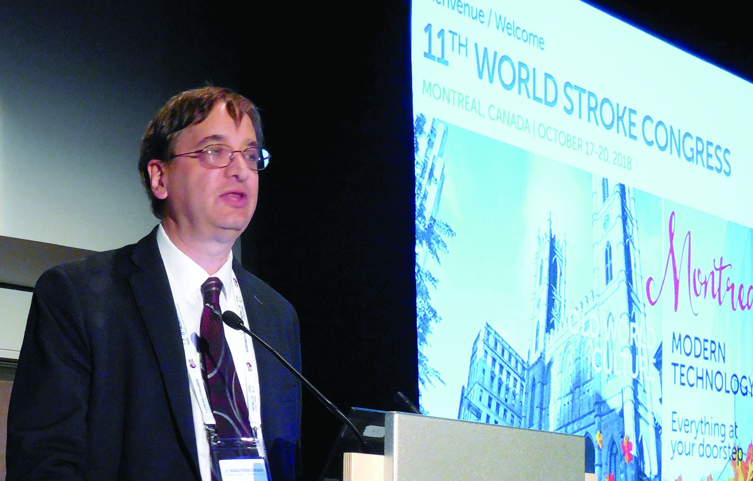
SPG stimulation started within 24 hours of an acute ischemic stroke “reduced poststroke disability over the entire outcome range and increased the proportion of patients who were alive and independent 3 months after their stroke” in the subgroup with a confirmed cortical infarction (CCI), Jeffrey L. Saver, MD, said at the World Stroke Congress. Five days of SPG stimulation, done once daily starting within 24 hours of stroke onset, “enhances ipsilateral collateral blood flow” and may also stabilize the blood brain barrier, explained Dr. Saver, professor of neurology and director of the stroke center at the University of California, Los Angeles. The study included a prespecified primary endpoint analysis that focused exclusively on the CCI subgroup, 52% of the total enrolled population.
If the reported data result in Food and Drug Administration marketing approval for the system, Dr. Saver said that he anticipated “substantial uptake” of the strategy, which he tested in patients who had not undergone thrombectomy or thrombolysis treatment. In current U.S. practice, there is “a large group of patients with a missed opportunity for recanalization” who would be candidates for treatment with SPG stimulation, a treatment that appeared to provide benefits beyond current standard care, he said in an interview.
Ongoing studies are also testing whether SPG stimulation can benefit acute ischemic stroke patients who have already undergone treatment with thrombectomy or thrombolysis, he added. The same SPG stimulation device is additionally undergoing U.S. testing as a treatment for headache and has regulatory approval in the European Union for treating headache and migraine.
The ImpACT-24B (Implant for Augmentation of Cerebral Blood Flow Trial, Effectiveness and Safety in a 24-Hour Window) trial involved 1,000 patients at 73 centers in 18 countries, including the United States. The investigators enrolled acute ischemic stroke patients 8-24 hours after stroke onset who had a National Institutes of Health Stroke Scale (NIHSS) score of 7-18.
Each patient received an implant of a short, thin metal electrode placed through the soft palate at the rear roof of the mouth, near the SPG. Neurologists primarily performed the implants in a procedure that had a “skin to skin” time of less than 5 minutes. Patients received either electrical stimulation or a sham stimulation through the electrode immediately after placement and then daily for the next 4 days. The investigators titrated the strength of the treatment stimulation in each patient to maximize its strength while maintaining patient comfort. Subsequent analysis of the results showed that the stronger the tolerated stimulation, the bigger the treatment effect in a clear dose-response pattern, Dr. Saver reported.
The study’s primary endpoint was improvement in the modified Rankin scale (mRS) score at 90 days after the index stroke when measured against historical expectations. By this measure, the overall study cohort showed a small, statistically insignificant improvement in actively treated patients, compared with sham-treated patients. However, in the prespecified, coprimary endpoint cohort of patients with a CCI, active treatment resulted in 50% of patients having a better-than-expected 90-day outcome, compared with 40% of controls, a 48% relative improvement in this measure that met the prespecified definition of statistical significance. The results also showed about a 50% relative improvement in each of three secondary outcomes in the CCI cohort: the percentage of patients with a mRS score of 0-2 after 90 days, the percentage with a mRS score of 0-3 after 90 days, and average stroke-related quality of life at 90 days.
Dr. Saver also reported results of a meta-analysis that combined the results he reported from 520 patients with CCI with results from 87 CCI patients enrolled in the preceding pilot study of this treatment strategy, ImpACT-1. The pilot findings were completely consistent and when combined with the current results strengthened the statistical significance of the primary and secondary endpoints.
“There is a compelling story” of efficacy based on the study results, the dose-response relationship, and the meta-analysis results, Dr. Saver said. “I think it’s a very strong case.”
He also reported “no safety concerns” raised in the new study, with no serious adverse effects seen in or experienced by the patients on active treatment.
“The data are compelling” for safety and efficacy, for this novel approach for treating acute ischemic stroke, commented Pooja Khatri, MD, professor of neurology and director of the acute stroke program at the University of Cincinnati.
The study was sponsored by BrainsGate, the company developing the tested device. Dr. Saver has been a consultant to BrainsGate. Dr. Khatri has been a consultant to Biogen, Greenwich, Lumosa, and PTC Therapeutics.
SOURCE: Saver J et al. Int J. Stroke. 2018 Oct;13(2S):28, Abstract 104.
MONTREAL – Stimulation of the sphenopalatine ganglion (SPG) using a small, implanted electrode for 5 days in patients who had just had an acute ischemic stroke led to statistically significant and clinically meaningful improvements in the subset of patients with confirmed cortical involvement in a pivotal, sham-controlled trial.
SPG stimulation started within 24 hours of an acute ischemic stroke “reduced poststroke disability over the entire outcome range and increased the proportion of patients who were alive and independent 3 months after their stroke” in the subgroup with a confirmed cortical infarction (CCI), Jeffrey L. Saver, MD, said at the World Stroke Congress. Five days of SPG stimulation, done once daily starting within 24 hours of stroke onset, “enhances ipsilateral collateral blood flow” and may also stabilize the blood brain barrier, explained Dr. Saver, professor of neurology and director of the stroke center at the University of California, Los Angeles. The study included a prespecified primary endpoint analysis that focused exclusively on the CCI subgroup, 52% of the total enrolled population.
If the reported data result in Food and Drug Administration marketing approval for the system, Dr. Saver said that he anticipated “substantial uptake” of the strategy, which he tested in patients who had not undergone thrombectomy or thrombolysis treatment. In current U.S. practice, there is “a large group of patients with a missed opportunity for recanalization” who would be candidates for treatment with SPG stimulation, a treatment that appeared to provide benefits beyond current standard care, he said in an interview.
Ongoing studies are also testing whether SPG stimulation can benefit acute ischemic stroke patients who have already undergone treatment with thrombectomy or thrombolysis, he added. The same SPG stimulation device is additionally undergoing U.S. testing as a treatment for headache and has regulatory approval in the European Union for treating headache and migraine.
The ImpACT-24B (Implant for Augmentation of Cerebral Blood Flow Trial, Effectiveness and Safety in a 24-Hour Window) trial involved 1,000 patients at 73 centers in 18 countries, including the United States. The investigators enrolled acute ischemic stroke patients 8-24 hours after stroke onset who had a National Institutes of Health Stroke Scale (NIHSS) score of 7-18.
Each patient received an implant of a short, thin metal electrode placed through the soft palate at the rear roof of the mouth, near the SPG. Neurologists primarily performed the implants in a procedure that had a “skin to skin” time of less than 5 minutes. Patients received either electrical stimulation or a sham stimulation through the electrode immediately after placement and then daily for the next 4 days. The investigators titrated the strength of the treatment stimulation in each patient to maximize its strength while maintaining patient comfort. Subsequent analysis of the results showed that the stronger the tolerated stimulation, the bigger the treatment effect in a clear dose-response pattern, Dr. Saver reported.
The study’s primary endpoint was improvement in the modified Rankin scale (mRS) score at 90 days after the index stroke when measured against historical expectations. By this measure, the overall study cohort showed a small, statistically insignificant improvement in actively treated patients, compared with sham-treated patients. However, in the prespecified, coprimary endpoint cohort of patients with a CCI, active treatment resulted in 50% of patients having a better-than-expected 90-day outcome, compared with 40% of controls, a 48% relative improvement in this measure that met the prespecified definition of statistical significance. The results also showed about a 50% relative improvement in each of three secondary outcomes in the CCI cohort: the percentage of patients with a mRS score of 0-2 after 90 days, the percentage with a mRS score of 0-3 after 90 days, and average stroke-related quality of life at 90 days.
Dr. Saver also reported results of a meta-analysis that combined the results he reported from 520 patients with CCI with results from 87 CCI patients enrolled in the preceding pilot study of this treatment strategy, ImpACT-1. The pilot findings were completely consistent and when combined with the current results strengthened the statistical significance of the primary and secondary endpoints.
“There is a compelling story” of efficacy based on the study results, the dose-response relationship, and the meta-analysis results, Dr. Saver said. “I think it’s a very strong case.”
He also reported “no safety concerns” raised in the new study, with no serious adverse effects seen in or experienced by the patients on active treatment.
“The data are compelling” for safety and efficacy, for this novel approach for treating acute ischemic stroke, commented Pooja Khatri, MD, professor of neurology and director of the acute stroke program at the University of Cincinnati.
The study was sponsored by BrainsGate, the company developing the tested device. Dr. Saver has been a consultant to BrainsGate. Dr. Khatri has been a consultant to Biogen, Greenwich, Lumosa, and PTC Therapeutics.
SOURCE: Saver J et al. Int J. Stroke. 2018 Oct;13(2S):28, Abstract 104.
REPORTING FROM THE WORLD STROKE CONGRESS
Key clinical point: Sphenopalatine ganglion stimulation of acute ischemic stroke patients boosted cerebral blood flow and improved 90-day outcomes in patients with confirmed cortical infarctions.
Major finding: For confirmed cortical infarctions ganglion stimulation led to a 48% higher rate of better-than-expected outcomes, compared with controls.
Study details: ImpACT-24B, a multicenter pivotal trial with 1,000 acute ischemic stroke patients.
Disclosures: The study was sponsored by BrainsGate, the company developing the tested device. Dr. Saver has been a consultant to BrainsGate. Dr. Khatri has been a consultant to Biogen, Greenwich, Lumosa, and PTC Therapeutics.
Source: Saver J et al. Int J. Stroke. 2018 Oct;13(2S):28, Abstract 104.