User login
Celiac disease may underlie seizures
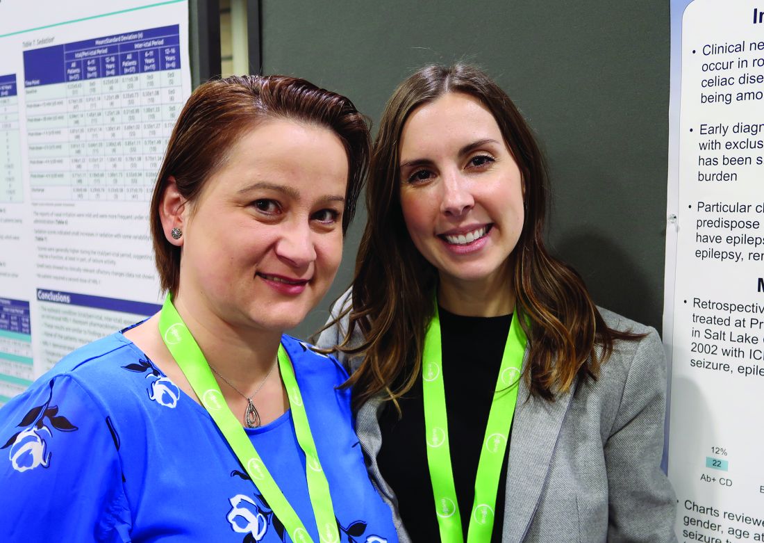
CHARLOTTE, N.C. – , according to a retrospective chart review presented at the annual meeting of the Child Neurology Society. Associations between celiac disease and seizures may have implications for screening and treatment, said study author Shanna Swartwood, MD, a fellow in the department of pediatric neurology at University of Utah in Salt Lake City.
“Screening for [celiac disease] early in patients with epilepsy, specifically with temporal EEG findings and intractable epilepsy, is warranted given the improvement of seizure burden that may result from exclusion of gluten from the diet,” said Dr. Swartwood and colleagues.
About 10% of patients with celiac disease have clinical neurologic manifestations, such as seizures. To characterize features of epilepsy in a pediatric population with celiac disease and to examine the effect of a gluten-free diet on seizure burden, Dr. Swartwood and colleagues reviewed patients treated at Primary Children’s Hospital in Salt Lake City since 2002. They identified patients with ICD-10 codes for seizures or epilepsy and celiac disease and reviewed 187 charts in all.
In all, 40 patients with seizures had biopsy-proven celiac disease, and 22 had a diagnosis of celiac disease based on the presence of antibodies. Among those with biopsy-proven celiac disease, 43% had intractable seizures. Among those with antibody-positive celiac disease, 31% had intractable seizures.
Among patients with intractable epilepsy, seizure onset preceded the diagnosis of celiac disease by an average of 5 years. For patients with nonintractable epilepsy, the first seizure occurred 1 year before the celiac disease diagnosis on average, but some patients received a celiac disease diagnosis first.
Focal seizures with secondary generalization and generalized tonic clonic seizures were the most common seizure types in this cohort. Epileptiform activity most often was seen in the temporal lobe. While other studies in patients with celiac disease have found occipital epileptiform activity to be the most common, only one patient in this cohort had activity in that location, Dr. Swartwood noted.
Patients with intractable seizures who adhered to a gluten-free diet “had a fairly robust response in terms of seizure improvement,” she said. Seizure improvement, including a decrease in seizure frequency or a decrease in antiepileptic medication dosage, occurred in seven of nine patients in the biopsy-proven group and in two of three patients in the antibody-positive group who adhered to a gluten-free diet and had intractable seizures. One patient was able to stop antiepileptic medication, and one patient had a complete resolution of seizure activity.
The researchers plan to further study the relationship between celiac disease and epilepsy, including whether various HLA subtypes of celiac disease correlate with seizures, said coinvestigator Cristina Trandafir, MD, PhD, assistant professor of pediatric neurology at University of Utah.
The chart review included relatively few patients with limited data. Nevertheless, the results suggest that there may be “substantial lag time” from first seizure to celiac disease diagnosis and that “earlier diagnosis and earlier placement on a gluten-free diet may be beneficial,” Dr. Swartwood said. Celiac disease may be asymptomatic, and screening for celiac disease with a blood test may make sense for patients with intractable seizures, she said.
The researchers had no relevant disclosures.
REPORTING FROM CNS 2019
Pediatric epilepsy surgery may improve cognition and behavior
CHARLOTTE, NC – according to a study presented at the annual meeting of the Child Neurology Society. The presence of comorbidities such as mood disorders and autism may influence the likelihood of perceived improvement, whereas the type of surgery may not.

“The parents and the families of the patients perceive that, even if the patients are not completely seizure free, the behavior and cognitive outcomes are better if there is some sort of seizure improvement,” said Trishna Kantamneni, MD, director of pediatric epilepsy at UC Davis in Sacramento.
To assess behavioral and cognitive outcomes following pediatric epilepsy surgery and to identify factors that predict improvement, Dr. Kantamneni and colleagues at the Cleveland Clinic Epilepsy Center retrospectively reviewed 126 patients younger than 18 years who underwent epilepsy surgery for medically refractory epilepsy during 2009-2016.
The primary outcome measure was the Impact of Childhood Neurologic Disability Scale (ICNDS), a parent-reported scale that assesses the behavior, cognition, and physical or neurologic disability of children with epilepsy. Parents completed the ICNDS preoperatively and at 6, 12, and 24 months after surgery. The researchers constructed separate linear mixed effects models to identify predictors of postoperative changes in ICNDS score.
Of the 126 patients, 62.7% were male, the median duration of epilepsy was 4.7 years, and 69.8% were seizure-free at the 2-year follow-up. Postoperative ICNDS scores were available for 103 patients at 6 months and for 54 patients at 24 months.
Before surgery, the average total ICNDS score was 55.7. At 6 months after surgery, the average score was 34.6, and at 24 months, it was 32.1, representing significant improvement from baseline.
In addition, behavior, cognition, and epilepsy subscores also improved post operatively, and the improvement persisted through 24 months. ICNDS scores significantly improved “even in patients who were not seizure-free after surgery,” by an average of about 22 points, the researchers said.
The absence of comorbid autism, cognitive impairment, and global developmental impairment and the absence of anxiety, depression, and ADHD were predictors of improved total ICNDS scores. Tumor pathology and being seizure free at 2 years also predicted improved scores. Duration and type of epilepsy, the number of antiepileptic drugs that patients were taking before surgery, and lobe of surgery were not predictive of improved ICNDS scores.
Dr. Kantamneni had no relevant disclosures.
SOURCE: Kantamneni T et al. CNS 2019, Abstract 51.
CHARLOTTE, NC – according to a study presented at the annual meeting of the Child Neurology Society. The presence of comorbidities such as mood disorders and autism may influence the likelihood of perceived improvement, whereas the type of surgery may not.
“The parents and the families of the patients perceive that, even if the patients are not completely seizure free, the behavior and cognitive outcomes are better if there is some sort of seizure improvement,” said Trishna Kantamneni, MD, director of pediatric epilepsy at UC Davis in Sacramento.
To assess behavioral and cognitive outcomes following pediatric epilepsy surgery and to identify factors that predict improvement, Dr. Kantamneni and colleagues at the Cleveland Clinic Epilepsy Center retrospectively reviewed 126 patients younger than 18 years who underwent epilepsy surgery for medically refractory epilepsy during 2009-2016.
The primary outcome measure was the Impact of Childhood Neurologic Disability Scale (ICNDS), a parent-reported scale that assesses the behavior, cognition, and physical or neurologic disability of children with epilepsy. Parents completed the ICNDS preoperatively and at 6, 12, and 24 months after surgery. The researchers constructed separate linear mixed effects models to identify predictors of postoperative changes in ICNDS score.
Of the 126 patients, 62.7% were male, the median duration of epilepsy was 4.7 years, and 69.8% were seizure-free at the 2-year follow-up. Postoperative ICNDS scores were available for 103 patients at 6 months and for 54 patients at 24 months.
Before surgery, the average total ICNDS score was 55.7. At 6 months after surgery, the average score was 34.6, and at 24 months, it was 32.1, representing significant improvement from baseline.
In addition, behavior, cognition, and epilepsy subscores also improved post operatively, and the improvement persisted through 24 months. ICNDS scores significantly improved “even in patients who were not seizure-free after surgery,” by an average of about 22 points, the researchers said.
The absence of comorbid autism, cognitive impairment, and global developmental impairment and the absence of anxiety, depression, and ADHD were predictors of improved total ICNDS scores. Tumor pathology and being seizure free at 2 years also predicted improved scores. Duration and type of epilepsy, the number of antiepileptic drugs that patients were taking before surgery, and lobe of surgery were not predictive of improved ICNDS scores.
Dr. Kantamneni had no relevant disclosures.
SOURCE: Kantamneni T et al. CNS 2019, Abstract 51.
REPORTING FROM CNS 2019
Delay in EEG monitoring associated with increased seizure duration in pediatric refractory status epilepticus
CHARLOTTE, NC – , according to a multicenter study that was presented at the annual meeting of the Child Neurology Society. Delays in initiating EEG monitoring are associated with longer seizure duration in this patient population.
Neurologists are advised to initiate continuous EEG monitoring rapidly for all cases of pediatric refractory status epilepticus. Little information is available, however, about patterns in the timing of EEG placement. In addition, the relationship between delays in the initiation of continuous EEG and outcomes of refractory status epilepticus are unknown. Dmitry Tchapyjnikov, MD, assistant professor of child neurology at Duke University in Durham, N.C., and colleagues evaluated trends in the time to continuous EEG initiation and examined whether delays are associated with longer seizure duration in children with refractory status epilepticus.
A retrospective analysis of pSERG data
Dr. Tchapyjnikov and colleagues analyzed data from 11 hospitals participating in the Pediatric Status Epilepticus Research Group (pSERG), a prospective, observational cohort. They focused on pediatric patients who were admitted from 2011 to 2017 with refractory status epilepticus, which they defined as a seizure that persisted after treatment with two or more antiseizure medications (ASMs), one of which had to be a nonbenzodiazepine ASM, or a continuous infusion. Eligible patients were between 1 month and 21 years old and had convulsive seizures at onset. Patients who had EEG placement before seizure onset were excluded.
The investigators included in their study 121 patients who had seizure durations of 3 or more hours. Based on an exploratory analysis of various time-point cutoffs, Dr. Tchapyjnikov and colleagues defined delayed continuous EEG placement as placement at more than 5 hours after seizure onset. They used the Kaplan–Meier estimator to assess time to continuous EEG and used covariate-adjusted proportional hazards models to examine the association between delay in continuous EEG placement and seizure duration.
EEG placement overall was delayed
The median time to continuous EEG placement after seizure onset was 9 hours. Approximately 4% of the children had continuous EEG placed within 1 hour, and 74% had it placed within 24 hours.
The investigators found that seizure onset outside the study hospital was associated with a higher likelihood of delayed time to EEG placement. “Females seemed to be more likely to have timely EEG placement,” said Dr. Tchapyjnikov. “I don’t have a physiological explanation for that.” The researchers saw no difference in treatment between patients who had timely EEG placement and those who had delayed EEG placement.
About 68% of children were having seizures at the time of continuous EEG placement. A presumed seizure etiology of CNS infection was associated with a higher likelihood of being in status epilepticus at the time of EEG placement. A history of epilepsy, developmental delay, or home ASM use, however, was associated with a lower likelihood of being in status epilepticus at time of EEG placement.
Dr. Tchapyjnikov’s group found that the 24-hour cumulative probability of seizure resolution was lower among patients who did not have continuous EEG initiation within 5 hours, compared with those who did (56% vs.70%). The association remained significant after the investigators adjusted the data for covariates that were independently associated with 24-hour seizure resolution (hazard ratio, 0.31).
The investigators included in their analysis patients who had seizure resolution before EEG placement, because restricting the analysis to patients who have persistent status epilepticus would have overemphasized the benefits of EEG, according to Dr. Tchapyjnikov. “Looking at the overall hazard ratios is a more conservative way of looking at these data.”
The study was not supported by external funding. Dr. Tchapyjnikov had no relevant disclosures.
REPORTING FROM CNS 2019
Does AED prophylaxis delay seizure onset in children with brain tumors?
CHARLOTTE, N.C. – according to research presented at the annual meeting of the Child Neurology Society. Levetiracetam, oxcarbazepine, and phenytoin are the most common initial prophylactic AEDs administered to children with brain tumors, the researchers said.
The literature indicates that between 20% and 35% of children with brain tumors have seizures, and up to half of these patients have seizure as their presenting symptom. Common practice is to prescribe antiseizure medication after a child has had a first seizure, because the risk for recurrence is high. In 2000, the American Academy of Neurology discouraged prophylactic use of AEDs in children, citing a lack of evidence for efficacy. Most of the data that it reviewed, however, came from adults.
Michelle Yun, a medical student at Weill Cornell Medical College, New York, and colleagues used national Medicaid claims data that had been collected between 2009 and 2012 for children with seizures to conduct a retrospective, observational, case-control study. They included children aged 0-20 years with a diagnosis of brain tumor, a seizure diagnosis within 6 months after brain tumor diagnosis, an AED prescription, and 12 continuous months of Medicaid coverage following brain tumor diagnosis in their analysis. The investigators defined seizure prophylaxis as AED prescription within 30 days after brain tumor diagnosis but before a first seizure diagnosis.
The exposure in the study was AED prescription within 45 days of diagnosis, and the outcome was the time to first seizure. Ms. Yun and colleagues also analyzed the most common initial prophylactic AEDs and the proportion of cases with first seizure diagnosis after prophylactic AED discontinuation, which was defined as a treatment gap longer than 30 days. The study covariates included age, sex, race, ethnicity, and medical comorbidities.
In all, 218 children were included in the study; 40 received AED prophylaxis and 26 received it within 45 days of brain tumor diagnosis. Patients with and without AED prophylaxis were well matched on all covariates.
At 1 year, Ms. Yun and colleagues saw no difference in time to first seizure between the two groups. The median time to first seizure was 75 days in the prophylaxis group and 80 days in the no-prophylaxis group. The researchers observed a transient separation between the two groups, however, in the early months after brain tumor diagnosis. When they examined children who had a seizure during the first 6 months of follow-up, the median time to diagnosis of first seizure was 68 days in children with prophylaxis and 34 days in the no-prophylaxis group. The difference between groups was statistically significant. “When we added all the covariates of interest, we found that there was a protective effect in these children with early seizures,” said Ms. Yun.
Among the study limitations that Ms. Yun acknowledged were its observational, retrospective design and its small sample size. Medicaid data themselves are limited, since states do not report them in a uniform manner, and the data do not include much clinical information. “Something that would be helpful is a prospective clinical study,” Ms. Yun concluded.
The Weill Cornell Clinical and Translational Science Center and the American Academy of Neurology provided funding for the study. The Pediatric Epilepsy Research Foundation provided the Medicaid data. Ms. Yun had no relevant disclosures.
SOURCE: Yun M et al. CNS 2019, Abstract PL2-1.
REPORTING FROM CNS 2019
Dr. Paul Aisen Q&A: Aducanumab for Alzheimer’s
In the wake of Biogen and Eisai’s Oct. 22 announcement about plans to apply to the Food and Drug Administration next year for the regulatory approval of the investigational monoclonal antibody aducanumab as a treatment for Alzheimer’s disease, we spoke with Paul Aisen, MD, the founding director of the Alzheimer’s Therapy Research Institute at the University of Southern California, Los Angeles, for his views on the news. He has been a consultant for Biogen and is a member of the aducanumab steering committee.
Q: What was your first reaction when you heard about the plan to submit an application for aducanumab to the FDA?
A: My initial reaction is that this provides terrific support for the amyloid hypothesis, and is consistent with the early aducanumab studies showing significant reductions in brain amyloid with resulting clinical improvement.
My next thought was that these data are going to be very, very challenging to analyze because both of these trials were stopped early, and one was clearly negative. We really need to scrutinize the data, but even at this point I would say this strongly supports targeting amyloid. The scrutiny will begin in detail at the Clinical Trials in Alzheimer’s Disease conference in December, when Biogen will likely release detailed data. A lot of people will analyze it, and I think that’s great. It’s beneficial to bring different perspectives.
We have had a terribly frustrating series of disappointments in the field. After the futility analysis of aducanumab and the multiple failures of BACE [beta-secretase] inhibitors, many were convinced we were barking up the wrong tree. I think these results, although complicated, should resurrect the enthusiasm for targeting amyloid.
Q: What is different about aducanumab from other antibodies tested – and rejected – in Alzheimer’s drug development?
A: There are lots of antibodies that have been tested in clinical trials. They all differ in terms of their affinity for amyloid beta. Some target monomers of the protein. Some target dimers. Some target fibrils. Some tie up amyloid and some reduce it. Aducanumab directly attacks brain plaques, reducing the plaque load in the brain. It carries a liability of amyloid-related imaging abnormalities [ARIA], but it also allows us to assess the impact that removing plaques might have on downstream events, including biomarkers. Overall, these data show that aducanumab did remove brain plaques and that removing them had a beneficial effect on cognition and function, and also a favorable effect on downstream biomarkers.
But again, we must be cautious because this is a complex data set taken from a post hoc analysis of two different terminated trials.
Q: We see some statistically significant differences in cognitive and functional outcomes. What would that mean for patients on an everyday basis?
A: Well, everyone is different, so that’s hard to say. A 25% slowing of functional decline on the Clinical Dementia Rating Scale sum of boxes (CDR-SB) might mean that, at the end of a year, there’s not a significant change in memory, or that there’s better social function. If both trials had been completed and if people had 18 months of high-dose aducanumab, the slowing of functional decline on the CDR-SB might in fact be greater than reported. Again, we’re having to draw conclusions from interrupted trials.
Q: This suggestion you make of a potentially continuous slowing of decline – are you suggesting that aducanumab might slow decline to the point of stopping it altogether? If an elderly patient has little or no progression until death would that, in effect, be considered a “cure?”
A: I don’t think it is possible to cure AD once the disease is clinically evident. These are studies of people with early AD, late mild cognitive impairment, and mild dementia. At that stage, there’s already a loss of synapses that won’t come back, and these studies don’t suggest that aducanumab can cure that. But what if people took it earlier, when the brain is still functioning normally? Some of us have argued for many years that earlier intervention is the way to go. And since we can now identify people [with brain plaques] before they become symptomatic, there is the possibility that if we removed them, we could stop progression.
Q: Are there any plans to study aducanumab as a preventive agent?
A: A grant has been awarded for this, but it was put on hold after the futility analysis. I don’t know when or if that will go forward.
(Editor’s note: The National Institutes of Health previously awarded Banner Health a $32 million, 5-year grant to examine this. The 2-year prevention study of aducanumab is aimed at cognitively unimpaired 65- to 80-year-old patients with PET-confirmed amyloid brain plaques. It was to be a multicenter, double-blind, placebo-controlled trial using Alzheimer’s biomarker endpoints as primary outcomes, along with cognitive and clinical changes, safety, and tolerability. The study was put on hold after Biogen discontinued the aducanumab development program in March. Investigators are considering whether to resurrect plans considering the new data. The study is intended to be a public-private partnership, with additional unspecified funding from Biogen plus $10 million from philanthropic sources. It has three intended goals: To find an approved prevention therapy as early as 2023, ahead of the National Plan to Address Alzheimer’s Disease’s goal of an effective prevention strategy by 2025; to advance the use of surrogate biomarkers to rapidly test and support accelerated approval of prevention therapies in almost everyone at biomarker or genetic risk, even in earlier preclinical Alzheimer’s stages when some treatments may have their greatest benefit; and to help make it possible to conduct prevention trials in at-risk persons even before they have extensive amyloid plaques, when some treatments may have their greatest benefit.)
Q: It seems like rolling this out to an enormous population of patients is going to be difficult, if not impossible. Are people really going to be able to commit to what could be a lifetime of monthly intravenous infusions of a medicine that could be expensive, as therapeutic antibodies generally are?
A: I would say, nothing about this disease is easy. It’s devastating and horrible. And if someone is diagnosed at this stage, I would think that individual would embrace any opportunity to treat it. My hope is that we will be able to prescreen people with an effective blood test for amyloid that would be part of a regular testing protocol once they reach a certain age. Those with positive results would be referred for more testing, including amyloid brain imaging.
CBD: What physicians need to know about it
Cannabidiol is a derivative of marijuana that is sold everywhere from medical marijuana stores to health food markets to gas stations. While this chemical is derived from marijuana plants, it can be sold in many states as a supplement and is largely unregulated. The ubiquity of cannabidiol (CBD) and its potential benefits means that doctors need to be able to counsel patients about what we know, what we don’t, and how to use it safely. For conditions such as chronic pain and addiction, where we have few safe and effective alternatives, CBD may be reasonable to recommend.

To find out what physicians need to know about CBD, Elisabeth Poorman, MD, a general internist at a University of Washington neighborhood clinic in Kent and member of the editorial advisory board of Internal Medicine News, interviewed Peter Grinspoon, MD, who provides free consultation to primary care patients on the benefits and risks of using various forms of cannabis, including CBD. Dr. Grinspoon is an internist at Massachusetts General Hospital Chelsea Healthcare Center and is an instructor at Harvard Medical School, Boston. He has contributed to the Harvard Health Blog on the topic of medical marijuana, delivered grand rounds on cannabis at Massachusetts General Hospital, and lectured at the American College of Physicians. Dr. Grinspoon is also medical director for Galenas, a medical marijuana company.
Dr. Grinspoon is the son of Lester Grinspoon, MD, associate professor emeritus of psychiatry at Harvard Medical School, who researched the medicinal legitimacy of marijuana prohibition and has authored books on the medical benefits of marijuana.
and his knowledge of CBD’s efficacy for various medical conditions. Below are excerpts from that conversation.
Dr. Poorman: How do you explain the difference between THC and CBD to patients?
Dr. Grinspoon: Cannabis contains at least a hundred different chemicals called cannabinoids, of which tetrahydrocannabinol (THC) and CBD are the most prevalent. THC is the one that gets you high and can be used recreationally and medically. The CBD molecule is not intoxicating, and people use it for a variety of medical purposes, most commonly to treat anxiety, insomnia, and pain.
Dr. Poorman: There are a lot of gaps in what we now about CBD’s potential benefits. Why don’t we know more?
Dr. Grinspoon: CBD has no abuse liability according to the World Health Organization, but because it is a cannabinoid, it is still technically a schedule I substance under the Controlled Substances Act, and that makes it difficult to study.
Dr. Poorman: What kinds of conditions can CBD treat?
Dr. Grinspoon: In anxiety, the enthusiasm has outpaced the science; there’s no question about that. And most of the studies have done in animals. That said, some studies have shown that CBD helps treat components of anxiety, like public speaking. Unlike THC, it is nonintoxicating and non–habit forming. But we don’t have the wealth of randomized controlled trials that we have for official psychiatric medications.
CBD’s benefits have been most extensively studied in pediatric epilepsy. The one Food and Drug Administration–approved drug derived from cannabis is Epidiolex, used to treat rare forms of childhood epilepsy. There is some evidence that as an adjunct, it can be used for glioblastoma multiforme in patients receiving other appropriate therapy. There is also some preliminary evidence that it can be used for addiction, including to opioids, cannabis, tobacco, and stimulants.
Most of the evidence for using CBD in chronic pain comes from animal studies, including a study published in the European Journal of Pain in 2016. Among my patients to whom I have suggested CBD for chronic pain, a few have noticed great benefit, a few have noticed some benefit, and a lot have noticed no benefit. For those who have said they noticed benefit it is unclear whether that benefit was just the placebo effect.
In insomnia, I usually have them take CBD under the tongue half an hour time before bedtime, or if it’s an edible, an hour before bedtime. I start with a lower dose and slowly try higher doses. I also encourage them to do the other sleep hygiene things, like no screens, increasing exercise, and decreasing caffeine. It seems that CBD helps them fall asleep, though it’s hard to know if it’s the CBD or the fact that they have started taking something, and have simultaneously made various lifestyle changes.
Dr. Poorman: Can CBD interfere with your normal sleep architecture, the way benzodiazepines and Benadryl can?
Dr. Grinspoon: We know that THC affects your sleep architecture and affects what percentage of REM sleep you have. But I don’t know if the effects of CBD on sleep architecture have been studied.
Dr. Poorman: What harms do you counsel patients about when discussing CBD?
Dr. Grinspoon: There are four main harms. The first is the price. It’s overpriced, and the doses are very low. In most animal studies, the doses are about 20 milligrams per kilogram of weight. And you go to the market, and it’s like a dollar for a hundredth of that.
Number two is that it’s not regulated; it’s a supplement. A few years ago, the government tested a bunch of samples of CBD, and some didn’t actually contain CBD, some didn’t have the right amount; and worse, some contained THC that had not been disclosed in the packaging. So you can’t just go to a roadside gas station and assume that if you buy CBD, it’s actually that. You want a place that has a certificate of assurance. Make sure third-party testing was done, including testing for pesticides and other heavy metals.
The third thing is drug interactions. It affects the body like grapefruit and inhibits the cytochrome P450 system. The medications doctors should be most concerned about are blood thinners like Coumadin. And if you’re on blood thinners, you definitely want to tell your doctor that you are on CBD and he or she might want to check your blood levels more frequently than they usually do.
The fourth concern is liver inflammation. In the childhood epilepsy studies, a bump in some liver enzymes was seen, although I haven’t heard of any clinically significant cases of chemical hepatitis related to CBD. But if someone has liver disease you want to keep an eye on their liver enzymes.
Dr. Poorman: What methods of ingestion do you recommend or not recommend?
Dr. Grinspoon: It’s basically trial and error, but I usually recommend oral form. If people feel comfortable taking a gummy bear, or a pill, I’m not particular about that. If the product being taken contains less than 0.3% THC, it won’t get you high.
The topical form probably works better for treating chronic pain if it contains some THC, suggests a review article published in the Cleveland Clinic Journal of Medicine. Topical THC is nonintoxicating, unless you managed to sit in a bathtub for 8 hours after applying it.
I don’t recommend smoking CBD, and right now, I don’t recommend vaping anything.
If people have severe pain, like moderately severe arthritis, CBD may not be enough, whereas medical cannabis with THC could help, a report suggests.
Dr. Poorman: Do you ever encourage patients to stop using CBD products?
Dr. Grinspoon: I work in a low-income area, and my patients don’t have a ton of disposable income. If it’s not working, I worry about the expense.
Dr. Poorman: The CBD industry is growing quickly. What changes are you seeing in what products are out there, and what changes would you like to see?
Dr. Grinspoon: CBD is being put in everything, and it’s comical. On the one hand, you can say if people want to waste their money on a CBD emitting pillowcase, that’s fine. On the other hand, you can say that certainly seems like misleading advertising, because a CBD emitting pillowcase isn’t going to help you sleep any better.
I think the purported benefits are far beyond what we can say scientifically. We do know that CBD has anti-inflammatory characteristics. But that doesn’t mean that putting CBD in all skin products is good for your skin. It’s bad for your pocketbook, though. I would like there to be less of a gap between the claims and the science.
Dr. Elisabeth Poorman has no conflicts to disclose.
Cannabidiol is a derivative of marijuana that is sold everywhere from medical marijuana stores to health food markets to gas stations. While this chemical is derived from marijuana plants, it can be sold in many states as a supplement and is largely unregulated. The ubiquity of cannabidiol (CBD) and its potential benefits means that doctors need to be able to counsel patients about what we know, what we don’t, and how to use it safely. For conditions such as chronic pain and addiction, where we have few safe and effective alternatives, CBD may be reasonable to recommend.
To find out what physicians need to know about CBD, Elisabeth Poorman, MD, a general internist at a University of Washington neighborhood clinic in Kent and member of the editorial advisory board of Internal Medicine News, interviewed Peter Grinspoon, MD, who provides free consultation to primary care patients on the benefits and risks of using various forms of cannabis, including CBD. Dr. Grinspoon is an internist at Massachusetts General Hospital Chelsea Healthcare Center and is an instructor at Harvard Medical School, Boston. He has contributed to the Harvard Health Blog on the topic of medical marijuana, delivered grand rounds on cannabis at Massachusetts General Hospital, and lectured at the American College of Physicians. Dr. Grinspoon is also medical director for Galenas, a medical marijuana company.
Dr. Grinspoon is the son of Lester Grinspoon, MD, associate professor emeritus of psychiatry at Harvard Medical School, who researched the medicinal legitimacy of marijuana prohibition and has authored books on the medical benefits of marijuana.
and his knowledge of CBD’s efficacy for various medical conditions. Below are excerpts from that conversation.
Dr. Poorman: How do you explain the difference between THC and CBD to patients?
Dr. Grinspoon: Cannabis contains at least a hundred different chemicals called cannabinoids, of which tetrahydrocannabinol (THC) and CBD are the most prevalent. THC is the one that gets you high and can be used recreationally and medically. The CBD molecule is not intoxicating, and people use it for a variety of medical purposes, most commonly to treat anxiety, insomnia, and pain.
Dr. Poorman: There are a lot of gaps in what we now about CBD’s potential benefits. Why don’t we know more?
Dr. Grinspoon: CBD has no abuse liability according to the World Health Organization, but because it is a cannabinoid, it is still technically a schedule I substance under the Controlled Substances Act, and that makes it difficult to study.
Dr. Poorman: What kinds of conditions can CBD treat?
Dr. Grinspoon: In anxiety, the enthusiasm has outpaced the science; there’s no question about that. And most of the studies have done in animals. That said, some studies have shown that CBD helps treat components of anxiety, like public speaking. Unlike THC, it is nonintoxicating and non–habit forming. But we don’t have the wealth of randomized controlled trials that we have for official psychiatric medications.
CBD’s benefits have been most extensively studied in pediatric epilepsy. The one Food and Drug Administration–approved drug derived from cannabis is Epidiolex, used to treat rare forms of childhood epilepsy. There is some evidence that as an adjunct, it can be used for glioblastoma multiforme in patients receiving other appropriate therapy. There is also some preliminary evidence that it can be used for addiction, including to opioids, cannabis, tobacco, and stimulants.
Most of the evidence for using CBD in chronic pain comes from animal studies, including a study published in the European Journal of Pain in 2016. Among my patients to whom I have suggested CBD for chronic pain, a few have noticed great benefit, a few have noticed some benefit, and a lot have noticed no benefit. For those who have said they noticed benefit it is unclear whether that benefit was just the placebo effect.
In insomnia, I usually have them take CBD under the tongue half an hour time before bedtime, or if it’s an edible, an hour before bedtime. I start with a lower dose and slowly try higher doses. I also encourage them to do the other sleep hygiene things, like no screens, increasing exercise, and decreasing caffeine. It seems that CBD helps them fall asleep, though it’s hard to know if it’s the CBD or the fact that they have started taking something, and have simultaneously made various lifestyle changes.
Dr. Poorman: Can CBD interfere with your normal sleep architecture, the way benzodiazepines and Benadryl can?
Dr. Grinspoon: We know that THC affects your sleep architecture and affects what percentage of REM sleep you have. But I don’t know if the effects of CBD on sleep architecture have been studied.
Dr. Poorman: What harms do you counsel patients about when discussing CBD?
Dr. Grinspoon: There are four main harms. The first is the price. It’s overpriced, and the doses are very low. In most animal studies, the doses are about 20 milligrams per kilogram of weight. And you go to the market, and it’s like a dollar for a hundredth of that.
Number two is that it’s not regulated; it’s a supplement. A few years ago, the government tested a bunch of samples of CBD, and some didn’t actually contain CBD, some didn’t have the right amount; and worse, some contained THC that had not been disclosed in the packaging. So you can’t just go to a roadside gas station and assume that if you buy CBD, it’s actually that. You want a place that has a certificate of assurance. Make sure third-party testing was done, including testing for pesticides and other heavy metals.
The third thing is drug interactions. It affects the body like grapefruit and inhibits the cytochrome P450 system. The medications doctors should be most concerned about are blood thinners like Coumadin. And if you’re on blood thinners, you definitely want to tell your doctor that you are on CBD and he or she might want to check your blood levels more frequently than they usually do.
The fourth concern is liver inflammation. In the childhood epilepsy studies, a bump in some liver enzymes was seen, although I haven’t heard of any clinically significant cases of chemical hepatitis related to CBD. But if someone has liver disease you want to keep an eye on their liver enzymes.
Dr. Poorman: What methods of ingestion do you recommend or not recommend?
Dr. Grinspoon: It’s basically trial and error, but I usually recommend oral form. If people feel comfortable taking a gummy bear, or a pill, I’m not particular about that. If the product being taken contains less than 0.3% THC, it won’t get you high.
The topical form probably works better for treating chronic pain if it contains some THC, suggests a review article published in the Cleveland Clinic Journal of Medicine. Topical THC is nonintoxicating, unless you managed to sit in a bathtub for 8 hours after applying it.
I don’t recommend smoking CBD, and right now, I don’t recommend vaping anything.
If people have severe pain, like moderately severe arthritis, CBD may not be enough, whereas medical cannabis with THC could help, a report suggests.
Dr. Poorman: Do you ever encourage patients to stop using CBD products?
Dr. Grinspoon: I work in a low-income area, and my patients don’t have a ton of disposable income. If it’s not working, I worry about the expense.
Dr. Poorman: The CBD industry is growing quickly. What changes are you seeing in what products are out there, and what changes would you like to see?
Dr. Grinspoon: CBD is being put in everything, and it’s comical. On the one hand, you can say if people want to waste their money on a CBD emitting pillowcase, that’s fine. On the other hand, you can say that certainly seems like misleading advertising, because a CBD emitting pillowcase isn’t going to help you sleep any better.
I think the purported benefits are far beyond what we can say scientifically. We do know that CBD has anti-inflammatory characteristics. But that doesn’t mean that putting CBD in all skin products is good for your skin. It’s bad for your pocketbook, though. I would like there to be less of a gap between the claims and the science.
Dr. Elisabeth Poorman has no conflicts to disclose.
Researchers seek to characterize pediatric new daily persistent headache
CHARLOTTE, N.C. – according to research presented at the 48th national meeting of the Child Neurology Society. Most children with NDPH fulfill criteria for its migraine subtype, and one-third of pediatric patients with NDPH have comorbid medication overuse headache (MOH).
NDPH is defined as a daily, unremitting headache that lasts for at least 3 months. “Not many studies on NDPH focus on pediatrics,” said Emily Pierce, from Children’s National Medical Center in Washington. NDPH “is considered to be one of the most intractable headaches in children. Children are able to tell that they’ve had this different type of headache because there’s some kind of onset that is very memorable.”
Ms. Pierce and colleagues conducted an observational study to describe the characteristics of NDPH in pediatric patients who presented to a headache program at a tertiary referral center. The researchers included pediatric patients who visited the headache clinic at Children’s National Medical Center between 2016 and 2018 in their analysis. All patients were enrolled in patient registry that had been approved by an independent review board. Ms. Pierce and colleagues queried the registry for NDPH and reviewed these records to examine participants’ clinical presentations.
The investigators identified 3,260 patient encounters during the study period. Of these encounters, 454 patients (13.9%) were identified as having NDPH. Patients with NDPH were predominantly female (78%) and white (72%). The median age of the sample was 14.8 years.
The frontal head region was the most common location of headache pain, which often had a throbbing quality and was associated with photophobia, phonophobia, nausea, and decreased activity. The median pain intensity was 6 of 10. Approximately 72% of patients had tried abortive medication, and 56% of patients had failed at least one abortive medication. Excedrin, ibuprofen, and acetaminophen were among the common failed abortive medications.
Furthermore, 36% of patients were diagnosed with MOH. The most commonly overused medication was ibuprofen. MOH “is also considered to be intractable for patients with NDPH,” said Ms. Pierce. “Typically, if the patient stops overusing that medication, they’ll find relief from their headaches. However, with our NDPH patients, when they stop overusing that medication, they still are having headaches associated with NDPH.”
The data indicated “a strong difference between our male and female patients,” said Ms. Pierce. Female patients reported significantly more instances of photophobia, phonophobia, nausea, and dizziness than did male patients. Overall, 78% of participants had a diagnosis of an additional comorbidity, such as head trauma (18%), anxiety (14%), depression (8%), or other (37%).
Observational studies of pediatric NDPH offer “a better way for our providers to diagnose these patients, and also to better understand them and help them figure out a treatment that may work,” said Ms. Pierce. In future research, she and her colleagues intend to examine blood work and potential genetic associations in pediatric patients with NDPH.
The study was not supported by funding, and the investigators had no disclosures.
SOURCE: Pierce E et al. CNS 2019, Abstract 100.
REPORTING FROM CNS 2019
Many children who present to headache clinics have joint hypermobility
CHARLOTTE, N.C. – according to data presented at the 48th national meeting of the Child Neurology Society. Furthermore, patients with joint hypermobility have a high rate of headache disability, while patients without joint hypermobility have less headache disability, according to Dhwani Sahjwani, MD, a resident at Inova Fairfax Hospital in Falls Church, Va., and colleagues.
While conducting research in the headache clinic at Children’s National Hospital in Washington, D.C., Dr. Sahjwani saw several children with joint hypermobility and a diagnosis of a disorder such as Ehlers-Danlos syndrome. She and her colleagues began analyzing patients to evaluate the potential association between joint hypermobility and headache disability in children. The investigators included pediatric patients examined in the headache clinic at Children’s National Medical Center between October 2018 and January 2019 in their study. All headache clinic patients were enrolled in a patient registry that had been approved by an independent review board.
Dr. Sahjwani and colleagues measured patients’ headache disability with the Headache Impact Test–6 (HIT-6) questionnaire. Scores of 60 or greater on this questionnaire indicate severe headache disability. The researchers assessed joint hypermobility using the Beighton scoring system. In this system, scores greater than 4 indicate joint hypermobility.
Dr. Sahjwani’s group scored 76 patients using the Beighton system and HIT-6 questionnaire. Participants’ median age was 13.7 years. Approximately 26% of patients had Beighton scores that indicated joint hypermobility. About 65% of the patients with joint hypermobility had a diagnosis of migraine without aura. In addition, 80% of patients with joint hypermobility had severe headache disability, according to the HIT-6 disability criteria. The average pain intensity in patients with hypermobile joints was 6.1 out of 10. Among participants without significant joint hypermobility, 90% had mild headache disability.
Patients with joint hypermobility and increased tissue elasticity “tend to have a lower threshold for pain, in general, in all parts of their bodies,” said Dr. Sahjwani. Greater headache severity might be expected in this population, “because they have more pain if they have hypermobile joints or tissue.”
Headache treatments for this population are based solely on the type of headache that each patient has. Patients with joint hypermobility and migraine, for example, are candidates for rescue medication and long-term prophylactic medications. “I don’t think the joint hypermobility is going to change how you manage their headaches,” said Dr. Sahjwani.
The study results suggest that, when children present with severely debilitating headaches, a neurologist should consider examining them for joint hypermobility “to see if they have another diagnosis, such as Ehlers-Danlos syndrome ... that has to be managed in addition to their headaches,” Dr. Sahjwani concluded.
The study was not supported by funding. The authors did not report any disclosures.
SOURCE: Sahjwani D et al. CNS 2019, Abstract 101.
REPORTING FROM CNS 2019
FDA approves onabotulinumtoxinA for pediatric lower limb spasticity
The Food and Drug Administration has approved onabotulinumtoxinA (Botox) for treatment of pediatric lower limb spasticity in patients aged 2-17 years, excluding those in whom it is associated with cerebral palsy, according to an announcement from Allergan.

The approval is based on a phase 3 study evaluating safety and efficacy in more than 300 patients with lower limb spasticity. Although patients with cerebral palsy were included in the study, they’re excluded from this indication. Orphan Drug Exclusivity prevents it from being indicated for lower limb spasticity in cerebral palsy because abobotulinumtoxinA (Dysport) already has marketing exclusivity for the indication. Botox also is indicated for children aged 2-17 years of age with upper limb spasticity, as well as nine other indications.
OnabotulinumtoxinA comes with warnings, including problems of swallowing, speaking, or breathing and even risk of spread of the toxin. It also may cause loss of strength or general muscle weakness, vision problems, or dizziness within hours or weeks of administration. Serious and sometimes immediate allergic reactions have been reported. Patients and health care professionals should discuss various concerns before treatment, including whether the patient has recently received antibiotics by injection, or has taken muscle relaxants, allergy or cold medicine, sleep medicine, and aspirinlike products or blood thinners. It’s important to note that the dose of onabotulinumtoxinA is not the same as that for other botulinum toxin products. The full prescribing information is available on the Allergan website.
The Food and Drug Administration has approved onabotulinumtoxinA (Botox) for treatment of pediatric lower limb spasticity in patients aged 2-17 years, excluding those in whom it is associated with cerebral palsy, according to an announcement from Allergan.
The approval is based on a phase 3 study evaluating safety and efficacy in more than 300 patients with lower limb spasticity. Although patients with cerebral palsy were included in the study, they’re excluded from this indication. Orphan Drug Exclusivity prevents it from being indicated for lower limb spasticity in cerebral palsy because abobotulinumtoxinA (Dysport) already has marketing exclusivity for the indication. Botox also is indicated for children aged 2-17 years of age with upper limb spasticity, as well as nine other indications.
OnabotulinumtoxinA comes with warnings, including problems of swallowing, speaking, or breathing and even risk of spread of the toxin. It also may cause loss of strength or general muscle weakness, vision problems, or dizziness within hours or weeks of administration. Serious and sometimes immediate allergic reactions have been reported. Patients and health care professionals should discuss various concerns before treatment, including whether the patient has recently received antibiotics by injection, or has taken muscle relaxants, allergy or cold medicine, sleep medicine, and aspirinlike products or blood thinners. It’s important to note that the dose of onabotulinumtoxinA is not the same as that for other botulinum toxin products. The full prescribing information is available on the Allergan website.
ACIP approves 2020 adult vaccination schedule
The Centers for Disease Control and Prevention’s Advisory Committee on Immunization Practices voted unanimously to approve the adult immunization schedule for 2020, although some fine-tuning may occur before publication.

“Some of the wordsmithing may be done later,” ACIP executive secretary Amanda Cohn, MD, said at the ACIP October meeting.
Key updates to the schedule included a change in wording for the definition of the red bars on the table to include “not recommended or contraindicated” instead of only the word “contraindicated.” Committee members were especially interested in changing this wording to guide clinicians in use of the live attenuated influenza vaccine because of its potential value in vaccinating health care personnel.
Other updates include language that vaccination of adolescents and young adults aged 16-23 years who are not at increased risk for meningococcal disease should be vaccinated as follows: “Based on shared clinical decision making, 2-dose series MenB-4C at least 1 month apart or 2-dose series MenB-FHbp at 0, 6 months.”
Similarly, clinical decision-making language was added to the notes for the pneumococcal polysaccharide vaccine (PPSV23) and the 13-valent pneumococcal conjugate vaccine (PCV13).
The routine vaccination calls for only one dose of PPSV23 given on or after the individual’s 65th birthday. Then, based on shared clinical decision making, a dose of PCV13 is recommended for immunocompetent individuals aged 65 years and older. The notes also state that, based on shared clinical decision making, PCV13 and PPSV23 should not be given in the same visit and, if both will be given, PCV13 should be first and should be given 1 year before PPSV23. In addition, “PPSV23 should be given at least 5 years after any previous PPSV23 dose.”
The schedule also adds shared clinical decision making to the notes on human papillomavirus vaccination for adults aged 27-45 years.
The committee members acknowledged the increasing complexity of the adult vaccination schedule, but several members agreed that it is accessible to many clinicians.
“We can’t let the perfect be the enemy of the good” said Jason Goldman, MD, liaison representing the American College of Physicians. “Those who want to learn the schedule will learn it; the health system will learn it,” even if not every specialist does.
The table “is something to draw you in,” said Sandra Fryhofer, MD, an internist who is liaison for the American Medical Association. The notes provide more details.
More specific information about contraindications for patients with cochlear implants, which also came up in the discussion, may be added to the schedule at a later date.
View the current adult vaccination schedule here.
The ACIP members had no financial conflicts to disclose.
The Centers for Disease Control and Prevention’s Advisory Committee on Immunization Practices voted unanimously to approve the adult immunization schedule for 2020, although some fine-tuning may occur before publication.
“Some of the wordsmithing may be done later,” ACIP executive secretary Amanda Cohn, MD, said at the ACIP October meeting.
Key updates to the schedule included a change in wording for the definition of the red bars on the table to include “not recommended or contraindicated” instead of only the word “contraindicated.” Committee members were especially interested in changing this wording to guide clinicians in use of the live attenuated influenza vaccine because of its potential value in vaccinating health care personnel.
Other updates include language that vaccination of adolescents and young adults aged 16-23 years who are not at increased risk for meningococcal disease should be vaccinated as follows: “Based on shared clinical decision making, 2-dose series MenB-4C at least 1 month apart or 2-dose series MenB-FHbp at 0, 6 months.”
Similarly, clinical decision-making language was added to the notes for the pneumococcal polysaccharide vaccine (PPSV23) and the 13-valent pneumococcal conjugate vaccine (PCV13).
The routine vaccination calls for only one dose of PPSV23 given on or after the individual’s 65th birthday. Then, based on shared clinical decision making, a dose of PCV13 is recommended for immunocompetent individuals aged 65 years and older. The notes also state that, based on shared clinical decision making, PCV13 and PPSV23 should not be given in the same visit and, if both will be given, PCV13 should be first and should be given 1 year before PPSV23. In addition, “PPSV23 should be given at least 5 years after any previous PPSV23 dose.”
The schedule also adds shared clinical decision making to the notes on human papillomavirus vaccination for adults aged 27-45 years.
The committee members acknowledged the increasing complexity of the adult vaccination schedule, but several members agreed that it is accessible to many clinicians.
“We can’t let the perfect be the enemy of the good” said Jason Goldman, MD, liaison representing the American College of Physicians. “Those who want to learn the schedule will learn it; the health system will learn it,” even if not every specialist does.
The table “is something to draw you in,” said Sandra Fryhofer, MD, an internist who is liaison for the American Medical Association. The notes provide more details.
More specific information about contraindications for patients with cochlear implants, which also came up in the discussion, may be added to the schedule at a later date.
View the current adult vaccination schedule here.
The ACIP members had no financial conflicts to disclose.