User login
Pathways to Melanoma
Justin M. Ko, MD, MBA, Nicole F. Velez, MD, and Hensin Tsao, MD, PhD
Melanoma is one of the most aggressive and yet poorly understood of human malignancies. Advances in genomics has allowed a more nuanced understanding of the disease, moving beyond the traditional dysplastic nevus-to-melanoma model and identifying multiple divergent oncogenic pathways leading to melanoma. An understanding of the molecular mechanisms driving melanoma has opened the doors for the development of targeted therapeutic approaches. As we enter the era of personalized medicine, it will be critical for clinicians to both appreciate and be able to determine the molecular profile of their patients’ melanoma because this profile will guide risk stratification, genetic counseling, and treatment customization. A review of the divergent pathways of melanoma development is presented here, with a particular emphasis on recently identified mutations, and their implications for patient care.
*For a PDF of the full article, click on the link to the left of this introduction.
Justin M. Ko, MD, MBA, Nicole F. Velez, MD, and Hensin Tsao, MD, PhD
Melanoma is one of the most aggressive and yet poorly understood of human malignancies. Advances in genomics has allowed a more nuanced understanding of the disease, moving beyond the traditional dysplastic nevus-to-melanoma model and identifying multiple divergent oncogenic pathways leading to melanoma. An understanding of the molecular mechanisms driving melanoma has opened the doors for the development of targeted therapeutic approaches. As we enter the era of personalized medicine, it will be critical for clinicians to both appreciate and be able to determine the molecular profile of their patients’ melanoma because this profile will guide risk stratification, genetic counseling, and treatment customization. A review of the divergent pathways of melanoma development is presented here, with a particular emphasis on recently identified mutations, and their implications for patient care.
*For a PDF of the full article, click on the link to the left of this introduction.
Justin M. Ko, MD, MBA, Nicole F. Velez, MD, and Hensin Tsao, MD, PhD
Melanoma is one of the most aggressive and yet poorly understood of human malignancies. Advances in genomics has allowed a more nuanced understanding of the disease, moving beyond the traditional dysplastic nevus-to-melanoma model and identifying multiple divergent oncogenic pathways leading to melanoma. An understanding of the molecular mechanisms driving melanoma has opened the doors for the development of targeted therapeutic approaches. As we enter the era of personalized medicine, it will be critical for clinicians to both appreciate and be able to determine the molecular profile of their patients’ melanoma because this profile will guide risk stratification, genetic counseling, and treatment customization. A review of the divergent pathways of melanoma development is presented here, with a particular emphasis on recently identified mutations, and their implications for patient care.
*For a PDF of the full article, click on the link to the left of this introduction.
Dermoscopy of Benign and Malignant Neoplasms in the Pediatric Population
Helen C. Haliasos, MD, Iris Zalaudek, MD, Josep Malvehy, MD, Christoph Lanschuetzer, MD, Helmut Hinter, MD, Rainer Hofmann-Wellenhof, MD, Ralph Braun, MD, and Ashfaq A. Marghoob, MD
Dermoscopy is a noninvasive technique that enables visualization of subsurface colors and structures within the skin that are imperceptible to the naked eye. The dermatoscope allows the physician to examine both the macroscopic and microscopic primary morphology of skin lesions, identify subtle clinical clues, confirm naked-eye clinical diagnoses, and monitor treatment progress while posing little threat to the young patient. Dermoscopic findings have been formulated into diagnostic criteria that assist experienced clinicians in differentiating benign and malignant neoplasms. In this review, clinical morphology of melanocytic nevi and melanoma in the pediatric population is examined and the relevant dermoscopic findings and histopathologic correlates that aid in the diagnosis and management of these lesions are described.
*For a PDF of the full article, click on the link to the left of this introduction.
Helen C. Haliasos, MD, Iris Zalaudek, MD, Josep Malvehy, MD, Christoph Lanschuetzer, MD, Helmut Hinter, MD, Rainer Hofmann-Wellenhof, MD, Ralph Braun, MD, and Ashfaq A. Marghoob, MD
Dermoscopy is a noninvasive technique that enables visualization of subsurface colors and structures within the skin that are imperceptible to the naked eye. The dermatoscope allows the physician to examine both the macroscopic and microscopic primary morphology of skin lesions, identify subtle clinical clues, confirm naked-eye clinical diagnoses, and monitor treatment progress while posing little threat to the young patient. Dermoscopic findings have been formulated into diagnostic criteria that assist experienced clinicians in differentiating benign and malignant neoplasms. In this review, clinical morphology of melanocytic nevi and melanoma in the pediatric population is examined and the relevant dermoscopic findings and histopathologic correlates that aid in the diagnosis and management of these lesions are described.
*For a PDF of the full article, click on the link to the left of this introduction.
Helen C. Haliasos, MD, Iris Zalaudek, MD, Josep Malvehy, MD, Christoph Lanschuetzer, MD, Helmut Hinter, MD, Rainer Hofmann-Wellenhof, MD, Ralph Braun, MD, and Ashfaq A. Marghoob, MD
Dermoscopy is a noninvasive technique that enables visualization of subsurface colors and structures within the skin that are imperceptible to the naked eye. The dermatoscope allows the physician to examine both the macroscopic and microscopic primary morphology of skin lesions, identify subtle clinical clues, confirm naked-eye clinical diagnoses, and monitor treatment progress while posing little threat to the young patient. Dermoscopic findings have been formulated into diagnostic criteria that assist experienced clinicians in differentiating benign and malignant neoplasms. In this review, clinical morphology of melanocytic nevi and melanoma in the pediatric population is examined and the relevant dermoscopic findings and histopathologic correlates that aid in the diagnosis and management of these lesions are described.
*For a PDF of the full article, click on the link to the left of this introduction.
Office Management of Melanoma Patients
Marc D. Brown, MD
As the incidence of melanoma continues to increase, so does the role of the dermatologist as both medical and surgical oncologist for these patients. The dermatologist holds a key role in all phases of care, including prevention, diagnosis, treatment, and follow-up. The dermatologist is best trained to complete a full and thorough skin examination and is best able to recognize a melanoma in its early stages of growth. Dermatologists have a unique opportunity to prevent melanoma through appropriate patient education concerning sun protection, self skin examinations, and the ABCDEs of melanoma recognition (ie, asymmetry, border irregularity, color variations, dimension and evolution). The dermatologist is well trained to obtain an appropriate full-thickness skin biopsy and is knowledgeable to interpret the pathologist report and understand the significance of the various histologic prognostic indexes. Most patients present with localized disease and with thinner Breslow depth and thus can be skillfully treated in an outpatient setting under local anesthesia by a dermatologist.
*For a PDF of the full article, click on the link to the left of this introduction.
Marc D. Brown, MD
As the incidence of melanoma continues to increase, so does the role of the dermatologist as both medical and surgical oncologist for these patients. The dermatologist holds a key role in all phases of care, including prevention, diagnosis, treatment, and follow-up. The dermatologist is best trained to complete a full and thorough skin examination and is best able to recognize a melanoma in its early stages of growth. Dermatologists have a unique opportunity to prevent melanoma through appropriate patient education concerning sun protection, self skin examinations, and the ABCDEs of melanoma recognition (ie, asymmetry, border irregularity, color variations, dimension and evolution). The dermatologist is well trained to obtain an appropriate full-thickness skin biopsy and is knowledgeable to interpret the pathologist report and understand the significance of the various histologic prognostic indexes. Most patients present with localized disease and with thinner Breslow depth and thus can be skillfully treated in an outpatient setting under local anesthesia by a dermatologist.
*For a PDF of the full article, click on the link to the left of this introduction.
Marc D. Brown, MD
As the incidence of melanoma continues to increase, so does the role of the dermatologist as both medical and surgical oncologist for these patients. The dermatologist holds a key role in all phases of care, including prevention, diagnosis, treatment, and follow-up. The dermatologist is best trained to complete a full and thorough skin examination and is best able to recognize a melanoma in its early stages of growth. Dermatologists have a unique opportunity to prevent melanoma through appropriate patient education concerning sun protection, self skin examinations, and the ABCDEs of melanoma recognition (ie, asymmetry, border irregularity, color variations, dimension and evolution). The dermatologist is well trained to obtain an appropriate full-thickness skin biopsy and is knowledgeable to interpret the pathologist report and understand the significance of the various histologic prognostic indexes. Most patients present with localized disease and with thinner Breslow depth and thus can be skillfully treated in an outpatient setting under local anesthesia by a dermatologist.
*For a PDF of the full article, click on the link to the left of this introduction.
Sentinel Node Biopsy for Melanoma: An Update After Two Decades of Experience
Merrick I. Ross, MD
When detected and treated early, melanoma has an excellent prognosis. Unfortunately, as the tumor invades deeper into tissue the risk of metastatic spread to regional lymph nodes and beyond increases and the prognosis worsens significantly. Therefore, accurately detecting any regional lymphatic metastasis would significantly aid in determining a patient’s prognosis and help guide his or her treatment plan. In 1991, Don Morton and colleagues presented new paradigm in diagnosing regional lymphatic involvement of tumors termed sentinel lymph node biopsy (SLNB). By mapping the regional lymph system around a tumor and tracing the lymphatic flow, a determination of the most likely lymph node or nodes the cancer will spread to first is made. Then, a limited biopsy of the most likely nodes is performed rather than a more-invasive removal of the entire local lymphatic chain. In 20 years that have followed, a great deal of information has been gained as to its accuracy, prognostic value, appropriate candidates, and its impact on regional disease control and survival. The SLNB has been shown to accurately stage regional lymph node basins in stage I and II melanoma patients with minimal morbidity. More sensitive histologic techniques are now being applied that may allow even greater accuracy in the staging of melanoma patients. Although specific percent risk thresholds are still in question, recommendation for SLNB when melanomas are 1 mm or thicker has gained wide acceptance. SLNB may also be appropriate for patients with melanomas that are between 0.76 and 1 mm thick and have ulceration, high mitotic rates, or reach a Clark level IV. Therefore, melanomas with IB or greater staging should be considered for SLNB.
*For a PDF of the full article, click on the link to the left of this introduction.
Merrick I. Ross, MD
When detected and treated early, melanoma has an excellent prognosis. Unfortunately, as the tumor invades deeper into tissue the risk of metastatic spread to regional lymph nodes and beyond increases and the prognosis worsens significantly. Therefore, accurately detecting any regional lymphatic metastasis would significantly aid in determining a patient’s prognosis and help guide his or her treatment plan. In 1991, Don Morton and colleagues presented new paradigm in diagnosing regional lymphatic involvement of tumors termed sentinel lymph node biopsy (SLNB). By mapping the regional lymph system around a tumor and tracing the lymphatic flow, a determination of the most likely lymph node or nodes the cancer will spread to first is made. Then, a limited biopsy of the most likely nodes is performed rather than a more-invasive removal of the entire local lymphatic chain. In 20 years that have followed, a great deal of information has been gained as to its accuracy, prognostic value, appropriate candidates, and its impact on regional disease control and survival. The SLNB has been shown to accurately stage regional lymph node basins in stage I and II melanoma patients with minimal morbidity. More sensitive histologic techniques are now being applied that may allow even greater accuracy in the staging of melanoma patients. Although specific percent risk thresholds are still in question, recommendation for SLNB when melanomas are 1 mm or thicker has gained wide acceptance. SLNB may also be appropriate for patients with melanomas that are between 0.76 and 1 mm thick and have ulceration, high mitotic rates, or reach a Clark level IV. Therefore, melanomas with IB or greater staging should be considered for SLNB.
*For a PDF of the full article, click on the link to the left of this introduction.
Merrick I. Ross, MD
When detected and treated early, melanoma has an excellent prognosis. Unfortunately, as the tumor invades deeper into tissue the risk of metastatic spread to regional lymph nodes and beyond increases and the prognosis worsens significantly. Therefore, accurately detecting any regional lymphatic metastasis would significantly aid in determining a patient’s prognosis and help guide his or her treatment plan. In 1991, Don Morton and colleagues presented new paradigm in diagnosing regional lymphatic involvement of tumors termed sentinel lymph node biopsy (SLNB). By mapping the regional lymph system around a tumor and tracing the lymphatic flow, a determination of the most likely lymph node or nodes the cancer will spread to first is made. Then, a limited biopsy of the most likely nodes is performed rather than a more-invasive removal of the entire local lymphatic chain. In 20 years that have followed, a great deal of information has been gained as to its accuracy, prognostic value, appropriate candidates, and its impact on regional disease control and survival. The SLNB has been shown to accurately stage regional lymph node basins in stage I and II melanoma patients with minimal morbidity. More sensitive histologic techniques are now being applied that may allow even greater accuracy in the staging of melanoma patients. Although specific percent risk thresholds are still in question, recommendation for SLNB when melanomas are 1 mm or thicker has gained wide acceptance. SLNB may also be appropriate for patients with melanomas that are between 0.76 and 1 mm thick and have ulceration, high mitotic rates, or reach a Clark level IV. Therefore, melanomas with IB or greater staging should be considered for SLNB.
*For a PDF of the full article, click on the link to the left of this introduction.
New Therapeutic Options in the Medical Management of Advanced Melanoma
Jose Lutzky, MD, FACP
During the past 3 decades, the incidence, morbidity, and mortality of malignant melanoma have increased dramatically. Advanced melanoma has remained a disease that is for the most part incurable and has challenged all therapeutic efforts to make a dent in its natural history. Recent advances in the understanding of the molecular alterations in melanoma and in the immunologic mechanisms playing a role in this malignancy have brought hope that significant progress can be achieved, as evidenced by early encouraging clinical data. This review will summarize these recent developments and their impact on current clinical practice.
*For a PDF of the full article, click on the link to the left of this introduction.
Jose Lutzky, MD, FACP
During the past 3 decades, the incidence, morbidity, and mortality of malignant melanoma have increased dramatically. Advanced melanoma has remained a disease that is for the most part incurable and has challenged all therapeutic efforts to make a dent in its natural history. Recent advances in the understanding of the molecular alterations in melanoma and in the immunologic mechanisms playing a role in this malignancy have brought hope that significant progress can be achieved, as evidenced by early encouraging clinical data. This review will summarize these recent developments and their impact on current clinical practice.
*For a PDF of the full article, click on the link to the left of this introduction.
Jose Lutzky, MD, FACP
During the past 3 decades, the incidence, morbidity, and mortality of malignant melanoma have increased dramatically. Advanced melanoma has remained a disease that is for the most part incurable and has challenged all therapeutic efforts to make a dent in its natural history. Recent advances in the understanding of the molecular alterations in melanoma and in the immunologic mechanisms playing a role in this malignancy have brought hope that significant progress can be achieved, as evidenced by early encouraging clinical data. This review will summarize these recent developments and their impact on current clinical practice.
*For a PDF of the full article, click on the link to the left of this introduction.
Managing Melanoma In Situ
Kristen L. Toren, MD, and Eric C. Parlette, MD
Melanoma is a highly aggressive skin cancer with an increasing incidence. Melanoma in situ is an early, non-invasive form in which the tumor is confined to the epidermis. Treatment of melanoma in situ is challenging due to the frequent subclinical microscopic spread and to the presentation on the head and neck in cosmetically sensitive areas with chronic sun damage. Optimizing tumor eradication is imperative to reduce the potential progression into invasive disease and metastasis, all while maintaining cosmesis. Multiple treatment regimens have been implemented for managing difficult melanoma in situ tumors. We provide a thorough review of surgical, and non-surgical, management of melanoma in situ which can pose therapeutic dilemmas due to size, anatomic location, and subclinical spread.
*For a PDF of the full article, click on the link to the left of this introduction.
Kristen L. Toren, MD, and Eric C. Parlette, MD
Melanoma is a highly aggressive skin cancer with an increasing incidence. Melanoma in situ is an early, non-invasive form in which the tumor is confined to the epidermis. Treatment of melanoma in situ is challenging due to the frequent subclinical microscopic spread and to the presentation on the head and neck in cosmetically sensitive areas with chronic sun damage. Optimizing tumor eradication is imperative to reduce the potential progression into invasive disease and metastasis, all while maintaining cosmesis. Multiple treatment regimens have been implemented for managing difficult melanoma in situ tumors. We provide a thorough review of surgical, and non-surgical, management of melanoma in situ which can pose therapeutic dilemmas due to size, anatomic location, and subclinical spread.
*For a PDF of the full article, click on the link to the left of this introduction.
Kristen L. Toren, MD, and Eric C. Parlette, MD
Melanoma is a highly aggressive skin cancer with an increasing incidence. Melanoma in situ is an early, non-invasive form in which the tumor is confined to the epidermis. Treatment of melanoma in situ is challenging due to the frequent subclinical microscopic spread and to the presentation on the head and neck in cosmetically sensitive areas with chronic sun damage. Optimizing tumor eradication is imperative to reduce the potential progression into invasive disease and metastasis, all while maintaining cosmesis. Multiple treatment regimens have been implemented for managing difficult melanoma in situ tumors. We provide a thorough review of surgical, and non-surgical, management of melanoma in situ which can pose therapeutic dilemmas due to size, anatomic location, and subclinical spread.
*For a PDF of the full article, click on the link to the left of this introduction.
Mohs Paste for Treating Melanoma: A Revival
SAN DIEGO - A remedy that has been used for nearly 2 centuries is still a valuable adjuvant therapy for melanoma, according to Dr. Norman Brooks.
Zinc chloride paste not only penetrates and kills even thick melanomas, it appears to stimulate a strong immune reaction that decreases the likelihood of metastasis and recurrence, Dr. Brooks said at the meeting sponsored by the American Society for Mohs Surgery.

"I believe chemosurgery with zinc chloride paste, followed by wide excision, is a better way to remove a melanoma than fresh tissue surgery," said Dr. Brooks, a dermatologist in Encino, Calif., who specializes in skin cancers. "It not only kills the tumor locally but stimulates total body resistance and immunity, supporting the idea that surgery actually enhances melanoma tumor growth and metastatic spread. That’s why I believe it's so important to use zinc chloride when treating melanoma."
History of Zinc Chloride
Zinc chloride was first used as a cancer treatment in 1815. In 1858, a U.S. physician added a powdered form of the bloodroot plant (Sanguinaria canadensis), which contains the anticancer alkaloid sanguinarine; the compound was used to treat breast cancer. The current paste, formulated by Dr. Frederic E. Mohs, is a compound of naturally occurring ingredients: the mineral stibnite, which forms the inert paste-like vehicle; powered root of the bloodroot plant, which has been shown to induce apoptosis in several types of cancer; and a saturated zinc chloride solution, which destructively penetrates tissue.
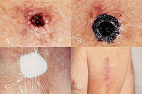
A 1997 study explored the idea that surgery can liberate atypical melanocytes and predispose a patient to metastatic disease, Dr. Brooks said (Dermatol. Surg. 1997;23:1043-6). The epidemiologic study of 1,224 primary melanomas concluded that surgery of primary melanoma may enhance tumor growth at metastatic sites.
Chemosurgery with zinc chloride paste, therefore, helps avoid this phenomenon, Dr. Brooks said. The paste not only destroys tissue where it is applied, it also fixes the tissue into a firm mass. A delayed excision allows the specimen to be removed en bloc and examined histopathologically to determine margins. However, leaving a portion of the fixed tissue in place for several more days causes a strong vaccine-like reaction on the skin surface, indicating, according to Dr. Brooks, the beginning of a systemic immunologic response.
The Evidence
According to Dr. Brooks, he and his colleagues published the only piece of objective evidence on this phenomenon in 1998 (Dermatol. Surg. 1998;24:1021-5).
The study involved mice that were injected with two different melanoma types: a poorly immunogenic (B16) type and a more immunogenic type (K1735p). The resultant tumors were freshly excised in some mice, or excised 24 hours after the application of zinc chloride paste. A week later, all the mice received a second injection of melanoma cells at a different site. K1735p tumors developed at the challenge sites in significantly fewer of the mice pretreated with zinc chloride (32% vs. 69%, respectively). The paste did not alter the development of B16 melanomas. The authors concluded: "Zinc chloride fixation of the more immunogenic K1735p melanoma increased resistance to subsequent tumor challenge, suggesting that zinc chloride fixative paste acts as an immune adjuvant."
Treatment Application
At the meeting, Dr. Brooks presented his most recent article describing a simplified method for using the paste (Dermatol. Surg. 2010;36:237-40).
His process begins with a fresh tissue biopsy to confirm melanoma. He recommended a shaved saucerized excision with a margin of 1-2 mm beyond the suspected tumor edge carried at least through the mid-dermis, or below the suspected thickness of the lesion.
After melanoma is histopathologically confirmed, he applies the Mohs paste. Since the compound will not penetrate an intact keratin layer, it must only be applied to the excised area. "If the biopsied area has developed a crust or eschar, this must also be removed," Dr. Brooks said.
He applies a 1-mm thick layer of paste with a cotton-tipped swab. More can be applied for thicker lesions, but less if the lesion overlies an important anatomic structure. If the biopsy is a small excisional one, a swab of 50% bi- or trichloracetic acid can remove the outer keratin layer and allow the paste to penetrate.
The next step is to apply a sterile cotton ball and cover the dressing with bio-occlusive tape for 24 hours – the time it takes the paste to achieve full penetration. After 24 hours, the patient can return for a conventional, wide excision with standard vertical or bevelled Mohs margins, followed by closure.
However, to take advantage of the paste's immunologic properties, the block of tissue should be left in place for a week or longer, or the main block can be removed, leaving a rim of the fixed tissue in place to stimulate the immune response. Dr. Brooks said the fixed tissue can then be histologically examined to confirm negative margins. For patients with raised lesions, he recommended debulking to create a flat tissue plane and they applying the paste as described.
He also stressed that the Mohs paste does not interfere in any way with conventional melanoma treatment. "About half of my patients end up going to [a cancer center] for excision, sentinel nodes, or other treatment," including interferon, he said. But some patients, after reviewing the comparatively small survival benefit interferon offers and its significant side effects, elect to have the fixed tissue excised and then continue with the practice of node palpation and regular ultrasounds to identify any early disease spread.
Dr. Brooks reported having no relevant financial disclosures.
"Mohs paste is a tried and true compound that does what it's purported to do," said Dr. Kenneth Gross.
"The first patient I [used the paste on] developed a sentinel node that was swollen and large, and I thought it was a metastatic, but it turned out to be clearly an immune response. On the flip side – I don't paste everyone. But if I have a high-risk patient who is well balanced and capable of understanding the implications, I do offer to paste them."
It is important to get informed consent and explain that zinc chloride is not an approved therapy; however, assure the patient it will not interfere with any other therapy they might want to pursue. Consider reserving it for high-risk patients, many of whom have melanoma that extends to the bone. "If you do your Mohs down to the periosteum and want to know if the bone is clear, you can paste it and a day or so later the bone will decalcify and a little chip will come out and you can examine that."
Mohs paste has fallen out of use over the years as fresh-tissue surgery has gained popularity. "Fresh-tissue replaced it because it allows immediate closure, while the paste causes a lot of inflammation and, the way Dr. Mohs did it, it left a wound that healed by secondary intention. It heals well but people won’t stand for that these days." Younger physicians, he said, "want to use what’s new, and what they were taught to use."
For many years, Dr. Mohs got the paste from just one pharmacist, who retired long ago. "Then there was just no reliable place to get the stuff. But recently we've been able to work with Delasco, [a medical supply company] that can compound it very reliably. It's cheap and a small amount will last virtually forever at room temperature."
Because Mohs paste is composed of natural ingredients and has been in use for so long, it is an unpatentable therapy. Therefore, it will probably always remain on the fringes of medicine.
"You will never find any large head-to-head studies with other therapies, and no pharmaceutical company is ever going to research it. It would cost millions of dollars to do the kind of studies you’d need to get approval and then what would you have at the end? Nothing you could make money off of. So it won't happen."
Aside from some slight discomfort and the inflammatory reaction, however, there are few down sides to using the paste. "If you look at it from a perspective of what can I do that won't hurt my patients, probably helps them, and won’t interfere with definitive, modern therapy, I would say Mohs paste is a shining example."
Kenneth Gross, M.D., was a course
director at the meeting, and is a surgical dermatologist in San Diego.
He reported having no relevant conflicts of interest.
"Mohs paste is a tried and true compound that does what it's purported to do," said Dr. Kenneth Gross.
"The first patient I [used the paste on] developed a sentinel node that was swollen and large, and I thought it was a metastatic, but it turned out to be clearly an immune response. On the flip side – I don't paste everyone. But if I have a high-risk patient who is well balanced and capable of understanding the implications, I do offer to paste them."
It is important to get informed consent and explain that zinc chloride is not an approved therapy; however, assure the patient it will not interfere with any other therapy they might want to pursue. Consider reserving it for high-risk patients, many of whom have melanoma that extends to the bone. "If you do your Mohs down to the periosteum and want to know if the bone is clear, you can paste it and a day or so later the bone will decalcify and a little chip will come out and you can examine that."
Mohs paste has fallen out of use over the years as fresh-tissue surgery has gained popularity. "Fresh-tissue replaced it because it allows immediate closure, while the paste causes a lot of inflammation and, the way Dr. Mohs did it, it left a wound that healed by secondary intention. It heals well but people won’t stand for that these days." Younger physicians, he said, "want to use what’s new, and what they were taught to use."
For many years, Dr. Mohs got the paste from just one pharmacist, who retired long ago. "Then there was just no reliable place to get the stuff. But recently we've been able to work with Delasco, [a medical supply company] that can compound it very reliably. It's cheap and a small amount will last virtually forever at room temperature."
Because Mohs paste is composed of natural ingredients and has been in use for so long, it is an unpatentable therapy. Therefore, it will probably always remain on the fringes of medicine.
"You will never find any large head-to-head studies with other therapies, and no pharmaceutical company is ever going to research it. It would cost millions of dollars to do the kind of studies you’d need to get approval and then what would you have at the end? Nothing you could make money off of. So it won't happen."
Aside from some slight discomfort and the inflammatory reaction, however, there are few down sides to using the paste. "If you look at it from a perspective of what can I do that won't hurt my patients, probably helps them, and won’t interfere with definitive, modern therapy, I would say Mohs paste is a shining example."
Kenneth Gross, M.D., was a course
director at the meeting, and is a surgical dermatologist in San Diego.
He reported having no relevant conflicts of interest.
"Mohs paste is a tried and true compound that does what it's purported to do," said Dr. Kenneth Gross.
"The first patient I [used the paste on] developed a sentinel node that was swollen and large, and I thought it was a metastatic, but it turned out to be clearly an immune response. On the flip side – I don't paste everyone. But if I have a high-risk patient who is well balanced and capable of understanding the implications, I do offer to paste them."
It is important to get informed consent and explain that zinc chloride is not an approved therapy; however, assure the patient it will not interfere with any other therapy they might want to pursue. Consider reserving it for high-risk patients, many of whom have melanoma that extends to the bone. "If you do your Mohs down to the periosteum and want to know if the bone is clear, you can paste it and a day or so later the bone will decalcify and a little chip will come out and you can examine that."
Mohs paste has fallen out of use over the years as fresh-tissue surgery has gained popularity. "Fresh-tissue replaced it because it allows immediate closure, while the paste causes a lot of inflammation and, the way Dr. Mohs did it, it left a wound that healed by secondary intention. It heals well but people won’t stand for that these days." Younger physicians, he said, "want to use what’s new, and what they were taught to use."
For many years, Dr. Mohs got the paste from just one pharmacist, who retired long ago. "Then there was just no reliable place to get the stuff. But recently we've been able to work with Delasco, [a medical supply company] that can compound it very reliably. It's cheap and a small amount will last virtually forever at room temperature."
Because Mohs paste is composed of natural ingredients and has been in use for so long, it is an unpatentable therapy. Therefore, it will probably always remain on the fringes of medicine.
"You will never find any large head-to-head studies with other therapies, and no pharmaceutical company is ever going to research it. It would cost millions of dollars to do the kind of studies you’d need to get approval and then what would you have at the end? Nothing you could make money off of. So it won't happen."
Aside from some slight discomfort and the inflammatory reaction, however, there are few down sides to using the paste. "If you look at it from a perspective of what can I do that won't hurt my patients, probably helps them, and won’t interfere with definitive, modern therapy, I would say Mohs paste is a shining example."
Kenneth Gross, M.D., was a course
director at the meeting, and is a surgical dermatologist in San Diego.
He reported having no relevant conflicts of interest.
SAN DIEGO - A remedy that has been used for nearly 2 centuries is still a valuable adjuvant therapy for melanoma, according to Dr. Norman Brooks.
Zinc chloride paste not only penetrates and kills even thick melanomas, it appears to stimulate a strong immune reaction that decreases the likelihood of metastasis and recurrence, Dr. Brooks said at the meeting sponsored by the American Society for Mohs Surgery.
"I believe chemosurgery with zinc chloride paste, followed by wide excision, is a better way to remove a melanoma than fresh tissue surgery," said Dr. Brooks, a dermatologist in Encino, Calif., who specializes in skin cancers. "It not only kills the tumor locally but stimulates total body resistance and immunity, supporting the idea that surgery actually enhances melanoma tumor growth and metastatic spread. That’s why I believe it's so important to use zinc chloride when treating melanoma."
History of Zinc Chloride
Zinc chloride was first used as a cancer treatment in 1815. In 1858, a U.S. physician added a powdered form of the bloodroot plant (Sanguinaria canadensis), which contains the anticancer alkaloid sanguinarine; the compound was used to treat breast cancer. The current paste, formulated by Dr. Frederic E. Mohs, is a compound of naturally occurring ingredients: the mineral stibnite, which forms the inert paste-like vehicle; powered root of the bloodroot plant, which has been shown to induce apoptosis in several types of cancer; and a saturated zinc chloride solution, which destructively penetrates tissue.
A 1997 study explored the idea that surgery can liberate atypical melanocytes and predispose a patient to metastatic disease, Dr. Brooks said (Dermatol. Surg. 1997;23:1043-6). The epidemiologic study of 1,224 primary melanomas concluded that surgery of primary melanoma may enhance tumor growth at metastatic sites.
Chemosurgery with zinc chloride paste, therefore, helps avoid this phenomenon, Dr. Brooks said. The paste not only destroys tissue where it is applied, it also fixes the tissue into a firm mass. A delayed excision allows the specimen to be removed en bloc and examined histopathologically to determine margins. However, leaving a portion of the fixed tissue in place for several more days causes a strong vaccine-like reaction on the skin surface, indicating, according to Dr. Brooks, the beginning of a systemic immunologic response.
The Evidence
According to Dr. Brooks, he and his colleagues published the only piece of objective evidence on this phenomenon in 1998 (Dermatol. Surg. 1998;24:1021-5).
The study involved mice that were injected with two different melanoma types: a poorly immunogenic (B16) type and a more immunogenic type (K1735p). The resultant tumors were freshly excised in some mice, or excised 24 hours after the application of zinc chloride paste. A week later, all the mice received a second injection of melanoma cells at a different site. K1735p tumors developed at the challenge sites in significantly fewer of the mice pretreated with zinc chloride (32% vs. 69%, respectively). The paste did not alter the development of B16 melanomas. The authors concluded: "Zinc chloride fixation of the more immunogenic K1735p melanoma increased resistance to subsequent tumor challenge, suggesting that zinc chloride fixative paste acts as an immune adjuvant."
Treatment Application
At the meeting, Dr. Brooks presented his most recent article describing a simplified method for using the paste (Dermatol. Surg. 2010;36:237-40).
His process begins with a fresh tissue biopsy to confirm melanoma. He recommended a shaved saucerized excision with a margin of 1-2 mm beyond the suspected tumor edge carried at least through the mid-dermis, or below the suspected thickness of the lesion.
After melanoma is histopathologically confirmed, he applies the Mohs paste. Since the compound will not penetrate an intact keratin layer, it must only be applied to the excised area. "If the biopsied area has developed a crust or eschar, this must also be removed," Dr. Brooks said.
He applies a 1-mm thick layer of paste with a cotton-tipped swab. More can be applied for thicker lesions, but less if the lesion overlies an important anatomic structure. If the biopsy is a small excisional one, a swab of 50% bi- or trichloracetic acid can remove the outer keratin layer and allow the paste to penetrate.
The next step is to apply a sterile cotton ball and cover the dressing with bio-occlusive tape for 24 hours – the time it takes the paste to achieve full penetration. After 24 hours, the patient can return for a conventional, wide excision with standard vertical or bevelled Mohs margins, followed by closure.
However, to take advantage of the paste's immunologic properties, the block of tissue should be left in place for a week or longer, or the main block can be removed, leaving a rim of the fixed tissue in place to stimulate the immune response. Dr. Brooks said the fixed tissue can then be histologically examined to confirm negative margins. For patients with raised lesions, he recommended debulking to create a flat tissue plane and they applying the paste as described.
He also stressed that the Mohs paste does not interfere in any way with conventional melanoma treatment. "About half of my patients end up going to [a cancer center] for excision, sentinel nodes, or other treatment," including interferon, he said. But some patients, after reviewing the comparatively small survival benefit interferon offers and its significant side effects, elect to have the fixed tissue excised and then continue with the practice of node palpation and regular ultrasounds to identify any early disease spread.
Dr. Brooks reported having no relevant financial disclosures.
SAN DIEGO - A remedy that has been used for nearly 2 centuries is still a valuable adjuvant therapy for melanoma, according to Dr. Norman Brooks.
Zinc chloride paste not only penetrates and kills even thick melanomas, it appears to stimulate a strong immune reaction that decreases the likelihood of metastasis and recurrence, Dr. Brooks said at the meeting sponsored by the American Society for Mohs Surgery.
"I believe chemosurgery with zinc chloride paste, followed by wide excision, is a better way to remove a melanoma than fresh tissue surgery," said Dr. Brooks, a dermatologist in Encino, Calif., who specializes in skin cancers. "It not only kills the tumor locally but stimulates total body resistance and immunity, supporting the idea that surgery actually enhances melanoma tumor growth and metastatic spread. That’s why I believe it's so important to use zinc chloride when treating melanoma."
History of Zinc Chloride
Zinc chloride was first used as a cancer treatment in 1815. In 1858, a U.S. physician added a powdered form of the bloodroot plant (Sanguinaria canadensis), which contains the anticancer alkaloid sanguinarine; the compound was used to treat breast cancer. The current paste, formulated by Dr. Frederic E. Mohs, is a compound of naturally occurring ingredients: the mineral stibnite, which forms the inert paste-like vehicle; powered root of the bloodroot plant, which has been shown to induce apoptosis in several types of cancer; and a saturated zinc chloride solution, which destructively penetrates tissue.
A 1997 study explored the idea that surgery can liberate atypical melanocytes and predispose a patient to metastatic disease, Dr. Brooks said (Dermatol. Surg. 1997;23:1043-6). The epidemiologic study of 1,224 primary melanomas concluded that surgery of primary melanoma may enhance tumor growth at metastatic sites.
Chemosurgery with zinc chloride paste, therefore, helps avoid this phenomenon, Dr. Brooks said. The paste not only destroys tissue where it is applied, it also fixes the tissue into a firm mass. A delayed excision allows the specimen to be removed en bloc and examined histopathologically to determine margins. However, leaving a portion of the fixed tissue in place for several more days causes a strong vaccine-like reaction on the skin surface, indicating, according to Dr. Brooks, the beginning of a systemic immunologic response.
The Evidence
According to Dr. Brooks, he and his colleagues published the only piece of objective evidence on this phenomenon in 1998 (Dermatol. Surg. 1998;24:1021-5).
The study involved mice that were injected with two different melanoma types: a poorly immunogenic (B16) type and a more immunogenic type (K1735p). The resultant tumors were freshly excised in some mice, or excised 24 hours after the application of zinc chloride paste. A week later, all the mice received a second injection of melanoma cells at a different site. K1735p tumors developed at the challenge sites in significantly fewer of the mice pretreated with zinc chloride (32% vs. 69%, respectively). The paste did not alter the development of B16 melanomas. The authors concluded: "Zinc chloride fixation of the more immunogenic K1735p melanoma increased resistance to subsequent tumor challenge, suggesting that zinc chloride fixative paste acts as an immune adjuvant."
Treatment Application
At the meeting, Dr. Brooks presented his most recent article describing a simplified method for using the paste (Dermatol. Surg. 2010;36:237-40).
His process begins with a fresh tissue biopsy to confirm melanoma. He recommended a shaved saucerized excision with a margin of 1-2 mm beyond the suspected tumor edge carried at least through the mid-dermis, or below the suspected thickness of the lesion.
After melanoma is histopathologically confirmed, he applies the Mohs paste. Since the compound will not penetrate an intact keratin layer, it must only be applied to the excised area. "If the biopsied area has developed a crust or eschar, this must also be removed," Dr. Brooks said.
He applies a 1-mm thick layer of paste with a cotton-tipped swab. More can be applied for thicker lesions, but less if the lesion overlies an important anatomic structure. If the biopsy is a small excisional one, a swab of 50% bi- or trichloracetic acid can remove the outer keratin layer and allow the paste to penetrate.
The next step is to apply a sterile cotton ball and cover the dressing with bio-occlusive tape for 24 hours – the time it takes the paste to achieve full penetration. After 24 hours, the patient can return for a conventional, wide excision with standard vertical or bevelled Mohs margins, followed by closure.
However, to take advantage of the paste's immunologic properties, the block of tissue should be left in place for a week or longer, or the main block can be removed, leaving a rim of the fixed tissue in place to stimulate the immune response. Dr. Brooks said the fixed tissue can then be histologically examined to confirm negative margins. For patients with raised lesions, he recommended debulking to create a flat tissue plane and they applying the paste as described.
He also stressed that the Mohs paste does not interfere in any way with conventional melanoma treatment. "About half of my patients end up going to [a cancer center] for excision, sentinel nodes, or other treatment," including interferon, he said. But some patients, after reviewing the comparatively small survival benefit interferon offers and its significant side effects, elect to have the fixed tissue excised and then continue with the practice of node palpation and regular ultrasounds to identify any early disease spread.
Dr. Brooks reported having no relevant financial disclosures.
EXPERT ANALYSIS FROM A MEETING SPONSORED BY THE AMERICAN SOCIETY FOR MOHS SURGERY
PV-10 Melanoma Drug Trial Enrolls Patients in Compassionate Use Program
Provectus Pharmaceuticals announced on Nov. 23 that it has enrolled at least 40 patients in its compassionate use program for PV-10, an experimental drug being studied primarily for melanoma.
PV-10 is an injectable form of Rose Bengal, a small molecule staining agent used to assess eye damage and liver ailments. Provectus determined that the drug selectively kills cancer cells and has been studying it in nonvisceral cancers.
Under the compassionate use program, patients who are not eligible for clinical trials and have certain breast cancers, basal cell carcinoma, squamous cell carcinoma, certain head and neck cancers, and melanoma can receive PV-10, the company announced.
Phase II studies of PV-10 in metastatic melanoma have just been completed, and 10 of the patients from the study joined the compassionate use program.
Patients in the program will have more frequent and extensive treatment over a longer duration than did those who received the drug in the phase II studies. The company hopes that the compassionate use program might help pinpoint a dosing regimen that can be used in a phase III trial in metastatic melanoma.
Provectus will also pursue the study of PV-10 for liver cancer.
Provectus Pharmaceuticals announced on Nov. 23 that it has enrolled at least 40 patients in its compassionate use program for PV-10, an experimental drug being studied primarily for melanoma.
PV-10 is an injectable form of Rose Bengal, a small molecule staining agent used to assess eye damage and liver ailments. Provectus determined that the drug selectively kills cancer cells and has been studying it in nonvisceral cancers.
Under the compassionate use program, patients who are not eligible for clinical trials and have certain breast cancers, basal cell carcinoma, squamous cell carcinoma, certain head and neck cancers, and melanoma can receive PV-10, the company announced.
Phase II studies of PV-10 in metastatic melanoma have just been completed, and 10 of the patients from the study joined the compassionate use program.
Patients in the program will have more frequent and extensive treatment over a longer duration than did those who received the drug in the phase II studies. The company hopes that the compassionate use program might help pinpoint a dosing regimen that can be used in a phase III trial in metastatic melanoma.
Provectus will also pursue the study of PV-10 for liver cancer.
Provectus Pharmaceuticals announced on Nov. 23 that it has enrolled at least 40 patients in its compassionate use program for PV-10, an experimental drug being studied primarily for melanoma.
PV-10 is an injectable form of Rose Bengal, a small molecule staining agent used to assess eye damage and liver ailments. Provectus determined that the drug selectively kills cancer cells and has been studying it in nonvisceral cancers.
Under the compassionate use program, patients who are not eligible for clinical trials and have certain breast cancers, basal cell carcinoma, squamous cell carcinoma, certain head and neck cancers, and melanoma can receive PV-10, the company announced.
Phase II studies of PV-10 in metastatic melanoma have just been completed, and 10 of the patients from the study joined the compassionate use program.
Patients in the program will have more frequent and extensive treatment over a longer duration than did those who received the drug in the phase II studies. The company hopes that the compassionate use program might help pinpoint a dosing regimen that can be used in a phase III trial in metastatic melanoma.
Provectus will also pursue the study of PV-10 for liver cancer.
FDA Panel Divided on MelaFind Device
Updated: 11/22/2010
COLLEGE PARK, MD. - A Food and Drug Administration advisory panel on Nov. 18 split on whether to support approval of a noninvasive device intended to help dermatologists and other physicians detect early melanomas.
At the meeting, the FDA's General and Plastic Surgery Devices Panel voted 8 to 7 with 1 abstention that the data available supported approval of the MelaFind device to evaluate clinically atypical cutaneous pigmented lesions that have one or more characteristics of melanoma, "when a physician chooses to obtain additional information before making a final decision to biopsy to rule out melanoma." That is the indication proposed for approval by the manufacturer, Mela Sciences Inc.
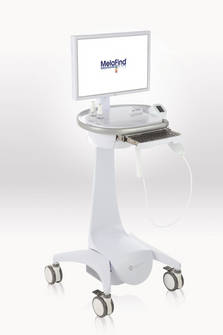
The first device of this kind, MelaFind is a multispectral computer vision system with a handheld component that captures the image of a lesion with a dermoscope through a thin layer of alcohol applied to the skin. The device has software that uses algorithms to analyze the image, indicating within 2 minutes whether a biopsy should be done, according to Mela Sciences. It is not intended to be used as a screening tool or in the evaluation of nonpigmented lesions; banal pigmented lesions; lesions considered definite melanomas; or mucosal, subungual and other lesions at different anatomic sites.
The device was used by dermatologists in a pivotal study of 1,257 patients (mean age 47 years) on 1,632 pigmented skin lesions that had at least one characteristic of melanoma, which were also biopsied. Of the 114 lesions that were positive for melanoma on biopsy, 112 were tagged as positive by MelaFind, for a sensitivity of 98.3%, which the company compared to the sensitivity of 70%-80% for dermatologists that has been reported in the literature. (Sensitivity for the dermatologists could not be determined because the decision to perform a biopsy had already been made by the clinician before referring to MelaFind.) Specificity was 9.5%.
Panelists voting for and against approval were concerned about the use of this device in the hands of nondermatologists, who, they said, might miss melanomas – a concern also raised by the FDA. But those voting in favor of approval said they believed with appropriate training, clinicians could learn to use the device effectively and that it would be a valuable tool. Concerns among those voting against approval included the need for more data, the risk of false negatives with the device, and issues with the data. It remained unclear how the results would guide decision-making.
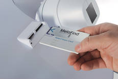
Dermatologists on the panel voted on both sides of the approval question. "I would not mind having this in my practice," said Dr. Wilma Bergfeld, senior dermatologist and co-director of dermatopathology at the Cleveland Clinic, Cleveland, who voted in favor of approval. While clinical trial data and protocol were confusing, she said, "I believe that with the appropriate training and use, we might be able to use this instrument very effectively in the clinic to diagnose these equivocal clinical lesions."
Dr. Lynn Drake, lecturer in dermatology, Harvard Medical School, who voted against approval, said that while there is a need for such a device, "this falls short right now." Among her concerns were the need for more data, the high false-positive rate, and possible widespread use by clinicians not adequately trained once it becomes available. "We have to be very careful about approving something that might replace clinical judgement," she said.
FDA reviewers raised several concerns about the study, including possible selection bias, and concluded that the available data were inadequate to determine whether the use of MelaFind would add any true value to evaluating clinically atypical lesions – and that a prospective study was needed before approval.
The FDA usually follows the recommendations of its advisory panels. Panel members have been cleared of potential conflicts of interest prior to the meeting.
Updated: 11/22/2010
COLLEGE PARK, MD. - A Food and Drug Administration advisory panel on Nov. 18 split on whether to support approval of a noninvasive device intended to help dermatologists and other physicians detect early melanomas.
At the meeting, the FDA's General and Plastic Surgery Devices Panel voted 8 to 7 with 1 abstention that the data available supported approval of the MelaFind device to evaluate clinically atypical cutaneous pigmented lesions that have one or more characteristics of melanoma, "when a physician chooses to obtain additional information before making a final decision to biopsy to rule out melanoma." That is the indication proposed for approval by the manufacturer, Mela Sciences Inc.
The first device of this kind, MelaFind is a multispectral computer vision system with a handheld component that captures the image of a lesion with a dermoscope through a thin layer of alcohol applied to the skin. The device has software that uses algorithms to analyze the image, indicating within 2 minutes whether a biopsy should be done, according to Mela Sciences. It is not intended to be used as a screening tool or in the evaluation of nonpigmented lesions; banal pigmented lesions; lesions considered definite melanomas; or mucosal, subungual and other lesions at different anatomic sites.
The device was used by dermatologists in a pivotal study of 1,257 patients (mean age 47 years) on 1,632 pigmented skin lesions that had at least one characteristic of melanoma, which were also biopsied. Of the 114 lesions that were positive for melanoma on biopsy, 112 were tagged as positive by MelaFind, for a sensitivity of 98.3%, which the company compared to the sensitivity of 70%-80% for dermatologists that has been reported in the literature. (Sensitivity for the dermatologists could not be determined because the decision to perform a biopsy had already been made by the clinician before referring to MelaFind.) Specificity was 9.5%.
Panelists voting for and against approval were concerned about the use of this device in the hands of nondermatologists, who, they said, might miss melanomas – a concern also raised by the FDA. But those voting in favor of approval said they believed with appropriate training, clinicians could learn to use the device effectively and that it would be a valuable tool. Concerns among those voting against approval included the need for more data, the risk of false negatives with the device, and issues with the data. It remained unclear how the results would guide decision-making.
Dermatologists on the panel voted on both sides of the approval question. "I would not mind having this in my practice," said Dr. Wilma Bergfeld, senior dermatologist and co-director of dermatopathology at the Cleveland Clinic, Cleveland, who voted in favor of approval. While clinical trial data and protocol were confusing, she said, "I believe that with the appropriate training and use, we might be able to use this instrument very effectively in the clinic to diagnose these equivocal clinical lesions."
Dr. Lynn Drake, lecturer in dermatology, Harvard Medical School, who voted against approval, said that while there is a need for such a device, "this falls short right now." Among her concerns were the need for more data, the high false-positive rate, and possible widespread use by clinicians not adequately trained once it becomes available. "We have to be very careful about approving something that might replace clinical judgement," she said.
FDA reviewers raised several concerns about the study, including possible selection bias, and concluded that the available data were inadequate to determine whether the use of MelaFind would add any true value to evaluating clinically atypical lesions – and that a prospective study was needed before approval.
The FDA usually follows the recommendations of its advisory panels. Panel members have been cleared of potential conflicts of interest prior to the meeting.
Updated: 11/22/2010
COLLEGE PARK, MD. - A Food and Drug Administration advisory panel on Nov. 18 split on whether to support approval of a noninvasive device intended to help dermatologists and other physicians detect early melanomas.
At the meeting, the FDA's General and Plastic Surgery Devices Panel voted 8 to 7 with 1 abstention that the data available supported approval of the MelaFind device to evaluate clinically atypical cutaneous pigmented lesions that have one or more characteristics of melanoma, "when a physician chooses to obtain additional information before making a final decision to biopsy to rule out melanoma." That is the indication proposed for approval by the manufacturer, Mela Sciences Inc.
The first device of this kind, MelaFind is a multispectral computer vision system with a handheld component that captures the image of a lesion with a dermoscope through a thin layer of alcohol applied to the skin. The device has software that uses algorithms to analyze the image, indicating within 2 minutes whether a biopsy should be done, according to Mela Sciences. It is not intended to be used as a screening tool or in the evaluation of nonpigmented lesions; banal pigmented lesions; lesions considered definite melanomas; or mucosal, subungual and other lesions at different anatomic sites.
The device was used by dermatologists in a pivotal study of 1,257 patients (mean age 47 years) on 1,632 pigmented skin lesions that had at least one characteristic of melanoma, which were also biopsied. Of the 114 lesions that were positive for melanoma on biopsy, 112 were tagged as positive by MelaFind, for a sensitivity of 98.3%, which the company compared to the sensitivity of 70%-80% for dermatologists that has been reported in the literature. (Sensitivity for the dermatologists could not be determined because the decision to perform a biopsy had already been made by the clinician before referring to MelaFind.) Specificity was 9.5%.
Panelists voting for and against approval were concerned about the use of this device in the hands of nondermatologists, who, they said, might miss melanomas – a concern also raised by the FDA. But those voting in favor of approval said they believed with appropriate training, clinicians could learn to use the device effectively and that it would be a valuable tool. Concerns among those voting against approval included the need for more data, the risk of false negatives with the device, and issues with the data. It remained unclear how the results would guide decision-making.
Dermatologists on the panel voted on both sides of the approval question. "I would not mind having this in my practice," said Dr. Wilma Bergfeld, senior dermatologist and co-director of dermatopathology at the Cleveland Clinic, Cleveland, who voted in favor of approval. While clinical trial data and protocol were confusing, she said, "I believe that with the appropriate training and use, we might be able to use this instrument very effectively in the clinic to diagnose these equivocal clinical lesions."
Dr. Lynn Drake, lecturer in dermatology, Harvard Medical School, who voted against approval, said that while there is a need for such a device, "this falls short right now." Among her concerns were the need for more data, the high false-positive rate, and possible widespread use by clinicians not adequately trained once it becomes available. "We have to be very careful about approving something that might replace clinical judgement," she said.
FDA reviewers raised several concerns about the study, including possible selection bias, and concluded that the available data were inadequate to determine whether the use of MelaFind would add any true value to evaluating clinically atypical lesions – and that a prospective study was needed before approval.
The FDA usually follows the recommendations of its advisory panels. Panel members have been cleared of potential conflicts of interest prior to the meeting.
FROM A MEETING OF THE FDA'S GENERAL AND PLASTIC SURGERY DEVICES PANEL
FDA Committee Considers Gardasil for Anal Cancer Prevention
SILVER SPRING, Md. - The human papillomavirus vaccine Gardasil moved closer to an indication for anal cancer prevention after a meeting of the Food and Drug Administration’s Vaccines and Related Biological Products Advisory Committee on Nov. 17.
The vaccine, manufactured by Merck & Co. Inc., is approved for the prevention of cervical, vulvar, and vaginal cancers and precancerous lesions in females aged 9-26 years, and for the prevention of genital warts in males and females aged 9-26 years. The company is seeking an indication for the prevention of anal cancer in males and females aged 9-26 years.
Previous studies have shown that anal cancer and cervical cancer are biologically similar, and both are associated with HPV infections, Dr. Joel Palefsky, professor of medicine at the University of California, San Francisco, said at the meeting.
The primary data supporting an anal cancer indication for Gardasil came from a randomized, controlled trial of 602 men who have sex with men (MSM), who were part of the larger study that led to the indication for preventing genital warts in boys and men. The participants received three doses of vaccine or a placebo.
The vaccine showed 78% effectiveness, compared with placebo in preventing anal intraepithelial neoplasms related to human papillomavirus types 6, 11, 16, and 18. These lesions are considered precursors to anal cancer, Dr. Palefsky said.
Although MSM are at increased risk for HPV-associated anal cancer, they are not the only population at risk. Data from the National Cancer Institute presented at the meeting showed that anal cancer incidence in the United States is increasing by a rate of approximately 2% per year, and that approximately 60% of cases and deaths occur in women.
A majority of the committee members expressed opinions in favor of the indication, but no formal vote was taken. The FDA will consider the committee’s recommendations. If the indication is approved before the February 2011 meeting of the Centers for Disease Control and Prevention’s Advisory Committee on Immunization Practices, the ACIP might revisit its previous recommendation against routine HPV vaccination for boys and men for genital wart prevention. ACIP has given a permissive recommendation for HPV vaccination for boys and men aged 9-26 years at the discretion of the physician.
Dr. Palefsky has served as a consultant and clinical investigator for Merck & Co. Inc.
Body text goes here
Doctor’s Bio
Body text goes here
Doctor’s Bio
Body text goes here
Doctor’s Bio
SILVER SPRING, Md. - The human papillomavirus vaccine Gardasil moved closer to an indication for anal cancer prevention after a meeting of the Food and Drug Administration’s Vaccines and Related Biological Products Advisory Committee on Nov. 17.
The vaccine, manufactured by Merck & Co. Inc., is approved for the prevention of cervical, vulvar, and vaginal cancers and precancerous lesions in females aged 9-26 years, and for the prevention of genital warts in males and females aged 9-26 years. The company is seeking an indication for the prevention of anal cancer in males and females aged 9-26 years.
Previous studies have shown that anal cancer and cervical cancer are biologically similar, and both are associated with HPV infections, Dr. Joel Palefsky, professor of medicine at the University of California, San Francisco, said at the meeting.
The primary data supporting an anal cancer indication for Gardasil came from a randomized, controlled trial of 602 men who have sex with men (MSM), who were part of the larger study that led to the indication for preventing genital warts in boys and men. The participants received three doses of vaccine or a placebo.
The vaccine showed 78% effectiveness, compared with placebo in preventing anal intraepithelial neoplasms related to human papillomavirus types 6, 11, 16, and 18. These lesions are considered precursors to anal cancer, Dr. Palefsky said.
Although MSM are at increased risk for HPV-associated anal cancer, they are not the only population at risk. Data from the National Cancer Institute presented at the meeting showed that anal cancer incidence in the United States is increasing by a rate of approximately 2% per year, and that approximately 60% of cases and deaths occur in women.
A majority of the committee members expressed opinions in favor of the indication, but no formal vote was taken. The FDA will consider the committee’s recommendations. If the indication is approved before the February 2011 meeting of the Centers for Disease Control and Prevention’s Advisory Committee on Immunization Practices, the ACIP might revisit its previous recommendation against routine HPV vaccination for boys and men for genital wart prevention. ACIP has given a permissive recommendation for HPV vaccination for boys and men aged 9-26 years at the discretion of the physician.
Dr. Palefsky has served as a consultant and clinical investigator for Merck & Co. Inc.
SILVER SPRING, Md. - The human papillomavirus vaccine Gardasil moved closer to an indication for anal cancer prevention after a meeting of the Food and Drug Administration’s Vaccines and Related Biological Products Advisory Committee on Nov. 17.
The vaccine, manufactured by Merck & Co. Inc., is approved for the prevention of cervical, vulvar, and vaginal cancers and precancerous lesions in females aged 9-26 years, and for the prevention of genital warts in males and females aged 9-26 years. The company is seeking an indication for the prevention of anal cancer in males and females aged 9-26 years.
Previous studies have shown that anal cancer and cervical cancer are biologically similar, and both are associated with HPV infections, Dr. Joel Palefsky, professor of medicine at the University of California, San Francisco, said at the meeting.
The primary data supporting an anal cancer indication for Gardasil came from a randomized, controlled trial of 602 men who have sex with men (MSM), who were part of the larger study that led to the indication for preventing genital warts in boys and men. The participants received three doses of vaccine or a placebo.
The vaccine showed 78% effectiveness, compared with placebo in preventing anal intraepithelial neoplasms related to human papillomavirus types 6, 11, 16, and 18. These lesions are considered precursors to anal cancer, Dr. Palefsky said.
Although MSM are at increased risk for HPV-associated anal cancer, they are not the only population at risk. Data from the National Cancer Institute presented at the meeting showed that anal cancer incidence in the United States is increasing by a rate of approximately 2% per year, and that approximately 60% of cases and deaths occur in women.
A majority of the committee members expressed opinions in favor of the indication, but no formal vote was taken. The FDA will consider the committee’s recommendations. If the indication is approved before the February 2011 meeting of the Centers for Disease Control and Prevention’s Advisory Committee on Immunization Practices, the ACIP might revisit its previous recommendation against routine HPV vaccination for boys and men for genital wart prevention. ACIP has given a permissive recommendation for HPV vaccination for boys and men aged 9-26 years at the discretion of the physician.
Dr. Palefsky has served as a consultant and clinical investigator for Merck & Co. Inc.
FROM A MEETING OF THE FOOD AND DRUG ADMINISTRATION’S VACCINE AND RELATED BIOLOGICAL PRODUCTS ADVISORY COMMITTEE