User login
Ipilimumab for Metastatic Melanoma
Dr. Richard D. Carvajal discusses ipilimumab's outcomes, patterns of response, and potential side effects as a treatment for metastatic melanoma.
Dr. Richard D. Carvajal discusses ipilimumab's outcomes, patterns of response, and potential side effects as a treatment for metastatic melanoma.
Dr. Richard D. Carvajal discusses ipilimumab's outcomes, patterns of response, and potential side effects as a treatment for metastatic melanoma.
Capecitabine May Protect Against Skin Cancers After Transplant
WAILEA, HAWAII – Oral capecitabine shows considerable early promise for the secondary prevention of nonmelanoma skin cancers in solid organ transplant recipients and other immunosuppressed individuals, according to Dr. Paul Nghiem.
Organ transplant recipients are at sharply increased risk of nonmelanoma skin cancer (NMSC). Many transplant recipients develop dozens of squamous cell carcinomas and/or basal cell carcinomas per year, and these tumors often behave very aggressively.

Dermatologists are sorely in need of additional tools to protect organ transplant recipients from NMSC – and capecitabine could fill the bill, he said at the SDEF Hawaii Dermatology Seminar.
Capecitabine (Xeloda) is a 5-fluorouracil (5-FU) precursor widely used for the treatment of colorectal cancer and metastatic breast cancer. Dr. Nghieim, a dermatologist at the University of Washington and the Fred Hutchinson Cancer Center in Seattle, credits University of Minnesota dermatologists with doing the pioneering work in developing capecitabine as a novel means of secondary prevention of NMSCs in the transplant population.
In a recent observational study, the Minnesota group reported on 15 solid organ transplant recipients, mean age 57 years, with recurrent NMSCs who were placed on low-dose capecitabine for the off-label purpose of preventing further NMSCs. The regimen was 1 g/m2 daily on days 1-14 of a 21-day cycle.
Comparing cumulative incidence rates for NMSC during the first year on capecitabine to those the year before, the investigators found the mean number of squamous cell carcinomas per month declined by 0.33, the mean number of actinic keratoses fell by 2.45 per month, and the mean number of basal cell carcinomas dropped by 0.04 per month. All these reductions were statistically significant.
Toxicities were deemed manageable. Grade 3/4 toxicities consisted of fatigue in 40% of patients, hand-foot syndrome in 20%, and diarrhea in 20%. One-third of subjects discontinued capecitabine by 1 year (Clin. Transplant. 2010 Nov. 2 [doi:10.1111/j.1399-0012.2010.01348.x]).
There are no definitive data yet, but Dr. Ngheim predicted there will not be a major rebound in NMSCs upon discontinuation of capecitabine, as occurs when systemic retinoids given for the treatment of multiple NMSCs are stopped. That’s because capecitabine is actually killing cancer cells.
"After all, we don’t see a rebound after we treat squamous cell carcinomas with topical 5-FU. I would suspect there is going to be a longer-lasting benefit than with retinoids, and that capecitabine really might be a boon to these folks," he said.
While studies of capecitabine for the secondary prevention of NMSCs continue, physicians can use several other means to protect transplant and other immunosuppressed patients. Dermatologic examinations at intervals of every 3 months or less are important to detect these cutaneous tumors before they metastasize. In addition, regular use of a broad-spectrum sunscreen has been shown to prevent the development of actinic keratoses and invasive squamous cell carcinomas in a randomized trial involving 120 immunocompromised organ transplant recipients (Br. J. Dermatol. 2009;161[suppl. 3]:78-84).
Also, Dr. Nghiem said, it has become clear in the last several years that the calcineurin inhibitors prescribed by transplant physicians to improve graft survival have a highly unwelcome ancillary effect: "They act directly on keratinocytes as a second-level fertilizer to make squamous cell carcinomas grow and become more aggressive."
Unlike the calcineurin inhibitors, sirolimus does not have a direct carcinogenic effect on keratinocytes. In animal studies, substituting sirolimus for calcineurin inhibitors in order to prevent graft rejection results in a striking reduction in NMSCs. Whether the same holds true in humans is now under formal study.
Systemic retinoids can be used in the treatment of multiple NMSCs. However, this is a therapy fraught with significant side effects, including an adverse effect on bone health, as well as a major rebound in squamous cell carcinomas after treatment cessation.
"I pretty much never use this," Dr. Nghiem said.
He said he had no relevant financial disclosures. SDEF and this news organization are owned by Elsevier.
WAILEA, HAWAII – Oral capecitabine shows considerable early promise for the secondary prevention of nonmelanoma skin cancers in solid organ transplant recipients and other immunosuppressed individuals, according to Dr. Paul Nghiem.
Organ transplant recipients are at sharply increased risk of nonmelanoma skin cancer (NMSC). Many transplant recipients develop dozens of squamous cell carcinomas and/or basal cell carcinomas per year, and these tumors often behave very aggressively.
Dermatologists are sorely in need of additional tools to protect organ transplant recipients from NMSC – and capecitabine could fill the bill, he said at the SDEF Hawaii Dermatology Seminar.
Capecitabine (Xeloda) is a 5-fluorouracil (5-FU) precursor widely used for the treatment of colorectal cancer and metastatic breast cancer. Dr. Nghieim, a dermatologist at the University of Washington and the Fred Hutchinson Cancer Center in Seattle, credits University of Minnesota dermatologists with doing the pioneering work in developing capecitabine as a novel means of secondary prevention of NMSCs in the transplant population.
In a recent observational study, the Minnesota group reported on 15 solid organ transplant recipients, mean age 57 years, with recurrent NMSCs who were placed on low-dose capecitabine for the off-label purpose of preventing further NMSCs. The regimen was 1 g/m2 daily on days 1-14 of a 21-day cycle.
Comparing cumulative incidence rates for NMSC during the first year on capecitabine to those the year before, the investigators found the mean number of squamous cell carcinomas per month declined by 0.33, the mean number of actinic keratoses fell by 2.45 per month, and the mean number of basal cell carcinomas dropped by 0.04 per month. All these reductions were statistically significant.
Toxicities were deemed manageable. Grade 3/4 toxicities consisted of fatigue in 40% of patients, hand-foot syndrome in 20%, and diarrhea in 20%. One-third of subjects discontinued capecitabine by 1 year (Clin. Transplant. 2010 Nov. 2 [doi:10.1111/j.1399-0012.2010.01348.x]).
There are no definitive data yet, but Dr. Ngheim predicted there will not be a major rebound in NMSCs upon discontinuation of capecitabine, as occurs when systemic retinoids given for the treatment of multiple NMSCs are stopped. That’s because capecitabine is actually killing cancer cells.
"After all, we don’t see a rebound after we treat squamous cell carcinomas with topical 5-FU. I would suspect there is going to be a longer-lasting benefit than with retinoids, and that capecitabine really might be a boon to these folks," he said.
While studies of capecitabine for the secondary prevention of NMSCs continue, physicians can use several other means to protect transplant and other immunosuppressed patients. Dermatologic examinations at intervals of every 3 months or less are important to detect these cutaneous tumors before they metastasize. In addition, regular use of a broad-spectrum sunscreen has been shown to prevent the development of actinic keratoses and invasive squamous cell carcinomas in a randomized trial involving 120 immunocompromised organ transplant recipients (Br. J. Dermatol. 2009;161[suppl. 3]:78-84).
Also, Dr. Nghiem said, it has become clear in the last several years that the calcineurin inhibitors prescribed by transplant physicians to improve graft survival have a highly unwelcome ancillary effect: "They act directly on keratinocytes as a second-level fertilizer to make squamous cell carcinomas grow and become more aggressive."
Unlike the calcineurin inhibitors, sirolimus does not have a direct carcinogenic effect on keratinocytes. In animal studies, substituting sirolimus for calcineurin inhibitors in order to prevent graft rejection results in a striking reduction in NMSCs. Whether the same holds true in humans is now under formal study.
Systemic retinoids can be used in the treatment of multiple NMSCs. However, this is a therapy fraught with significant side effects, including an adverse effect on bone health, as well as a major rebound in squamous cell carcinomas after treatment cessation.
"I pretty much never use this," Dr. Nghiem said.
He said he had no relevant financial disclosures. SDEF and this news organization are owned by Elsevier.
EXPERT ANALYSIS FROM SDEF HAWAII DERMATOLOGY SEMINAR
SDEF: Capecitabine May Protect Against Skin Cancers After Transplant
WAILEA, HAWAII – Oral capecitabine shows considerable early promise for the secondary prevention of nonmelanoma skin cancers in solid organ transplant recipients and other immunosuppressed individuals, according to Dr. Paul Nghiem.
Organ transplant recipients are at sharply increased risk of nonmelanoma skin cancer (NMSC). Many transplant recipients develop dozens of squamous cell carcinomas and/or basal cell carcinomas per year, and these tumors often behave very aggressively.
Dermatologists are sorely in need of additional tools to protect organ transplant recipients from NMSC – and capecitabine could fill the bill, he said at the SDEF Hawaii Dermatology Seminar.
Capecitabine (Xeloda) is a 5-fluorouracil (5-FU) precursor widely used for the treatment of colorectal cancer and metastatic breast cancer. Dr. Nghiem, a dermatologist at the University of Washington and the Fred Hutchinson Cancer Center in Seattle, credits University of Minnesota dermatologists with doing the pioneering work in developing capecitabine as a novel means of secondary prevention of NMSCs in the transplant population.
In a recent observational study, the Minnesota group reported on 15 solid organ transplant recipients, mean age 57 years, with recurrent NMSCs who were placed on low-dose capecitabine for the off-label purpose of preventing further NMSCs. The regimen was 1 g/m2 daily on days 1-14 of a 21-day cycle.
Comparing cumulative incidence rates for NMSC during the first year on capecitabine to those the year before, the investigators found the mean number of squamous cell carcinomas per month declined by 0.33, the mean number of actinic keratoses fell by 2.45 per month, and the mean number of basal cell carcinomas dropped by 0.04 per month. All these reductions were statistically significant.
Toxicities were deemed manageable. Grade 3/4 toxicities consisted of fatigue in 40% of patients, hand-foot syndrome in 20%, and diarrhea in 20%. One-third of subjects discontinued capecitabine by 1 year (Clin. Transplant. 2010 Nov. 2 [doi:10.1111/j.1399-0012.2010.01348.x]).
There are no definitive data yet, but Dr. Ngheim predicted there will not be a major rebound in NMSCs upon discontinuation of capecitabine, as occurs when systemic retinoids given for the treatment of multiple NMSCs are stopped. That’s because capecitabine is actually killing cancer cells.
"After all, we don’t see a rebound after we treat squamous cell carcinomas with topical 5-FU. I would suspect there is going to be a longer-lasting benefit than with retinoids, and that capecitabine really might be a boon to these folks," he said.
While studies of capecitabine for the secondary prevention of NMSCs continue, physicians can use several other means to protect transplant and other immunosuppressed patients. Dermatologic examinations at intervals of every 3 months or less are important to detect these cutaneous tumors before they metastasize. In addition, regular use of a broad-spectrum sunscreen has been shown to prevent the development of actinic keratoses and invasive squamous cell carcinomas in a randomized trial involving 120 immunocompromised organ transplant recipients (Br. J. Dermatol. 2009;161[suppl. 3]:78-84).
Also, Dr. Nghiem said, it has become clear in the last several years that the calcineurin inhibitors prescribed by transplant physicians to improve graft survival have a highly unwelcome ancillary effect: "They act directly on keratinocytes as a second-level fertilizer to make squamous cell carcinomas grow and become more aggressive."
Unlike the calcineurin inhibitors, sirolimus does not have a direct carcinogenic effect on keratinocytes. In animal studies, substituting sirolimus for calcineurin inhibitors in order to prevent graft rejection results in a striking reduction in NMSCs. Whether the same holds true in humans is now under formal study.
Systemic retinoids can be used in the treatment of multiple NMSCs. However, this is a therapy fraught with significant side effects, including an adverse effect on bone health, as well as a major rebound in squamous cell carcinomas after treatment cessation.
"I pretty much never use this," Dr. Nghiem said.
He said he had no relevant financial disclosures. SDEF and this news organization are owned by Elsevier.
EXPERT ANALYSIS FROM SDEF HAWAII DERMATOLOGY SEMINAR
Capecitabine to Prevent Squamous Cell Carcinoma
Oral capecitabine shows considerable early promise for the secondary prevention of nonmelanoma skin cancers in solid organ transplant recipients and other immunosuppressed individuals, according to Dr. Paul Nghiem.
Oral capecitabine shows considerable early promise for the secondary prevention of nonmelanoma skin cancers in solid organ transplant recipients and other immunosuppressed individuals, according to Dr. Paul Nghiem.
Oral capecitabine shows considerable early promise for the secondary prevention of nonmelanoma skin cancers in solid organ transplant recipients and other immunosuppressed individuals, according to Dr. Paul Nghiem.
SDEF: Consider Potential Risks of Newly Approved Therapeutics
The limitations of the drug approval process in fully characterizing a new drug's safety profile are clearly illustrated in the dermatologic therapeutics arena and clinicians should keep these limitations in mind when prescribing and counseling patients about a relatively new therapeutic agent, according to Dr. Joel M. Gelfand.
Previously unknown risks of new drugs are routinely identified after Food and Drug Administration approval, when "rare" adverse events are more likely to be detected, said Dr. Gelfand at the Hawaii Dermatology Seminar sponsored by Skin Disease Education Foundation (SDEF). Examples of rare adverse events – those that occur at a rate of less than 1 per 1,000 – include fatal arrhythmias associated with terfenadine and astemizole, lymphomas associated with biologics, and progressive multifocal leukoencephalopathy (PML) associated with efalizumab.

The clinical trials that are the basis of drug approvals evaluate short-term safety only and lack the statistical power to detect rare events, he said, pointing out that a study of 3,000 patients can only detect adverse events that occur at a rate of more than 1 per 1,000.
The types of adverse effects detected pre-approval are pharmacologic side effects, which are common and dose dependent, such as isotretinoin-induced cheilitis, said Dr. Gelfand, of the department of dermatology at the University of Pennsylvania in Philadelphia.
Adverse effects that are detected after approval fall into two categories: idiosyncratic or allergic reactions, which are rare and "occur in close proximity to exposure," he said, such as dapsone-induced agranulocytosis, and new morbidities, which are delayed and uncommon. An example of the latter is skin cancers associated with psoralen and ultraviolet A (PUVA) treatment, which was associated with melanoma in 1997, more than 2 decades after it was found to be effective for treating psoriasis.
There is a need for ongoing risk assessment throughout the life cycle of a drug, Dr. Gelfand said, which includes MedWatch, the FDA's voluntary adverse event reporting program that relies on spontaneous reporting of adverse events. MedWatch's advantages include that it is inexpensive and can identify safety signals; however, it is limited by under-reporting and MedWatch reports usually cannot be used to determine causality, he noted.
The association between efalizumab (Raptiva) and PML, an untreatable and often fatal central nervous system infection that occurs primarily in the setting of immunosuppression, is an example of "signal detection in action," Dr. Gelfand said. Efalizumab was approved in 2003 for psoriasis, based on studies of about 2,700 people, including 218 treated for more than 1 year. By 2008, after 46,000 people had been treated, including 3,000 for at least 2 years, there had been 3 confirmed cases and one suspected case of PML. All four were spontaneous reports, which "can be useful for very rare diseases such as PML," he said.
With efalizumab treatment, the overall estimated risk of PML is 1 in 15,000 patients per year and one in 1,000 patients treated for more than 2 years. These are likely underestimates because of incomplete reporting, and is a relationship that is "likely causal," he said. In the case of efalizumab, the risk was "judged unacceptable given treatment alternatives and disease indication" and the drug was withdrawn from the market in 2009.
A causal association between isotretinoin and inflammatory bowel disease (IBD) has not been established, but the drug's prescribing information includes a warning about the association.
The data on isotretinoin and IBD include a large administrative claims database of over 8,000 cases of IBD, and 21,832 controls published in 2010 (Am. J. Gastroenterol. 2010;105:1986-93). Analysis of the database found that the risk of IBD within 12 months of being treated with isotretinoin was 1.7; for ulcerative colitis, the risk was increased fourfold; for Crohn's disease the risk was slightly reduced.
In that study, which controlled for age, sex, and geographic region, the dose response was evident for ulcerative colitis only, Dr. Gelfand said.
In a review of 85 spontaneous reports of IBD in isotretinoin patients to the FDA between 1997-2002, isotretinoin was considered "highly probable" as the cause in 4 cases (5%), "probable" in 58 cases (68%), and "possible" in 23 cases (27%). These included three cases with a positive dechallenge and rechallenge.
When in comes to ulcerative colitis, the overall data available "suggest a specific association" between isotretinoin and the disease, but the data are conflicting, he said. Studies have not addressed the possibility that patients with severe acne are at an increased risk of ulcerative colitis, or of confounding variables including oral antibiotics and smoking, he said. If the risk is real, it is small, with a number needed to harm that exceeds 3,300.
With these examples in mind, Dr. Gelfand advised clinicians to "use the science of medicine in judging safety... [and] the art of medicine" when communicating the risk of therapies to their patients.
"The benefits of treatments are well-characterized relative to the long-term safety and the risk of rare but serious medical events," he said. "Sir William Osler was the first to recognize the need to be cautious when prescribing new medications advising physicians 'Do not be the first to prescribe a new drug and do not be the last to stop prescribing an old drug.' "
Dr. Gelfand disclosed that he has been an investigator and/or consultant to Amgen, Abbott, Centocor, Pfizer, Celegene, Novartis, and Genentech, and that his presentation was his work only.
SDEF and this news organization are owned by Elsevier.
The limitations of the drug approval process in fully characterizing a new drug's safety profile are clearly illustrated in the dermatologic therapeutics arena and clinicians should keep these limitations in mind when prescribing and counseling patients about a relatively new therapeutic agent, according to Dr. Joel M. Gelfand.
Previously unknown risks of new drugs are routinely identified after Food and Drug Administration approval, when "rare" adverse events are more likely to be detected, said Dr. Gelfand at the Hawaii Dermatology Seminar sponsored by Skin Disease Education Foundation (SDEF). Examples of rare adverse events – those that occur at a rate of less than 1 per 1,000 – include fatal arrhythmias associated with terfenadine and astemizole, lymphomas associated with biologics, and progressive multifocal leukoencephalopathy (PML) associated with efalizumab.
The clinical trials that are the basis of drug approvals evaluate short-term safety only and lack the statistical power to detect rare events, he said, pointing out that a study of 3,000 patients can only detect adverse events that occur at a rate of more than 1 per 1,000.
The types of adverse effects detected pre-approval are pharmacologic side effects, which are common and dose dependent, such as isotretinoin-induced cheilitis, said Dr. Gelfand, of the department of dermatology at the University of Pennsylvania in Philadelphia.
Adverse effects that are detected after approval fall into two categories: idiosyncratic or allergic reactions, which are rare and "occur in close proximity to exposure," he said, such as dapsone-induced agranulocytosis, and new morbidities, which are delayed and uncommon. An example of the latter is skin cancers associated with psoralen and ultraviolet A (PUVA) treatment, which was associated with melanoma in 1997, more than 2 decades after it was found to be effective for treating psoriasis.
There is a need for ongoing risk assessment throughout the life cycle of a drug, Dr. Gelfand said, which includes MedWatch, the FDA's voluntary adverse event reporting program that relies on spontaneous reporting of adverse events. MedWatch's advantages include that it is inexpensive and can identify safety signals; however, it is limited by under-reporting and MedWatch reports usually cannot be used to determine causality, he noted.
The association between efalizumab (Raptiva) and PML, an untreatable and often fatal central nervous system infection that occurs primarily in the setting of immunosuppression, is an example of "signal detection in action," Dr. Gelfand said. Efalizumab was approved in 2003 for psoriasis, based on studies of about 2,700 people, including 218 treated for more than 1 year. By 2008, after 46,000 people had been treated, including 3,000 for at least 2 years, there had been 3 confirmed cases and one suspected case of PML. All four were spontaneous reports, which "can be useful for very rare diseases such as PML," he said.
With efalizumab treatment, the overall estimated risk of PML is 1 in 15,000 patients per year and one in 1,000 patients treated for more than 2 years. These are likely underestimates because of incomplete reporting, and is a relationship that is "likely causal," he said. In the case of efalizumab, the risk was "judged unacceptable given treatment alternatives and disease indication" and the drug was withdrawn from the market in 2009.
A causal association between isotretinoin and inflammatory bowel disease (IBD) has not been established, but the drug's prescribing information includes a warning about the association.
The data on isotretinoin and IBD include a large administrative claims database of over 8,000 cases of IBD, and 21,832 controls published in 2010 (Am. J. Gastroenterol. 2010;105:1986-93). Analysis of the database found that the risk of IBD within 12 months of being treated with isotretinoin was 1.7; for ulcerative colitis, the risk was increased fourfold; for Crohn's disease the risk was slightly reduced.
In that study, which controlled for age, sex, and geographic region, the dose response was evident for ulcerative colitis only, Dr. Gelfand said.
In a review of 85 spontaneous reports of IBD in isotretinoin patients to the FDA between 1997-2002, isotretinoin was considered "highly probable" as the cause in 4 cases (5%), "probable" in 58 cases (68%), and "possible" in 23 cases (27%). These included three cases with a positive dechallenge and rechallenge.
When in comes to ulcerative colitis, the overall data available "suggest a specific association" between isotretinoin and the disease, but the data are conflicting, he said. Studies have not addressed the possibility that patients with severe acne are at an increased risk of ulcerative colitis, or of confounding variables including oral antibiotics and smoking, he said. If the risk is real, it is small, with a number needed to harm that exceeds 3,300.
With these examples in mind, Dr. Gelfand advised clinicians to "use the science of medicine in judging safety... [and] the art of medicine" when communicating the risk of therapies to their patients.
"The benefits of treatments are well-characterized relative to the long-term safety and the risk of rare but serious medical events," he said. "Sir William Osler was the first to recognize the need to be cautious when prescribing new medications advising physicians 'Do not be the first to prescribe a new drug and do not be the last to stop prescribing an old drug.' "
Dr. Gelfand disclosed that he has been an investigator and/or consultant to Amgen, Abbott, Centocor, Pfizer, Celegene, Novartis, and Genentech, and that his presentation was his work only.
SDEF and this news organization are owned by Elsevier.
EXPERT ANALYSIS FROM SDEF HAWAII DERMATOLOGY SEMINAR
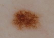
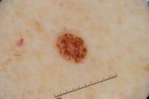
SDEF: Abtropfung, Hochsteigerung Theories of Nevus Evolution Questioned
Theories of how nevi develop have received little attention in the last 100 years, but that appears to be changing, according to Dr. Ashfaq A. Marghoob.
Dr. Marghoob, a dermatologist at Memorial Sloan-Kettering Cancer Center in New York, discussed what is currently known about nevogenesis. "Since nevi are associated with an increased risk of melanoma, understanding nevogenesis may help to unravel some of the mysteries of melanomagenesis," he noted at the Hawaii Dermatology Seminar sponsored by Skin Disease Education Foundation (SDEF).

German physician Paul Gerson Unna proposed the "Abtropfung" theory of nevogenesis in 1893. The theory holds that melanocytic nevus cells develop in the epidermis and drop off to the dermis over time. "For almost a century this theory of nevogenesis was accepted as truth and remained uncontested," Dr. Marghoob said.
During the past few decades, however, newly acquired observations from histopathology and embryogenesis have led some researchers to question the validity of the "Abtropfung" theory in favor of the "Hochsteigerung" theory.
Dr. Stewart F. Cramer, a pathologist, proposed the latter in 1984. It essentially is the reverse of the Abtropfung theory, meaning that nevus cells migrate from the dermis to the epidermis.
For example, in one histopathology study, no child younger than 10 had a purely junctional nevus, 52% had compound nevi, and 48% had dermal nevi (Am. J. Dermatopathol. 1998;20:135-9). In patients older than age 60, 12% had junctional nevi, 23% had compound nevi, and 65% had dermal nevi. Had the Abtropfung theory been correct, most childhood nevi would be junctional, and most nevi in late adult life would be intradermal. The researchers said their findings better fit the theory of upward migration of nevus cells.
In another study, researchers reviewed all biopsy reports for junctional nevi from 2001 at the Penn State Milton S. Hershey Medical Center and found that these lesions occur with similar frequency in the young and in the elderly (J. Am. Acad. Dermatol 2007;56:825-7).
"However, new insights gained from the epidemiology of nevi, cross-sectional and longitudinal study of nevi, dermoscopy and confocal microscopy investigation of nevi, as well as the cellular and molecular study of nevi, brings into question the aforementioned theories," Dr. Marghoob says.
In fact, he added, there is insufficient evidence that either theory is the norm in postnatal life. More longitudinal studies are needed to determine nevus evolution.
Dr. Marghoob reported having no relevant conflicts of interest. SDEF and this news organization are owned by Elsevier.
Theories of how nevi develop have received little attention in the last 100 years, but that appears to be changing, according to Dr. Ashfaq A. Marghoob.
Dr. Marghoob, a dermatologist at Memorial Sloan-Kettering Cancer Center in New York, discussed what is currently known about nevogenesis. "Since nevi are associated with an increased risk of melanoma, understanding nevogenesis may help to unravel some of the mysteries of melanomagenesis," he noted at the Hawaii Dermatology Seminar sponsored by Skin Disease Education Foundation (SDEF).
German physician Paul Gerson Unna proposed the "Abtropfung" theory of nevogenesis in 1893. The theory holds that melanocytic nevus cells develop in the epidermis and drop off to the dermis over time. "For almost a century this theory of nevogenesis was accepted as truth and remained uncontested," Dr. Marghoob said.
During the past few decades, however, newly acquired observations from histopathology and embryogenesis have led some researchers to question the validity of the "Abtropfung" theory in favor of the "Hochsteigerung" theory.
Dr. Stewart F. Cramer, a pathologist, proposed the latter in 1984. It essentially is the reverse of the Abtropfung theory, meaning that nevus cells migrate from the dermis to the epidermis.
For example, in one histopathology study, no child younger than 10 had a purely junctional nevus, 52% had compound nevi, and 48% had dermal nevi (Am. J. Dermatopathol. 1998;20:135-9). In patients older than age 60, 12% had junctional nevi, 23% had compound nevi, and 65% had dermal nevi. Had the Abtropfung theory been correct, most childhood nevi would be junctional, and most nevi in late adult life would be intradermal. The researchers said their findings better fit the theory of upward migration of nevus cells.
In another study, researchers reviewed all biopsy reports for junctional nevi from 2001 at the Penn State Milton S. Hershey Medical Center and found that these lesions occur with similar frequency in the young and in the elderly (J. Am. Acad. Dermatol 2007;56:825-7).
"However, new insights gained from the epidemiology of nevi, cross-sectional and longitudinal study of nevi, dermoscopy and confocal microscopy investigation of nevi, as well as the cellular and molecular study of nevi, brings into question the aforementioned theories," Dr. Marghoob says.
In fact, he added, there is insufficient evidence that either theory is the norm in postnatal life. More longitudinal studies are needed to determine nevus evolution.
Dr. Marghoob reported having no relevant conflicts of interest. SDEF and this news organization are owned by Elsevier.
EXPERT ANALYSIS FROM SDEF HAWAII DERMATOLOGY SEMINAR
New Melanoma Treatments Show Improved Survival
During the past 30 years, only two drugs – dacarbazine and interleukin-2 – have been approved by the Food and Drug Administration for treating metastatic melanoma, and while these drugs help control the disease in a small percentage of patients, they do not appear to improve survival.
However, agents introduced within the last 2 years are reported to improve survival, Dr. Richard D. Carvajal of Memorial Sloan-Kettering Cancer Center, said at the Hawaii Dermatology Seminar sponsored by Skin Disease Education Foundation (SDEF).

The first is ipilimumab (also known as MDX-010 or MDX-101), a monoclonal antibody that binds to cytotoxic T-lymphocyte-associated antigen 4 (CTLA-4). Normally, CTLA-4 slows the immune system, but this treatment allows the immune system to recognize melanoma, Dr. Carvajal said.
In one study reported last year, ipilimumab improved overall survival in patients with previously treated metastatic melanoma (N. Engl. J. Med. 2010;363;711-23). Specifically, patients who received ipilimumab plus glycoprotein 100 (gp100) had an overall survival of 10 months, while those who received gp100 alone had a survival rate of 6.4 months. The median overall survival for patients taking ipilimumab alone was 10.1 months.
The second treatment, PLX4032 (also known as RG7204 or RO5185426), works by inhibiting the mutated BRAF protein found in about half of all cases of metastatic melanoma. In January, the manufacturer, Plexxikon, announced that a phase III clinical study showed a significant survival benefit in people with previously untreated BRAF V600 mutation-positive metastatic melanoma.
Study participants who received PLX4032 lived longer (overall survival) and also lived longer without their disease getting worse (progression-free survival), compared with participants who received dacarbazine, the current standard of care, Dr. Carvajal said, adding "these are really large breakthroughs."
Dr. Carvajal disclosed that he has served as a consultant for Novartis. SDEF and this news organization are owned by Elsevier.
During the past 30 years, only two drugs – dacarbazine and interleukin-2 – have been approved by the Food and Drug Administration for treating metastatic melanoma, and while these drugs help control the disease in a small percentage of patients, they do not appear to improve survival.
However, agents introduced within the last 2 years are reported to improve survival, Dr. Richard D. Carvajal of Memorial Sloan-Kettering Cancer Center, said at the Hawaii Dermatology Seminar sponsored by Skin Disease Education Foundation (SDEF).
The first is ipilimumab (also known as MDX-010 or MDX-101), a monoclonal antibody that binds to cytotoxic T-lymphocyte-associated antigen 4 (CTLA-4). Normally, CTLA-4 slows the immune system, but this treatment allows the immune system to recognize melanoma, Dr. Carvajal said.
In one study reported last year, ipilimumab improved overall survival in patients with previously treated metastatic melanoma (N. Engl. J. Med. 2010;363;711-23). Specifically, patients who received ipilimumab plus glycoprotein 100 (gp100) had an overall survival of 10 months, while those who received gp100 alone had a survival rate of 6.4 months. The median overall survival for patients taking ipilimumab alone was 10.1 months.
The second treatment, PLX4032 (also known as RG7204 or RO5185426), works by inhibiting the mutated BRAF protein found in about half of all cases of metastatic melanoma. In January, the manufacturer, Plexxikon, announced that a phase III clinical study showed a significant survival benefit in people with previously untreated BRAF V600 mutation-positive metastatic melanoma.
Study participants who received PLX4032 lived longer (overall survival) and also lived longer without their disease getting worse (progression-free survival), compared with participants who received dacarbazine, the current standard of care, Dr. Carvajal said, adding "these are really large breakthroughs."
Dr. Carvajal disclosed that he has served as a consultant for Novartis. SDEF and this news organization are owned by Elsevier.
EXPERT ANALYSIS FROM SDEF HAWAII DERMATOLOGY SEMINAR
SDEF: New Melanoma Treatments Show Improved Survival
During the past 30 years, only two drugs – dacarbazine and interleukin-2 – have been approved by the Food and Drug Administration for treating metastatic melanoma, and while these drugs help control the disease in a small percentage of patients, they do not appear to improve survival.
However, agents introduced within the last 2 years are reported to improve survival, Dr. Richard D. Carvajal of Memorial Sloan-Kettering Cancer Center, said at the Hawaii Dermatology Seminar sponsored by Skin Disease Education Foundation (SDEF).
The first is ipilimumab (also known as MDX-010), a monoclonal antibody that binds to cytotoxic T-lymphocyte-associated antigen 4 (CTLA4). Normally, CTLA4 functions as one of several negative feedback mechanisms within the immune system. Treatment with ipilimumab takes away this negative feedback mechanism and allows the immune system to recognize and combat melanoma, Dr. Carvajal said.

In one study reported last year, ipilimumab improved overall survival in patients with previously treated metastatic melanoma (N. Engl. J. Med. 2010;363;711-23). Specifically, patients who received ipilimumab plus glycoprotein 100 (gp100) vaccine had an overall survival of 10 months, while those who received gp100 alone had a survival rate of 6.4 months. The median overall survival for patients taking ipilimumab alone was 10.1 months.
Even more striking, the 1-year overall survival rate for patients receiving ipilimumab alone or in combination with the vaccine was approximately 45%, significantly better than the 1-year overall survival rate of 25% achieved with the vaccine alone.
The second agent, PLX4032 (also known as RG7204 or RO5185426), works by inhibiting the mutated BRAF protein found in about half of all cases of metastatic melanoma. In January, the manufacturer, Plexxikon, announced that a phase III clinical study showed a significant survival benefit in people with previously untreated BRAF V600 mutation-positive metastatic melanoma.
Study participants who received PLX4032 lived longer (overall survival) and also lived longer without their disease getting worse (progression-free survival), compared with participants who received dacarbazine, the current standard of care.
"As these are the first two drugs ever demonstrated to improve survival in patients with advanced melanoma, these recent developments are extremely significant breakthroughs for the management of this disease," Dr. Carvajal said.
Dr. Carvajal disclosed that he has served as a consultant for Novartis. SDEF and this news organization are owned by Elsevier.
During the past 30 years, only two drugs – dacarbazine and interleukin-2 – have been approved by the Food and Drug Administration for treating metastatic melanoma, and while these drugs help control the disease in a small percentage of patients, they do not appear to improve survival.
However, agents introduced within the last 2 years are reported to improve survival, Dr. Richard D. Carvajal of Memorial Sloan-Kettering Cancer Center, said at the Hawaii Dermatology Seminar sponsored by Skin Disease Education Foundation (SDEF).
The first is ipilimumab (also known as MDX-010), a monoclonal antibody that binds to cytotoxic T-lymphocyte-associated antigen 4 (CTLA4). Normally, CTLA4 functions as one of several negative feedback mechanisms within the immune system. Treatment with ipilimumab takes away this negative feedback mechanism and allows the immune system to recognize and combat melanoma, Dr. Carvajal said.
In one study reported last year, ipilimumab improved overall survival in patients with previously treated metastatic melanoma (N. Engl. J. Med. 2010;363;711-23). Specifically, patients who received ipilimumab plus glycoprotein 100 (gp100) vaccine had an overall survival of 10 months, while those who received gp100 alone had a survival rate of 6.4 months. The median overall survival for patients taking ipilimumab alone was 10.1 months.
Even more striking, the 1-year overall survival rate for patients receiving ipilimumab alone or in combination with the vaccine was approximately 45%, significantly better than the 1-year overall survival rate of 25% achieved with the vaccine alone.
The second agent, PLX4032 (also known as RG7204 or RO5185426), works by inhibiting the mutated BRAF protein found in about half of all cases of metastatic melanoma. In January, the manufacturer, Plexxikon, announced that a phase III clinical study showed a significant survival benefit in people with previously untreated BRAF V600 mutation-positive metastatic melanoma.
Study participants who received PLX4032 lived longer (overall survival) and also lived longer without their disease getting worse (progression-free survival), compared with participants who received dacarbazine, the current standard of care.
"As these are the first two drugs ever demonstrated to improve survival in patients with advanced melanoma, these recent developments are extremely significant breakthroughs for the management of this disease," Dr. Carvajal said.
Dr. Carvajal disclosed that he has served as a consultant for Novartis. SDEF and this news organization are owned by Elsevier.
EXPERT ANALYSIS FROM SDEF HAWAII DERMATOLOGY SEMINAR
SDEF: Preconceived Melanoma Pathogenesis Called Into Question
Clinical observation calls into question some of the preconceived notions about melanoma pathogenesis, according to Dr. James M. Grichnik.
The traditional model is that melanomas initially develop from differentiated melanocytes in the epidermis and then invade the dermis. This is largely based on pathologic features, said Dr. Grichnik, director of the melanoma program at Sylvester Cancer Comprehensive Center, and professor of dermatology at the University of Miami. Specifically, the lowest-risk melanoma in situ tumors are thought to remain in the epidermis, while higher-risk tumors invade the deeper dermal tissues.
However, there is a newer model of melanomagenesis that is based on stem cell biology, Dr. Grichnik said at the Hawaii Dermatology Seminar sponsored by Skin Disease Education Foundation (SDEF). The model suggests that stem cells in the dermis can become mature epidermal melanocytes, and that early epigenetic or genetic alterations leading to transformation may take place in the dermis rather than in the epidermis (J. Invest. Dermatol. 2008;128:2365-80).
Also, stem cell markers CD166, CD133, and nestin are expressed at significantly higher levels in melanoma compared to banal nevi. Nestin was found to be significantly increased in metastatic melanoma, compared with primary melanoma, suggesting this subpopulation of cells may be particularly virulent (Mod. Pathol. 2007;20:102-7).
The problem with tumor stem cell components, however, is that they may not have normal antigens, and components may escape into the lymph system rather than adhere to the lymph nodes.
"So, the immune system doesn't do a good job of catching them," Dr. Grichnik said, adding that tumors contain a heterogeneous population of cells. "They're trying to differentiate towards a normal pigment of cells, the melanocytes. Some of the heterogeneous population has the antigens that the immune system destroys, but the stem cell population doesn't."
Recognition of these pathways may lead to better diagnostic and therapeutic tools. A multiple treatment approach most likely will be needed.
"One approach is to destroy proliferative cells and another is to destroy the nonproliferative tumor stem cells," Dr. Grichnik said. "We'll have to develop therapies that are specific to the tumor stem cells."
Dr. Grichnik disclosed that he is a major shareholder and founder of DigitalDerm, and serves in a consultant role for Electro-Optical Systems and Spectral Image.
SDEF and this news organization are owned by Elsevier.
EXPERT ANALYSIS FROM SDEF HAWAII DERMATOLOGY SEMINAR
Serum Test Could Define Need for SLN Biopsy in Melanoma
SAN ANTONIO – The development of a serum glycoprotein microarray has led to the discovery of four proteins for which antibodies significantly predict nodal metastases in patients with melanoma.
If validated, the data could form the basis of a blood test to select patients for sentinel lymph node biopsy, Dr. Michael Sabel said during a symposium sponsored by the Society of Surgical Oncology.
In an internal validation cohort of 79 patients with newly diagnosed melanoma, 22 patients (28%) had serum antibodies to acid ceramidase (ASAH1), cathepsin D (CTSD), or lactate dehydrogenase B (LDHB) that were significantly associated with being node negative (P value less than .0001; hazard ratio, 0.173).
"This isn't a small percentage of patients, so if validated, this test could significantly reduce the number of patients to whom we offer sentinel node biopsy," he said in an interview.
Antibodies to a fourth protein, GRP94 (also known as heat shock protein 90), were detected in nine patients (11%), and were significantly associated with being node positive (P less than .0001; HR, 3.04).
"Theoretically, this could be used in patients who are on the borderline for the procedure, where the presence of antibodies to GRP94 could prompt you to use sentinel node biopsy," Dr. Sabel said.
Surprisingly, the presence of antibodies to GRP94 did not correlate with the volume of disease burden within the sentinel node. The antibodies were equally present in patients who had microscopic disease and in those with more significant tumor burden within their sentinel node.
The bigger picture, however, is the potential for glycoprotein microarray analysis to identify predictive and prognostic biomarkers in melanoma, suggested Dr. Sabel, a surgical oncologist with the University of Michigan Health Systems in Ann Arbor.
Studies are ongoing to find other antibodies and their relevant glycoproteins that may differentiate benign from malignant skin lesions, identify patients at high risk of distant metastases, and signal response to adjuvant therapies.
"Glycoproteins have apparently important roles in melanoma progression, and also may serve as targets for therapy," he said.
The use of serum autoantibody profiling would be particularly attractive in clinical practice because it does not require primary tumor tissue. Unlike most other solid tumors, roughly 90% of a primary melanoma tumor is removed during the initial biopsy, leaving little tissue for subsequent testing once the diagnosis has been made, Dr. Sabel said.
Sentinel node biopsy with selective lymph node dissection was endorsed in the latest American Joint Commission on Cancer staging guidelines for melanoma, but it is positive in only 20% of patients with melanoma, he observed.
The researchers studied glycoproteins rather than a broad array of proteins because glycoproteins have a strong interaction with the immune system. This is particularly advantageous in melanoma because the interaction between the immune system and melanoma is well documented. In addition, glycoproteins undergo distinct changes during cancer progression, including precancerous states and posttranslational modifications that are associated with disease progression, Dr. Sabel explained.
In order to perform serum autoantibody profiling, the researchers used dual-lectin affinity and reverse-phase chromatography to separate out glycoproteins from a whole cell lysate created from a metastatic melanoma cell line. The glycoproteins were spotted on a microarray and tested using serum from 43 melanoma patients, with anti–human IgG used to detect response, Dr. Sabel said.
Wilcoxon rank-sum testing and outlier analysis identified nine fractions that significantly separated 27 node-negative patients from 16 node-positive patients. Mass spectrometry was then used to identify the relevant proteins of interest. Prevalidation testing with recombinant proteins boiled down the nine fractions to the four proteins used in the validation set of 79 patients.
Interestingly, although LDHB, CTSD, and GRP94 have strong associations with melanoma, the ASAH1 protein has been associated with breast, thyroid, and prostate cancers, but not melanoma, Dr. Sabel said.
In multivariate analysis of the validation cohort, there was no significant correlation among the four proteins or between the proteins and known melanoma prognostic factors. Only a mild, negative correlation was observed between ASAHI and increasing age, he said.
All four antibodies remained significant after adjustment for age, sex, Breslow thickness, ulceration, and mitotic rate, with a receiver operating characteristic area under the curve of 0.8690.
Dr. Sabel disclosed research funding from Merck Oncology. His coauthors report no conflicts.
FROM A SYMPOSIUM SPONSORED BY THE SOCIETY OF SURGICAL ONCOLOGY
Major Finding: A glycoprotein microarray identified four serum antibodies that were present and significantly predicted sentinel lymph node status in 39% of patients.
Data Source: Serum samples from 79 patients with newly diagnosed melanoma.
Disclosures: Dr. Sabel disclosed research funding from Merck Oncology. His coauthors report no conflicts.