User login
‘Extremely exciting’ results from phase I trial of novel cancer metabolism agent
SAN DIEGO – Five of seven evaluable patients with advanced hematologic malignancies demonstrated complete remission or complete remission with partial platelet count recovery after treatment with AG-221, an oral first-in-class drug that exploits cancer metabolism, according to preliminary findings from a phase I trial.
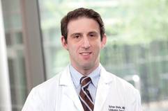
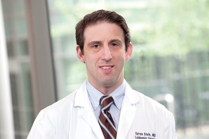
"This data is kind of unheard of," Dr. Eytan M. Stein said during a press briefing at the annual meeting of the American Association for Cancer Research. "It’s early clinical data – we’ll have to see what happens to the other patients who are on study – but I would say that this is an extremely exciting result."
A novel agent being developed by Cambridge, Mass.–based Agios Pharmaceuticals, AG-221 acts by interfering with cancer metabolism to halt tumor growth. It is an oral inhibitor of the isocitrate dehydrogenase-2 (IDH2) protein and is administered once or twice daily in a 28-day cycle.
IDH2 mutations are found in 10%-15% of acute myelogenous leukemias (AML), 5% of myelodysplastic syndromes/multiple primary neoplasms (MDS/MPN), and 25% of angioimmunoblastic non-Hodgkin lymphomas.
Mutations in the genes for the metabolic enzyme are thought to be the drivers of distinct subsets of AML, by allowing increased production of 2-hydroxyglutarate, an oncometabolite. Researchers hypothesized that blocking production of the enzyme may lead to clinical benefit in patients with these mutations.
In September 2013, Dr. Stein and his associates conducted a single-arm, phase I, open-label study of AG-221 as single-agent therapy with continuous oral daily dosing in 28-day cycles. Patients received the drug at a dose of 30 mg b.i.d., 50 mg b.i.d., 75 mg b.i.d., or 100 mg b.i.d. The median age of the population was 62.5 years, and participants included those with relapsed or refractory AML and MDS, or patients over age 60 who were unable to be treated with conventional therapy because of comorbid medical conditions.
"They had to be IDH2 mutation positive to get into the trial," Dr. Stein explained. "The key objectives were to assess the safety and tolerability; determine maximum tolerated dose and recommended phase II dose; determine dose-limiting toxicity, pharmacokinetics, and pharmacodynamics; and characterize differentiation effect and clinical activity."
As of March 20, 2014, there were 22 patients enrolled and 16 patients remain on study. Dr. Stein reported that there have been two possible treatment-related serious adverse events to date: one grade 2 hyperleukocytosis and differentiation syndrome, and one case of grade 3 confusion in the setting of respiratory failure in a patient with sepsis. There have been four deaths within 30 days of study drug termination, all stemming from complications of disease-related sepsis. "This is not unusual for patients with refractory AML, who are often very ill," Dr. Stein said.
The researchers observed a greater than 90% plasma 2-hydroxyglutarate reduction after multiple doses, providing proof of concept for the drug mechanism. In the 30-mg b.i.d. cohort, there was one complete remission and one complete remission with incomplete platelet recovery. In the 50-mg b.i.d. cohort, there were two complete remissions and one complete remission with an incomplete platelet count recovery, one partial remission, and one patient with progressive disease.
"Overall, this shows that out of the seven patients who were evaluable for efficacy, five of the patients achieved complete remission or complete remission with partial platelet count recovery," Dr. Stein said. "What that means is that there is no more leukemia in the bone marrow; their platelet count just has not risen to above 100,000/mcL."
Preliminary analysis of pharmacokinetics at the 30- and 50-mg dose levels showed a mean plasma half-life of greater than 40 hours. Moving forward, dose escalation will continue, he said, as the maximum tolerated dose has not yet been realized. Expansion cohorts are scheduled to begin in late 2014.
"These data provide early validation of mutant IDH-2 as a therapeutic target in AML and MDS," Dr. Stein said. "We’re going to be further characterizing the safety, pharmacokinetics/pharmacodynamics, and response rate of AG-221 in AML. Down the road, we’re hoping to evaluate this agent in combinations and in earlier lines to treatment. We’re also going to be exploring the activity of AG-221 in other IDH2-mutation-positive hematologic malignancies and solid tumors."
In an interview, Dr. Patricia M. LoRusso, director of the Center for Translational Therapeutics at Wayne State University’s Karmanos Cancer Institute, Detroit, said that she was impressed by the study’s findings in the context of refractory AML. "To be able to get responses with a drug that’s not nearly as toxic as chemotherapy, that is much more tolerable than chemotherapy, is exciting," she said.
"We don’t have long enough follow-up to know what those complete responses mean in terms of long-term survival. It is a phase I trial, but to see these kinds of responses in an AML refractory population with a selective drug against a mutated target, it’s a sign that there are some pretty exciting things happening in oncology these days."
Agios Pharmaceuticals funded the study. Dr. Stein disclosed that he has been a consultant for Janssen.
Agios Pharmaceuticals, halt tumor growth, oral inhibitor, isocitrate dehydrogenase-2 (IDH2) protein,
SAN DIEGO – Five of seven evaluable patients with advanced hematologic malignancies demonstrated complete remission or complete remission with partial platelet count recovery after treatment with AG-221, an oral first-in-class drug that exploits cancer metabolism, according to preliminary findings from a phase I trial.
"This data is kind of unheard of," Dr. Eytan M. Stein said during a press briefing at the annual meeting of the American Association for Cancer Research. "It’s early clinical data – we’ll have to see what happens to the other patients who are on study – but I would say that this is an extremely exciting result."
A novel agent being developed by Cambridge, Mass.–based Agios Pharmaceuticals, AG-221 acts by interfering with cancer metabolism to halt tumor growth. It is an oral inhibitor of the isocitrate dehydrogenase-2 (IDH2) protein and is administered once or twice daily in a 28-day cycle.
IDH2 mutations are found in 10%-15% of acute myelogenous leukemias (AML), 5% of myelodysplastic syndromes/multiple primary neoplasms (MDS/MPN), and 25% of angioimmunoblastic non-Hodgkin lymphomas.
Mutations in the genes for the metabolic enzyme are thought to be the drivers of distinct subsets of AML, by allowing increased production of 2-hydroxyglutarate, an oncometabolite. Researchers hypothesized that blocking production of the enzyme may lead to clinical benefit in patients with these mutations.
In September 2013, Dr. Stein and his associates conducted a single-arm, phase I, open-label study of AG-221 as single-agent therapy with continuous oral daily dosing in 28-day cycles. Patients received the drug at a dose of 30 mg b.i.d., 50 mg b.i.d., 75 mg b.i.d., or 100 mg b.i.d. The median age of the population was 62.5 years, and participants included those with relapsed or refractory AML and MDS, or patients over age 60 who were unable to be treated with conventional therapy because of comorbid medical conditions.
"They had to be IDH2 mutation positive to get into the trial," Dr. Stein explained. "The key objectives were to assess the safety and tolerability; determine maximum tolerated dose and recommended phase II dose; determine dose-limiting toxicity, pharmacokinetics, and pharmacodynamics; and characterize differentiation effect and clinical activity."
As of March 20, 2014, there were 22 patients enrolled and 16 patients remain on study. Dr. Stein reported that there have been two possible treatment-related serious adverse events to date: one grade 2 hyperleukocytosis and differentiation syndrome, and one case of grade 3 confusion in the setting of respiratory failure in a patient with sepsis. There have been four deaths within 30 days of study drug termination, all stemming from complications of disease-related sepsis. "This is not unusual for patients with refractory AML, who are often very ill," Dr. Stein said.
The researchers observed a greater than 90% plasma 2-hydroxyglutarate reduction after multiple doses, providing proof of concept for the drug mechanism. In the 30-mg b.i.d. cohort, there was one complete remission and one complete remission with incomplete platelet recovery. In the 50-mg b.i.d. cohort, there were two complete remissions and one complete remission with an incomplete platelet count recovery, one partial remission, and one patient with progressive disease.
"Overall, this shows that out of the seven patients who were evaluable for efficacy, five of the patients achieved complete remission or complete remission with partial platelet count recovery," Dr. Stein said. "What that means is that there is no more leukemia in the bone marrow; their platelet count just has not risen to above 100,000/mcL."
Preliminary analysis of pharmacokinetics at the 30- and 50-mg dose levels showed a mean plasma half-life of greater than 40 hours. Moving forward, dose escalation will continue, he said, as the maximum tolerated dose has not yet been realized. Expansion cohorts are scheduled to begin in late 2014.
"These data provide early validation of mutant IDH-2 as a therapeutic target in AML and MDS," Dr. Stein said. "We’re going to be further characterizing the safety, pharmacokinetics/pharmacodynamics, and response rate of AG-221 in AML. Down the road, we’re hoping to evaluate this agent in combinations and in earlier lines to treatment. We’re also going to be exploring the activity of AG-221 in other IDH2-mutation-positive hematologic malignancies and solid tumors."
In an interview, Dr. Patricia M. LoRusso, director of the Center for Translational Therapeutics at Wayne State University’s Karmanos Cancer Institute, Detroit, said that she was impressed by the study’s findings in the context of refractory AML. "To be able to get responses with a drug that’s not nearly as toxic as chemotherapy, that is much more tolerable than chemotherapy, is exciting," she said.
"We don’t have long enough follow-up to know what those complete responses mean in terms of long-term survival. It is a phase I trial, but to see these kinds of responses in an AML refractory population with a selective drug against a mutated target, it’s a sign that there are some pretty exciting things happening in oncology these days."
Agios Pharmaceuticals funded the study. Dr. Stein disclosed that he has been a consultant for Janssen.
Agios Pharmaceuticals, halt tumor growth, oral inhibitor, isocitrate dehydrogenase-2 (IDH2) protein,
Agios Pharmaceuticals, halt tumor growth, oral inhibitor, isocitrate dehydrogenase-2 (IDH2) protein,
AT THE AACR ANNUAL MEETING
Major finding: Among seven patients evaluable for efficacy, five achieved complete remission or complete remission with partial platelet count recovery after treatment.
Data source: Preliminary results from a phase I trial of the novel agent AG-221 in patients with advanced hematologic malignancies with an isocitrate dehydrogenase-2 mutation.
Disclosures: Agios Pharmaceuticals funded the study. Dr. Stein disclosed that he has been a consultant for Janssen.
Compound is potent FLT3 inhibitor, team says

Credit: Rhoda Baer
An experimental compound called TTT-3002 could be “one of the most potent FLT3 inhibitors to date,” according to researchers.
In preclinical experiments, TTT-3002 proved more active than the most potent FLT3 inhibitor currently in clinical trials.
TTT-3002 blocked FLT3 activity in human FLT3/ITD mutant leukemia cell lines, prolonged survival in a mouse model of FLT3-associated acute myeloid leukemia (AML), and proved toxic to leukemic cells from patients with AML.
Donald Small, MD, PhD, of Johns Hopkins University School of Medicine in Baltimore, and his colleagues reported these results in Blood.
“We’re very excited about TTT-3002, because it appears in our tests so far to be the most potent FLT3 inhibitor to date,” Dr Small said. “It showed activity against FLT3-mutated cells taken from patients and with minimal toxicity to normal bone marrow cells, making it a promising new candidate for the treatment of AML.”
In a series of experiments, the researchers found that the amount of TTT-3002 needed to block FLT3 activity in human leukemia cell lines was 6- to 7-fold lower than for ACC220, the most potent FLT3 inhibitor currently in clinical trials.
TTT-3002 also inhibited proteins made by genes further down the FLT3 signaling pathway, including STAT5, AKT, and MAPK. And it showed activity against the most frequently occurring FLT3 mutations, FLT3/ITD and FLT3/D835Y. (Many other inhibitors are ineffective against FLT3/D835Y mutations.)
When the researchers tested TTT-3002 in a mouse model of leukemia, they found the drug eliminated the presence of leukemic cells within 10 days of treatment.
Mice lived an average of more than 100 days after TTT-3002 treatment and resumed normal bone marrow activity. In comparison, mice treated with a placebo died an average of 18 days after treatment.
The researchers also found that TTT-3002 was toxic to leukemia samples taken from newly diagnosed and relapsed patients with AML, but it did not affect normal bone marrow cells taken from healthy donors.
A single dose of TTT-3002 caused more than 90% inhibition against FLT3 signaling that lasted for 12 hours. ![]()
How NK cells kill abnormal blood cells

Credit: Bjorn Onfelt/Dan Davis
New research provides additional insight into how natural killer (NK) cells eliminate abnormal hematopoietic cells.
The investigators evaluated 2 molecules that are known to play important roles in this process.
Ewing’s sarcoma-associated transcript 2 (EAT-2) and signaling lymphocytic activation molecule (SLAM)–associated protein (SAP) are expressed in NK cells, and their combined expression is essential for NK cells to kill abnormal hematopoietic cells.
“We knew that EAT-2 cooperates with SAP, and, with this research project, we wanted to better understand why they are both required for the proper functioning of NK cells,” said study author André Veillette, PhD, of the Institut de Recherches Cliniques de Montréal (IRCM) in Canada.
Dr Veillette and his colleagues described this research in the Journal of Experimental Medicine.
“We identified the molecular chain of events that occur and showed that EAT-2 and SAP perform different functions using distinct mechanisms,” Dr Veillette said. “These findings explain the cooperative and essential function of these 2 molecules in activating NK cells, thereby allowing them to kill abnormal blood cells.”
The investigators noted that SAP couples SLAM family receptors to the protein tyrosine kinase Fyn and the exchange factor Vav, thereby promoting conjugate formation between NK cells and target hematopoietic cells.
EAT-2, on the other hand, works by accelerating the polarization and exocytosis of cytotoxic granules toward hematopoietic cells.
EAT-2 mediates its effects in NK cells by linking SLAM family receptors to phospholipase Cγ, calcium fluxes, and Erk kinase. These signals are triggered by 1 or 2 tyrosines that are located in the carboxyl-terminal tail of EAT-2.
Dr Veillete pointed out that, although EAT-2 and SAP behave differently, both are linked to receptors of the SLAM family on the cell surface.
“Because they can make better drug targets, our future work will focus on these receptors,” he said, “which could eventually lead to identifying new potential treatment avenues for blood cancers such as leukemia and lymphoma.” ![]()
Enrollment stalled for CAR T-cell study
Update: The hold on this trial has been lifted. Click here for additional details.
Memorial Sloan-Kettering Cancer Center has temporarily suspended enrollment in a study of chimeric antigen receptor (CAR) T-cell therapy, due to 2 patient deaths.
The study is an evaluation of CD19-targeted CAR T cells in patients with B-cell acute lymphoblastic leukemia (ALL).
Of the 22 patients enrolled on the study to date, 10 have died. But only 2 of these deaths gave researchers pause and made them question enrollment criteria.
Six deaths were a result of disease relapse or progression, and 2 patients died of complications from stem cell transplant.
The 2 deaths that prompted the suspension of enrollment occurred within 2 weeks of the patients receiving CAR T cells.
“The first of these patients had a prior history of cardiac disease, while the second patient died due to complications associated with persistent seizure activity,” said Renier Brentjens, MD, PhD, of Memorial Sloan-Kettering in New York.
“As a matter of routine review at Sloan-Kettering for adverse events on-study, our center made the decision to pause enrollment and review these 2 patients in greater detail.”
“And as a consequence of this review, we’ve amended the enrollment criteria in regards to comorbidities, thereby excluding patients with cardiac disease, and adjusted the T-cell dose based on the extent of disease, [in the] hope that this modification will reduce the cytokine release syndrome that these patients with morphological disease have experienced.”
The researchers expect the trial to resume enrollment soon.
Some results from this study were recently published in Science Translational Medicine, and Dr Brentjens presented the latest results at the AACR Annual Meeting 2014 in San Diego (abstract CT102*).
Thus far, the researchers have enrolled 22 adult patients who had relapsed or refractory B-ALL, were minimal residual disease-positive, or were in the first complete remission (CR1) at enrollment. Patients in CR1 were monitored and only received CAR T cells if they relapsed.
The remaining patients received re-induction chemotherapy (physician’s choice), followed by CAR T-cell infusion. After treatment, the options were allogeneic transplant, a different salvage therapy, or monitoring.
In all, 20 patients received a CAR T-cell dose of 3 x 10⁶ T cells/kg. Eighty-two percent of patients initially achieved a CR, and 72% had a morphologic CR. The average time to CR was about 24.5 days.
Twelve of the responders were eligible for transplant. Of the 8 patients who ultimately underwent transplant and survived, 1 relapsed, but the rest remain in remission.
Dr Brentjens noted that some patients developed cytokine release syndrome, and this was related to the amount of disease present at the time of CAR T-cell infusion.
“Those patients that had only minimal residual disease at the time of CAR T-cell infusion . . . less than 5% blasts, generally had either no fever or very transient, low-grade fever,” he said.
“In contrast, all those patients that had morphologic residual disease at the time of CAR T-cell infusion demonstrated a high, persistent spike in fevers . . . , became hypotensive, and required transfer—for additional, closer monitoring—to our ICU.”
The researchers initially treated these patients with high-dose steroids, which reduced cytokine levels in the serum and ameliorated fevers. But it also rapidly reduced T-cell populations to undetectable levels.
Fortunately, another group of researchers subsequently discovered that the monoclonal antibody tocilizumab can treat cytokine release syndrome without inducing this side effect. So Dr Brentjens and his colleagues began using this drug and found it both safe and effective. ![]()
*Information in the abstract differs from that presented at the meeting.
How autophagy helps cancer cells evade death

Credit: Sarah Pfau
SAN DIEGO—New research suggests that autophagy may allow cancer cells to recover and divide, rather than die, when faced with chemotherapy.
“What we showed is that if this mechanism doesn’t work right—for example, if autophagy is too high or if the target regulated by autophagy isn’t around—cancer cells may be able to rescue themselves from death caused by chemotherapies,” said study author Andrew Thorburn, PhD, of the University of Colorado Denver.
He and his colleagues believe this finding has important implications. It demonstrates a mechanism whereby autophagy controls cell death, and it further reinforces the clinical potential of inhibiting autophagy to sensitize cancer cells to chemotherapy.
Dr Thorburn and his colleagues recounted their research in Cell Reports and presented it in an education session at the AACR Annual Meeting 2014.
The researchers had set out to examine how autophagy affects canonical death receptor-induced mitochondrial outer membrane permeabilization (MOMP) and apoptosis. They found that MOMP occurs at variable times in cells, and it’s delayed by autophagy.
Furthermore, autophagy leads to inefficient MOMP. This causes some cells to die via a slower process than typical apoptosis, which allows them to eventually recover and divide.
Specifically, the researchers found that, as a cancer cell begins to die, mitochondrial cell walls break down. And the cell’s mitochondria release proteins via MOMP.
But then, high autophagy allows the cell to encapsulate and “digest” these released proteins before MOMP can keep the cell well and truly dead. The cell recovers and goes on to divide.
“The implication here is that if you inhibit autophagy, you’d make this less likely to happen; ie, when you kill cancer cells, they would stay dead,” Dr Thorburn said.
He and his colleagues also found that autophagy depends on the target PUMA to regulate cell death. When PUMA is absent, it doesn’t matter if autophagy is inhibited. Without the communicating action of PUMA, cancer cells evade apoptosis and continue to survive.
The researchers said this suggests autophagy can control apoptosis via a regulator that makes MOMP faster and more efficient, thus ensuring the rapid completion of apoptosis.
“Autophagy is complex and, as yet, not fully understood,” Dr Thorburn said. “But now that we see a molecular mechanism whereby cell fate can be determined by autophagy, we hope to discover patient populations that could benefit from drugs that inhibit this action.” ![]()
Nanocapsules prevent release of nontargeted radiation

Credit: Rhoda Baer
A novel type of nanocapsule can safely and effectively store isotopes that emit ionizing radiation, according to a paper published in Biochimica et Biophysica Acta.
In experiments, the nanocapsules were taken up by cells, accumulated near the perinuclear region, and persisted without degrading.
They also prevented nontargeted, radioactive daughter ions from escaping. These ions could cause significant damage if released, such as prompting the development of leukemia.
This research suggests the nanocapsules have the potential to advance radiation therapy, according to study author John M. Tomich, PhD, of Kansas State University’s Johnson Cancer Research Center.
Dr Tomich and his colleagues created the nanocapsules, called branched amphiphilic peptide capsules (BAPCs), by combining 2 related sequences of amino acids—bis(FLIVI)-K-KKKK and bis(FLIVIGSII)-K-KKKK.
“We found that the 2 sequences come together to form a thin membrane that assembled into little spheres, which we call capsules,” Dr Tomich said. “While other vesicles have been created from lipids, most are much less stable and break down. Ours are like stones, though. They’re incredibly stable and are not destroyed by cells in the body.”
The capsules’ ability to stay intact with the isotope inside and remain undetected by the body’s clearance systems prompted Dr Tomich to investigate using BAPCs for radiation therapies.
“The problem with current alpha-particle radiation therapies used to treat cancer is that they lead to the release of nontargeted, radioactive daughter ions into the body,” Dr Tomich said. “Radioactive atoms break down to form new atoms, called daughter ions, with the release of some form of energy or energetic particles. Alpha emitters give off an energetic particle that comes off at nearly the speed of light.”
The alpha particle destroys DNA and whatever vital cellular components are in its path. Similarly, the daughter ions recoil with high energy on ejection of the alpha particle. The daughter ions have enough energy to escape the targeting and containment molecules that are currently in use.
“Once freed, the daughter isotopes can end up in places you don’t want them, like bone marrow, which can then lead to leukemia and new challenges,” Dr Tomich said.
To see if the BAPCs could prevent the release of daughter isotopes, the researchers loaded the nanoparticles with 225Actinium. Upon decay, this compound releases 4 alpha particles and numerous daughter ions.
The team found that BAPCs loaded with the compound readily entered cells and migrated to a position alongside the nucleus.
As the alpha-particle-emitting isotopes decayed, the recoiled daughter ions collided with the capsule walls, essentially bouncing off them, and remained trapped inside the BAPCs. This completely blocked the release of the daughter ions, which prevented uptake in nontarget tissues.
Dr Tomich said more studies are needed to add target molecules to the surface of the BAPCs. But he believes the particles could provide a safer option for treating tumors with radiation therapy by reducing the amount of radioisotope required for killing cancer cells and reducing the side effects caused by off-target accumulation of radioisotopes.
“These capsules are easy to make and easy to work with,” Dr Tomich said. “I think we’re just scratching the surface of what we can do with them to improve human health and nanomaterials.” ![]()
Product gets orphan designation for AML

The US Food and Drug Administration (FDA) has granted orphan designation for eryaspase to treat acute myeloid leukemia (AML).
Eryaspase, also known as ERY-ASP or GRASPA, is L-asparaginase encapsulated in red blood cells.
These donor-derived, enzyme-loaded red blood cells function as bioreactors to eliminate circulating asparagine and “starve”
leukemic cells, thereby inducing their death.
Research has suggested this delivery system provides improved pharmacodynamics. It protects L-aspariginase from circulating proteolytic enzymes and prevents early liver or renal clearance.
The system also appears to reduce the risk of adverse events.
Eryaspase is currently under investigation in a phase 3 trial for acute lymphoblastic leukemia (ALL) and a phase 2b trial for AML in Europe. A phase 1 study in adult ALL is being launched in the US.
Eryaspase now has orphan designation for ALL, AML and pancreatic cancer, both in Europe and the US.
In the US, orphan designation is generally granted for drugs or biologics intended to treat disorders of high unmet medical need that affect fewer than 200,000 people.
This designation conveys special incentives to the product’s sponsor, including 7 years of US market exclusivity for the drug or biologic upon FDA approval, a prescription drug user fee waiver, and certain tax credits. ![]()
Study reveals events leading to ribosomopathies

yeast buds before dividing
Credit: Carolyn Larabell
Research conducted in yeast suggests ribosomopathies are caused by a sequence of mistakes at the molecular level.
First, a genetic mutation prompts the production of defective ribosomes.
Then, a quality-control system eliminates most of these faulty ribosomes. This leaves few available for cells to produce required proteins, which causes anemia and bone marrow failure.
Next, a second mutation suppresses the quality-control system, making more ribosomes available to cells. However, these ribosomes are defective and cause changes in gene expression patterns that can result in cancer.
Jonathan Dinman, PhD, of the University of Maryland, and his colleagues described this chain of events in Proceedings of the National Academy of Sciences.
The researchers set out to investigate the structural, biochemical, and other defects in ribosomes that may lead to cancer. They selected budding yeast as their model system, as the assembly of its ribosomes shares many characteristics with human cells.
The team used the rpL10-R98S (uL16-R98S) mutant yeast model of the most commonly identified ribosomal mutation in T-cell acute lymphoblastic leukemia (T-ALL). They showed that the rpl10-R98S mutation causes a late-stage 60S subunit maturation failure that targets mutant ribosomes for degradation (the quality-control system).
When the researchers grew the mutant yeast cells on a petri dish, the cells grew very slowly. The team suggested that, because of the cells’ quality-control system, the majority of defective ribosomes carrying the T-ALL mutation do not “pass inspection.”
This severely limits the supply of ribosomes available to produce proteins, only providing enough ribosomes for cells to barely survive. This supply-and-demand problem hits rapidly dividing cells like blood cells particularly hard, and can therefore cause anemia and bone marrow failure in humans.
The bone marrow cells are subjected to selective pressure, an evolutionary process that favors the reproduction of things that resolve problems limiting their ability to thrive. In this case, cells would be favored that could circumvent the rpl10-R98S mutation.
After a few weeks, a group of fast-growing cells appeared on the petri dish containing the rpl10-R98S mutant yeast cells. The researchers sequenced the genomes of these cells and found a mutation in a second gene, NMD3, which suppresses the growth and ribosome biogenesis defects of rpl10-R98S cells.
So the mutation, NMD3-Y379D, increased the total number of ribosomes available to the cells, enabling cells with the mutation to make more protein, grow quickly, and take over the population. However, the available ribosomes were still defective.
NMD3-Y379D did not suppress the structural, biochemical, and translational fidelity defects of rpL10-R98S ribosomes. And the translational defects affected telomere maintenance. The mutant cells exhibited shortened telomeres, which have been linked to cancer.
The researchers proposed 2 different, but not mutually exclusive, explanations for these effects. The rpL10-R98S ribosomes could be directly changing patterns of gene expression and promoting T-ALL, and/or NMD3-Y379D could be driving T-ALL.
“Our yeast work has established a new paradigm that we are now translating to humans,” Dr Dinman said. “Once we determine which ribosomal mutations suppress the quality-control system in humans, we may be able to identify a potential drug target.” ![]()
Testing reveals abnormalities in CN-AML/MDS

Credit: NIGMS
NASHVILLE—New research suggests we may need to use more sensitive methods to analyze patients with cytogenetically normal acute myeloid leukemia or myelodysplastic syndrome (CN-AML/MDS).
Using “highly sensitive” microarray technology, researchers found a distinct pattern of genetic abnormalities in 22 patients diagnosed with CN-AML/MDS.
The team identified 3 overlapping regions of homozygosity in 3 genes, 2 of which are known to be involved in carcinogenesis.
This suggests that using karyotyping or FISH, or simply looking for known mutations, is not sufficient for evaluating patients with CN-AML/MDS, according to Ravindra Kolhe, MD, PhD, of the Medical College of Georgia at Georgia Regents University.
“The technology we currently use can’t identify specifically what’s wrong,” Dr Kolhe said. “We have to use more sensitive tests to give patients the proper answer.”
Dr Kolhe presented this finding, and the research to support it, at the American College of Medical Genetics and Genomics Annual Clinical Genetics Meeting.
He and his colleagues analyzed 22 patients. Seventeen had AML, and 5 had MDS, including 1 with refractory anemia with excess blasts-2. All patients had normal karyotype and FISH and had greater than 20% blasts in the bone marrow.
The researchers analyzed samples from these patients using a high-resolution, single-nucleotide polymorphism (SNP) microarray called CytoScanHD.
According to the company that markets this technology (Affymetrix, Inc.), the assay includes 750,000 SNPs with over 99% accuracy to detect accurate breakpoint estimation, loss of heterozygosity determination, regions identical-by-descent, maternal contamination, and low-level mosaicism.
For Dr Kolhe and his colleagues, the assay revealed small, previously undetectable changes in patients thought to be cytogenetically normal.
Specifically, the researchers identified 3 overlapping regions of homozygosity in all 22 cases—chromosome 1p34.3, chromosome 1p32.3, and chromosome 16q22.1 in the SFPQ, EPS15, and CTCF genes, respectively.
SFPQ and CTCF are already known to be involved in carcinogenesis, and Dr Kolhe and his colleagues are now investigating the role of EPS15 in leukemogenesis.
The researchers also identified additional abnormalities and are investigating these as well. They are sequencing the genes to identify homozygous or compound heterozygous mutations, performing expression studies to confirm that these mutations are leukemic, and conducting experiments in knockout mice to demonstrate that these genes produce the same leukemia phenotype.
The materials and reagents for this study were provided by Affymetrix. The test design, experimentation, data collection, analysis, and interpretation were done independently by the researchers. ![]()
Credit: NIGMS
NASHVILLE—New research suggests we may need to use more sensitive methods to analyze patients with cytogenetically normal acute myeloid leukemia or myelodysplastic syndrome (CN-AML/MDS).
Using “highly sensitive” microarray technology, researchers found a distinct pattern of genetic abnormalities in 22 patients diagnosed with CN-AML/MDS.
The team identified 3 overlapping regions of homozygosity in 3 genes, 2 of which are known to be involved in carcinogenesis.
This suggests that using karyotyping or FISH, or simply looking for known mutations, is not sufficient for evaluating patients with CN-AML/MDS, according to Ravindra Kolhe, MD, PhD, of the Medical College of Georgia at Georgia Regents University.
“The technology we currently use can’t identify specifically what’s wrong,” Dr Kolhe said. “We have to use more sensitive tests to give patients the proper answer.”
Dr Kolhe presented this finding, and the research to support it, at the American College of Medical Genetics and Genomics Annual Clinical Genetics Meeting.
He and his colleagues analyzed 22 patients. Seventeen had AML, and 5 had MDS, including 1 with refractory anemia with excess blasts-2. All patients had normal karyotype and FISH and had greater than 20% blasts in the bone marrow.
The researchers analyzed samples from these patients using a high-resolution, single-nucleotide polymorphism (SNP) microarray called CytoScanHD.
According to the company that markets this technology (Affymetrix, Inc.), the assay includes 750,000 SNPs with over 99% accuracy to detect accurate breakpoint estimation, loss of heterozygosity determination, regions identical-by-descent, maternal contamination, and low-level mosaicism.
For Dr Kolhe and his colleagues, the assay revealed small, previously undetectable changes in patients thought to be cytogenetically normal.
Specifically, the researchers identified 3 overlapping regions of homozygosity in all 22 cases—chromosome 1p34.3, chromosome 1p32.3, and chromosome 16q22.1 in the SFPQ, EPS15, and CTCF genes, respectively.
SFPQ and CTCF are already known to be involved in carcinogenesis, and Dr Kolhe and his colleagues are now investigating the role of EPS15 in leukemogenesis.
The researchers also identified additional abnormalities and are investigating these as well. They are sequencing the genes to identify homozygous or compound heterozygous mutations, performing expression studies to confirm that these mutations are leukemic, and conducting experiments in knockout mice to demonstrate that these genes produce the same leukemia phenotype.
The materials and reagents for this study were provided by Affymetrix. The test design, experimentation, data collection, analysis, and interpretation were done independently by the researchers.
Adult dosing regimen of pegaspargase achieves high remission rates
Researchers have identified an optimal pegaspargase dosing regimen for the treatment of adult acute lymphoblastic leukemia using up to six intravenous 2,000 IU/m2-doses given at 4-week intervals and synchronized with other chemotherapy drugs to avoid overlapping toxicities.
Of 51 adults with newly diagnosed acute lymphoblastic leukemia who were treated with some or all of six doses, 49 (96%) achieved remission – most within 4 weeks – and the 7-year disease-free and overall survival were 58% and 51% respectively, according to a study published in the March 20 issue of the Journal of Clinical Oncology (doi:10.1200/JCO.2013.50.2708).
Dr. Dan Douer of Memorial Sloan-Kettering Cancer Center, New York, and his colleagues reported that there were no pegaspargase-related deaths, with the most common grade three or four toxicities being hyperbilirubinemia (31.3%), transaminitis (62.7%), hyperglycemia (33.3%), and hypertriglyceridemia (17.6%), and the most serious being pancreatitis (13.7%) and anaphylaxis (5.9%).
Pegaspargase is standard in the treatment of pediatric acute lymphoblastic leukemia but is used sparingly in adults because of its potential higher toxicity, however in this study, the drug was discontinued in only 20% of patients after serious pegaspargase-related toxicity.
Dr. Douer declared honoraria and research funding from Sigma Tau Pharmaceuticals, which supported the study with a research grant.
FROM THE JOURNAL OF CLINICAL ONCOLOGY
Major finding: A dosing regimen of up to six intravenous 2,000 IU/m2-doses, given at 4-week intervals, achieved a 96% remission rate in adults newly diagnosed with acute lymphoblastic leukemia.
Data source: Prospective study of 51 adults.
Disclosures: Dr. Douer declared honoraria and research funding from Sigma Tau Pharmaceuticals, which supported the study with a research grant.