User login
Genetic mutation identifies favorable prognosis MDS
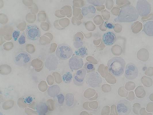
Among patients with refractory anemia with ring sideroblasts, the presence of a common mutation in SF3B1 appears to be a marker for an indolent clinical course and favorable outcome compared to patients with wild-type SF3B1, European investigators reported.
The gene SF3B1, which encodes for a splicing factor subunit, is frequently mutated in cases of chronic lymphocytic leukemia and myelodysplastic syndromes.
“SF3B1 mutation is a major determinant of disease phenotype and clinical outcome in MDS [myelodysplastic syndrome] with ring sideroblasts. SF3B1-mutated MDS is characterized by homogeneous hematologic features, favorable prognosis, and restricted patterns of co-mutated genes and clonal evolution. Overall, these results strongly support the recognition of MDS associated with SF3B1 mutation as a distinct MDS subtype. Conversely, SF3B1-negative MDS with ring sideroblasts represents a subset with a high prevalence of TP53 mutations and worse outcome that should be taken into consideration in clinical decision-making,” the study authors conclude.

Dr. Luca Malcovati and his colleagues from the University of Pavia, Italy, and other European centers, conducted a mutational analysis of 293 patients with myeloid neoplasms and 1% or more ring sideroblasts. They found somatic mutations in SF3B1 in 129 of 159 patients with refractory anemia with ring sideroblasts (RARS) or refractory cytopenia with multilineage dysplasia and ring sideroblasts (RCMD-RS). In contrast, there was a significantly lower prevalence of SF3B1 mutations among 50 patients with myelodysplastic/myeloproliferative neoplasm (MDS/MPN), and among 84 additional patients with other myeloid diseases under the World Health Organization classification of disorders of hematopoietic and lymphoid tissues (P < .001).
In multivariable analyses controlling for demographic and disease-related factors, patients with SF3B1 mutations had significantly better overall survival (hazard ratio, 0.37; P = .003), as well as a lower cumulative incidence of disease progression (HR, 0.31; P = .018), compared with patients with wild-type SF3B1 (Blood 2015;126[2]:233-41).
Mutations in SF3B1 were predictive of better outcomes among patients with RARS, RCMD-RS, and in patients with MDS without excess blasts.
When they looked at other mutations, the investigators found that in patients with SF3B1 mutations, the mutations in DNA methylation genes were associated with the presence of multilineage dysplasia, but this association had no significant effect on clinical outcomes.
Among patients with wild-type SB3B1, mutations in TP53 were frequently seen, and these mutations were associated with poor outcomes.
Gene sequencing efforts in myeloid malignancies have largely charted the mutational “landscape.” This map allows us to (1) have some idea of the fundamental biology underlying the disease, (2) define potential drug targets, and (3) refine outcome expectations, especially when there are no “knockout” therapies (as in chronic myeloid leukemia). The consequence is also the further subclassification of myeloid malignancies, thus making relatively rare diseases into extremely rare ones. One obvious challenge is to cleverly design clinical studies given the myriad subcategories of disease. The higher bar is understanding the biology of how the various mutations and pathways merge to cause disease. The work Malcovati et al., along with the other fine studies noted above, gets us one step farther down the road to cures.
Dr. Jerald Radich of Fred Hutchinson Cancer Research Center, Seattle, made his comment in an accompanying editorial.
Among patients with refractory anemia with ring sideroblasts, the presence of a common mutation in SF3B1 appears to be a marker for an indolent clinical course and favorable outcome compared to patients with wild-type SF3B1, European investigators reported.
The gene SF3B1, which encodes for a splicing factor subunit, is frequently mutated in cases of chronic lymphocytic leukemia and myelodysplastic syndromes.
“SF3B1 mutation is a major determinant of disease phenotype and clinical outcome in MDS [myelodysplastic syndrome] with ring sideroblasts. SF3B1-mutated MDS is characterized by homogeneous hematologic features, favorable prognosis, and restricted patterns of co-mutated genes and clonal evolution. Overall, these results strongly support the recognition of MDS associated with SF3B1 mutation as a distinct MDS subtype. Conversely, SF3B1-negative MDS with ring sideroblasts represents a subset with a high prevalence of TP53 mutations and worse outcome that should be taken into consideration in clinical decision-making,” the study authors conclude.
Dr. Luca Malcovati and his colleagues from the University of Pavia, Italy, and other European centers, conducted a mutational analysis of 293 patients with myeloid neoplasms and 1% or more ring sideroblasts. They found somatic mutations in SF3B1 in 129 of 159 patients with refractory anemia with ring sideroblasts (RARS) or refractory cytopenia with multilineage dysplasia and ring sideroblasts (RCMD-RS). In contrast, there was a significantly lower prevalence of SF3B1 mutations among 50 patients with myelodysplastic/myeloproliferative neoplasm (MDS/MPN), and among 84 additional patients with other myeloid diseases under the World Health Organization classification of disorders of hematopoietic and lymphoid tissues (P < .001).
In multivariable analyses controlling for demographic and disease-related factors, patients with SF3B1 mutations had significantly better overall survival (hazard ratio, 0.37; P = .003), as well as a lower cumulative incidence of disease progression (HR, 0.31; P = .018), compared with patients with wild-type SF3B1 (Blood 2015;126[2]:233-41).
Mutations in SF3B1 were predictive of better outcomes among patients with RARS, RCMD-RS, and in patients with MDS without excess blasts.
When they looked at other mutations, the investigators found that in patients with SF3B1 mutations, the mutations in DNA methylation genes were associated with the presence of multilineage dysplasia, but this association had no significant effect on clinical outcomes.
Among patients with wild-type SB3B1, mutations in TP53 were frequently seen, and these mutations were associated with poor outcomes.
FROM BLOOD
Key clinical point: Mutations in SF3B1 identify a subset of patients with MDS with favorable prognosis.
Major finding: Patients with SF3B1 had a hazard ratio for death of 0.37, compared with patients with unmutated (wild-type) SF3B1.
Data source: Mutational analysis of 293 patients with myeloid neoplasms with 1% of more ring sideroblasts followed in centers in Italy, Sweden, and Denmark.
Disclosures: The study was supported by grants from Associazione Italiana per la Ricerca sul Cancro, Fondo per gli Investimenti della Ricerca di Base, and Ministero dell’Istruzione, dell’Università e della Ricerca PRIN 2010-2011, Fondazione Veronesi and Regione Lombardia/Fondazione Cariplo, and Associazione Italiana per la Ricerca sul Cancro IG. The authors and Dr. Radich reported no conflicts of interest.
Team synthesizes compounds that induce rapid apoptosis in leukemia
apoptosis in cancer cells
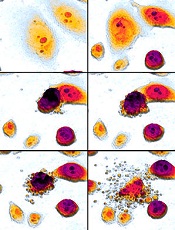
For the first time, researchers have synthesized compounds that induce rapid apoptosis in leukemic cells.
The team synthesized several members of the family of dimeric nuphar alkaloids, which are compounds previously isolated from the yellow pond lily.
These structurally complex molecules have proven capable of inducing apoptosis in human leukemia cell lines faster than any other small molecule tested to date.
The researchers were also able to synthesize some related structures that they predict might exist in nature but have not yet been found.
Their work is published in Angewandte Chemie International Edition.
“We anticipate that these compounds will serve as useful tools for dissecting an important, but as yet undefined, step in the regulation of apoptosis,” said study author Jimmy Wu, PhD, of Dartmouth College in Hanover, New Hampshire.
The research also provides a means to a steady supply of the active compounds for further study.
Preliminary biological tests conducted by Alan Eastman, PhD, also of Dartmouth College, suggest the compounds, both the naturally occurring ones and those predicted to exist in nature, are capable of inducing extremely rapid apoptosis in leukemic cells.
“Studies to clarify the biological mechanism by which they operate are ongoing,” Dr Wu said.
He noted that there have been 2 reports that attempt to explain the molecules’ mechanism of action. But these are incomplete, and more research is required to fully reveal how these compounds work.
“A better understanding of the biological basis of how the dimeric nuphar alkaloids can so rapidly induce cell death may lead to novel points of intervention for the design of prospective therapeutics,” Dr Wu concluded. ![]()
apoptosis in cancer cells
For the first time, researchers have synthesized compounds that induce rapid apoptosis in leukemic cells.
The team synthesized several members of the family of dimeric nuphar alkaloids, which are compounds previously isolated from the yellow pond lily.
These structurally complex molecules have proven capable of inducing apoptosis in human leukemia cell lines faster than any other small molecule tested to date.
The researchers were also able to synthesize some related structures that they predict might exist in nature but have not yet been found.
Their work is published in Angewandte Chemie International Edition.
“We anticipate that these compounds will serve as useful tools for dissecting an important, but as yet undefined, step in the regulation of apoptosis,” said study author Jimmy Wu, PhD, of Dartmouth College in Hanover, New Hampshire.
The research also provides a means to a steady supply of the active compounds for further study.
Preliminary biological tests conducted by Alan Eastman, PhD, also of Dartmouth College, suggest the compounds, both the naturally occurring ones and those predicted to exist in nature, are capable of inducing extremely rapid apoptosis in leukemic cells.
“Studies to clarify the biological mechanism by which they operate are ongoing,” Dr Wu said.
He noted that there have been 2 reports that attempt to explain the molecules’ mechanism of action. But these are incomplete, and more research is required to fully reveal how these compounds work.
“A better understanding of the biological basis of how the dimeric nuphar alkaloids can so rapidly induce cell death may lead to novel points of intervention for the design of prospective therapeutics,” Dr Wu concluded. ![]()
Database may help predict cancer patients’ survival

Photo by Darren Baker
A newly developed database may help physicians predict survival outcomes in patients with hematologic and solid tumor malignancies, according to a paper published in Nature Medicine.
The database, known as PRECOG, integrates gene expression patterns of 39 types of cancer from nearly 18,000 patients with data about how long those patients lived.
By combining these data, researchers were able to see broad patterns that correlate with survival. They also believe this information could help them pinpoint potential therapeutic targets for a range of cancers.
“We were able to identify key pathways that can dramatically stratify survival across diverse cancer types,” said Ash Alizadeh, MD, PhD, of Stanford University in California.
“The patterns were very striking, especially because few such examples are currently available for the use of genes or immune cells for cancer prognosis.”
In addition to identifying potentially useful gene expression patterns, the researchers used an analytical tool called CIBERSORT to determine the composition of leukocytes that flock to a tumor.
“We were able to infer which immune cells are present or absent in individual solid tumors, to estimate their prevalence, and to correlate that information with patient survival,” said Aaron Newman, PhD, of Stanford University.
“We found you can even broadly distinguish cancer types just based on what kind of immune cells have infiltrated the tumor.”
Compiling the data
Researchers have tried for years to identify specific patterns of gene expression in cancerous tumors that differ from those in normal tissue. But the extreme variability among individual patients and tumors has made the process difficult, even when focused on particular cancer types.
“There are many more genes in a cell than there are patients with any one type of cancer, and this makes discovering the important genes for cancer outcomes a tough problem,” said Andrew Gentles, PhD, of Stanford University.
“Because it’s easy to find spurious associations that don’t hold up in follow-up studies, we combined information from a vast array of cancer types to better see meaningful correlations.”
The researchers first collected publicly available data on gene expression patterns of many types of cancers.
They then matched the gene expression profiles with clinical information about the patients, including their age, disease status, and how long they survived after diagnosis. Finally, the team combined the studies in a database.
“We wanted to be able to connect gene expression data with patient outcome for thousands of people at once,” Dr Alizadeh said. “Then, we could ask what we could learn more broadly.”
Surprising findings
The researchers were surprised to find that prognostic genes were often shared among distinct cancer types, suggesting that similar biological programs impact survival across cancers.
They were able to identify the top 10 genes that seemed to confer adverse outcomes—FOXM1, BIRC5, TOP2A, TPX2, NME1, CCNB1, CEP55, TYMS, CENPF, and CDKN3—and the top 10 genes associated with more positive outcomes—KLRB1, ITM2B, CBX7, CD2, CREBL2, SATB1, NR3C1, TMEM66, KLRK1, and FUCA1.
Many of these genes are involved in aspects of cell division or are associated with distinct leukocytes that flood a tumor.
The researchers were also able to identify combinations of leukocytes that appear to be correlated with outcomes.
In particular, elevated numbers of plasma cells and certain types of T cells correlated with better patient survival rates across many different solid tumors. But a high proportion of granulocytes was associated with adverse outcomes.
The researchers hope that PRECOG and CIBERSORT will increase our understanding of cancer biology and aid the development of new therapies for cancer patients. The team is applying these tools to better predict which patients will respond to new and emerging anticancer therapies.
Dr Alizadeh said this is especially important given recent advances in the development of drugs that engage immune responses but work well only for a subset of cancer patients. ![]()
Group creates mouse model of RUNX1-mutated AML

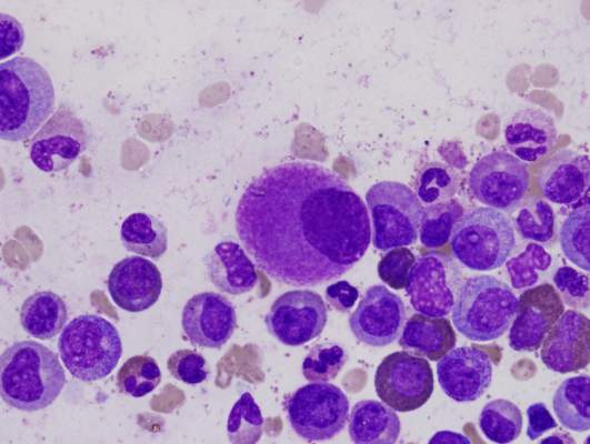
Researchers have developed a mouse model to help them understand why patients with RUNX1-mutated acute myeloid leukemia (AML) respond poorly to chemotherapy.
Approximately 15% of AML patients harbor a mutation in the RUNX1 gene.
In these patients, anthracycline/cytarabine-based chemotherapy does not eradicate AML cells from the bone marrow.
But scientists don’t fully understand the underlying mechanisms protecting these residual cells.
Jason H. Mendler, MD, PhD, of the University of Rochester Medical Center in Rochester, New York, and his colleagues have suggested that a genetically defined mouse model of RUNX1-mutated AML is the ideal platform to investigate the cellular mechanisms protecting residual AML cells in this disease subtype.
“Like all cancers, leukemia is not a one-size-fits-all, and, therefore, it’s important to find better ways to study high-risk subtypes of the disease,” Dr Mendler said. “We believe our mouse model will allow us to quickly define new ways to target this challenging disease.”
Dr Mendler and his colleagues described their model in PLOS ONE.
The researchers began with a patient-derived cell line of RUNX1-mutated, cytogenetically normal AML. They injected these cells into NOD-SCID-γ mice and observed leukemic engraftment in the bone marrow, spleen, and peripheral blood within 6 weeks.
When the researchers treated the mice with anthracycline/cytarabine-based chemotherapy, they saw AML clearance in the spleen and peripheral blood. But leukemic cells remained in the bone marrow.
Dr Mendler and his colleagues also found their mouse model contained mutations in 5 genes aside from RUNX1—ASXL1, CEBPA, GATA2, NRAS, and SETBP1.
The team said further investigation will be focused on identifying the interplay of genes and pathways that are critical to mediating chemotherapy resistance in this model. ![]()
EHA: High-risk APL curable with chemo-free combo
VIENNA – Patients with high-risk newly diagnosed acute promyelocytic leukemia derive the same survival benefit from a chemotherapy-free combination as an anthracycline-containing standard of care, according to results of the AML17 APL study.
The 4-year overall survival rates in high-risk patients (WBC > 10 x 109/L) were 87% with arsenic trioxide plus all-trans retinoic acid and 84% with the standard all-trans retinoic acid and idarubicin schedule.
Relapse-free survival rates were superior with the chemotherapy-free combination (100% vs. 74%; P = .008), Dr. Alan Burnett reported at the annual congress of the European Hematology Association.
“One of our rationales for using arsenic as first-line (therapy) was to try and get at the early death that remains a major problem in this disease,” he said.
Arsenic trioxide and gemtuzumab ozogamicin are effective as single agents with the former approved for relapsed disease in patients with APL. The GIMEMA-AMLSG-SAL trial indicated that a daily schedule of arsenic trioxide plus all-trans retinoic acid was at least as effective and may be superior to all-trans retinoic acid plus chemotherapy in low to moderate risk APL patients (N. Engl. J. Med. 2013;369: 111-21).
The AML17 APL trial was designed by the U.K. National Cancer Research Institute with the aim of comparing all-trans retinoic acid and idarubicin with arsenic trioxide in an attenuated dosing schedule plus all-trans retinoic acid. Importantly, high-risk patients were included, with the option to receive a single dose of gemtuzumab ozogamicin (Mylotarg) 6 mg/m2 within the first 4 days of induction, said Dr. Burnett, who performed the research as head of hematology at Cardiff University in Wales and is now global lead for myeloid diseases at CTI BioPharma in Seattle. In the United States, gemtuzumab was withdrawn from the market in 2010 because of safety concerns.
From May 2009 to October 2013, 235 patients with molecularly confirmed APL were randomized at 81 centers to all-trans retinoic acid 45 mg/m2 as a divided daily oral dose to day 60 or complete remission plus arsenic trioxide 0.3 mg/kg on days 1-5 of week 1 and then 0.25 mg/kg twice per week for 7 weeks plus all-trans retinoic acid 45 mg/m2 as a divided daily oral dose to day 60 or complete remission or to idarubicin 12 mg/m2 on days 2, 4, 6, 8 plus all-trans retinoic acid 45 mg/m2 as a divided daily oral dose to day 60.
In the arsenic trioxide plus all-trans retinoic acid arm, this was followed by all-trans retinoic acid 45 mg/m2 as a divided daily dose 2 weeks on and 2 weeks off plus four consolidation courses of arsenic trioxide 0.3 mg/kg days 1-5 of week 1 and then 0.25 mg/kg twice per week for 3 weeks (total 63 days of arsenic trioxide).
Consolidation in the all-trans retinoic acid and idarubicin arm was all-trans retinoic acid 45 mg/m2 as a divided daily dose on days 1-15 plus idarubicin 5 mg/m2 days 1-4 in course 2, mitoxantrone 10mg/m2 days 1-4 in course 3, and idarubicin 12 mg/m2 day 1 in course 4.
No maintenance was given in either arm. The median patient age was 47 years, with about 20% of patients over age 60; over 20% of the patients were high-risk, and they were equally balanced in the two treatment groups.
At 4 years, the overall survival rate among all patients was comparable – 93% with arsenic trioxide plus all-trans retinoic acid and 89% with all-trans retinoic acid and idarubicin.
However, event-free survival was significantly better in the arsenic trioxide plus all-trans retinoic acid cohort (91% vs. 74%; hazard ratio, 0.36; P = .003), as were frank relapse-free survival (97% vs. 83%; HR, 0.24; P = .004) and molecular relapse-free survival (98% vs. 70%; HR, 0.17; P < .0001), Dr. Burnett said.
One patient on arsenic trioxide plus all-trans retinoic acid experienced frank relapse, compared with 13 on all-trans retinoic acid and idarubicin, plus a further 19 molecular relapses occurred on this arm (cumulative incidence of molecular and hematologic relapse 0% vs. 27%, HR, 0.12; P < .0001).
“Once a patient was in molecular remission there were no further relapses in patients on (arsenic trioxide plus all-trans retinoic acid),” he said.
Of the 30 high-risk patients allocated to the chemo-free arm, 28 received gemtuzumab ozogamicin as per protocol. The overall survival at 4 years for these patients was 89%. Of the two patients not treated with gemtuzumab ozogamicin, one died on day 12 due to causes unrelated to treatment.
Among the 49 patients older than 60 years, overall survival was 80% with arsenic trioxide plus all-trans retinoic acid and 74% with all-trans retinoic acid and idarubicin. Similarly, among good-risk patients, relapse-free survival was significantly improved (96% vs. 79%; HR, 0.33; P = .04). Also, overall survival was not inferior at 95% vs. 90%, “very much replicating the outcomes seen in the GIMEMA study,” Dr. Burnett said.
The benefits were also achieved with significantly less grade 3-4 liver toxicity than observed in the GIMEMA study (<10% vs 63%).
There was, however, an excess of cardiac toxicity in course 2 with arsenic trioxide plus all-trans retinoic acid, compared with all-trans retinoic acid and idarubicin (P = .001).
“We’re not totally sure what that’s all due to, but it doesn’t look to be due to a QC prolongation,” he said.
The arsenic trioxide plus all-trans retinoic acid regimen was associated with significant reductions in supportive care requirements including fewer blood and platelet transfusions, days on antibiotics, and days in hospital, with “many patients treated exclusively as outpatients,” he added.
The low risk of relapse with arsenic trioxide plus all-trans retinoic acid also negates the need for minimal residual disease monitoring.
Finally, compared with the GIMEMA study protocol, the attenuated arsenic dosing schedule in AML17 APL resulted in less frequent dosing of arsenic trioxide (63 doses vs. 140 doses) and less drug required (151 vials vs. 280 vials for a 70-kg patient). At an acquisition cost of £350 per vial, this represents a cost savings of £46,000 (nearly $72,000) per patient, not to mention the added convenience to patients, Dr. Burnett observed.
Cancer Research U.K. funded the study. Cephalon provided the arsenic trioxide. Dr. Burnett disclosed part-time employment with CTI LifeSciences and in the last 12 months serving on the advisory boards of Celgene, Agios, Pfizer, and Bristol-Myers Squibb.
AT THE EHA CONGRESS
Key clinical point: Arsenic trioxide plus all-trans retinoic acid is at least equivalent to all-trans retinoic acid and idarubicin in terms of overall survival in high-risk patients given gemtuzumab ozogamicin prophylaxis.
Major finding: At 4 years, the overall survival rate among all patients was comparable – 93% with arsenic trioxide plus all-trans retinoic acid and 89% with all-trans retinoic acid and idarubicin.
Data source: Prospective, randomized trial in 235 patients with acute promyelocytic leukemia.
Disclosures: Cancer Research U.K. funded the study. Cephalon provided the arsenic trioxide. Dr. Burnett disclosed part-time employment with CTI LifeSciences and in the last 12 months serving on the advisory boards of Celgene, Agios, Pfizer, and Bristol-Myers Squibb.
CML patients die from comorbidities, not leukemia
Patients with chronic myeloid leukemia (CML) treated with imatinib are much more likely to die from their comorbid conditions than from the leukemia, according to a report published in Blood.
During the past decade, CML has been transformed from a routinely fatal disease to a chronic condition controlled by regular drug therapy using imatinib and newer tyrosine kinase inhibitors. The influence of comorbidities on survival outcomes has not been studied until now, said Dr. Susanne Saussele of Heidelberg University, Mannheim (Germany), and her associates.

They used data from a large German study of first-line imatinib therapy, focusing on 1,519 CML patients who were evaluable after a median follow-up of 68 months. Approximately 40% of these study participants had one or more of 511 evaluable comorbidities. The most common conditions relevant to CML and its treatment were diabetes, nonactive cancer other than CML, chronic pulmonary disease, renal insufficiency, MI, cerebrovascular disease, heart failure, and peripheral vascular disease.
Study participants were categorized by the number and severity of their comorbidities using the Charlson Comorbidity Index as CCI 2 (589 patients), CCI 3 or 4 (599 patients), CCI 5 or 6 (229 patients), or CCI 7 and above (102 patients), with higher levels indicating a greater burden of comorbidity. Overall 8-year survival probabilities directly correlated with CCI category, at 94% for CCI 2, 89% for CCI 3 or 4, 78% for CCI 5 or 6, and 46% for CCI 7 or above. In addition, CCI score was the most powerful predictor of overall survival, the researchers said (Blood 2015;126:42-9).
Comorbidities had no impact on the success of imatinib therapy. Even patients with multiple or severe comorbidities derived significant benefit from imatinib, and comorbidities had no negative effect on remission rates or disease progression. Taken together with comorbidities’ strong influence on mortality, this indicates that patients’ survival is determined more by their comorbidities than by CML itself, Dr. Saussele and her associates said.
Their findings also showed that overall survival alone is no longer an appropriate endpoint for assessing treatment efficacy in CML. Progression-free survival seems to be a more accurate measure of treatment effect, since it was not influenced by comorbidities in this study, they added.
Patients with chronic myeloid leukemia (CML) treated with imatinib are much more likely to die from their comorbid conditions than from the leukemia, according to a report published in Blood.
During the past decade, CML has been transformed from a routinely fatal disease to a chronic condition controlled by regular drug therapy using imatinib and newer tyrosine kinase inhibitors. The influence of comorbidities on survival outcomes has not been studied until now, said Dr. Susanne Saussele of Heidelberg University, Mannheim (Germany), and her associates.
They used data from a large German study of first-line imatinib therapy, focusing on 1,519 CML patients who were evaluable after a median follow-up of 68 months. Approximately 40% of these study participants had one or more of 511 evaluable comorbidities. The most common conditions relevant to CML and its treatment were diabetes, nonactive cancer other than CML, chronic pulmonary disease, renal insufficiency, MI, cerebrovascular disease, heart failure, and peripheral vascular disease.
Study participants were categorized by the number and severity of their comorbidities using the Charlson Comorbidity Index as CCI 2 (589 patients), CCI 3 or 4 (599 patients), CCI 5 or 6 (229 patients), or CCI 7 and above (102 patients), with higher levels indicating a greater burden of comorbidity. Overall 8-year survival probabilities directly correlated with CCI category, at 94% for CCI 2, 89% for CCI 3 or 4, 78% for CCI 5 or 6, and 46% for CCI 7 or above. In addition, CCI score was the most powerful predictor of overall survival, the researchers said (Blood 2015;126:42-9).
Comorbidities had no impact on the success of imatinib therapy. Even patients with multiple or severe comorbidities derived significant benefit from imatinib, and comorbidities had no negative effect on remission rates or disease progression. Taken together with comorbidities’ strong influence on mortality, this indicates that patients’ survival is determined more by their comorbidities than by CML itself, Dr. Saussele and her associates said.
Their findings also showed that overall survival alone is no longer an appropriate endpoint for assessing treatment efficacy in CML. Progression-free survival seems to be a more accurate measure of treatment effect, since it was not influenced by comorbidities in this study, they added.
FROM BLOOD
Key clinical point: Patients with chronic myeloid leukemia treated with imatinib are much more likely to die from comorbid conditions than from leukemia.
Major finding: Overall 8-year survival probabilities directly correlated with CCI category, at 94% for CCI 2, 89% for CCI 3 or 4, 78% for CCI 5 or 6, and 46% for CCI 7 or above.
Data source: A secondary analysis of data in a nationwide German study involving 1,519 patients treated with imatinib and followed for a median of 68 months.
Disclosures: This study was supported by the Deutsche Krebshilfe, Novartis, Kompetenznetz für Akute und Chronische Leukämien, Deutsche Jose-Carreras Leukämiestiftung, European LeukemiaNet, Roche, and Essex Pharma. Dr. Saussele reported honoraria and research funding from Pfizer, Novartis, and Bristol-Myers Squibb, and her associates reported ties to these companies and ARIAD.
Polyphenols may enhance doxorubicin treatment

Photo by Rhoda Baer
New research suggests the polyphenols resveratrol and quercetin could be used to augment treatment with the anthracycline doxorubicin.
Investigators found they could increase the bioavailability of resveratrol and quercetin using copolymers that make the compounds water soluble and allow for their injection into the blood stream.
The team then showed the compounds synergize with doxorubicin while also reducing cardiac toxicity.
Although doxorubicin has proven effective against lymphomas, leukemias, and other cancers, the drug can only be used for a limited time because it confers cardiotoxicity.
The co-administration of resveratrol and quercetin might allow for much more extensive use of doxorubicin, while at the same time improving its efficacy and demonstrating the polyphenols’ own anticancer properties, investigators said.
They described research supporting this idea in the Journal of Controlled Release.
“This has great potential to improve chemotherapeutic cancer treatment,” said Adam Alani, PhD, of Oregon State University in Portland.
“The co-administration of high levels of resveratrol and quercetin, in both in vitro and in vivo studies, shows that it significantly reduces the cardiac toxicity of [doxorubicin]. And these compounds have a synergistic effect that enhances the efficacy of the cancer drug, by sensitizing the cancer cells to the effects of the drug.”
Dr Alani said further research may demonstrate that these compounds can completely eliminate the cardiotoxicity of doxorubicin, as they scavenge the toxic free radicals produced by this drug.
It’s also possible, he said, that administration of these natural polyphenols could have value in cancer therapy by themselves or in combination with a wider range of other chemotherapeutic drugs.
Increasing bioavailability
Resveratrol is a natural compound found in foods such as grapes, red wine, green tea, berries, and dark chocolate. Quercetin reaches some of its highest natural levels in capers, some berries, and leafy greens.
When consumed via food or taken as supplements, these polyphenol compounds reach only a tiny fraction of the level that’s possible with direct injection. Such injection was not possible until Dr Alani and his colleagues adapted the use of polymeric micelles.
Specifically, the investigators combined resveratrol and quercetin in Pluronic F127 micelles (mRQ). Pluronics are triblock copolymers consisting of a polypropylene oxide chain flanked with 2 polyethylene oxide chains that can self-assemble into polymeric micelles. The micelles have hydrophobic cores that help solubilize compounds with poor aqueous solubility.
“There are several advantages with this system,” Dr Alani said. “We can finally reach clinical levels of these polyphenols in the body. We can load both the compounds at one time to help control the cardiotoxicity of the cancer drug, and we can help the polyphenols accumulate in cancer cells where they have their own anticancer properties.”
In combination with doxorubicin
The investigators prepared mRQ micelles that were capable of retaining 1.1 mg/mL of resveratrol and 1.42 mg/mL of quercetin. They then tested mRQ in combination with doxorubicin in human ovarian cancer cells (SKOV-3) and rat cardiomyocytes (H9C2).
The team found that a resveratrol-quercetin-doxorubicin ratio of 10:10:1 was synergistic in SKOV-3 cells and antagonistic in H9C2 cells.
mRQ did not interfere with doxorubicin’s caspase activity in SKOV-3 cells but significantly decreased the activity in H9C2 cells. Likewise, there were no changes in the generation of reactive oxygen species in SKOV-3 cells, but there was significant scavenging in H9C2 cells.
The investigators also administered doxorubicin, with or without mRQ, to healthy mice and found that mRQ “conferred full cardioprotection.”
Dr Alani noted that previous research suggested resveratrol and quercetin are safe when given at high concentrations, but additional research is needed. ![]()
Prenatal test results linked to cancer in mothers

Photo by Nina Matthews
Researchers have again found evidence to suggest that tests used to identify chromosomal fetal disorders can detect occult malignancies in pregnant women.
In a study made public last month, non-invasive prenatal tests (NIPTs) revealed 2 cases of lymphoma and a case of ovarian cancer in expectant mothers.
In the new study, researchers showed that positive NIPT results were due to leukemia, lymphoma, or solid tumors in 10 expectant mothers.
The research was published in JAMA and presented at the 19th International Conference on Prenatal Diagnosis and Therapy in Washington, DC. Funding for the study was provided by Illumina, and company employees were involved in the research.
“We did this study because noninvasive prenatal testing using sequencing of cell-free DNA in the mother’s plasma is the fastest-growing area of prenatal testing and, indeed, of genomic medicine,” said study author Diana W. Bianchi, MD, of Tufts Medical Center in Boston, Massachusetts.
“As the volume of tests has expanded, we’ve become increasingly aware of the so-called “false-positive” cases. [A]pproximately 0.2% of the time, there is a discrepancy between the results of the prenatal test—in which an aneuploidy is reported—and the result from the diagnostic fetal procedure, the amniocentesis or the chorionic villus sampling.”
“So we’re interested in the situation where the fetal chromosomes are normal, but the prenatal test shows that there’s an aneuploidy detected. We’re interested in the possible explanations for that discrepancy.”
To gain some insight, Dr Bianchi and her colleagues evaluated 125,426 samples from asymptomatic pregnant women who underwent plasma cell-free DNA sequencing for clinical prenatal aneuploidy screening using Illumina’s verifi Prenatal Test.
In all, 3757 samples (3%) were positive for 1 or more aneuploidies involving chromosomes 13, 18, 21, X, or Y. These were reported to the ordering physician with recommendations for further evaluation.
“In a small minority of women, [subsequent tests analyzing only fetal DNA] showed that the fetal chromosomes were normal, and that disagreed with [results of the NIPT],” Dr Bianchi said. “We were examining whether cancer could explain the discrepancy between these two test results.”
The researchers found that 10 of the women with discordant test results were subsequently diagnosed with cancer. There were 3 cases of B-cell lymphoma and 1 case each of T-cell leukemia, Hodgkin lymphoma, unspecified adenocarcinoma, leiomyosarcoma, and neuroendocrine, colorectal, and anal carcinomas.
Dr Bianchi and her colleagues were able to obtain detailed clinical and sequencing data for 8 of these cases. In the other 2 cases (leiomyosarcoma and unspecified adenocarcinoma), the women were critically ill and were not approached about participating in the study.
The researchers found that maternal cancers most frequently occurred when the NIPT detected more than 1 aneuploidy. There were 7 known cancers among 39 cases of multiple aneuploidies by NIPT. In 1 case, blood was sampled after the patient completed treatment for colorectal cancer, and the abnormal pattern was no longer evident.
When the researchers examined additional genetic information for the women with cancer, they found unique patterns of nonspecific copy-number gains and losses across multiple chromosomes.
“[These women] had DNA imbalances all across the genome,” Dr Bianchi said. “The [NIPT] normally is only looking at DNA material from the chromosomes of clinical interest—chromosomes 13, 18, 21, X, and Y.”
“When we opened up their results to look at all of the chromosomes, there were multiple abnormalities in other places, such as chromosome 8, chromosome 6, etc. Each woman had a unique pattern that was abnormal in many places. This suggested that it was the tumor DNA that was being shed into her blood and was contributing to the abnormal pattern.”
Dr Bianchi stressed that the tumor DNA did not affect the babies. She said all were born healthy, although labor was induced early in one mother to facilitate her cancer treatment. ![]()
YA cancer survivors hospitalized more often
Photo courtesy of
St. Michael’s Hospital
Up to 20 years after they are declared cancer-free, young adult (YA) cancer survivors are still hospitalized more often than the general population, according to research published in the Journal of Clinical Oncology.
Overall, the cancer survivors, who were ages 20 to 44 at diagnosis, were hospitalized about 1.5 times as often as control subjects.
“Even when young adults survive cancer, the cancer still has an impact on their lives and their long-term health, and this age group still has a lot of life to live,” said study author Nancy Baxter, MD, PhD, of St. Michael’s Hospital in Toronto, Ontario, Canada.
To conduct this study, Dr Baxter and her colleagues examined data from the Ontario Cancer Registry spanning the period from 1992 to 1999.
This included 20,275 patients who had their first cancer diagnosis between the ages of 20 and 44 and had lived for 5 years cancer-free. The researchers compared hospitalizations among these patients to hospitalizations in 101,344 non-cancer controls.
During the study period, 34.3% of cancer survivors (n=6948) were admitted to the hospital. The adjusted relative rate (ARR) of hospitalizations in survivors compared to controls was 1.51.
There was a significant decrease in hospitalizations among cancer survivors from the first time point the researchers analyzed to the last time point (P<0.0001).
But hospitalizations were more common among cancer survivors regardless of the time point. The ARR was 1.67 at 5 to 8 years after cancer diagnosis and 1.22 at 18 to 20 years after diagnosis.
When the researchers looked at individual malignancies, they found that survivors of melanoma or testicular cancer did not have higher rates of hospitalization than the control population. The ARRs were 0.97 and 1.07, respectively.
However, the rate of hospitalization was at least twice as high as the control population for survivors of leukemia (ARR=2.23) and lymphoma (ARR=2.02), as well as gastrointestinal (ARR=2.49), urologic (ARR=2.20), colorectal (ARR=2.10), and brain cancers (ARR=2.04).
Dr Baxter said having a better understanding of healthcare utilization and late effects in the YA cancer population may help healthcare providers counsel YA survivors on their future quality of life, identify areas where preventative strategies could be employed, and highlight the need to consider treatments that are not associated with long-term health consequences. ![]()
JAK2 inhibitor could treat B-ALL
Photo courtesy of the
Dana-Farber Cancer Institute
A type II JAK2 inhibitor has shown activity against B-cell acute lymphoblastic leukemia (B-ALL) in preclinical experiments.
The inhibitor, known as CHZ868, works by binding JAK2 into a tightly clenched position, which prevents the protein from functioning.
Researchers tested CHZ868 in samples from patients with CRLF2-rearranged B-ALL, in mice with the disease, and in mice implanted with human B-ALL tissue.
“In each case, we saw good activity: leukemia cells died, JAK2 signaling was suspended, and survival rates increased,” said David Weinstock, MD, of Dana-Farber Cancer Institute in Boston, Massachusetts.
“When we combined CHZ868 with the steroid dexamethasone, the killing of leukemia cells was much more extensive, and the animals lived longer than they did with CHZ868 alone.”
Dr Weinstock and his colleagues reported these results in Cancer Cell. Some of the researchers involved in this work are employees of, or have received research funding from, Novartis.
The team found that CHZ868 inhibited JAK2 signaling in B-ALL, both in vitro and in vivo. CHZ868 could overcome persistent JAK2 signaling where type I JAK2 inhibitors (BSK805 and BVB808) could not.
However, the researchers also identified a mutation—JAK2 L884P—that conferred resistance to CHZ868 and another type II JAK2 inhibitor, BBT594.
Nevertheless, CHZ868 suppressed the growth of CRLF2-rearranged human B-ALL cells and improved survival in mice with human or murine B-ALL.
CHZ868 worked synergistically with dexamethasone to induce apoptosis in JAK2-dependent B-ALL. The combination also improved survival in mice with B-ALL, when compared to either dexamethasone or CHZ868 alone.
The researchers noted that, when given at 30 mg/kg/day, CHZ868 was tolerated in NSG mice for up to 25 days and in immunocompetent mice for up to 44 days. And the drug had “essentially no effects” on peripheral blood counts.
This result and the tolerability of dexamethasone make CHZ868 and dexamethasone a “particularly attractive” combination that should be investigated in clinical trials, the team said.
They also speculated that CHZ868 or other type II JAK2 inhibitors could prove effective against malignancies other than B-ALL.
“JAK2 abnormalities are found in some cases of triple-negative breast cancer and Hodgkin lymphoma,” Dr Weinstock noted. “The success of CHZ868 in B-ALL suggests that it, or a compound that works by a similar mechanism, may also be effective in these cancers.” ![]()
Photo courtesy of the
Dana-Farber Cancer Institute
A type II JAK2 inhibitor has shown activity against B-cell acute lymphoblastic leukemia (B-ALL) in preclinical experiments.
The inhibitor, known as CHZ868, works by binding JAK2 into a tightly clenched position, which prevents the protein from functioning.
Researchers tested CHZ868 in samples from patients with CRLF2-rearranged B-ALL, in mice with the disease, and in mice implanted with human B-ALL tissue.
“In each case, we saw good activity: leukemia cells died, JAK2 signaling was suspended, and survival rates increased,” said David Weinstock, MD, of Dana-Farber Cancer Institute in Boston, Massachusetts.
“When we combined CHZ868 with the steroid dexamethasone, the killing of leukemia cells was much more extensive, and the animals lived longer than they did with CHZ868 alone.”
Dr Weinstock and his colleagues reported these results in Cancer Cell. Some of the researchers involved in this work are employees of, or have received research funding from, Novartis.
The team found that CHZ868 inhibited JAK2 signaling in B-ALL, both in vitro and in vivo. CHZ868 could overcome persistent JAK2 signaling where type I JAK2 inhibitors (BSK805 and BVB808) could not.
However, the researchers also identified a mutation—JAK2 L884P—that conferred resistance to CHZ868 and another type II JAK2 inhibitor, BBT594.
Nevertheless, CHZ868 suppressed the growth of CRLF2-rearranged human B-ALL cells and improved survival in mice with human or murine B-ALL.
CHZ868 worked synergistically with dexamethasone to induce apoptosis in JAK2-dependent B-ALL. The combination also improved survival in mice with B-ALL, when compared to either dexamethasone or CHZ868 alone.
The researchers noted that, when given at 30 mg/kg/day, CHZ868 was tolerated in NSG mice for up to 25 days and in immunocompetent mice for up to 44 days. And the drug had “essentially no effects” on peripheral blood counts.
This result and the tolerability of dexamethasone make CHZ868 and dexamethasone a “particularly attractive” combination that should be investigated in clinical trials, the team said.
They also speculated that CHZ868 or other type II JAK2 inhibitors could prove effective against malignancies other than B-ALL.
“JAK2 abnormalities are found in some cases of triple-negative breast cancer and Hodgkin lymphoma,” Dr Weinstock noted. “The success of CHZ868 in B-ALL suggests that it, or a compound that works by a similar mechanism, may also be effective in these cancers.”