User login
Cancer drug prices vary widely from country to country

Photo by Bill Branson
The price of cancer drugs varies widely between European countries, Australia, and New Zealand, according to a study published in The Lancet Oncology.
The study indicates that, overall, the UK and Mediterranean countries such as Greece, Spain, and Portugal pay the lowest average unit manufacturer prices for a group of 31 originator cancer drugs (new drugs under patent).
And Sweden, Switzerland, and Germany pay the highest prices.
The greatest differences in price were noted for gemcitabine, which costs €209 per vial in New Zealand and €43 in Australia, and zoledronic acid, which costs €330 per vial in New Zealand but €128 in Greece.*
“Public payers in Germany are paying 223% more in terms of official prices for interferon alfa 2b for melanoma and leukemia treatment than those in Greece,” noted study author Sabine Vogler, PhD, of the WHO Collaborating Centre for Pharmaceutical Pricing and Reimbursement Policies in Vienna, Austria.
“For gefitinib to treat non-small-lung cancer, the price in Germany is 172% higher than in New Zealand.”
To uncover these price differences, Dr Vogler and her colleagues reviewed official drug price data from the Pharma Price Information (PPI) service of the Austrian Public Health Institute for 16 European countries**, and from the pharmaceutical schedules in Australia and New Zealand.
The researchers compared what manufacturers charged for a unit (ie, price per tablet or vial) of 31 originator cancer drugs in June 2013.
None of these drugs had a unit price lower than €10. Four drugs (13%) had an average unit manufacturer price between €250 and €500, and 2 drugs (6%) had an average unit price between €500 and €1000.
Seven drugs (23%) had an average unit price higher than €1000. For example, plerixafor cost over €5000 per injection.
The price differences between the highest- and lowest-priced countries ranged from 28% to 50% for a third of the drugs sampled, between 50% and 100% for half of the drugs, and between 100% and 200% for 3 drugs (10%).
The researchers noted that information on real drug prices is scarce. The cancer drug prices they surveyed did not include confidential discounts such as those agreed upon in managed-entry arrangements that are increasingly used in countries such as Australia, Italy, the UK, and the Netherlands.
“Some high-income countries have managed to barter the manufacturers down to lower prices, but these agreements, including the agreed prices, are confidential,” Dr Vogler explained.
“Although these agreements ensure patient access to new drugs, other countries risk overpaying when setting drug prices through the common practice of external price referencing, or international price comparison, because they can only use the official undiscounted prices as a benchmark. There needs to be far more transparency.”
“We hope that our findings will provide concrete evidence for policymakers to take action to address high prices and ensure more transparency in cancer drug pricing so that costs and access to new drugs does not depend on where a patient lives.” ![]()
*Gemcitabine and zoledronic acid have generic versions in several countries, and originator prices were decreased in some countries following patent expiry but not in others.
**Austria, Belgium, Denmark, Germany, Greece, Finland, France, Italy, Ireland, the Netherlands, Norway, Portugal, Spain, Sweden, Switzerland, and the UK.
ASH: Genes tag increased risk for avascular necrosis in ALL patients under age 10
ORLANDO – Variants in mesenchymal stem cell genes that are important to bone and fat differentiation were associated with the risk of developing osteonecrosis in children under age 10 with acute lymphocytic leukemia, Dr. Seth E. Karol, of St. Jude Children’s Research Hospital in Memphis, reported at the annual meeting of the American Society of Hematology.

In a second study of children with ALL, Dr. Peter D. Cole, of Montefiore Medical Center, Albert Einstein College of Medicine, Bronx, NY, reported that patients under age 10 and homozygous for the 2R polymorphism in thymidylate synthase were at increased risk for avascular necrosis. For children over age 10 with ALL, homozygosity for the 2R polymorphism was linked with an increased risk of fracture.
The pathophysiology of bony morbidity differs depending on the patient’s age, Dr. Cole noted. “This is the first report identifying a genetic risk factor for avascular necrosis specifically among children younger than 10 years, a group where AVN is significantly less frequent.”
Bony morbidity is more common in children over age 10 with ALL, and related to exposure to crucial components of leukemia therapy including corticosteroids, asparaginase, and methotrexate. Therapy-induced osteonecrosis has become a limiting toxicity in the intensification of treatment for pediatric acute lymphoblastic leukemia (ALL), particularly among patients 10-20 years old.
However, children under 10 make up 75% of new pediatric ALL diagnoses, Dr. Karol said. So even though osteonecrosis occurs in just 3%-10% of children under age 10, those patients make up 40% of the cases.
In Dr. Cole’s study, research focused on 19 common genetic polymorphisms in peripheral blood or bone marrow collected at remission from 637 children treated for ALL.
The research targeted common variants within genes related to glucocorticoid metabolism, oxidative damage, and folate physiology. Children above and below the age of 10 years were evaluated separately. Multivariable models for bony morbidity included sex, WBC at diagnosis, race, final risk group, and asparaginase randomization. Blood was collected during treatment for analysis of serum and RBC folate.
Of the 637 patients tested, 627 achieved a complete remission and received post-induction therapy; about 10% of patients experienced avascular necrosis and 22% had at least 1 fracture. Both morbidities were significantly more common in patients aged 10 and older; 23% had avascular necrosis and 31% had a fracture. In younger children, 5.5% had avascular necrosis and 19% had fractures.
Of 626 patients tested, about 21% were homozygous for the 2R polymorphism in thymidylate synthase. In children less than age 10, this 2R/2R genotype was associated with nearly three times the rate of 5-year estimated incidence of avascular necrosis, almost 12%, as compared to the 4% rate seen in children with 2R/3R or 3R/3R genotypes.
Among children over age 10, this polymorphism was associated with an increased risk of bony fracture (multivariable HR 2.13; 95% CI 1.13-3.99; P=0.019).
Bony morbidity was not associated with the other tested polymorphisms previously linked to risk of avascular necrosis, including PAI-1 (rs6092), ABCB1 (rs1045642), and the vitamin D receptor Fok1 restriction site (rs228570). No significant association was observed between thymidylate synthase genotype and serum or RBC folate at any time point during therapy.
In the study reported by Dr. Karol, the researchers studied a discovery cohort of 82 cases of osteonecrosis and 287 controls treated on the Children’s Oncology Group (COG) NCI standard risk ALL protocol and tested for replication in 817 children less than 10 treated on the COG high risk ALL protocol.
Both discovery and replication genome-wide association studies adjusted for demographic and therapy variables known to modify the risk of osteonecrosis. Genes associated with the identified single nucleotide polymorphisms were evaluated for enrichment in biologically relevant pathways.
Within the discovery cohort and replication cohorts, top ranked variants were located near bone morphogenic protein 7 (BMP7) and PROX1-antisense RNA1. The top replicated non-synonymous SNP, rs34144324, was in a glutamate receptor gene, and the genotyping of this variant was verified in the whole exome sequencing data.
Pathway analysis of genes linked to top SNPs demonstrated enrichment in glutamate receptor signaling and adipogenesis pathways.
“Patients with osteonecrosis are 8-15 times more likely to possesses genetic variants near a gene important to bone development (BMP7) and between 3-6 times more likely to have variants near a gene important to fat levels in the blood (PROX1),” Dr. Karol reported.
Dr. Cole and Dr. Karol had no relevant financial disclosures.
On Twitter @maryjodales
ORLANDO – Variants in mesenchymal stem cell genes that are important to bone and fat differentiation were associated with the risk of developing osteonecrosis in children under age 10 with acute lymphocytic leukemia, Dr. Seth E. Karol, of St. Jude Children’s Research Hospital in Memphis, reported at the annual meeting of the American Society of Hematology.
In a second study of children with ALL, Dr. Peter D. Cole, of Montefiore Medical Center, Albert Einstein College of Medicine, Bronx, NY, reported that patients under age 10 and homozygous for the 2R polymorphism in thymidylate synthase were at increased risk for avascular necrosis. For children over age 10 with ALL, homozygosity for the 2R polymorphism was linked with an increased risk of fracture.
The pathophysiology of bony morbidity differs depending on the patient’s age, Dr. Cole noted. “This is the first report identifying a genetic risk factor for avascular necrosis specifically among children younger than 10 years, a group where AVN is significantly less frequent.”
Bony morbidity is more common in children over age 10 with ALL, and related to exposure to crucial components of leukemia therapy including corticosteroids, asparaginase, and methotrexate. Therapy-induced osteonecrosis has become a limiting toxicity in the intensification of treatment for pediatric acute lymphoblastic leukemia (ALL), particularly among patients 10-20 years old.
However, children under 10 make up 75% of new pediatric ALL diagnoses, Dr. Karol said. So even though osteonecrosis occurs in just 3%-10% of children under age 10, those patients make up 40% of the cases.
In Dr. Cole’s study, research focused on 19 common genetic polymorphisms in peripheral blood or bone marrow collected at remission from 637 children treated for ALL.
The research targeted common variants within genes related to glucocorticoid metabolism, oxidative damage, and folate physiology. Children above and below the age of 10 years were evaluated separately. Multivariable models for bony morbidity included sex, WBC at diagnosis, race, final risk group, and asparaginase randomization. Blood was collected during treatment for analysis of serum and RBC folate.
Of the 637 patients tested, 627 achieved a complete remission and received post-induction therapy; about 10% of patients experienced avascular necrosis and 22% had at least 1 fracture. Both morbidities were significantly more common in patients aged 10 and older; 23% had avascular necrosis and 31% had a fracture. In younger children, 5.5% had avascular necrosis and 19% had fractures.
Of 626 patients tested, about 21% were homozygous for the 2R polymorphism in thymidylate synthase. In children less than age 10, this 2R/2R genotype was associated with nearly three times the rate of 5-year estimated incidence of avascular necrosis, almost 12%, as compared to the 4% rate seen in children with 2R/3R or 3R/3R genotypes.
Among children over age 10, this polymorphism was associated with an increased risk of bony fracture (multivariable HR 2.13; 95% CI 1.13-3.99; P=0.019).
Bony morbidity was not associated with the other tested polymorphisms previously linked to risk of avascular necrosis, including PAI-1 (rs6092), ABCB1 (rs1045642), and the vitamin D receptor Fok1 restriction site (rs228570). No significant association was observed between thymidylate synthase genotype and serum or RBC folate at any time point during therapy.
In the study reported by Dr. Karol, the researchers studied a discovery cohort of 82 cases of osteonecrosis and 287 controls treated on the Children’s Oncology Group (COG) NCI standard risk ALL protocol and tested for replication in 817 children less than 10 treated on the COG high risk ALL protocol.
Both discovery and replication genome-wide association studies adjusted for demographic and therapy variables known to modify the risk of osteonecrosis. Genes associated with the identified single nucleotide polymorphisms were evaluated for enrichment in biologically relevant pathways.
Within the discovery cohort and replication cohorts, top ranked variants were located near bone morphogenic protein 7 (BMP7) and PROX1-antisense RNA1. The top replicated non-synonymous SNP, rs34144324, was in a glutamate receptor gene, and the genotyping of this variant was verified in the whole exome sequencing data.
Pathway analysis of genes linked to top SNPs demonstrated enrichment in glutamate receptor signaling and adipogenesis pathways.
“Patients with osteonecrosis are 8-15 times more likely to possesses genetic variants near a gene important to bone development (BMP7) and between 3-6 times more likely to have variants near a gene important to fat levels in the blood (PROX1),” Dr. Karol reported.
Dr. Cole and Dr. Karol had no relevant financial disclosures.
On Twitter @maryjodales
AT ASH 2015
Key clinical point: Gene profiles can identify ALL patients under age 10 at increased risk for avascular necrosis.
Major finding: In children under 10 who were homozygous for the 2R polymorphism in thymidylate synthase the rate of 5-year estimated incidence of avascular necrosis was nearly 12%, three times higher than the 4% rate seen in children with 2R/3R or 3R/3R genotypes.
Data source: Research focused on 19 common genetic polymorphisms in peripheral blood or bone marrow collected at remission from 637 children treated for ALL.
Disclosures: Dr. Cole and Dr. Karol had no relevant financial disclosures.
ASH: Pill bottles flag noncompliance in patients on ALL maintenance
ORLANDO – Pill containers are not known for eloquence, but they tell a disturbing tale of patient over-reporting of adherence to essential leukemia maintenance therapy.
Among 416 children with acute lymphoblastic leukemia (ALL) in first remission, the number of days patients or their guardians reported that the child took his/her daily oral 6-mercaptopurine (6MP) for maintenance was significantly higher than the number of days the pill bottles were opened, as reported by an electronic medication management system.
“Over-reporting was more likely in patients who were non-adherent, older, of non-white race, and came from households with lower paternal education,” said Dr. Wendy Landier from the Institute for Cancer Outcomes and Survivorship at the University of Alabama at Birmingham.
The results suggest that patient self-reports of adherence to 6MP oral maintenance therapy may be unreliable, and should be taken with a grain of salt, Dr. Landier said in a briefing at the American Society of Hematology annual meeting.
The investigators had previously reported that poor adherence to oral 6MP maintenance therapy was associated with a nealy four-fold increased risk for relapse (JCO 30[17]:2094-101, Blood 124[15]:2345-53).
To better understand why some patients are poorly adherent to 6MP maintenance therapy, the investigator provided 416 children with ALL in first remission with pill containers equipped with a Medication Event Management System (MEMS) that electronically recorded the dates and times that each pill bottle was opened over a 16-week period. Patients age 12 and older or the parents/guardians of younger children were also asked to report the date and time of each daily oral dose at the end of each 28-day study month.
The authors collected and evaluated a total of 1344 patient-months of self-report and MEMS data, compared the records, and stratified patients as either “perfect reporters”, whose self-reports matched the objective MEMS data; “over-reporters”, who self-report exceeded the MEMS data on 5 or more days per month for more than half of study months, and “others.”The median patient age at study entry was 6 years (range 2 to 20 years).
The authors reported in their study that 40.4% of patients were not adherent to oral 6MP therapy, as evidence by a mismatch between self-report and objective (MEMS) reporting.
The overall adjusted mean number of self-reported days per month that patients took their pills ranged from a low of 25.8+5.3 to a high of 26.1+4.5. The pill bottles, however, told a different tale, reporting that they had been opened from a low of 22.8 ± 6.4 to a high of 25.4 ± 4.5 days per month.
Month-by-month correlations between self-report and MEMS 0.36 to 0.58, and were all statistically significant.
In logistic regression models adjusted for thiopurine methyltransferase (TMPT) genotype, 6MP dose intensity, and 6-thiogaunine levels in red cells, significant predictors for over-reporting included older age, with an odds ratio (OR) of 1.07 for every 1-year increase in age (P = .04); Hispanic origin (OR 2.4, P = .02); Asian origin, (OR 3.1 P = .02; African-American origin (OR 5.3, P < .001); paternal education level lower than college (OR 2.1, P = .02); and 6MP non-adherence (OR 8.6, P < .0001).
Dr. Landier noted that 78.6% of over-reporters were non-adherent, compared with only 2% of perfect reporters.
“The real importance of this paper from my perspective is this: there is a famous quote that if the patient doesn’t take the medication it can’t work and in an era where we’re trying to improve the care of patients, one of the things we oftentimes forget that if the medication isn’t being taken, the patient can’t get better from it,” said Dr. Mark Crowther, Professor and Chair in the Department of Pathology and Molecular Medicine of McMaster University in Hamilton, Ontario, Canada. Dr. Crowther moderated a briefing where Dr. Cserti-Gazdewich presented the data.
The study was supported by grants from the National Institutes of Health and from St. Baldrick’s Foundation. Dr. Landier and Dr. Crowther reported no relevant conflicts of interest.
AT ASH 2015
Key clinical point: Patients in first ALL remission may not be as adherent to 6MP maintenance therapy as they claim.
Major finding: In all, 40.4% of patients were not adherent to oral 6MP maintenance therapy.
Data source: Observational study comparing patient reported and electronically monitored dosing in 416 children/young adults with ALL in first remission.
Disclosures: The study was supported by grants from the National Institutes of Health and from St. Baldrick’s Foundation. Dr. Landier and Dr. Crowther reported no relevant conflicts of interest.

FDA approves generic imatinib

Photo by Steven Harbour
The US Food and Drug Administration (FDA) has approved the use of imatinib mesylate, a generic version of Novartis’s Gleevec being developed by a subsidiary of Sun Pharmaceuticals Limited.
Under the terms of a settlement agreement with Novartis, the Sun Pharma subsidiary is allowed to launch its generic imatinib in the US on February 1, 2016.
The drug will be available in 100 mg and 400 mg tablets.
The Sun Pharma subsidiary was the first company to file an abbreviated new drug application for generic imatinib with a para IV certification and is therefore eligible for 180 days of marketing exclusivity in the US.
Sun Pharma’s imatinib mesylate is approved for the following indications:
- Newly diagnosed adult and pediatric patients with Philadelphia chromosome positive chronic myeloid leukemia (Ph+ CML) in chronic phase
- Patients with (Ph+ CML) in blast crisis, accelerated phase, or in chronic phase after failure of interferon-alpha therapy
- Adult patients with relapsed or refractory Ph+ acute lymphoblastic leukemia
- Adult patients with myelodysplastic/myeloproliferative diseases associated with PDGFR gene re-arrangements
- Adult patients with aggressive systemic mastocytosis without the D816V c-Kit mutation or with c-Kit mutational status unknown
- Adult patients with hypereosinophilic syndrome (HES) and/or chronic eosinophilic leukemia (CEL) who have the FIP1L1-PDGFRα fusion kinase and for patients with HES and/or CEL who are FIP1L1- PDGFRα fusion kinase negative or unknown
- Adult patients with unresectable, recurrent and/or metastatic dermatofibrosarcoma protuberans.
The drug is not approved to treat patients with KIT (CD117)-positive unresectable and/or metastatic malignant gastrointestinal stromal tumors. ![]()
ASH: Genes, induction response identify high risk childhood B-lymphoblastic leukemia patients with good outcomes
ORLANDO – Cytogenetic profile and response to initial induction therapy can identify a subgroup of National Cancer Institute (NCI) high risk childhood B-lymphoblastic leukemia patients who have good outcomes, Dr. Elizabeth Raetz said, reporting results for her colleagues in the Children’s Oncology Group (COG).
Outcomes were excellent for patients with favorable cytogenetic features and rapid minimal residual disease responses during induction therapy, said Dr. Raetz, of Huntsman Cancer Institute and Primary Children’s Hospital, University of Utah, Salt Lake City. Notably, 5-year overall survival (OS) was over 98% for those with favorable cytogenetic subsets, nearly half of all patients, in a combined analysis of standard risk and high risk patients.

The findings suggest that these patients would not benefit from further intensification of their chemotherapy, sparing them possible toxicity and late effects.
The COG used clinical, biologic, and early disease response measures to study 11,144 patients, aged 1-30 years, enrolled on the COG AALL03B1 classification study. Patients began either a standard risk 3-drug or high risk 4-drug induction therapy based on NCI risk group, she said at the annual meeting of the American Society of Hematology.
After induction therapy, patients were classified into low (29%), standard (33%), high (34%), or very high (4%) risk groups for treatment allocation. Variables used for risk classification included age, initial WBC, extramedullary disease status, blast cytogenetics, early treatment response based on bone marrow morphology and minimal residual disease in marrow at day 29.
Rapid early response was defined as having less than 5% blasts in bone marrow by day 15 plus flow cytometry-based minimal residual disease less than 0.1% on day 29 of induction. Those with either 5% or more blasts in their marrow at day 15 or minimal residual disease of 0.1% or more at day 29 were deemed slow early responders.
Of the 96% of patients evaluable for post-induction treatment assignment, 5104 (65%) were treated for NCI standard risk and 2791 were treated for NCI high-risk B-ALL.
Rapid early responses were seen in 84% and slow early responses in 16%. For those with rapid early responses, the 5-year event-free survival rates was 89% and the overall survival rate was 95%. For those with slow early responses, EFS was 68% and OS was 84%.
The standard risk and high risk groups were then combined and analyzed based on cytogenetic subtype. The favorable gene ETV6-RUNX1 was seen in 26% (1,928 patients) and associated with an EFS of 93% as compared to an EFS of 84% for the 5578 patients without ETV6-RUNX1. OS was 98% for positive patients and 92% for negative patients. In the 21% (1,483 patients) with the triple trisomy 4/10/17, EFS was 95% as compared to an EFS of 84% for the 5,603 patients without the trisomy. OS was 99% and 92%, respectively.
In a subsequent analysis using current COG minimum residual disease response measures – with rapid early response defined as day 8 blood minimal residual disease less than 1% and day 29 marrow minimal residual disease less than 0.01% – the 243 high risk patients who had favorable cytogenetics and no detectable leukemia cells in their CSF had 5 years EFS of 95% and OS of 98%.
Dr. Raetz had no relevant financial disclosures.
Abstract 807: Genetic and Response-Based Risk Classification Identifies a Subgroup of NCI High Risk Childhood B-Lymphoblastic Leukemia (HR B-ALL) with Outstanding Outcomes: A Report from the Children’s Oncology Group (COG).
On Twitter @maryjodales
ORLANDO – Cytogenetic profile and response to initial induction therapy can identify a subgroup of National Cancer Institute (NCI) high risk childhood B-lymphoblastic leukemia patients who have good outcomes, Dr. Elizabeth Raetz said, reporting results for her colleagues in the Children’s Oncology Group (COG).
Outcomes were excellent for patients with favorable cytogenetic features and rapid minimal residual disease responses during induction therapy, said Dr. Raetz, of Huntsman Cancer Institute and Primary Children’s Hospital, University of Utah, Salt Lake City. Notably, 5-year overall survival (OS) was over 98% for those with favorable cytogenetic subsets, nearly half of all patients, in a combined analysis of standard risk and high risk patients.
The findings suggest that these patients would not benefit from further intensification of their chemotherapy, sparing them possible toxicity and late effects.
The COG used clinical, biologic, and early disease response measures to study 11,144 patients, aged 1-30 years, enrolled on the COG AALL03B1 classification study. Patients began either a standard risk 3-drug or high risk 4-drug induction therapy based on NCI risk group, she said at the annual meeting of the American Society of Hematology.
After induction therapy, patients were classified into low (29%), standard (33%), high (34%), or very high (4%) risk groups for treatment allocation. Variables used for risk classification included age, initial WBC, extramedullary disease status, blast cytogenetics, early treatment response based on bone marrow morphology and minimal residual disease in marrow at day 29.
Rapid early response was defined as having less than 5% blasts in bone marrow by day 15 plus flow cytometry-based minimal residual disease less than 0.1% on day 29 of induction. Those with either 5% or more blasts in their marrow at day 15 or minimal residual disease of 0.1% or more at day 29 were deemed slow early responders.
Of the 96% of patients evaluable for post-induction treatment assignment, 5104 (65%) were treated for NCI standard risk and 2791 were treated for NCI high-risk B-ALL.
Rapid early responses were seen in 84% and slow early responses in 16%. For those with rapid early responses, the 5-year event-free survival rates was 89% and the overall survival rate was 95%. For those with slow early responses, EFS was 68% and OS was 84%.
The standard risk and high risk groups were then combined and analyzed based on cytogenetic subtype. The favorable gene ETV6-RUNX1 was seen in 26% (1,928 patients) and associated with an EFS of 93% as compared to an EFS of 84% for the 5578 patients without ETV6-RUNX1. OS was 98% for positive patients and 92% for negative patients. In the 21% (1,483 patients) with the triple trisomy 4/10/17, EFS was 95% as compared to an EFS of 84% for the 5,603 patients without the trisomy. OS was 99% and 92%, respectively.
In a subsequent analysis using current COG minimum residual disease response measures – with rapid early response defined as day 8 blood minimal residual disease less than 1% and day 29 marrow minimal residual disease less than 0.01% – the 243 high risk patients who had favorable cytogenetics and no detectable leukemia cells in their CSF had 5 years EFS of 95% and OS of 98%.
Dr. Raetz had no relevant financial disclosures.
Abstract 807: Genetic and Response-Based Risk Classification Identifies a Subgroup of NCI High Risk Childhood B-Lymphoblastic Leukemia (HR B-ALL) with Outstanding Outcomes: A Report from the Children’s Oncology Group (COG).
On Twitter @maryjodales
AT ASH 2015
Key clinical point: Knowing which patients would not benefit from further intensification of their chemotherapy might spare them possible toxicity and late effects.
Major finding: The 243 high risk patients who had favorable cytogenetics and no detectable leukemia cells in their CSF had 5 year EFS of 94% and OS of 98%.
Data source: 11,144 patients, aged 1-30 years, enrolled on the COG AALL03B1 classification study.
Disclosures: Dr. Raetz had no relevant financial disclosures.
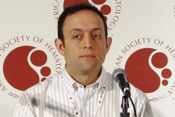
ASH: Donor CAR-T cells elicit responses in mixture of progressive B-cell cancers
ORLANDO – A single infusion of donor-derived chimeric antigen receptor (CAR)-modified T cells targeting CD19 achieved remission in 9 of 20 patients with B-cell malignancies that progressed after allogeneic stem cell transplant, a study shows.
The seven complete remissions and two partial remissions occurred without any chemotherapy and without causing acute graft-versus-host disease (GVHD).

The experimental anti-CD 19 CAR T-cells seem particularly effective against acute lymphoid leukemia (ALL) and chronic lymphocytic leukemia (CLL), but responses also occurred in lymphoma, Dr. James Kochenderfer of the Center for Cancer Research, National Cancer Institute, in Bethesda, Md., reported at the annual meeting of the American Society of Hematology.
B-cell malignancies that persist after transplantation are often treated with unmanipulated donor lymphocytes, but these infusions are often ineffective and associated with significant morbidity and mortality from GVHD.
To improve on this approach, 20 patients were infused with T cells obtained from the original stem cell donor and transduced with a CD19-directed CAR that was encoded by a gamma-retroviral vector and included a CD28 co-stimulatory domain. The highest dose reached in the phase I study was 107 total cell/kg. Production of the anti-CD19 CAR T cells took only eight days for each patient, Dr. Kochenderfer said at a press briefing.
The patients had received at least one standard donor-leukocyte infusion, had to have minimal or no GVHD, and could not be receiving systemic immunosuppressive drugs.
The video associated with this article is no longer available on this site. Please view all of our videos on the MDedge YouTube channel
The highest response rates were in ALL, where four of five patients obtained complete remission (CR) with no detectable minimal residual disease by multi-color flow cytometry, Dr. Kochenderfer said. Two of these patients later relapsed, one is in ongoing CR at 18 months, and one went on to a second allogeneic transplant and continues in complete remission.
The longest ongoing CR at 36 months occurred in a patient treated for CLL. Another patient achieved a partial remission (PR) ongoing at 18 months, two patients progressed, and one has stable disease.
In five patients treated for Mantle cell lymphoma, there is one CR ongoing at 31 months, one PR, and three stable diseases.
Three of the five patients treated for diffuse large B-cell lymphoma experienced stable disease, one progressive disease, and one obtained a CR, but is no longer evaluable because she received other therapies for chronic GVHD. Dr. Kochenderfer went on describe an impressive response in this patient, who had large lymphoma masses at the back of her head and in her eye socket before infusion.
“Amazingly, the tumor masses completely disappeared within five days of CAR T-cell infusion,” he said.
Patients with high tumor burdens, however, experienced severe cytokine-release syndrome with fever, tachycardia, and hypertension that was treated with the interleukin-6 receptor antagonist tocilizumab (Actemra).
Only one case of mild aphasia occurred, which contrasts with other CAR T-cell therapies where neurotoxicity is common, Dr. Kochenderfer said.
One patient had continued worsening of pre-existing chronic GVHD after CAR T-cell therapy, and one patient developed very mild chronic eye GVHD more than a year after infusion.
The press corps was not fully convinced by the findings, however, asking Dr. Kochenderfer why they should be excited by the 40% remission rate when other CAR T-cell therapies have yielded remission rates as high as 90%.
Dr. Kochenderfer pointed out that four of the five ALL patients (80%) achieved a MRD-negative complete response, which compares favorably with other protocols. The remaining patients had far more advanced, treatment-resident disease of varying histologies than evaluated in other trials and, unlike most trials, all patients had received an allogeneic transplant. Further, the investigators used no chemotherapy whatsoever, whereas other CAR T-cells trials have used chemotherapy, sometimes in huge does, he said.
ORLANDO – A single infusion of donor-derived chimeric antigen receptor (CAR)-modified T cells targeting CD19 achieved remission in 9 of 20 patients with B-cell malignancies that progressed after allogeneic stem cell transplant, a study shows.
The seven complete remissions and two partial remissions occurred without any chemotherapy and without causing acute graft-versus-host disease (GVHD).
The experimental anti-CD 19 CAR T-cells seem particularly effective against acute lymphoid leukemia (ALL) and chronic lymphocytic leukemia (CLL), but responses also occurred in lymphoma, Dr. James Kochenderfer of the Center for Cancer Research, National Cancer Institute, in Bethesda, Md., reported at the annual meeting of the American Society of Hematology.
B-cell malignancies that persist after transplantation are often treated with unmanipulated donor lymphocytes, but these infusions are often ineffective and associated with significant morbidity and mortality from GVHD.
To improve on this approach, 20 patients were infused with T cells obtained from the original stem cell donor and transduced with a CD19-directed CAR that was encoded by a gamma-retroviral vector and included a CD28 co-stimulatory domain. The highest dose reached in the phase I study was 107 total cell/kg. Production of the anti-CD19 CAR T cells took only eight days for each patient, Dr. Kochenderfer said at a press briefing.
The patients had received at least one standard donor-leukocyte infusion, had to have minimal or no GVHD, and could not be receiving systemic immunosuppressive drugs.
The video associated with this article is no longer available on this site. Please view all of our videos on the MDedge YouTube channel
The highest response rates were in ALL, where four of five patients obtained complete remission (CR) with no detectable minimal residual disease by multi-color flow cytometry, Dr. Kochenderfer said. Two of these patients later relapsed, one is in ongoing CR at 18 months, and one went on to a second allogeneic transplant and continues in complete remission.
The longest ongoing CR at 36 months occurred in a patient treated for CLL. Another patient achieved a partial remission (PR) ongoing at 18 months, two patients progressed, and one has stable disease.
In five patients treated for Mantle cell lymphoma, there is one CR ongoing at 31 months, one PR, and three stable diseases.
Three of the five patients treated for diffuse large B-cell lymphoma experienced stable disease, one progressive disease, and one obtained a CR, but is no longer evaluable because she received other therapies for chronic GVHD. Dr. Kochenderfer went on describe an impressive response in this patient, who had large lymphoma masses at the back of her head and in her eye socket before infusion.
“Amazingly, the tumor masses completely disappeared within five days of CAR T-cell infusion,” he said.
Patients with high tumor burdens, however, experienced severe cytokine-release syndrome with fever, tachycardia, and hypertension that was treated with the interleukin-6 receptor antagonist tocilizumab (Actemra).
Only one case of mild aphasia occurred, which contrasts with other CAR T-cell therapies where neurotoxicity is common, Dr. Kochenderfer said.
One patient had continued worsening of pre-existing chronic GVHD after CAR T-cell therapy, and one patient developed very mild chronic eye GVHD more than a year after infusion.
The press corps was not fully convinced by the findings, however, asking Dr. Kochenderfer why they should be excited by the 40% remission rate when other CAR T-cell therapies have yielded remission rates as high as 90%.
Dr. Kochenderfer pointed out that four of the five ALL patients (80%) achieved a MRD-negative complete response, which compares favorably with other protocols. The remaining patients had far more advanced, treatment-resident disease of varying histologies than evaluated in other trials and, unlike most trials, all patients had received an allogeneic transplant. Further, the investigators used no chemotherapy whatsoever, whereas other CAR T-cells trials have used chemotherapy, sometimes in huge does, he said.
AT ASH 2015
Key clinical point: Allogeneic anti-CD19 CAR T-cell therapy showed promise in a treatment approach for B-cell malignancies persisting after allogeneic transplantation.
Major finding: Nine of 20 patients achieved remission with anti-CD19 CAR T-cell therapy.
Data source: Phase I study in 20 patients with CD19-positive B-cell malignancies progressing after allogeneic transplant.
Disclosures: Dr. Kochenderfer reported research funding from Bluebird bio, the study sponsor.
Combo could target LSCs, treat CML

Image by Difu Wu
Researchers say they have identified a mechanism governing malignant reprogramming of progenitors into self-renewing leukemia stem cells (LSCs).
And their discovery has revealed a potential new therapeutic approach for chronic myeloid leukemia (CML).
Experiments in mice showed that a targeted monoclonal antibody could impair LSCs’ ability to regenerate and made them easier to eradicate with a tyrosine kinase inhibitor.
Catriona Jamieson, MD, PhD, of the University of California, San Diego, and her colleagues described this research in PNAS.
The researchers found that downregulation of Muscleblind-like 3 (MBNL3) RNA binding proteins resulted in re-expression of a human embryonic stem cell-specific alternative splicing gene regulatory network—a mechanism that controls embryonic stem cell pluripotency and fate. One effect of this was reprogramming of progenitor cells into LSCs in blast crisis CML.
“This is the first description of cancer stem cell generation through decreased expression of a transcriptional repressor of an embryonic pattern of alternative splicing that enhances stem cell self-renewal and survival,” Dr Jamieson said.
“Rather than acquiring multiple DNA mutations, as was previously thought, cancer stem cells in chronic myeloid leukemia switch to embryonic RNA splicing, which enhances their capacity to self-renew or clone themselves.”
“If we can detect and turn off embryonic splicing, we may be able to prevent cancer stem cells from propagating themselves. Also, if we target embryonic versions of proteins that are re-expressed by cancer, like CD44 variant 3, with specific antibodies together with tyrosine kinase inhibitors, we may be able to circumvent cancer relapse—a leading cause of cancer-related mortality.”
The researchers tested this theory in mice. They found that treatment with a humanized pan-CD44 monoclonal antibody and a targeted tyrosine kinase antagonist disrupted the development of LSCs in their protected microenvironment.
This forced the cells to enter the bloodstream, where dasatinib could effectively target them. ![]()
Team identifies potential biomarkers for AML therapy

Photo by Kristin Gladney
New research published in Cell Reports has revealed biomarkers that may help predict which acute myeloid leukemia (AML) patients will respond to treatment with PU-H71.
This drug targets a tumor-enriched form of the protein Hsp90 called teHSP90, which is critical to the growth of cancer cells.
However, previous preclinical experiments showed that PU-H71 only kills some AML cells.
“We observed that only a subset of leukemia patient samples were sensitive to the drug,” said study author Monica Guzman, PhD, of Weill Cornell Medical College in New York, New York.
“We wanted to be able to identify which patients with leukemia would respond to this drug.”
So Dr Guzman and her colleagues focused on groups of proteins that function within signaling networks in leukemia cells.
The researchers found that 2 of these networks, JAK-STAT and PI3K-AKT, were important for leukemia cells to function. These signaling pathways are critical for the survival of AML cells, and they, in turn, are dependent on teHsp90.
The team treated AML cells with PU-H71 and found that cells with greater JAK-STAT and PI3K-AKT activity were killed by the drug. Cells with less active signaling networks did not respond to PU-H71.
“Higher activation of these networks makes the leukemia cells more dependent on Hsp90,” Dr Guzman said. “Since PU-H71 targets teHsp90, leukemia samples with these features are good targets for treatment with the drug.”
The next step for Dr Guzman’s team is to test their findings in patients. Ideally, the JAK-STAT and PI3K-AKT signaling pathways will serve as biomarkers for identifying patients whose leukemias will be sensitive to PU-H71.
“We are working on a tool that will quickly and easily identify patients whose cancers will respond to PU-H71,” Dr Guzman said. “We are really looking forward to seeing this in leukemic patients and being able to offer them a new treatment.” ![]()
Expert panel issues guidelines for treatment of hematologic cancers in pregnancy
Consensus guidelines for the perinatal management of hematologic malignancies detected during pregnancy have been issued by a panel of international experts.
The guidelines, published online in the Journal of Clinical Oncology, aim to ensure that timely treatment of the cancers is not delayed in pregnant women (doi: 10.1200/JCO.2015.62.4445).
While rare, hematologic malignancies in pregnancy introduce clinical, social, ethical, and moral dilemmas. Evidence-based data are scarce, according to the researchers, who note the International Network on Cancer, Infertility and Pregnancy registers all cancers occurring during gestation.
“Patient accrual is ongoing and essential, because registration of new cases and long-term follow-up will improve clinical knowledge and increase the level of evidence,” Dr. Michael Lishner of Tel Aviv University and Meir Medical Center, Kfar Saba, Israel, and his fellow panelists wrote.
Hodgkin lymphoma
The researchers note that Hodgkin lymphoma is the most common hematologic cancer in pregnancy, and the prognosis for these patients is excellent. When diagnosed during the first trimester, a regimen based on vinblastine monotherapy has been used. ABVD (doxorubicin, bleomycin, vinblastine, dacarbazine) therapy can be used postpartum and has been used in cases of progression during pregnancy, the panelists wrote.
“The limited data available suggest that ABVD may be administered safely and effectively during the latter phases of pregnancy,” the panel wrote. “Although it may be associated with prematurity and lower birth weights, studies have not reported significant disadvantages.”
Non-Hodgkin lymphoma
The second most common cancer in pregnancy is non-Hodgkin lymphoma. In the case of indolent disease, watchful waiting is possible, with the intent to treat with monoclonal antibodies – with or without chemotherapy – if symptoms or evidence of disease progression are noted. Steroids can be administered during the first trimester as a bridge to the second trimester, when chemotherapy can be used with relatively greater safety, the panelists noted.
Aggressive lymphomas diagnosed before 20 weeks’ gestation warrant pregnancy termination and treatment, they recommend. When diagnosed after 20 weeks, therapy should be comparable to that given a nonpregnant woman, including monoclonal antibodies (R-CHOP).
Chronic myeloid leukemia
Chronic myeloid leukemia occurs in approximately 1 in 100,000 pregnancies and is typically diagnosed during routine blood testing in an asymptomatic patient. As a result, treatment may not be needed until the patient’s white count or platelet count have risen to levels associated with the onset of symptoms. An approximate guideline is a white cell count greater than 100 X 109/L and a platelet count greater than 500 X 109/L.
Therapeutic approaches in pregnancy include interferon-a (INF-a), which does not inhibit DNA synthesis or readily cross the placenta, and leukapheresis, which is frequently required two to three times per week during the first and second trimesters. Counts tend to drop during the third trimester, allowing less frequent intervention.
Consideration should be given to adding aspirin or low-molecular-weight heparin (LMWH) when the platelet count exceeds 1,000 X 109/L.
Myeloproliferative neoplasms
The most common myeloproliferative neoplasm seen in women of childbearing age is essential thrombocytosis.
“A large meta-analysis of pregnant women with essential thrombocytosis reported a live birth rate of 50%-70%, first trimester loss in 25%-40%, late pregnancy loss in 10%, placental abruption in 3.6%, and intrauterine growth restriction in 4.5%. Maternal morbidity is rare, but stroke has been reported,” according to the panelists.
Limited literature suggests similar outcomes for pregnant women with polycythemia vera and primary myelofibrosis.
In low-risk pregnancies, aspirin (75 mg/day) should be offered unless clearly contraindicated. For women with polycythemia vera, venesection may be continued when tolerated to maintain the hematocrit within the gestation-appropriate range.
Fetal ultrasound scans should be performed at 20, 26, and 34 weeks of gestation and uterine artery Doppler should be performed at 20 weeks’ gestation. If the mean pulsatility index exceeds 1.4, the pregnancy may be considered high risk, and treatment and monitoring should be increased.
In high-risk pregnancies, additional treatment includes cytoreductive therapy with or without LMWH. If cytoreductive therapy is required, INF-a should be titrated to maintain a platelet count of less than 400 X 109/L and hematocrit within appropriate range.
Local protocols regarding interruption of LMWH should be adhered to during labor, and dehydration should be avoided. Platelet count and hematocrit may increase postpartum, requiring cytoreductive therapy. Thromboprophylaxis should be considered at 6 weeks’ postpartum because of the increased risk of thrombosis, the guidelines note.
Acute leukemia
“The remarkable anemia, thrombocytopenia, and neutropenia that characterize acute myeloid and lymphoblastic leukemia” require prompt treatment. Leukapheresis in the presence of clinically significant evidence of leukostasis can be considered, regardless of gestational stage. When patients are diagnosed with acute myeloid leukemia during the first trimester, pregnancy termination followed by conventional induction therapy (cytarabine/anthracycline) is recommended.
Those diagnosed later in pregnancy can receive conventional induction therapy, although this seems to be associated with increased risk of fetal growth restriction and even fetal loss. “Notably, neonates rarely experience neutropenia and cardiac impairment unless exposed to lipophilic idarubicin, which should not be used,” the panelists wrote.
When acute promyelocytic leukemia is diagnosed in the first trimester, pregnancy termination is recommended before initiating conventional ATRA-anthracycline therapy. Later in pregnancy, the regimen demonstrates low teratogenicity and can be used in women diagnosed after that stage. Arsenic treatment is highly teratogenic and is prohibited throughout gestation.
Acute lymphocytic leukemia (ALL) requires prophylactic CNS therapy, including methotrexate and L-asparaginase, which are fetotoxic. Methotrexate interferes with organogenesis and is prohibited before week 20 of gestation. L-asparaginase may increase the high risk for thromboembolic events attributed to the combination of pregnancy and malignancy.
Notably, tyrosine kinase inhibitors, essential for patients with Philadelphia chromosome–positive ALL, are teratogenic. Given these limitations, women diagnosed with ALL before 20 weeks’ gestation should undergo termination of the pregnancy and start conventional treatment. After week 20, conventional chemotherapy can be administered during pregnancy. Tyrosine kinase inhibitors can be initiated postpartum.
The guidelines also contain recommendations on diagnostic testing and radiotherapy, maternal supportive care, and perinatal and pediatric aspects of hematologic malignancies in pregnancy. An online appendix offers recommendations on the treatment of rare hematologic malignancies, including hairy cell leukemia, multiple myeloma, and myelodysplastic syndromes.
Dr. Lishner and nine of his coauthors had no financial relationships to disclose. Three coauthors received honoraria and research funding or are consultants to a wide variety of drug makers.
On Twitter @maryjodales
FROM JOURNAL OF CLINICAL ONCOLOGY
A different approach to AML treatment

Image by Tom Ellenberger
PARP inhibitors could prove useful for treating aggressive acute myeloid leukemia (AML), according to preclinical research published in Nature Medicine.
Experiments showed that AML driven by leukemia-associated transcription factors, including AML1-ETO and PML-RARα, was “extremely sensitive” to PARP inhibition.
Mixed-lineage leukemia (MLL) was initially resistant to treatment with PARP inhibitors, but inhibiting Hoxa9 changed that.
“PARP inhibitors could potentially offer a completely different approach to specifically target certain subtypes of acute myeloid leukemia,” said study author Chi Wai Eric So, PhD, of King’s College London in the UK.
He and his colleagues first tested the PARP inhibitor olaparib on primary mouse hematopoietic cells transformed by the leukemia-associated transcription factors AML1-ETO, PML-RARα, MLL-AF9, and E2A-PBX.
The drug suppressed the colony-forming ability of cells transformed by AML1-ETO or PML-RARα by about 90% but had little impact on MLL-AF9- or E2A-PBX-transformed cells. Olaparib also had minimal effects on normal bone marrow.
The investigators observed similar results in experiments with the PARP inhibitor veliparib.
They then tested PARP inhibitors in patient-derived leukemic cell lines carrying AML1-ETO (Kasumi), mutated PML-RARα that is resistant to standard all-trans retinoic acid treatment (NB4-LR2), or MLL-AF9 (THP1).
Similar to the previous experiments, PARP inhibition reduced the colony-forming ability of Kasumi and NB4-LR2 cells but not THP1 cells.
Next, the investigators transplanted the 3 cell lines into immunocompromised mice and treated the mice with olaparib. The drug delayed disease onset and prolonged survival in both the Kasumi and NB4-LR2 models but had no effect on disease onset or survival in the THP1 model.
Dr So and his colleagues said PARP inhibition was likely effective in AML driven by AML1-ETO and PML-RARα because of the suppressed expression of key homologous recombination-associated genes and the compromised DNA-damage response observed in these malignancies.
The team noted that PARP has proven critical in a variety of DNA repair processes, including as a sensor of single-strand breaks in base-excision repair, as a mediator for restarting stalled replication forks of homologous recombination-mediated double-strand break repair, and as a means of preventing the binding of Ku proteins to DNA ends in non-homologous end-joining pathways.
It therefore follows that MLL-driven AML was resistant to treatment with PARP inhibitors because DNA repair is not suppressed in this malignancy. The Hoxa9 protein activates other DNA damage repair pathways in these leukemia cells, keeping them alive.
So the investigators tested drugs that could (indirectly) inhibit Hoxa9—the GSK3 inhibitors LiCl and Li2CO3—in combination with olaparib. Both in vitro and in vivo, treatment with a GSK3 inhibitor and olaparib proved effective against MLL-driven AML.
“While this approach seems promising from these laboratory studies, it is still very early days,” said Matt Kaiser, PhD, head of research at Bloodwise, a charity that helped fund this study.
“We need to see how real patients respond to these drugs and how effective they are. PARP inhibitors such as olaparib are already being used to treat patients with other cancers and have been shown to be safe. This should speed up the time it takes for this potentially effective new treatment to enter clinical trials.” ![]()