User login
WOSCOPS 20-year follow-up shows impressive statin ‘legacy effect’
CHICAGO – Five years of statin therapy for primary prevention provided an impressive lifetime benefit expressed as reduced risks of a range of cardiovascular disease outcomes in a 20-year follow-up of the landmark WOSCOPS trial.
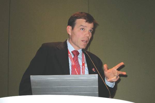
“There is a rather remarkable persistence of benefit in terms of risk reduction over a long period. You’ve changed the natural history of the disease in some way by lowering LDL,” Dr. Chris J. Packard said in presenting the 20-year WOSCOPS (West of Scotland Coronary Prevention Study) follow-up at the American Heart Association scientific sessions.
The primary prevention study randomized 6,595 middle-aged Scotsmen with an average baseline LDL cholesterol of 190 mg/dL to 4 years of pravastatin at 40 mg/day or placebo. This was one of the early large statin trials, and publication of the 5-year outcomes showing a 31% reduction in the relative risk of cardiovascular death or MI (N. Engl. J. Med. 1995;333:1301-8) caused a great stir.
At that point, WOSCOPS leaders advised study participants’ primary care physicians to seriously consider putting their patients on long-term statin therapy. However, only an identically low 31% of subjects in each of the two study arms did so.
Because the Scottish national health care system effectively captures all utilization of medical services, Dr. Packard and coinvestigators were able to analyze 20-year outcomes in the former study participants. No other major statin trial has come close in terms of length of reported follow-up.
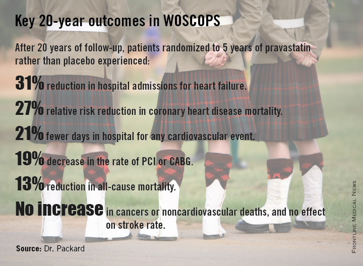
At 20 years, with the only treatment difference between the two original study arms being that the pravastatin group had been on statin therapy for an additional 5 years, the 20-year coronary heart disease mortality rate in the original statin group was reduced by 27%, compared with the original controls. All-cause mortality was reduced by 13%. And numerous other benefits were noted at 20 years with 5 years of statin therapy.
Indeed, patients treated with pravastatin for 5 years during the trial gained an average of 5 extra years free of nonfatal MI or cardiovascular death at the 20-year mark, he said.
“The average age of the men was 55 years during the trial and 20 years on they’re now 75 years old. This covers the entire period of premature cardiovascular morbidity and mortality. We would argue that this is a good picture of the lifetime benefit, which is different from lifetime risk. This is real events happening to real people, not predictions,” observed Dr. Packard, professor of cardiovascular and medical sciences at the University of Glasgow, Scotland.
The group given pravastatin for 5 years collectively spent 24,038 days in the hospital for any cardiovascular event, compared with 30,342 days for controls.
Particularly noteworthy was the divergence in the risk of heart failure, with 96 cases being diagnosed during 20 years in patients who received 5 years of pravastatin, compared with 128 in controls.
“Heart failure was our biggest surprise,” he said. “We got no result for heart failure at all at 5 years; these were middle-aged men with just high cholesterol, so we didn’t see much in the way of incident heart failure. But extrapolating 20 years, there is a 31% risk reduction in the incidence of hospitalization for heart failure in the statin-treated group, compared to the placebo-treated group. This is a remarkable finding.”
For WOSCOPS participants who got a 20% drop in LDL during 5 years of pravastatin from a baseline of 190 mg/dL, as was typical, the number of patients who needed to be treated (NNT) for 5 years to prevent one cardiovascular hospital admission over a 20-year period was six. Moreover, for every 10 patients on statin therapy for 5 years, an average of 19 hospital days were avoided over the course of 20 years. For patients with a baseline LDL of 120 mg/dL, the NNT was 10, and 12 hospital days were saved over a 20-year period for every 10 patients treated for 5 years.
“This is a real savings to the health service and the health care providers to offset the cost of drugs,” Dr. Packard continued.
He urged physicians to look beyond the initial reports of primary outcomes of the statin clinical trials and take a long-term view.
“If you take a lifetime approach to benefit so you’re looking not only at the first event, but the second event, the third, at heart failure, and at death, you can see tremendous benefits, whereas usually we only focus on the first event. And that’s not the full cost evaluation that you need to do,” according to Dr. Packard.
The overall incidence of cancer during 20 years of follow-up was 24.8% in the initial placebo arm and 24.5% in the pravastatin group, with no differences between the two groups in any type of cancer. Nor did the original pravastatin group show any increase in noncardiovascular mortality.
“This is a very important study – the first study to show a legacy effect, with reduced mortality and a gain of 5 event-free years over 20 years attributable to a 5-year treatment allocation,” said discussant Harvey White of Auckland (New Zealand) City Hospital.
This legacy effect, he added, can be viewed as an ongoing carryover effect related to statin-induced slowing and/or stabilization of existing coronary artery plaque. The mechanism is unknown, Dr. White said, but the key to why the legacy effect was seen in WOSCOPS despite the use of pravastatin – a less potent statin – but not to date in other statin trials may lie in the fact that WOSCOPS was a primary prevention study and its participants had the youngest mean age of all the major statin trials.
“Their plaques may not have been calcified yet and therefore were more able to be modified and stabilized. If you treat very early you might get a bigger effect,” said to Dr. White.
Undercutting that argument, however, was the WOSCOPS finding that the long-term benefits of 5 years of pravastatin were independent of age at treatment, Dr. Packard said.
He believes based upon other studies that statins’ coronary disease prevention benefits are expressed within the first 12 months after starting therapy.
“It suggests that whatever is happening to the pathobiology of atherosclerosis happens within a year, and somehow a statis is introduced into plaque. That’s my guess, that an unstable plaque is reduced to a stable one. People then form a new trajectory going forward and they never catch up. We should think of atherosclerosis as a rate effect rather than something that either happens or doesn’t happen. It’s a rate of happening,” Dr. Packard asserted.
Asked why in the aftermath of the strongly positive 5-year results of WOSCOPS only 31% of patients in each treatment arm were on long-term statin therapy, he replied that 20 years ago in Scotland there really was no push for primary prevention.
“The 4S trial had come out the year before [Lancet 1994;344:1383-9] and placed the focus on secondary prevention. Statins were relatively expensive and everybody was putting their money into secondary prevention. Our health care system, which is socialized, had not put any emphasis at all on primary prevention. We were actually amazed that even 31% got treated,” the physician explained.
Dr. Packard reported serving as a consultant to Merck, Roche, and AstraZeneca.
CHICAGO – Five years of statin therapy for primary prevention provided an impressive lifetime benefit expressed as reduced risks of a range of cardiovascular disease outcomes in a 20-year follow-up of the landmark WOSCOPS trial.
“There is a rather remarkable persistence of benefit in terms of risk reduction over a long period. You’ve changed the natural history of the disease in some way by lowering LDL,” Dr. Chris J. Packard said in presenting the 20-year WOSCOPS (West of Scotland Coronary Prevention Study) follow-up at the American Heart Association scientific sessions.
The primary prevention study randomized 6,595 middle-aged Scotsmen with an average baseline LDL cholesterol of 190 mg/dL to 4 years of pravastatin at 40 mg/day or placebo. This was one of the early large statin trials, and publication of the 5-year outcomes showing a 31% reduction in the relative risk of cardiovascular death or MI (N. Engl. J. Med. 1995;333:1301-8) caused a great stir.
At that point, WOSCOPS leaders advised study participants’ primary care physicians to seriously consider putting their patients on long-term statin therapy. However, only an identically low 31% of subjects in each of the two study arms did so.
Because the Scottish national health care system effectively captures all utilization of medical services, Dr. Packard and coinvestigators were able to analyze 20-year outcomes in the former study participants. No other major statin trial has come close in terms of length of reported follow-up.
At 20 years, with the only treatment difference between the two original study arms being that the pravastatin group had been on statin therapy for an additional 5 years, the 20-year coronary heart disease mortality rate in the original statin group was reduced by 27%, compared with the original controls. All-cause mortality was reduced by 13%. And numerous other benefits were noted at 20 years with 5 years of statin therapy.
Indeed, patients treated with pravastatin for 5 years during the trial gained an average of 5 extra years free of nonfatal MI or cardiovascular death at the 20-year mark, he said.
“The average age of the men was 55 years during the trial and 20 years on they’re now 75 years old. This covers the entire period of premature cardiovascular morbidity and mortality. We would argue that this is a good picture of the lifetime benefit, which is different from lifetime risk. This is real events happening to real people, not predictions,” observed Dr. Packard, professor of cardiovascular and medical sciences at the University of Glasgow, Scotland.
The group given pravastatin for 5 years collectively spent 24,038 days in the hospital for any cardiovascular event, compared with 30,342 days for controls.
Particularly noteworthy was the divergence in the risk of heart failure, with 96 cases being diagnosed during 20 years in patients who received 5 years of pravastatin, compared with 128 in controls.
“Heart failure was our biggest surprise,” he said. “We got no result for heart failure at all at 5 years; these were middle-aged men with just high cholesterol, so we didn’t see much in the way of incident heart failure. But extrapolating 20 years, there is a 31% risk reduction in the incidence of hospitalization for heart failure in the statin-treated group, compared to the placebo-treated group. This is a remarkable finding.”
For WOSCOPS participants who got a 20% drop in LDL during 5 years of pravastatin from a baseline of 190 mg/dL, as was typical, the number of patients who needed to be treated (NNT) for 5 years to prevent one cardiovascular hospital admission over a 20-year period was six. Moreover, for every 10 patients on statin therapy for 5 years, an average of 19 hospital days were avoided over the course of 20 years. For patients with a baseline LDL of 120 mg/dL, the NNT was 10, and 12 hospital days were saved over a 20-year period for every 10 patients treated for 5 years.
“This is a real savings to the health service and the health care providers to offset the cost of drugs,” Dr. Packard continued.
He urged physicians to look beyond the initial reports of primary outcomes of the statin clinical trials and take a long-term view.
“If you take a lifetime approach to benefit so you’re looking not only at the first event, but the second event, the third, at heart failure, and at death, you can see tremendous benefits, whereas usually we only focus on the first event. And that’s not the full cost evaluation that you need to do,” according to Dr. Packard.
The overall incidence of cancer during 20 years of follow-up was 24.8% in the initial placebo arm and 24.5% in the pravastatin group, with no differences between the two groups in any type of cancer. Nor did the original pravastatin group show any increase in noncardiovascular mortality.
“This is a very important study – the first study to show a legacy effect, with reduced mortality and a gain of 5 event-free years over 20 years attributable to a 5-year treatment allocation,” said discussant Harvey White of Auckland (New Zealand) City Hospital.
This legacy effect, he added, can be viewed as an ongoing carryover effect related to statin-induced slowing and/or stabilization of existing coronary artery plaque. The mechanism is unknown, Dr. White said, but the key to why the legacy effect was seen in WOSCOPS despite the use of pravastatin – a less potent statin – but not to date in other statin trials may lie in the fact that WOSCOPS was a primary prevention study and its participants had the youngest mean age of all the major statin trials.
“Their plaques may not have been calcified yet and therefore were more able to be modified and stabilized. If you treat very early you might get a bigger effect,” said to Dr. White.
Undercutting that argument, however, was the WOSCOPS finding that the long-term benefits of 5 years of pravastatin were independent of age at treatment, Dr. Packard said.
He believes based upon other studies that statins’ coronary disease prevention benefits are expressed within the first 12 months after starting therapy.
“It suggests that whatever is happening to the pathobiology of atherosclerosis happens within a year, and somehow a statis is introduced into plaque. That’s my guess, that an unstable plaque is reduced to a stable one. People then form a new trajectory going forward and they never catch up. We should think of atherosclerosis as a rate effect rather than something that either happens or doesn’t happen. It’s a rate of happening,” Dr. Packard asserted.
Asked why in the aftermath of the strongly positive 5-year results of WOSCOPS only 31% of patients in each treatment arm were on long-term statin therapy, he replied that 20 years ago in Scotland there really was no push for primary prevention.
“The 4S trial had come out the year before [Lancet 1994;344:1383-9] and placed the focus on secondary prevention. Statins were relatively expensive and everybody was putting their money into secondary prevention. Our health care system, which is socialized, had not put any emphasis at all on primary prevention. We were actually amazed that even 31% got treated,” the physician explained.
Dr. Packard reported serving as a consultant to Merck, Roche, and AstraZeneca.
AT THE AHA SCIENTIFIC SESSIONS
Key clinical point: Five years of statin therapy during midlife for primary prevention may reset the clock for atherosclerotic progression, providing persistently lower cardiovascular event rates 20 years later.
Major finding: The number of patients who needed to be treated with pravastatin for 5 years to prevent one cardiovascular hospitalization over a 20-year period was six.
Data source: A 20-year follow-up of the West of Scotland Coronary Prevention Study, in which 6,595 middle-aged men with high cholesterol were randomized to 5 years of pravastatin or placebo.
Disclosures: WOSCOPS was sponsored by Bristol-Myers Squibb. The presenter reported serving as a consultant to Merck, Roche, and AstraZeneca.

Elevated troponin present in 40% with T2D and stable heart disease
CHICAGO– Abnormal levels of high-sensitivity cardiac troponin T are present in 40% of type 2 diabetic patients with stable ischemic heart disease, and they do not bode well, according to a new secondary analysis of the BARI 2D study.
In BARI 2D, an abnormal high-sensitivity cardiac troponin T (hsTnT), defined as 14 ng/L or greater, a powerful marker of ongoing myocardial injury, was independently associated with a doubled 5-year risk of the composite endpoint of cardiovascular death, MI, or stroke. Moreover, and discouragingly so, prompt coronary revascularization did nothing to mitigate that risk, Dr. Brendan M. Everett reported at the American Heart Association Scientific Sessions.
Further, early coronary revascularization did not result in a reduction in abnormal hsTnT at 1 year of follow-up, said Dr. Everett, director of the general cardiology inpatient service at Brigham and Women’s Hospital, Boston.

“To better address the risk represented by an abnormal hsTnT, we need to gain an improved understanding of the biology of troponin release in this population,” he observed. “The fact that we saw an overall decrease of about 0.5% in hemoglobin A1c and an LDL reduction of 16 mg/dL at 1 year and still there was no change in hsTnT leaves me scratching my head. The abnormal hsTnT is clearly a marker of badness, but where is it coming from? Can we address it? Or are we just left to look at it and worry about our patients who have an abnormal hsTnT?”
The BARI 2D trial was designed to learn whether patients with type 2 diabetes and stable ischemic heart disease benefit from prompt coronary revascularization plus intensive medical therapy as compared with intensive medical therapy alone. As previously reported (N. Engl. J. Med. 2009;360:2503-15), this proved not to be the case; prompt revascularization conferred no outcome advantage.
The aim of Dr. Everett’s new secondary analysis of BARI 2D was to learn if the hsTnT assay can be used to identify a subgroup of patients with type 2 diabetes and stable ischemic heart disease who might benefit from prompt coronary revascularization. The rationale was that, in patients with acute coronary syndromes, it’s well established that an abnormal hsTnT is associated with poor prognosis, and such patients would benefit from early revascularization.
The secondary analysis included 2,285 type 2 diabetics with stable ischemic heart disease whose physicians first decided whether they were better candidates for percutaneous coronary intervention or CABG surgery. Patients were then randomized to prompt revascularization by the preferred method plus intensive medical therapy or to intensive medical therapy alone.
Forty percent of participants had an abnormal hsTnT at baseline. Their 5-year rate of the composite primary endpoint of cardiovascular death, MI, or stroke was 27.1%, compared with 12.9% in patients with a baseline hsTnT below 14 ng/mL. After adjusting in a multivariate analysis for various potential confounders – including age, race, and the standard cardiovascular risk factors – the group with an abnormal baseline hsTnT had a 2.09-fold increased risk of a major cardiovascular event.
Early revascularization, regardless of whether by percutaneous coronary intervention or coronary artery bypass graft surgery, provided no benefit no matter what the patient’s baseline hsTnT level. In patients with an hsTnT of 14 ng/L or greater, the 5-year rate of the composite outcome was 26.5% with early revascularization compared with 27.6% with intensive medical therapy. In those with an hsTnT below 14 ng/L, the rate was 11.8% in the early revascularization group and 14% with medical management, a trend favoring prompt revascularization that didn’t achieve statistical significance, according to Dr. Everett.
Of patients with an abnormal hsTnT at baseline, 77% still had an abnormal value at 1 year, regardless of whether they underwent prompt revascularization or intensive medical therapy alone.
Session moderator Dr. Mikhail N. Kosiborod commented that the new BARI 2D substudy highlights a dilemma: “We know that a large population of patients with diabetes, and to some extent those with prediabetes, have elevated hsTnT levels, and we know those patients don’t do well. What we don’t know is what to do about it.”
“What [Dr. Everett’s] study clearly demonstrates is that this does not appear to be driven by epicardial coronary artery disease. If we fix the epicardial CAD, it has absolutely no impact on the outcomes nor on the actual troponin level at follow-up. As far as I can tell, it doesn’t appear to be a glycemic control issue, either. It appears that this is a humoral issue. There are ‘evil humors’ – whatever they are – and we don’t really understand what they are or what to do about it,” said Dr. Kosiborod, professor of medicine at the University of Missouri, Kansas City.
“The truth of the matter is we have no idea what’s causing this low-grade myocardial necrosis, and it’s a hugely important thing,” he continued. “There is absolutely no question that elevated hsTnT, even at very low levels, has a huge impact on subsequent risk of heart failure. We know what the public health effects of heart failure are. And patients with diabetes and heart failure tend to do particularly poorly.”
The BARI 2D trial was funded by the National Institutes of Health. Dr. Everett’s secondary analysis was funded by Roche Diagnostics. He reported receiving research grants from Roche and Novartis.
CHICAGO– Abnormal levels of high-sensitivity cardiac troponin T are present in 40% of type 2 diabetic patients with stable ischemic heart disease, and they do not bode well, according to a new secondary analysis of the BARI 2D study.
In BARI 2D, an abnormal high-sensitivity cardiac troponin T (hsTnT), defined as 14 ng/L or greater, a powerful marker of ongoing myocardial injury, was independently associated with a doubled 5-year risk of the composite endpoint of cardiovascular death, MI, or stroke. Moreover, and discouragingly so, prompt coronary revascularization did nothing to mitigate that risk, Dr. Brendan M. Everett reported at the American Heart Association Scientific Sessions.
Further, early coronary revascularization did not result in a reduction in abnormal hsTnT at 1 year of follow-up, said Dr. Everett, director of the general cardiology inpatient service at Brigham and Women’s Hospital, Boston.
“To better address the risk represented by an abnormal hsTnT, we need to gain an improved understanding of the biology of troponin release in this population,” he observed. “The fact that we saw an overall decrease of about 0.5% in hemoglobin A1c and an LDL reduction of 16 mg/dL at 1 year and still there was no change in hsTnT leaves me scratching my head. The abnormal hsTnT is clearly a marker of badness, but where is it coming from? Can we address it? Or are we just left to look at it and worry about our patients who have an abnormal hsTnT?”
The BARI 2D trial was designed to learn whether patients with type 2 diabetes and stable ischemic heart disease benefit from prompt coronary revascularization plus intensive medical therapy as compared with intensive medical therapy alone. As previously reported (N. Engl. J. Med. 2009;360:2503-15), this proved not to be the case; prompt revascularization conferred no outcome advantage.
The aim of Dr. Everett’s new secondary analysis of BARI 2D was to learn if the hsTnT assay can be used to identify a subgroup of patients with type 2 diabetes and stable ischemic heart disease who might benefit from prompt coronary revascularization. The rationale was that, in patients with acute coronary syndromes, it’s well established that an abnormal hsTnT is associated with poor prognosis, and such patients would benefit from early revascularization.
The secondary analysis included 2,285 type 2 diabetics with stable ischemic heart disease whose physicians first decided whether they were better candidates for percutaneous coronary intervention or CABG surgery. Patients were then randomized to prompt revascularization by the preferred method plus intensive medical therapy or to intensive medical therapy alone.
Forty percent of participants had an abnormal hsTnT at baseline. Their 5-year rate of the composite primary endpoint of cardiovascular death, MI, or stroke was 27.1%, compared with 12.9% in patients with a baseline hsTnT below 14 ng/mL. After adjusting in a multivariate analysis for various potential confounders – including age, race, and the standard cardiovascular risk factors – the group with an abnormal baseline hsTnT had a 2.09-fold increased risk of a major cardiovascular event.
Early revascularization, regardless of whether by percutaneous coronary intervention or coronary artery bypass graft surgery, provided no benefit no matter what the patient’s baseline hsTnT level. In patients with an hsTnT of 14 ng/L or greater, the 5-year rate of the composite outcome was 26.5% with early revascularization compared with 27.6% with intensive medical therapy. In those with an hsTnT below 14 ng/L, the rate was 11.8% in the early revascularization group and 14% with medical management, a trend favoring prompt revascularization that didn’t achieve statistical significance, according to Dr. Everett.
Of patients with an abnormal hsTnT at baseline, 77% still had an abnormal value at 1 year, regardless of whether they underwent prompt revascularization or intensive medical therapy alone.
Session moderator Dr. Mikhail N. Kosiborod commented that the new BARI 2D substudy highlights a dilemma: “We know that a large population of patients with diabetes, and to some extent those with prediabetes, have elevated hsTnT levels, and we know those patients don’t do well. What we don’t know is what to do about it.”
“What [Dr. Everett’s] study clearly demonstrates is that this does not appear to be driven by epicardial coronary artery disease. If we fix the epicardial CAD, it has absolutely no impact on the outcomes nor on the actual troponin level at follow-up. As far as I can tell, it doesn’t appear to be a glycemic control issue, either. It appears that this is a humoral issue. There are ‘evil humors’ – whatever they are – and we don’t really understand what they are or what to do about it,” said Dr. Kosiborod, professor of medicine at the University of Missouri, Kansas City.
“The truth of the matter is we have no idea what’s causing this low-grade myocardial necrosis, and it’s a hugely important thing,” he continued. “There is absolutely no question that elevated hsTnT, even at very low levels, has a huge impact on subsequent risk of heart failure. We know what the public health effects of heart failure are. And patients with diabetes and heart failure tend to do particularly poorly.”
The BARI 2D trial was funded by the National Institutes of Health. Dr. Everett’s secondary analysis was funded by Roche Diagnostics. He reported receiving research grants from Roche and Novartis.
AT THE AHA SCIENTIFIC SESSIONS
Key clinical point: Forty percent of patients with type 2 diabetes and stable ischemic heart disease are walking around with abnormal levels of hsTnT, placing them at substantial risk of a major cardiovascular event within 5 years.
Major finding: Prompt coronary revascularization does not reduce this risk, nor does it reduce the elevated troponin T level.
Data source: A secondary analysis of the randomized, prospective BARI 2D study involving 2,285 participants with type 2 diabetes and stable ischemic heart disease.
Disclosures: The BARI 2D study was funded by the National Institutes of Health. This new secondary analysis was funded by a research grant from Roche Diagnostics.
Insulin glargine shows cardiac safety in ORIGIN-ECHO
CHICAGO – Insulin glargine showed no effects on left ventricular mass or function during 3 years of follow-up in dysglycemic patients at high cardiovascular risk in the ORIGIN echocardiographic substudy.
This echocardiographic study of the ORIGIN (Outcome Reduction With an Initial Glargine Intervention) trial, the largest reported study of the effects of exogenous insulin on left ventricular mass and LV systolic and diastolic function, provides reassuring new evidence that insulin glargine is safe from a cardiac standpoint, Dr. Michelle Haroun said at the American Heart Association scientific sessions.
“The key here is that we didn’t see any signal whatsoever to suggest that insulin is putting patients at increased risk. We think that this finding is important. While you need to follow patients for a very long time to detect changes in clinical heart failure outcomes, we think we’d be able to detect subtle changes in endpoints like LV mass over a 3-year period if insulin was of harm to patients,” said Dr. Haroun of the Population Health Research Institute at McMaster University in Hamilton, Ont.
ORIGIN-ECHO involved 564 dysglycemic patients at high cardiovascular risk who were randomized to insulin glargine (Lantus) or standard therapy. All had echocardiograms at baseline and after 3 years of therapy. Participants had to have impaired fasting blood glucose, impaired glucose tolerance, or early type 2 diabetes managed with no more than one oral antiglycemic drug at baseline. This was a group at high cardiovascular risk: 32% had a prior MI, 84% had a history of hypertension, obesity was common, and the average age was 64. However, none of the participants had heart failure at baseline.
The study was undertaken because some of the medications used to treat hyperglycemia are associated with increased risk of heart failure. Regulatory agencies, physicians, and patients want to see evidence of cardiovascular safety, and until ORIGIN-ECHO, the effects of exogenous insulin on LV mass and function hadn’t been well studied.
Baseline LV mass and function values were within normal range and did not change significantly over 3 years of follow-up in either treatment arm. For example, left ventricular mass/height averaged 116 g/m at baseline and 115 g/m after 3 years on insulin glargine, and was comparable at 113 and 114 g/m, respectively, with standard therapy. This was an unexpected finding, according to Dr. Haroun.
“We thought patients with diabetes on standard therapy were going to develop left ventricular hypertrophy over a 3-year follow-up period, and they didn’t. That came as a bit of a surprise to us. We expected to see a lower rate of LVH in the patients on insulin glargine. This patient population was relatively early in their course of diabetes, and we believe our findings suggest that adequate management of cardiovascular risk factors – especially hypertension– and the use of cardioprotective drugs in this population may prevent or delay abnormalities in LV structure and function,” she said.
The primary outcomes of the full ORIGIN study involving more than 12,000 patients have previously been published (N. Engl. J. Med. 2012; 367:319-28). ORIGIN was sponsored by Sanofi. Dr. Haroun reported having no financial conflicts.
AT THE AHA SCIENTIFIC SESSIONS
Key clinical point: The cardiac safety of insulin glargine in dysglycemic patients at high cardiovascular risk has received strong support from a 3-year echocardiographic study.
Major finding: Left ventricular mass over height was 116 g/m at baseline and 115 g/m after 3 years on insulin glargine.
Data source: The ORIGIN-ECHO substudy included 564 dysglycemic patients at high cardiovascular risk who were randomized to 3 years of insulin glargine or standard therapy.
Disclosures: The ORIGIN trial was sponsored by Sanofi. The presenter reported having no financial conflicts.
Key definitions, data standards established for CV endpoints
Universal definitions and data standards have now been established for clinical research involving cardiovascular endpoints, according to a report published online Dec. 29 in both the Journal of the American College of Cardiology and Circulation.
The American College of Cardiology/American Heart Association Task Force on Clinical Data Standards, in collaboration with the Food and Drug Administration and the Standardized Data Collection for Cardiovascular Trials Initiative (SCTI), described the standards as “a first step in developing a universal language for clinical trials and other types of health-related research.” The aim is to make patient data regarding CV endpoints as consistent as possible across electronic health records, clinical trial databases, registries, drug/device surveillance programs, and other health-related research, to facilitate information sharing, said Dr. Karen A. Hicks, chair of the task force’s writing committee to develop cardiovascular endpoints data standards, and her associates.

The report defines CV endpoints such as cardiovascular cause of death (as opposed to noncardiovascular or undetermined causes of death), MI, hospitalization for unstable angina, TIA, stroke, heart failure event, percutaneous coronary intervention, peripheral vascular intervention, and stent thrombosis. For example, the report notes that the outcome “hospitalization for unstable angina” is frequently assessed in research evaluating the safety of CV therapies, but by necessity involves subjective assessment of the most likely etiology of the symptoms that trigger hospitalization. To clarify that this outcome is truly due to cardiovascular ischemia, the definition must now include the presence of ECG abnormalities, such as deviations in the ST segment, the morphology of ST-segment changes (horizontal or downsloping vs. upsloping), and the magnitude of the deviation.
Similarly, the report’s definition of TIA now emphasizes the patient’s clinical presentation rather than the anatomic location of the lesion, because the availability of imaging modalities varies so much across medical centers. And, because heart failure can result from so many different etiologies, the key element in the definition of a heart failure event now is “the need for a resource-intensive response to failure of the primary therapeutic management strategy.”
“What makes this work unique is that it reviews and refines the terms as developed by the SCTI explicitly for use in reporting clinical trial results and in regulatory submissions, and it delineates where these concepts could or should not be used as the foundational vocabulary in routine clinical care,” Dr. Hicks, a cardiologist at the FDA, and her associates stated.
The SCTI is a working group comprised of experts from academia, professional societies, the FDA, and manufacturers of pharmaceuticals and CV devices. The ACC/AHA task force included 16 people with expertise in internal medicine, cardiovascular medicine, neurology, clinical research, epidemiology, invasive and interventional therapies, outcomes assessment, medical informatics, health information management, and healthcare services research and delivery.
The full report is available from the American College of Cardiology at www.acc.org and from the American Heart Association at www.myamericanheart.org.
This work received no commercial support. Dr. Hicks and her associates volunteered their time and were supported exclusively by the ACC and AHA.
Universal definitions and data standards have now been established for clinical research involving cardiovascular endpoints, according to a report published online Dec. 29 in both the Journal of the American College of Cardiology and Circulation.
The American College of Cardiology/American Heart Association Task Force on Clinical Data Standards, in collaboration with the Food and Drug Administration and the Standardized Data Collection for Cardiovascular Trials Initiative (SCTI), described the standards as “a first step in developing a universal language for clinical trials and other types of health-related research.” The aim is to make patient data regarding CV endpoints as consistent as possible across electronic health records, clinical trial databases, registries, drug/device surveillance programs, and other health-related research, to facilitate information sharing, said Dr. Karen A. Hicks, chair of the task force’s writing committee to develop cardiovascular endpoints data standards, and her associates.
The report defines CV endpoints such as cardiovascular cause of death (as opposed to noncardiovascular or undetermined causes of death), MI, hospitalization for unstable angina, TIA, stroke, heart failure event, percutaneous coronary intervention, peripheral vascular intervention, and stent thrombosis. For example, the report notes that the outcome “hospitalization for unstable angina” is frequently assessed in research evaluating the safety of CV therapies, but by necessity involves subjective assessment of the most likely etiology of the symptoms that trigger hospitalization. To clarify that this outcome is truly due to cardiovascular ischemia, the definition must now include the presence of ECG abnormalities, such as deviations in the ST segment, the morphology of ST-segment changes (horizontal or downsloping vs. upsloping), and the magnitude of the deviation.
Similarly, the report’s definition of TIA now emphasizes the patient’s clinical presentation rather than the anatomic location of the lesion, because the availability of imaging modalities varies so much across medical centers. And, because heart failure can result from so many different etiologies, the key element in the definition of a heart failure event now is “the need for a resource-intensive response to failure of the primary therapeutic management strategy.”
“What makes this work unique is that it reviews and refines the terms as developed by the SCTI explicitly for use in reporting clinical trial results and in regulatory submissions, and it delineates where these concepts could or should not be used as the foundational vocabulary in routine clinical care,” Dr. Hicks, a cardiologist at the FDA, and her associates stated.
The SCTI is a working group comprised of experts from academia, professional societies, the FDA, and manufacturers of pharmaceuticals and CV devices. The ACC/AHA task force included 16 people with expertise in internal medicine, cardiovascular medicine, neurology, clinical research, epidemiology, invasive and interventional therapies, outcomes assessment, medical informatics, health information management, and healthcare services research and delivery.
The full report is available from the American College of Cardiology at www.acc.org and from the American Heart Association at www.myamericanheart.org.
This work received no commercial support. Dr. Hicks and her associates volunteered their time and were supported exclusively by the ACC and AHA.
FROM JACC AND CIRCULATION
Key clinical point: The ACC and AHA collaborated with the FDA to establish a common vocabulary and data standards for research involving cardiovascular endpoints.
Major finding: The report defines CV endpoints such as cardiovascular cause of death (as opposed to noncardiovascular or undetermined causes of death), MI, hospitalization for unstable angina, TIA, stroke, heart failure event, PCI, peripheral vascular intervention, and stent thrombosis.
Data source: A document compiled by experts in academia, professional societies, the FDA, and manufacturers of pharmaceuticals and devices, which establishes universal definitions and practices for capturing CV event information.
Disclosures: This work received no commercial support. Dr. Hicks and her associates volunteered their time and were supported exclusively by the ACC and AHA.

When cardiologists attend meetings, do patients benefit?
Can the high-intensity care given acutely ill, high-risk U.S. patients with cardiac disease actually harm them?
Results from an unusual analysis of cardiology-meeting times seem to suggest that sobering possibility. Patient outcomes improved when thousands of high-level, American cardiologists left their practices for a few days each year to attend either of the two major U.S. heart disease meetings.
Researchers led by Dr. Anupam B. Jena of Harvard University, Boston, used Medicare data to examine mortality rates among patients hospitalized for cardiac arrest, heart failure, or acute myocardial infarction during 2002-2011. They focused on patients admitted during the annual meetings of the American College of Cardiology (usually in March) or the American Heart Association (during November).
As controls in their case-control analyses, they used data from patients admitted on similar days of the week during the 3 weeks immediately before or after these two meetings. This gave them roughly 11,000 total patients with cardiac arrest, nearly 134,000 with heart failure, and about 60,000 with acute MI – about 14% of patients in each disease category admitted during a meeting and the other 86% (controls) admitted when there was no meeting.
The results showed some statistically significant differences indicating that patients did better during the meetings, presumably when many cardiologists were away from their hospitals. These associations only occurred at teaching hospitals and among patients at high risk for inpatient mortality. The investigators saw no statistically significant differences, after adjustment, in mortality during meetings among patients treated at nonteaching hospitals or among patients with a low risk for inpatient mortality.
In analyses that adjusted for baseline differences in risk factors, the 30-day mortality rate for patients admitted to teaching hospitals with cardiac arrest was 69% during control dates and 59% during the meetings. Thirty-day mortality for patients admitted with heart failure was 25% during control dates and 18% during the meetings, researchers reported in an article published online on Dec. 22 (JAMA Intern. Med. 2014 [doi:10.1001/jamainternmed.2014.6781]).
Although 30-day mortality among patients admitted with an acute MI did not differ significantly at teaching hospitals between patients who presented during a major meeting and those who did not, the results showed that these similar mortality rates were achieved despite a statistically significant difference in the rate of percutaneous coronary interventions (PCI) that patients received: During the major meetings, 21% of the acute MI patients underwent PCI, but when a meeting was not in progress, the PCI rate jumped to 28% of all acute MI patients.
“One explanation for these findings is that the intensity of care provided during meeting dates is lower, and that for high-risk patients with cardiovascular disease, the harms of this care may unexpectedly outweigh the benefits,” Dr. Jena concluded.
It’s a remarkable and surprising finding, but can it be taken seriously? At least one expert said yes, at least seriously enough to warrant further study and consideration.
An editor’s note published with the new report suggested a plausible explanation for the findings is that “more interventions in high-risk patients with heart failure and cardiac arrest leads to higher mortality.” In her note, Dr. Rita F. Redberg, a cardiologist at the University of California, San Francisco, and editor of JAMA Internal Medicine, concluded, “It is reassuring that patient outcomes do not suffer while many cardiologists are away. More important, this analysis may help us to understand how we could lower mortality throughout the year.”
It will be interesting to see if anyone takes up the challenge to further explore this relationship and tries to find ways to apply throughout the year the protective effect of having fewer teaching-hospital cardiologists around. If a drug had this beneficial effect on mortality, the pharmaceutical industry would be all over it.
On Twitter @mitchelzoler

Takotsubo cardiomyopathy: predicting in-hospital mortality
CHICAGO – A novel risk score has been developed for predicting the risk of in-hospital mortality in patients with takotsubo cardiomyopathy.
The new risk score’s unique strength is that it was developed using hospital data on a huge patient population with this rare and incompletely understood cardiac condition: 10,582 patients hospitalized for takotsubo cardiomyopathy in seven U.S. states, Dr. David P. Kao reported at the American Heart Association scientific sessions.
He used multivariate logistic regression analysis to identify seven independent characteristics predictive of mortality in the study population. Their collective area under the curve as predictors was 0.70, which is considered good. And while the overall in-hospital mortality rate was low at 4.4%, it varied enormously depending upon how many of the seven risk factors were present, according to Dr. Kao of the University of Colorado, Denver.
In-hospital mortality ranged from 1.6% in the 2,585 patients with none of the risk factors to 19.6% in those with three or more. The 616 patients with at least three risk factors accounted for 22% of all in-hospital deaths in patients with takotsubo cardiomyopathy in California, New York, New Jersey, Colorado, West Virginia, New Hampshire, and Vermont during 2006-2012.
Intracranial hemorrhage, which was present in 2% of hospitalized takotsubo cardiomyopathy patients, was the most potent predictor both of in-hospital mortality and major adverse events. In multivariate analysis, intracranial hemorrhage was independently associated with a 6.8-fold increased risk of in-hospital mortality. The other mortality risk factors and their associated odds ratios were age 60 years or older, with a 1.8-fold risk; Asian race, 1.8-fold; male sex, 1.9-fold; acute renal failure, 4.1-fold; atrial fibrillation or flutter, 1.7-fold; and stroke, 2.9-fold.
The simple, user-friendly risk score is derived by totaling the number of risk factors present in a given hospitalized patient. The presence of each additional risk factor increased the odds of in-hospital death by 2.2-fold, according to Dr. Kao.

Takotsubo cardiomyopathy is marked by acute, typically rapidly reversible left ventricular dysfunction without evidence of epicardial coronary artery occlusion. It occurs most often in postmenopausal women in response to emotional or physical stress. Indeed, 89% of the more than 10,000 affected patients in this series were women.
Most adverse events in patients with takotsubo cardiomyopathy occur during their first hospitalization for the disorder. In this large series, 22% of patients experienced major adverse events, including ventricular arrhythmias, acute heart failure, cardiogenic shock, pulmonary edema, or ventricular rupture.
In a separate multiple logistic regression analysis, Dr. Kao and coinvestigator Dr. JoAnn Lindenfeld, also of the University of Colorado, identified eight characteristics independently predictive of in-hospital major adverse events. Five of them were also predictors of in-hospital mortality: intracranial hemorrhage, male gender, stroke, atrial fibrillation/flutter, and acute renal failure. The other three were age less than 60, substance abuse, and anemia. The major adverse event rate ranged from 10% in patients with none of the risk factors to 56% in the 242 patients having four or more. The 1,057 patients with three or more risk factors had a 47% major adverse event rate and accounted for 20% of all such events.
Dr. Kao reported having no financial conflicts related to this study.
CHICAGO – A novel risk score has been developed for predicting the risk of in-hospital mortality in patients with takotsubo cardiomyopathy.
The new risk score’s unique strength is that it was developed using hospital data on a huge patient population with this rare and incompletely understood cardiac condition: 10,582 patients hospitalized for takotsubo cardiomyopathy in seven U.S. states, Dr. David P. Kao reported at the American Heart Association scientific sessions.
He used multivariate logistic regression analysis to identify seven independent characteristics predictive of mortality in the study population. Their collective area under the curve as predictors was 0.70, which is considered good. And while the overall in-hospital mortality rate was low at 4.4%, it varied enormously depending upon how many of the seven risk factors were present, according to Dr. Kao of the University of Colorado, Denver.
In-hospital mortality ranged from 1.6% in the 2,585 patients with none of the risk factors to 19.6% in those with three or more. The 616 patients with at least three risk factors accounted for 22% of all in-hospital deaths in patients with takotsubo cardiomyopathy in California, New York, New Jersey, Colorado, West Virginia, New Hampshire, and Vermont during 2006-2012.
Intracranial hemorrhage, which was present in 2% of hospitalized takotsubo cardiomyopathy patients, was the most potent predictor both of in-hospital mortality and major adverse events. In multivariate analysis, intracranial hemorrhage was independently associated with a 6.8-fold increased risk of in-hospital mortality. The other mortality risk factors and their associated odds ratios were age 60 years or older, with a 1.8-fold risk; Asian race, 1.8-fold; male sex, 1.9-fold; acute renal failure, 4.1-fold; atrial fibrillation or flutter, 1.7-fold; and stroke, 2.9-fold.
The simple, user-friendly risk score is derived by totaling the number of risk factors present in a given hospitalized patient. The presence of each additional risk factor increased the odds of in-hospital death by 2.2-fold, according to Dr. Kao.
Takotsubo cardiomyopathy is marked by acute, typically rapidly reversible left ventricular dysfunction without evidence of epicardial coronary artery occlusion. It occurs most often in postmenopausal women in response to emotional or physical stress. Indeed, 89% of the more than 10,000 affected patients in this series were women.
Most adverse events in patients with takotsubo cardiomyopathy occur during their first hospitalization for the disorder. In this large series, 22% of patients experienced major adverse events, including ventricular arrhythmias, acute heart failure, cardiogenic shock, pulmonary edema, or ventricular rupture.
In a separate multiple logistic regression analysis, Dr. Kao and coinvestigator Dr. JoAnn Lindenfeld, also of the University of Colorado, identified eight characteristics independently predictive of in-hospital major adverse events. Five of them were also predictors of in-hospital mortality: intracranial hemorrhage, male gender, stroke, atrial fibrillation/flutter, and acute renal failure. The other three were age less than 60, substance abuse, and anemia. The major adverse event rate ranged from 10% in patients with none of the risk factors to 56% in the 242 patients having four or more. The 1,057 patients with three or more risk factors had a 47% major adverse event rate and accounted for 20% of all such events.
Dr. Kao reported having no financial conflicts related to this study.
AT THE AHA SCIENTIFIC SESSIONS
Key clinical point: The risk of in-hospital mortality in patients with takotsubo cardiomyopathy can be predicted using a simple risk score based upon information readily available at the bedside.
Major finding: In-hospital mortality ranged from just 1.6% in patients with no risk factors to 19.6% in those with any three or more.
Data source: The seven independent characteristics associated with in-hospital mortality were identified through multivariate logistic regression analysis applied to the hospital records of 10,582 patients with takotsubo cardiomyopathy in seven states.
Disclosures: The presenter reported having no financial conflicts in connection with this study, conducted free of commercial interests.

VIDEO: Focused cardiac ultrasound aids acute heart failure patients
VIENNA – Bedside echocardiography has become a key part of quickly assessing patients with acute heart failure to decide the best management strategy.
But bedside echo images often are challenging to interpret, so physicians performing an initial work-up of an acute heart failure patient need training in a basic echocardiography examination, Dr. Nuno Cardim said in an interview during the annual meeting of the European Association of Cardiovascular Imaging.
Known as Focused Cardiac Ultrasound (FoCUS), this triage-level exam is distinct from a comprehensive echocardiography assessment. Instead, FoCUS addresses some basic, yes-or-no, present-or-absent questions regarding systolic function, hypovolemia, tamponade, pleural effusion, and pulmonary embolus.
In a position paper and then in practice recommendations issued in 2014, the European Association of Cardiovascular Imaging acknowledged that use of FoCUS in acute heart failure patients was “irreversible,” said Dr. Cardim, professor and director of echocardiography and cardiac imaging at Hospital da Luz in Lisbon.
Cardiologists and echocardiography specialists need to make sure that FoCUS training is available to all physicians who might see suspected acute heart failure patients in emergency medicine settings, Dr. Cardim said.
Dr. Cardim had no disclosures.
The video associated with this article is no longer available on this site. Please view all of our videos on the MDedge YouTube channel
On Twitter @mitchelzoler
AT EUROECHO-IMAGING 2014
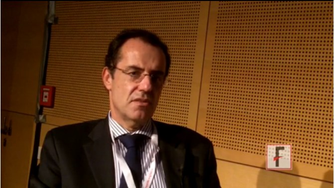
CABG plus mitral repair put under spotlight
CHICAGO – Early results from a randomized trial cast doubt on the benefits of routinely repairing a leaky mitral valve during coronary artery bypass grafting in patients with moderate ischemic mitral regurgitation.
At 1 year, there was no significant difference between patients undergoing CABG alone or CABG plus mitral repair in the primary endpoint of left ventricular reverse modeling, as defined by changes in LV end-systolic volume index (LVESVI) at 1 year (z score 0.50).

Both groups achieved significant reductions in LVESVI, with a median reduction of about 6 mL/m2 from baseline, Dr. Robert Michler, chair of cardiovascular and thoracic surgery at Montefiore Medical Center, New York, reported at the American Heart Association scientific sessions.
At 1 year, patients who underwent CABG and mitral valve repair had significantly less residual moderate or severe mitral regurgitation (11% vs. 31%; P < .001).
On the other hand, the combination procedure was associated with significantly higher rates of any neurologic event (9.6% vs. 3.1%; P = .03), longer cross-clamp (117 vs. 74 minutes) and cardiopulmonary bypass times (163 vs. 106 minutes; P values both < .001), and longer ICU (4.8 vs. 4.0 days; P = .006) and hospital length of stay (11.3 vs. 9.4 days; P = .002), according to the results, also published online (N. Engl. J. Med. 2014 [doi: 10.1056/NEJMoa1410490]).
“The trial did not demonstrate a clinically meaningful advantage to the routine addition of mitral valve repair to CABG,” Dr. Michler said, on behalf of the Cardiothoracic Surgical Trials Network investigators.
The 2014 AHA/American College of Cardiology mitral valve guidelines give a weak class IIb recommendation for mitral valve repair for patients with chronic mitral regurgitation (MR) who are undergoing other cardiac surgery, he noted.
The study evenly randomized 301 patients with moderate ischemic mitral regurgitation to CABG alone or CABG plus valve repair with an undersized ring (average, 28.3 mm for men and 27.1 mm for women).
The mean LVESVI at baseline was 54.8 mL/m2 in the CABG-alone group and 59.6 mL/m2 in the combined procedure group.
At 12 months, mean LVESVIs were 46.1 mL/m2 and 49.6 mL/m2, respectively.
The trial lacked a clinical primary endpoint, and longer follow-up is ongoing to determine whether the lower incidence of moderate or severe MR at 1 year will translate into a net clinical benefit for patients undergoing CABG plus mitral repair, Dr. Michler said.
Designated discussant Dr. David Adams, director of Mount Sinai Hospital’s mitral valve repair reference center in New York, cautioned the audience on the length of the study and called for a full 5 years of follow-up rather than the 2 years as planned.
“Ischemic mitral regurgitation is a disease that hurts you over time. That’s in patients that have had MI, had previous CABG, had attempted mitral valve repair, and had PCI [percutaneous coronary intervention]. So we need much longer term follow-up of this very important data set to really understand its implications,” he said.
In light of roughly half of the patients being in heart failure at baseline, session cochair Dr. Robert Bonow, vice chair of medicine, Northwestern University, Chicago, questioned whether there were differences in outcomes related to changes in baseline ejection fraction (EF) or whether improvement in EF in patients with low EF correlated with reduction in mitral regurgitation with CABG alone.
What is known right now is that mean LVEF increased to the same degree after CABG in both groups, Dr. Michler responded. This was true despite this being a “sick population of patients,” with more than half having diabetes, 50% with heart failure, 20% with renal insufficiency, 10% with prior stroke, and a mean ejection fraction of 40% in both groups.
“What we have yet to identify and plan to explore is the correlation between reverse ventricular remodeling, ejection fraction, and outcome, meaning both the degree of mitral regurgitation and whether there is any signal with respect to repeat hospitalizations, heart failure, or possibly even mortality,” Dr. Michler said.
Both Dr. Michler and Dr. Adams remarked that surgery was extremely safe for the CABG alone and CABG plus MR groups, as reflected by the low mortality at 30 days (2.7% vs. 1.3%) and 1 year (7.3% vs. 6.7%).
The composite endpoint of major adverse cardiac or cerebrovascular events was also similar between groups.
The trial was funded by the National Institutes of Health and the Canadian Institutes for Health Research. Dr. Michler reported grant support from NIH during the conduct of the study. Dr. Adams reported coinventing a mitral valve repair ring.
CHICAGO – Early results from a randomized trial cast doubt on the benefits of routinely repairing a leaky mitral valve during coronary artery bypass grafting in patients with moderate ischemic mitral regurgitation.
At 1 year, there was no significant difference between patients undergoing CABG alone or CABG plus mitral repair in the primary endpoint of left ventricular reverse modeling, as defined by changes in LV end-systolic volume index (LVESVI) at 1 year (z score 0.50).
Both groups achieved significant reductions in LVESVI, with a median reduction of about 6 mL/m2 from baseline, Dr. Robert Michler, chair of cardiovascular and thoracic surgery at Montefiore Medical Center, New York, reported at the American Heart Association scientific sessions.
At 1 year, patients who underwent CABG and mitral valve repair had significantly less residual moderate or severe mitral regurgitation (11% vs. 31%; P < .001).
On the other hand, the combination procedure was associated with significantly higher rates of any neurologic event (9.6% vs. 3.1%; P = .03), longer cross-clamp (117 vs. 74 minutes) and cardiopulmonary bypass times (163 vs. 106 minutes; P values both < .001), and longer ICU (4.8 vs. 4.0 days; P = .006) and hospital length of stay (11.3 vs. 9.4 days; P = .002), according to the results, also published online (N. Engl. J. Med. 2014 [doi: 10.1056/NEJMoa1410490]).
“The trial did not demonstrate a clinically meaningful advantage to the routine addition of mitral valve repair to CABG,” Dr. Michler said, on behalf of the Cardiothoracic Surgical Trials Network investigators.
The 2014 AHA/American College of Cardiology mitral valve guidelines give a weak class IIb recommendation for mitral valve repair for patients with chronic mitral regurgitation (MR) who are undergoing other cardiac surgery, he noted.
The study evenly randomized 301 patients with moderate ischemic mitral regurgitation to CABG alone or CABG plus valve repair with an undersized ring (average, 28.3 mm for men and 27.1 mm for women).
The mean LVESVI at baseline was 54.8 mL/m2 in the CABG-alone group and 59.6 mL/m2 in the combined procedure group.
At 12 months, mean LVESVIs were 46.1 mL/m2 and 49.6 mL/m2, respectively.
The trial lacked a clinical primary endpoint, and longer follow-up is ongoing to determine whether the lower incidence of moderate or severe MR at 1 year will translate into a net clinical benefit for patients undergoing CABG plus mitral repair, Dr. Michler said.
Designated discussant Dr. David Adams, director of Mount Sinai Hospital’s mitral valve repair reference center in New York, cautioned the audience on the length of the study and called for a full 5 years of follow-up rather than the 2 years as planned.
“Ischemic mitral regurgitation is a disease that hurts you over time. That’s in patients that have had MI, had previous CABG, had attempted mitral valve repair, and had PCI [percutaneous coronary intervention]. So we need much longer term follow-up of this very important data set to really understand its implications,” he said.
In light of roughly half of the patients being in heart failure at baseline, session cochair Dr. Robert Bonow, vice chair of medicine, Northwestern University, Chicago, questioned whether there were differences in outcomes related to changes in baseline ejection fraction (EF) or whether improvement in EF in patients with low EF correlated with reduction in mitral regurgitation with CABG alone.
What is known right now is that mean LVEF increased to the same degree after CABG in both groups, Dr. Michler responded. This was true despite this being a “sick population of patients,” with more than half having diabetes, 50% with heart failure, 20% with renal insufficiency, 10% with prior stroke, and a mean ejection fraction of 40% in both groups.
“What we have yet to identify and plan to explore is the correlation between reverse ventricular remodeling, ejection fraction, and outcome, meaning both the degree of mitral regurgitation and whether there is any signal with respect to repeat hospitalizations, heart failure, or possibly even mortality,” Dr. Michler said.
Both Dr. Michler and Dr. Adams remarked that surgery was extremely safe for the CABG alone and CABG plus MR groups, as reflected by the low mortality at 30 days (2.7% vs. 1.3%) and 1 year (7.3% vs. 6.7%).
The composite endpoint of major adverse cardiac or cerebrovascular events was also similar between groups.
The trial was funded by the National Institutes of Health and the Canadian Institutes for Health Research. Dr. Michler reported grant support from NIH during the conduct of the study. Dr. Adams reported coinventing a mitral valve repair ring.
AT THE AHA SCIENTIFIC SESSIONS 2014
Key clinical point: CABG plus mitral repair did not significantly improve left ventricular remodeling at 1 year, and was associated with some untoward events.
Major finding: At 12 months, mean LVESVI was 46.1 mL/m2 with CABG alone and 49.6 mL/m2 with CABG plus mitral repair.
Data source: A randomized trial in 301 patients with moderate mitral regurgitation.
Disclosures: The trial was funded by the National Institutes of Health and the Canadian Institutes for Health Research. Dr. Michler reported grant support from NIH during the conduct of the study. Dr. Adams reported coinventing a mitral valve repair ring.
Losartan fails in hypertrophic cardiomyopathy
CHICAGO – A year of losartan had no effect on left ventricular mass in patients with overt hypertrophic cardiomyopathy, compared with placebo, in the INHERIT trial.
The angiotensin receptor blocker did not alter any secondary endpoint, including maximal wall thickness, left ventricular fibrosis, diastolic function, and exercise capacity.
There also was no significant change in symptoms or resting left ventricular outflow tract (LVOT) gradient, Dr. Anna Axelsson reported at the American Heart Association scientific sessions.

Current therapies for hypertrophic cardiomyopathy (HCM) are palliative for symptoms only.
Losartan is known to reduce heart wall thickening in patients with hypertension, but is only used with caution in patients with HCM because of a suspicion of deleterious side effects.
INHERIT was initiated based on promising effects with angiotensin receptor blockade in animal and human studies, said Dr. Axelsson of Rigshospitalet, University of Copenhagen, and Harvard Medical School, Boston.
Losartan (Cozaar) has been shown to prevent the development of hypertrophy and fibrosis in HCM mice when started in the prehypertrophic phase. Candesartan (Atacand) use in humans was associated with regression of LV hypertrophy and improvement in LV function and exercise capacity in a small pilot study (J. Mol. Diagn. 2009;11:35-41).
INHERIT (Inhibition of the Renin Angiotensin System With Losartan in Patients With Hypertrophic Cardiomyopathy) evenly allocated 133 adults with overt HCM to daily losartan 100 mg or placebo for 12 months. LV mass was 108 g/m2 in the placebo group and 105 g/m2 in the losartan group, and 83% of patients had fibrosis.
Of the 124 patients completing the study, 93% were compliant (> 80%,) as assessed by pill count. Corroborating this was a significant decrease in blood pressure in the losartan group compared with the placebo group (P value = .001), Dr. Axelsson said.
At 12 months, both groups had a small decrease from baseline in LV mass, as assessed by magnetic resonance imaging or computed tomography, but the difference between groups was not statistically significant or clinically relevant (mean difference 1 g/m2), she said.
Adverse events were few, and comparable between the losartan and control groups. Sudden cardiac death occurred in two patients on placebo. One patient in each group had worsening of New York Heart Association class. Two controls and one losartan-treated patient had increases in LVOT gradient of at least 10 mm Hg. One patient discontinued losartan because of angioedema and two discontinued because of unspecified symptoms leading to withdrawal of consent.
“The observed safety suggests that losartan may be used for other indications in patients with obstructive physiology,” Dr. Axelsson said.
When asked whether clinicians can conclude that it’s safe to give patients with obstructive physiology a vasodilator, Dr. Axelsson said, “perhaps it shouldn’t be considered such a contraindication anymore, but it should still be done cautiously.”
Invited discussant Dr. Euan Ashley of Stanford (Calif.) University, said that only 15% of patients in the trial had an LVOT gradient of more than 30 mm Hg at baseline, “so to try and extrapolate that vasodilators are safe in older hypertrophics may be going too far.”
Finally, Dr. Axelsson said the overall results do not “close the door” to treatment with angiotensin receptor blockers in HCM, and that future trials may determine whether ARBs can prevent development of disease in preclinical or earlier stages of HCM.
The ongoing VANISH trial is evaluating valsartan (Diovan) in younger patients with early sarcomeric HCM. Also, the Liberty-HCM study will evaluate the effects of the investigational late sodium channel inhibitor, GS-6615, in patients aged 18-65 years, with symptomatic HCM.
INHERIT was funded by the Danish Heart Foundation and several other Danish research organizations. Dr. Axelsson holds stock with AstraZeneca.
CHICAGO – A year of losartan had no effect on left ventricular mass in patients with overt hypertrophic cardiomyopathy, compared with placebo, in the INHERIT trial.
The angiotensin receptor blocker did not alter any secondary endpoint, including maximal wall thickness, left ventricular fibrosis, diastolic function, and exercise capacity.
There also was no significant change in symptoms or resting left ventricular outflow tract (LVOT) gradient, Dr. Anna Axelsson reported at the American Heart Association scientific sessions.
Current therapies for hypertrophic cardiomyopathy (HCM) are palliative for symptoms only.
Losartan is known to reduce heart wall thickening in patients with hypertension, but is only used with caution in patients with HCM because of a suspicion of deleterious side effects.
INHERIT was initiated based on promising effects with angiotensin receptor blockade in animal and human studies, said Dr. Axelsson of Rigshospitalet, University of Copenhagen, and Harvard Medical School, Boston.
Losartan (Cozaar) has been shown to prevent the development of hypertrophy and fibrosis in HCM mice when started in the prehypertrophic phase. Candesartan (Atacand) use in humans was associated with regression of LV hypertrophy and improvement in LV function and exercise capacity in a small pilot study (J. Mol. Diagn. 2009;11:35-41).
INHERIT (Inhibition of the Renin Angiotensin System With Losartan in Patients With Hypertrophic Cardiomyopathy) evenly allocated 133 adults with overt HCM to daily losartan 100 mg or placebo for 12 months. LV mass was 108 g/m2 in the placebo group and 105 g/m2 in the losartan group, and 83% of patients had fibrosis.
Of the 124 patients completing the study, 93% were compliant (> 80%,) as assessed by pill count. Corroborating this was a significant decrease in blood pressure in the losartan group compared with the placebo group (P value = .001), Dr. Axelsson said.
At 12 months, both groups had a small decrease from baseline in LV mass, as assessed by magnetic resonance imaging or computed tomography, but the difference between groups was not statistically significant or clinically relevant (mean difference 1 g/m2), she said.
Adverse events were few, and comparable between the losartan and control groups. Sudden cardiac death occurred in two patients on placebo. One patient in each group had worsening of New York Heart Association class. Two controls and one losartan-treated patient had increases in LVOT gradient of at least 10 mm Hg. One patient discontinued losartan because of angioedema and two discontinued because of unspecified symptoms leading to withdrawal of consent.
“The observed safety suggests that losartan may be used for other indications in patients with obstructive physiology,” Dr. Axelsson said.
When asked whether clinicians can conclude that it’s safe to give patients with obstructive physiology a vasodilator, Dr. Axelsson said, “perhaps it shouldn’t be considered such a contraindication anymore, but it should still be done cautiously.”
Invited discussant Dr. Euan Ashley of Stanford (Calif.) University, said that only 15% of patients in the trial had an LVOT gradient of more than 30 mm Hg at baseline, “so to try and extrapolate that vasodilators are safe in older hypertrophics may be going too far.”
Finally, Dr. Axelsson said the overall results do not “close the door” to treatment with angiotensin receptor blockers in HCM, and that future trials may determine whether ARBs can prevent development of disease in preclinical or earlier stages of HCM.
The ongoing VANISH trial is evaluating valsartan (Diovan) in younger patients with early sarcomeric HCM. Also, the Liberty-HCM study will evaluate the effects of the investigational late sodium channel inhibitor, GS-6615, in patients aged 18-65 years, with symptomatic HCM.
INHERIT was funded by the Danish Heart Foundation and several other Danish research organizations. Dr. Axelsson holds stock with AstraZeneca.
AT THE AHA SCIENTIFIC SESSIONS
Key clinical point: Losartan 100 mg daily did not alter LV mass or any secondary endpoint in patients with hypertrophic cardiomyopathy.
Major finding: At 12 months, the change in LV mass from baseline was similar between patients receiving losartan and those on placebo.
Data source: INHERIT, a double-blind, randomized, placebo controlled trial of 133 patients with hypertrophic cardiomyopathy.
Disclosures: The Danish Heart Foundation and several other Danish research organizations funded the study. Dr. Axelsson holds stock with AstraZeneca.

TOPCAT reconsidered: Say ‘nyet’ to Russian data
CHICAGO – Something smells fishy about the results from Russia and Georgia in the TOPCAT trial, according to a study reappraisal conducted by the trial’s leaders.
TOPCAT (Treatment of Preserved Cardiac Function Heart Failure with an Aldosterone Antagonist) was a randomized, double-blind, placebo-controlled trial of spironolactone for the treatment of heart failure with preserved ejection fraction (HFpEF) in 3,445 patients in six countries. The primary outcome was negative, as presented at last year’s AHA scientific sessions and later published (N. Engl. J. Med. 2014;370:1383-92). However, a new post hoc analysis casts doubt on the validity of the results reported from Russia and Georgia, countries that contributed 49% of TOPCAT participants, Dr. Marc A. Pfeffer reported at the American Heart Association scientific sessions.
The patient outcomes reported from Russia and Georgia were spectacularly at odds with those reported from the United States, Canada, Argentina, and Brazil. Upon careful scrutiny, it appears likely that many Russian and Georgian patients either did not actually have HFpEF or were not taking their spironolactone. And if the results from the two Eastern European countries are put aside, then spironolactone markedly reduced the rate of the primary composite outcome – cardiovascular death or hospitalization for management of heart failure – in the 1,767 study participants in the Americas.
Indeed, the primary composite outcome occurred in 27.3% of spironolactone-treated subjects in the Western Hemisphere during a mean 3.3 years of follow-up, for an event rate of 10.4 per 100 patient-years, compared with rates of 31.8% and 12.6 per 100 patient-years in placebo-treated controls. That translates to a highly significant, 18% relative risk reduction (P = .026) in the primary outcome in spironolactone-treated HFpEF patients in the Americas. Cardiovascular mortality was reduced by 26% and heart failure hospitalization was reduced by 18%, according to Dr. Pfeffer, professor of medicine at Harvard University, Boston.
A post hoc analysis such as this would ordinarily be viewed as hypothesis-generating and nondefinitive. But this is a special situation, according to the cardiologist, who noted that guidelines offer no recommendations for the treatment of HFpEF, which now accounts for roughly 50% of all cases of heart failure.
“HFpEF is a growing part of the heart failure syndrome; it’s a frustrating part of the heart failure syndrome. And if we can improve the prognosis of the 40%-50% of people with symptomatic HFpEF by stating that our observation in the Americas was that spironolactone was associated with reduced cardiovascular deaths as well as hospitalizations for heart failure, then this should be taken into account. Since we don’t have other things we can do for these patients, I bring this to your attention,” Dr. Pfeffer said.
Among the major tip-offs that the Russian/Georgian TOPCAT data were dodgy was the post hoc finding that all-cause mortality, irrespective of treatment, was 21.8% in the Americas but a mere 8.4% in Eastern Europe.
“When I look at anyone’s clinical trial, if I want to ask about the severity of illness, I go to all-cause mortality. And here it was markedly different,” he observed.
Indeed, life-table analyses showed that the Russian/Georgian HFpEF subjects had a life expectancy typical of the region’s general population, whereas death rates for HFpEF enrollees from the Americas were several-fold higher than expected for age- and gender-matched controls.
Discussant Dr. Judith S. Hochman issued the usual caveats about the hazards of drawing conclusions from post hoc analyses of overall negative clinical trials, but then went on to agree with Dr. Pfeffer’s conclusions.
“I conclude, as does Dr. Pfeffer, that it’s reasonable to try mineralocorticoid receptor antagonists for symptomatic HFpEF patients with anticipated risks similar to those enrolled in the Americas. ... This is a growing condition and we have no other treatments,” said Dr. Hochman, professor and associate director of cardiology at New York University.
A key factor in her thinking was the mechanistic plausibility of the differential subgroup treatment effects, she added. For example, hyperkalemia – a well-known side effect of spironolactone – was 3.5-fold more common in patients randomized to spironolactone than in placebo patients in the Americas, as would be expected; but hyperkalemia was equally infrequent in both treatment arms in Russia and Georgia. Conversely, hypokalemia was 49% less likely with spironolactone than placebo in the Americas, yet there was no difference in the incidence of this side effect according to treatment status in Eastern Europe.
Moreover, systolic blood pressure fell by a placebo-subtracted 4.2 mm Hg at 1 year in spironolactone-treated patients in the Americas but was unchanged in Russian and Georgian patients. And while doubling of creatinine levels to above normal range was 60% more common with spironolactone than placebo in the Americas, rates were identical in the two treatment arms in Russia and Georgia.
“There was a physiologic response to spironolactone that paralleled an apparent outcome response,” Dr. Hochman concluded.
As for the discordant Eastern European data, she observed that confirming the diagnosis of HFpEF may be difficult: “Dyspnea, orthopnea, fatigue, lower extremity edema – all of these may be caused by other conditions. So it’s very complicated.”
In contrast to heart failure with reduced ejection fraction, which has seen enormous treatment advances in recent years, not much progress has been made in HFpEF. For that to occur, Dr. Hochman said, it will be essential to come up with refined, objective diagnostic criteria for use in clinical trials. Perhaps echocardiographic findings or elevated natriuretic peptide levels will fill that role, she added.
However, Dr. Pfeffer said that, much to his disappointment, he and other investigators have not seen a consistent correlation between higher baseline brain natriuretic peptide levels and greater clinical response to mineralocorticoid receptor antagonist therapy.
Asked what sort of oversight he and the other TOPCAT leaders had over the Russian and Georgian study sites, Dr. Pfeffer replied that the National Institutes of Health–sponsored study was underfunded for such monitoring.
“As one of the leaders of the trial, there are a lot of things that I would have wished to have done differently,” he said. “I have to stand here and say the amount that you get is inadequate to do what happens in industry-sponsored trials if there’s a perceived problem.”
Both Dr. Pfeffer and Dr. Hochman emphasized the critical importance of careful monitoring of serum potassium and creatinine when prescribing spironolactone in patients with HFpEF. But with regular monitoring, Dr. Pfeffer observed, the risk of major elevations is reassuringly low. For example, with monitoring of serum creatinine at every clinic visit and dose change as per TOPCAT protocol, the incidence of a level of 3.0 mg/dL or more was 10% with spironolactone and not significantly different at 9% with placebo in the Americas.
Dr. Pfeffer reported having received consultant fees from 20 pharmaceutical or medical device companies.
AT THE AHA SCIENTIFIC SESSIONS
Key clinical point: Prescribing spironolactone for patients with symptomatic heart failure with preserved ejection fraction may reduce their risks of cardiovascular death and hospitalizations for heart failure.
Major finding: After exclusion of highly suspect Russian and Georgian data on 1,678 patients, the primary composite outcome of cardiovascular death or heart failure hospitalization in the remaining 1,767 patients with heart failure with preserved ejection fraction in four Western Hemisphere countries occurred in 27.3% of patients on spironolactone, for a significant 18% relative risk reduction compared with the 31.8% rate in placebo-treated controls.
Data source: TOPCAT, a randomized, double-blind, placebo-controlled, six-nation study involving 3,445 patients with heart failure with preserved ejection fraction treated for a mean of 3.3 years.
Disclosures: TOPCAT was sponsored by the National Heart, Lung, and Blood Institute. The presenter has received consulting fees from 20 pharmaceutical or medical device companies.
