User login
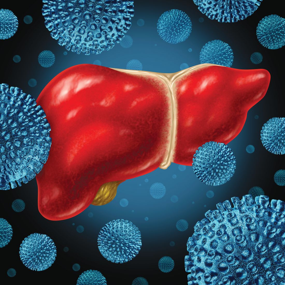
Glecaprevir/pibrentasvir highly effective in HCV genotype 3, among others
BOSTON – Specifically in patients with hepatitis C virus (HCV) genotype 3 infection and compensated cirrhosis, glecaprevir/pibrentasvir (Mavyret) was safe and had high efficacy in a phase 3 clinical trial, echoing an earlier report describing clinical results for the fixed-dose combination in multiple other genotypes.
Treatment with glecaprevir/pibrentasvir was safe and produced high rates of sustained virologic response 12 weeks after treatment (SVR12) for the genotype 3 patients and compensated cirrhosis in the recent results from the EXPEDITION-8 study, which will be presented at the annual meeting of the American Association for the Study of Liver Diseases.
In a previous report from EXPEDITION-8, investigators said that the treatment was well tolerated and effective in patients with HCV genotypes 1, 2, 4, 5, and 6.
“The availability of an 8-week, pangenotypic regimen for all treatment-naive HCV-infected patients regardless of cirrhosis status may simplify the HCV care pathway, furthering progress towards HCV elimination,” said investigator Robert S. Brown Jr., MD, MPH, and coinvestigators in a late-breaking abstract of the latest study results.
The nonrandomized, multicenter, phase 3b study included 343 adults with HCV genotypes 1-6 who received glecaprevir 300 mg and pibrentasvir 120 mg once daily for 8 weeks. A total of 63% of patients were male, 83% were white; 18% had HCV genotype 3, while the majority (67%) had HCV genotype 1.
For the genotype 3 patients, SVR12 rates were 95.2% in the intention-to-treat population, and 98.4% in the per-protocol population, Dr. Brown and coauthors said in their report on the study. For genotype 1, 2, 4, 5, and 6 patients, the intention-to-treat and per-protocol SVR12 rates were 98.2% and 100%.
Taken together, the SVR12 rates for all genotypes were 97.7% and 99.7%, respectively, for the intention-to-treat and per-protocol populations, according to the investigators.
There were no virologic failures on treatment, and one patients with genotype 3 relapsed in week 4 posttreatment, while one genotype 1 patient discontinued treatment though not because of adverse events, they said.
Most adverse events were grade 1 in severity, and included fatigue, pruritus, headache, and nausea. There were no liver-related toxicities, and no serious adverse events that were related to the study treatment, according to the investigators.
Glecaprevir/pibrentasvir is indicated for patients aged 12 years and older with treatment-naive HCV genotype 1-6 infection without cirrhosis or with compensated cirrhosis, and in patients with HCV genotype 1 infection previously treated with an HCV NS5A inhibitor or an NS3/4A protease inhibitor.
Dr. Brown reported disclosures related to pharmaceutical companies including AbbVie, which markets Mavyret.
SOURCE: Brown RS et al. The Liver Meeting 2019. Abstract LP9.
REPORTING FROM THE LIVER MEETING 2019
Key clinical point: In patients with hepatitis C virus (HCV) genotype 3 infection and compensated cirrhosis, glecaprevir/pibrentasvir was safe and had high efficacy, echoing an earlier result for the fixed-dose combination in genotypes 1, 2, and 4-6.
Major finding: For the genotype 3 patients, the rate of sustained virologic response 12 weeks after treatment (SVR12) was 95.2% in the intention-to-treat population, and 98.4% in the per-protocol population.
Study details: Further results from EXPEDITION-8, a single-arm phase 3b study including 343 adult patients with HCV genotypes 1-6.
Disclosures: Dr. Brown reported disclosures related to AbbVie, Gilead, Intercept, Dova, Shionogi, Merck, and Bristol-Myers Squibb.
Source: Brown RS et al. The Liver Meeting 2019. Abstract LP9.
Hepatitis C vaccine alters viral trajectory, but fails in chronic infection protection
BOSTON – A prime-boost hepatitis C virus (HCV) vaccine regimen did not protect against chronic infection, but it did evoke immune responses and differences in viral trajectory, according to investigators in what is believed to be the first randomized, placebo-controlled efficacy trial in this setting.
There were no apparent safety concerns with the vaccine according to investigators, led by Kimberly Page, PhD, MPH, of the University of New Mexico, Albuquerque.
“A safe and effective vaccine to prevent chronic hepatitis C virus infection is essential to reduce transmission,” Dr. Page and coauthors said in a late-breaking abstract of the study results, which will be presented at the annual meeting of the American Association for the Study of Liver Diseases.
The phase 1/2 trial described by Dr. Page and colleagues included 455 adults at risk of HCV infection because of injection drug use. They were randomized to vaccine, which consisted of a recombinant chimpanzee adenovirus-3 vectored vaccine prime plus a recombinant Modified Vaccinia virus Ankara boost, or to two doses of placebo at days 0 and 56 of the study.
There was no difference in chronic HCV infection at 6 months, the primary endpoint of the study. There were 14 chronically infected participants in the vaccine group, as well as 14 in the placebo group, for an overall incidence of infection of 13.0/100 person-years, Dr. Page and coauthors reported in the abstract.
However, there were significant differences in HCV RNA geometric mean peak at 1 month, which was 193,795 IU/L in the vaccine group and 1,078,092 IU/L in the placebo group, according to investigators. Similarly, geometric mean fold rise after infection was 0.2 in the vaccine group and 13.5 in the placebo group.
A total of 78% of vaccinated individuals had T-cell responses to at least one vaccine antigen pool, investigators said, adding that the vaccine was safe, well tolerated, and not associated with any serious adverse events.
Dr. Page had no disclosures related to the abstract.
SOURCE: Page K et al. The Liver Meeting 2019. Abstract LP17.
REPORTING FROM THE LIVER MEETING 2019
Key clinical point: A prime-boost HCV vaccine altered viral trajectory but did not protect against chronic infection.
Major finding: At 6 months after vaccination, there were 14 chronically infected participants in the vaccine group, and 14 in the placebo group.
Study details: A randomized, placebo controlled phase 1/2 trial including 455 adults at risk of HCV infection.
Disclosures: The first author reported no disclosures.
Source: Page K et al. The Liver Meeting 2019. Abstract LP17.
Dupilumab effective in early- and late-onset asthma
NEW ORLEANS – A new analysis suggests
Dupilumab may be more effective in reducing severe asthma exacerbations in patients with late-onset asthma, but the drug’s effect on lung function appeared the same regardless of asthma onset. Nicola Hanania, MD, of Baylor College of Medicine in Houston presented these results at the annual meeting of the American College of Chest Physicians.
Dr. Hanania and colleagues conducted a subanalysis of the LIBERTY ASTHMA QUEST study (NCT02414854). Previous data from this study showed that patients with uncontrolled, moderate to severe asthma who received dupilumab had fewer exacerbations and better lung function than did patients who received placebo (N Engl J Med. 2018;378:2486-96).
In their subanalysis, Dr. Hanania and his colleagues evaluated the efficacy of dupilumab, given at 200 mg or 300 mg every 2 weeks, in patients with early-onset asthma (at 40 years of age or younger) and late-onset asthma (at 41 years or older). The analysis included 919 patients with early-onset asthma who received dupilumab and 450 early-onset patients who received placebo. There were 345 patients with late-onset asthma who received dupilumab and 188 late-onset patients who received placebo.
Exacerbations
Dupilumab significantly reduced the adjusted annualized severe exacerbation rates during the 52-week treatment period. Significant reductions occurred in both early- and late-onset patients, though reductions were greater in the late-onset group.
In early-onset patients, dupilumab reduced severe exacerbations by 38% when given at 200 mg and by 37% when given at 300 mg (P less than .001 vs. placebo). In late-onset patients, dupilumab reduced exacerbations by 64% and 69%, respectively (P less than .001 vs. placebo).
Dr. Hanania said it isn’t clear why late-onset patients appear to derive more benefit with regard to exacerbations. It may be because these patients have more comorbidities or because they aren’t using their inhalers correctly. The researchers are investigating these possibilities.
Dr. Hanania went on to note that reductions in exacerbation rates were greatest in patients with elevated blood eosinophils (150 cells/mcL or greater) or fractional exhaled nitric oxide (FeNO; 25 ppb or greater).
In patients with early-onset asthma and elevated eosinophils, dupilumab reduced severe exacerbations by 50% when given at 200 mg and by 55% when given at 300 mg (P less than .001 vs. placebo). In late-onset patients with elevated eosinophils, dupilumab reduced exacerbations by 65% and 73%, respectively (P less than .001 vs. placebo).
In patients with early-onset asthma and elevated FeNO, dupilumab reduced severe exacerbations by 56% when given at 200 mg and by 52% when given at 300 mg (P less than .001 vs. placebo). In late-onset patients with elevated FeNO, dupilumab reduced exacerbations by 79% and 71%, respectively (P less than .001 vs. placebo).
Lung function
Dupilumab also improved prebronchodilator forced expiratory volume in 1 second (pre-BD FEV1), compared with placebo, with similar results in early- and late-onset patients.
In early-onset patients, the P values were less than .001 for both doses of dupilumab at weeks 12 and 52. In late-onset patients, the P values were less than .001 for the 300-mg dose at week 12 and the 200-mg dose at week 52, less than .01 for the 200-mg dose at week 12, and less than .05 for the 300-mg dose at week 52.
The effects of dupilumab on pre-BD FEV1 were greatest in patients with elevated eosinophils or FeNO. At week 12, the P value was less than .001 for both doses of dupilumab in early-onset patients with elevated eosinophils or FeNO. The P value was less than .01 for both doses in late-onset patients with elevated eosinophils. And the P value was less than .001 for both doses in late-onset patients with elevated FeNO.
This research was sponsored by Sanofi and Regeneron. Dr. Hanania disclosed relationships with Genentech, Novartis, AstraZeneca, Boehringer Ingelheim, GSK, Regeneron, and Sanofi.
SOURCE: Hanania N et al. CHEST 2019. Abstract, doi: 10.1016/j.chest.2019.08.870.
REPORTING FROM CHEST 2019
Better overall survival with nivolumab vs. chemo for advanced ESCC
BARCELONA – Nivolumab was associated with improved overall survival and a favorable safety profile, compared with chemotherapy, in patients with previously treated advanced esophageal squamous cell carcinoma (ESCC) in the open-label phase 3 ATTRACTION-3 study.
The overall survival (OS) benefit was observed regardless of tumor programmed death-ligand 1 (PD-L1) expression, Byoung Chul Cho, MD, reported at the European Society for Medical Oncology Congress.
The findings were reported online simultaneously in The Lancet Oncology.
Median OS at a minimum follow-up of 17.6 months was 10.9 vs. 8.4 months in 210 patients randomized to receive treatment with the PD-1 inhibitor nivolumab and 209 who received chemotherapy, respectively (hazard ratio, 0.77), said Dr. Cho of Yonsei Cancer Center, Yonsei University College of Medicine, Seoul, South Korea.
“Notably, there was a 13% and 10% improvement in overall survival rates at 12 months (47% vs. 34%) and 18 months (31% vs. 21%), respectively,” he said, also noting that the HRs for death favored nivolumab vs. chemotherapy across multiple prespecified subgroups, including those based on tumor PD-L1 expression (HRs, 0.69 and 0.84 for PD-L1 of 1% or greater and less than 1%, respectively).
No meaningful difference was seen in progression-free survival between the nivolumab and chemotherapy groups (12% vs. 7%; HR, 1.08), or in objective response rates (19% vs. 22%), he said.
“However, responses were substantially more durable with nivolumab, compared to chemotherapy; duration of response was 6.9 months with nivolumab vs. 3.9 months in the chemotherapy arm,” he said. “Notably, 21% of patients in the nivolumab arm were still in response, compared to only 6% in the chemotherapy arm.”
Patients enrolled in the open label study had unresectable advanced or recurrent ESCC refractory or intolerant to one prior fluoropyrimidine/platinum-based therapy. They were randomized 1:1 to receive 240 mg of nivolumab every 2 weeks or investigators’ choice of paclitaxel or docetaxel.
Fewer treatment-related adverse events (TRAEs) were reported with nivolumab, Dr. Cho said.
Any grade TRAEs occurred in 66% vs. 95% of patients in the groups, respectively, and grade 3-4 TRAEs occurred in 18% vs. 63%. The majority of select TRAEs – defined as those with potential immunologic etiology, including endocrine, gastrointestinal, hepatic, pulmonary, renal, and skin effects – were grade 1 or 2, and the only difference between the nivolumab and chemotherapy groups with respect to those was in endocrine effects, which affected 11% vs. less than 1% of patients, respectively.
Grade 3/4 select TRAEs occurred in less than 2% of patients, Dr. Cho noted.
An exploratory analysis further showed significant overall improvement in health-related quality of life with nivolumab through week 42 on treatment, he added.
The findings are of note, because metastatic esophageal cancer has a 5-year relative survival rate of less than 8%, and ESCC accounts for about 90% of cases worldwide, he said, adding that current second-line chemotherapy options for ESCC offer poor long-term survival and are associated with toxicity.
Nivolumab, which showed promising antitumor activity and manageable toxicity for advanced ESCC in patients who were refractory to or intolerant of standard chemotherapies in the phase 2 ATTRACTION-1 study, is the first immune checkpoint inhibitor to demonstrate a statistically significant, clinically meaningful improvement in OS vs. chemotherapy in this setting, he said.
The findings of this final analysis of ATTRACTION-3, which shows a 23% reduction in the risk of death, a 2.5-month improvement in median OS, benefit across PD-L1 subgroups, and a favorable safety profile, suggest that nivolumab represents a new standard second-line treatment option for patients with advanced ESCC, he concluded.
ATTRACTION-3 was funded by Ono Pharmaceutical Co., in collaboration with Bristol-Myers Squibb. Dr. Cho reported relationships with Bristol-Myers Squibb, Ono Pharmaceutical, and others. He also reported stock ownership and/or patents with TheraCanVac and Champions Oncology.
SOURCE: Cho B et al. ESMO 2019, Abstract LBA11.
REPORTING FROM ESMO 2019
Key clinical point: Nivolumab was associated with improved OS vs. chemotherapy, in previously treated advanced ESCC.
Major finding: Median OS was 10.9 vs. 8.4 months with nivolumab vs. chemotherapy, respectively (hazard ratio, 0.77).
Study details: A randomized, open-label, phase 3 study of 419 patients.
Disclosures: ATTRACTION-3 was funded by Ono Pharmaceutical Co., in collaboration with Bristol-Myers Squibb. Dr. Cho reported relationships with Bristol-Myers Squibb, Ono Pharmaceutical, and others. He reported stock ownership and/or patents with TheraCanVac and Champions Oncology.
Source: Cho B et al. ESMO 2019, Abstract LBA11.
More evidence that statins reduce HCC risk
SAN ANTONIO – The evidence that statin therapy reduces the risk of developing hepatocellular carcinoma, while not rising to the highest-level 1A strata, is nonetheless sufficiently persuasive at this point that consideration should be given to prescribing a statin in all patients with risk factors for the malignancy, regardless of their cardiovascular risk profile, Muhammad Talal Sarmini, MD, asserted at the annual meeting of the American College of Gastroenterology.
This includes individuals with hepatitis B or C virus infection as well as those with cirrhosis. The jury is still out as to whether nonalcoholic steatohepatitis is a risk factor for hepatocellular carcinoma (HCC), observed Dr. Sarmini of the Cleveland Clinic.
He presented a new meta-analysis, which concluded that patients on statin therapy had a 43% lower risk of new-onset HCC than persons not taking a statin. This meta-analysis – the largest ever addressing the issue – included 20 studies totaling more than 2.6 million patients and 24,341 cases of new-onset HCC. There were 11 retrospective case-control studies, 6 cohort studies, and 3 randomized trials. Five studies were from the United States, nine from Asia, and six were European.
In subgroup analyses aimed at assessing the consistency of the study results across various domains, there was a 45% reduction in the risk of HCC in association with statin therapy in the three studies of patients with hepatitis B virus, and significant reductions as well in Asia, Europe, and the United States when those participants were evaluated separately. The reduction was significant in both the case-control and cohort studies, but not when the three randomized, controlled trials (RCTs) were analyzed collectively. However, Dr. Sarmini shrugged off the neutral RCT results.
“It’s worth noting that the RCTs reported data from patients who were on statins with 4-5 years of follow-up. They were not at high risk for HCC. Given the nature of the disease and the relatively short period of follow-up, these studies only reported 81 cases of HCC. So they were very limited,” he said.
Audience members were eager to learn if Dr. Sarmini had found a differential preventive effect for lipophilic statins, such as atorvastatin or simvastatin, versus hydrophilic statins. He replied that, unfortunately, the published study results don’t allow for such an analysis. However, a large, propensity-matched cohort study published too recently for inclusion in his meta-analysis shed light on this matter. This Swedish national registry study included 16,668 propensity score–matched adults with chronic hepatitis B or C infection, of whom 6,554 initiated lipophilic statin therapy, 1,780 began treatment with a hydrophilic statin, and the rest were statin nonusers. The lipophilic statin users had an adjusted 44% reduction in 10-year HCC risk, compared with nonusers, while hydrophilic statins weren’t associated with a significant preventive effect (Ann Intern Med. 2019 Sep 3;171[5]:318-27).
Dr. Sarmini said that the meta-analysis results, together with the Swedish registry findings, highlight the need for additional well-designed cohort studies and RCTs of statins in populations at high risk for HCC in order to verify the existence of an HCC preventive effect and pinpoint which statins are effective at what dosages.
HCC is the fourth-leading cause of cancer-related mortality globally, accounting for 800,000 deaths annually. And the incidence is rising on a year-by-year basis.
Dr. Sarmini reported having no financial conflicts regarding his study, which was conducted free of commercial support.
REPORTING FROM ACG 2019
Probiotics with Lactobacillus reduce loss in spine BMD for postmenopausal women
, according to recent research published in The Lancet Rheumatology.

“The menopausal and early postmenopausal lumbar spine bone loss is substantial in women, and by using a prevention therapy with bacteria naturally occurring in the human gut microbiota we observed a close to complete protection against lumbar spine bone loss in healthy postmenopausal women,” Per-Anders Jansson, MD, chief physician at the University of Gothenburg (Sweden), and colleagues wrote in their study.
Dr. Jansson and colleagues performed a double-blind trial at four centers in Sweden in which 249 postmenopausal women were randomized during April-November 2016 to receive probiotics consisting of three Lactobacillus strains or placebo once per day for 12 months. Participants were healthy women, neither underweight nor overweight, and were postmenopausal, which was defined as being 2-12 years or less from last menstruation. The Lactobacillus strains, L. paracasei 8700:2 (DSM 13434), L. plantarum Heal 9 (DSM 15312), and L. plantarum Heal 19 (DSM 15313), were equally represented in a capsule at a dose of 1 x 10¹⁰ colony-forming unit per capsule. The researchers measured the lumbar spine bone mineral density (LS-BMD) at baseline and at 12 months, and also evaluated the safety profile of participants in both the probiotic and placebo groups.
Overall, 234 participants (94%) had data available for analysis at the end of the study. There was a significant reduction in LS-BMD loss for participants who received the probiotic treatment, compared with women in the control group (mean difference, 0.71%; 95% confidence interval, 0.06%-1.35%), while there was a significant loss in LS-BMD for participants in the placebo group (percentage change, –0.72%; 95% CI, –1.22% to –0.22%) compared with loss in the probiotic group (percentage change, –0.01%; 95% CI, –0.50% to 0.48%). Using analysis of covariance, the researchers found the probiotic group had reduced LS-BMD loss after adjustment for factors such as study site, age at baseline, BMD at baseline, and number of years from menopause (mean difference, 7.44 mg/cm2; 95% CI, 0.38 to 14.50).
In a subgroup analysis of women above and below the median time since menopause at baseline (6 years), participants in the probiotic group who were below the median time saw a significant protective effect of Lactobacillus treatment (mean difference, 1.08%; 95% CI, 0.20%-1.96%), compared with women above the median time (mean difference, 0.31%; 95% CI, –0.62% to 1.23%).
Researchers also examined the effects of probiotic treatment on total hip and femoral neck BMD as secondary endpoints. Lactobacillus treatment did not appear to affect total hip (–1.01%; 95% CI, –1.65% to –0.37%) or trochanter BMD (–1.13%; 95% CI, –2.27% to 0.20%), but femoral neck BMD was reduced in the probiotic group (–1.34%; 95% CI, –2.09% to –0.58%), compared with the placebo group (–0.88%; 95% CI, –1.64% to –0.13%).
Limitations of the study included examining only one dose of Lactobacillus treatment and no analysis of the effect of short-chain fatty acids on LS-BMD. The researchers noted that “recent studies have shown that short-chain fatty acids, which are generated by fermentation of complex carbohydrates by the gut microbiota, are important regulators of both bone formation and resorption.”
The researchers also acknowledged that the LS-BMD effect size for the probiotic treatment over the 12 months was a lower magnitude, compared with first-line treatments for osteoporosis in postmenopausal women using bisphosphonates. “Further long-term studies should be done to evaluate if the bone-protective effect becomes more pronounced with prolonged treatment with the Lactobacillus strains used in the present study,” they said.
In a related editorial, Shivani Sahni, PhD, of Harvard Medical School, Boston, and Connie M. Weaver, PhD, of Purdue University, West Lafayette, Ind., reiterated that the effect size of probiotics is “of far less magnitude” than such treatments as bisphosphonates and expressed concern about the reduction of femoral neck BMD in the probiotic group, which was not explained in the study (Lancet Rheumatol. 2019 Nov;1[3]:e135-e137. doi: 10.1016/S2665-9913(19)30073-6). There is a need to learn the optimum dose of probiotics as well as which Lactobacillus strains should be used in future studies, as the strains chosen by Jansson et al. were based on results in mice.
In the meantime, patients might be better off choosing dietary interventions with proven bone protection and no documented negative effects on the hip, such as prebiotics like soluble corn fiber and dried prunes, in tandem with drug therapies, Dr. Sahni and Dr. Weaver said.
“Although Jansson and colleagues’ results are important, more work is needed before such probiotics are ready for consumers,” they concluded.
This study was funded by Probi, which employs two of the study’s authors. Three authors reported being coinventors of a patent involving the effects of probiotics in osteoporosis treatment, and one author is listed as an inventor on a pending patent application on probiotic compositions and uses. Dr. Sahni reported receiving grants from Dairy Management. Dr. Weaver reported no relevant conflicts of interest.
SOURCE: Jansson P-A et al. Lancet Rheumatol. 2019 Nov;1(3):e154-e162. doi: 10.1016/S2665-9913(19)30068-2
, according to recent research published in The Lancet Rheumatology.
“The menopausal and early postmenopausal lumbar spine bone loss is substantial in women, and by using a prevention therapy with bacteria naturally occurring in the human gut microbiota we observed a close to complete protection against lumbar spine bone loss in healthy postmenopausal women,” Per-Anders Jansson, MD, chief physician at the University of Gothenburg (Sweden), and colleagues wrote in their study.
Dr. Jansson and colleagues performed a double-blind trial at four centers in Sweden in which 249 postmenopausal women were randomized during April-November 2016 to receive probiotics consisting of three Lactobacillus strains or placebo once per day for 12 months. Participants were healthy women, neither underweight nor overweight, and were postmenopausal, which was defined as being 2-12 years or less from last menstruation. The Lactobacillus strains, L. paracasei 8700:2 (DSM 13434), L. plantarum Heal 9 (DSM 15312), and L. plantarum Heal 19 (DSM 15313), were equally represented in a capsule at a dose of 1 x 10¹⁰ colony-forming unit per capsule. The researchers measured the lumbar spine bone mineral density (LS-BMD) at baseline and at 12 months, and also evaluated the safety profile of participants in both the probiotic and placebo groups.
Overall, 234 participants (94%) had data available for analysis at the end of the study. There was a significant reduction in LS-BMD loss for participants who received the probiotic treatment, compared with women in the control group (mean difference, 0.71%; 95% confidence interval, 0.06%-1.35%), while there was a significant loss in LS-BMD for participants in the placebo group (percentage change, –0.72%; 95% CI, –1.22% to –0.22%) compared with loss in the probiotic group (percentage change, –0.01%; 95% CI, –0.50% to 0.48%). Using analysis of covariance, the researchers found the probiotic group had reduced LS-BMD loss after adjustment for factors such as study site, age at baseline, BMD at baseline, and number of years from menopause (mean difference, 7.44 mg/cm2; 95% CI, 0.38 to 14.50).
In a subgroup analysis of women above and below the median time since menopause at baseline (6 years), participants in the probiotic group who were below the median time saw a significant protective effect of Lactobacillus treatment (mean difference, 1.08%; 95% CI, 0.20%-1.96%), compared with women above the median time (mean difference, 0.31%; 95% CI, –0.62% to 1.23%).
Researchers also examined the effects of probiotic treatment on total hip and femoral neck BMD as secondary endpoints. Lactobacillus treatment did not appear to affect total hip (–1.01%; 95% CI, –1.65% to –0.37%) or trochanter BMD (–1.13%; 95% CI, –2.27% to 0.20%), but femoral neck BMD was reduced in the probiotic group (–1.34%; 95% CI, –2.09% to –0.58%), compared with the placebo group (–0.88%; 95% CI, –1.64% to –0.13%).
Limitations of the study included examining only one dose of Lactobacillus treatment and no analysis of the effect of short-chain fatty acids on LS-BMD. The researchers noted that “recent studies have shown that short-chain fatty acids, which are generated by fermentation of complex carbohydrates by the gut microbiota, are important regulators of both bone formation and resorption.”
The researchers also acknowledged that the LS-BMD effect size for the probiotic treatment over the 12 months was a lower magnitude, compared with first-line treatments for osteoporosis in postmenopausal women using bisphosphonates. “Further long-term studies should be done to evaluate if the bone-protective effect becomes more pronounced with prolonged treatment with the Lactobacillus strains used in the present study,” they said.
In a related editorial, Shivani Sahni, PhD, of Harvard Medical School, Boston, and Connie M. Weaver, PhD, of Purdue University, West Lafayette, Ind., reiterated that the effect size of probiotics is “of far less magnitude” than such treatments as bisphosphonates and expressed concern about the reduction of femoral neck BMD in the probiotic group, which was not explained in the study (Lancet Rheumatol. 2019 Nov;1[3]:e135-e137. doi: 10.1016/S2665-9913(19)30073-6). There is a need to learn the optimum dose of probiotics as well as which Lactobacillus strains should be used in future studies, as the strains chosen by Jansson et al. were based on results in mice.
In the meantime, patients might be better off choosing dietary interventions with proven bone protection and no documented negative effects on the hip, such as prebiotics like soluble corn fiber and dried prunes, in tandem with drug therapies, Dr. Sahni and Dr. Weaver said.
“Although Jansson and colleagues’ results are important, more work is needed before such probiotics are ready for consumers,” they concluded.
This study was funded by Probi, which employs two of the study’s authors. Three authors reported being coinventors of a patent involving the effects of probiotics in osteoporosis treatment, and one author is listed as an inventor on a pending patent application on probiotic compositions and uses. Dr. Sahni reported receiving grants from Dairy Management. Dr. Weaver reported no relevant conflicts of interest.
SOURCE: Jansson P-A et al. Lancet Rheumatol. 2019 Nov;1(3):e154-e162. doi: 10.1016/S2665-9913(19)30068-2
FROM THE LANCET RHEUMATOLOGY
Strategy critical to surviving drug shortages
NATIONAL HARBOR, MD. –
“Statistically speaking, there is no proof that patients are worse off from drug shortages,” Matt Grissinger, RPh, director of error-reporting programs at the Institute for Safe Medication Practices, told the audience at the annual conference of the Academy of Managed Care Pharmacy. The data and anecdotes he presented suggest the contrary.
As Mr. Grissinger pointed out, drug shortages can create a sequela of events that stress health care workers seeking to find the next-best available and most appropriate therapy for their patients. In the process, numerous medication-related errors can occur, resulting in patient harm, including adverse drug events and even death.
One potential problems is erroneous or inappropriate drug substitution stemming from mis- or uncalculated doses because of factors such as incorrect labeling and lack of knowledge regarding acceptable therapeutic interchanges. Other potential errors include non–therapeutically equivalent drug substitutions, resulting in supraoptimal therapy or overdoses, and unfamiliarity with drug labeling from outsourced facilities.
As a result, patients may experience worse outcomes as a consequence of the drug shortage: Worsening of the disease, disease prolongation, side effects stemming from alternative drug selections, untreated pain, psychological effects, severe electrolyte imbalances, severe acid/base imbalances, and death.
While a paper trail can help piece together clues regarding how a medication error occurred, documentation or lack thereof can also introduce errors when drug shortages occur.
Any changes to a drug order or prescription that deviate from the prescriber’s original request require prescriber approval but can still create opportunities for error. While documenting these changes and updating labeling is essential, appropriate documentation does not always occur and raises the question of who is responsible for making such changes.
Drug shortages also challenge a clinician’s professional judgment. Mr. Grissinger cited an example in which a nurse used half of a 0.5-mg single-use vial of promethazine for a patient requiring a 0.25 mg dose. The nurse wrote on the label that the remainder should be saved. While the vial was manufactured for one-time use, whether to discard the unused contents in a situation of drug shortages required the nurse to make a judgment call. In this case, the nurse chose to save the balance of the drug – a choice Mr. Grissinger stated he might have made had he been in a similar situation.
Additionally, drug shortages can create a climate in which more ethical questions arise – especially with regard to disease states such as cancer.
“If you only have 10 vials of vincristine, who gets it?” Mr. Grissinger asked the audience.
To help answer these difficult life-or-death questions, hospital settings need to engage the ethics committees and social workers.
While education plays a vital role in bringing attention to and addressing errors stemming from drug shortages, Mr. Grissinger cautioned the audience not to rely on education as the solution.
“Education is a poor strategy for addressing drug shortages,” he said. While education can draw awareness to drug shortages and subsequent medication-related errors, Mr. Grissinger recommends that organizations implement strategies to help ameliorate the havoc created by drug shortages.
Drug shortage assessment checklists can help organizations evaluate the impact of shortages by verifying inventory, and proactively searching for alternatives. From there, they can enact strategies such as assigning priority to patients who have the greatest need, altering packaging and concentrations, and finding suitable therapeutic substitutions.
REPORTING FROM AMCP NEXUS 2019
Levothyroxine dose for checkpoint inhibitor toxicity may be too high
CHICAGO – both for patients with preexisting and de novo hypothyroidism.

The real-world data, presented by Megan Kristan, MD, at the annual meeting of the American Thyroid Association, refine recommendations for dosing by body weight for levothyroxine in patients receiving checkpoint inhibitor therapy.
Immune checkpoint inhibitors stand a good chance of turning the tide against melanoma, some lung cancers, and other malignancies that have long been considered lethal. However, as more patients are exposed to the therapies, endocrinologists are seeing a wave of thyroid abnormalities, and must decide when, and at what doses, to treat hypothyroidism, said Dr. Kristan, a diabetes, endocrinology, and nutrition fellow at the University of Maryland, Baltimore.
Six checkpoint inhibitors are currently approved to hit a variety of molecular targets, and the prevalence of thyroid toxicity and hypothyroidism across the drug class ranges from a reported 9% to 40%, said Dr. Kristan.
The acknowledged thyroid toxicity of these drugs led the American Society for Clinical Oncology (ASCO) to issue guidelines advising that oncologists obtain baseline thyroid function tests before initiating checkpoint inhibitors, and that values be rechecked frequently – every 4-6 weeks – during therapy.
The guidelines advise dosing levothyroxine at approximately 1.6 mcg/kg per day, based on ideal patient body weight. The recommendation is limited to patients without risk factors, and approximates full levothyroxine replacement.
However, some patients enter cancer treatment with hypothyroidism, and some develop it de novo after beginning checkpoint inhibitor therapy. It is not known how best to treat each group, said Dr. Kristan.
To help answer that question, she and her collaborators at Georgetown University Hospital, McLean, Va., made use of a database drawn from five hospitals to perform a retrospective chart review. They looked at 822 patients who had received checkpoint inhibitor therapy, and from those patients, they selected 118 who had a diagnosis of hypothyroidism, or who received a prescription for levothyroxine during the 8-year study period.
The investigators assembled all available relevant data for each patient, including thyroid function tests, levothyroxine dosing, type of cancer, and type of therapy. They sorted participants into those who had received a diagnosis of hypothyroidism before or after receiving the first dose of checkpoint inhibitor therapy.
At baseline, 81 patients had preexisting hypothyroidism and were receiving a mean levothyroxine dose of 88.2 mcg. After treatment, the mean dose was 94.3 mcg, a nonsignificant difference. The median dose for this group remained at 88 mcg through treatment.
For the 37 patients who developed hypothyroidism de novo during checkpoint inhibitor therapy, the final observed levothyroxine dose was 71.2 mcg.
The mean age of the patients at baseline was 69 years. About half were women, and 91% were white. Either nivolumab or pembrolizumab was used in 72% of patients, making them the most commonly used checkpoint inhibitors, though 90% of patients received combination therapy. Taken together, melanoma and lung cancer accounted for about two-thirds of the cancers seen.
For both groups, the on-treatment levothyroxine dose was considerably lower than the ASCO-recommended, weight-based dosing, which would have been 122.9 mcg for those with preexisting hypothyroidism and 115.7 mcg for those who developed hypothyroidism on treatment (P less than .001 for both).
Dr. Kristan noted that thyroid stimulating hormone (TSH) values for patients with pretreatment hypothyroidism peaked between weeks 12 and 20, though there was no preemptive adjustment of levothyroxine dosing.
For those who developed on-treatment hypothyroidism, TSH values peaked at a series of times, at about weeks 8, 16, and 32. These waves of TSH elevation, she said, support the 4- to 6-week follow-up interval recommended in the ASCO guidelines.
However, she said, patients with de novo hypothyroidism “should not be started on the 1.6-mcg/kg-a-day weight-based dosing.” The cohort with de novo hypothyroidism in Dr. Kristan’s analysis required a daily dose of about 1 mcg/kg, she said. These real-world results support the idea that many patients on checkpoint inhibitors retain some thyroid reserve.
Dr. Kristan said that based on these findings, she and her collaborators recommend monitoring thyroid function every 4-6 weeks for patients taking immune checkpoint inhibitors. Patients with preexisting thyroid disease should not have an empiric adjustment of levothyroxine dose on checkpoint inhibitor initiation. For patients who develop thyroiditis after starting therapy, initiating a dose at 1 mcg/kg per day of ideal body weight is a good place to start, and treatment response should be monitored.
The study was limited by its retrospective nature and the small sample size, acknowledged Dr. Kristan. In addition, there were confounding variables and different frequencies of testing across institutions, and antibody status was not available and may have affected the results. Testing was performable for all participants.
Dr. Kristan said that the analysis opens up areas for further study, such as which patient populations are at risk for developing thyroid toxicity, what baseline characteristics can help predict which patients develop toxicity, and whether particular checkpoint inhibitors are more likely to cause toxicity. In addition, she said, a subset of patients will develop hyperthyroidism on checkpoint inhibitor therapy, and little is known about how to treat that complication.
Dr. Kristan reported no conflicts of interest. The research she presented was completed during her residency at Georgetown University.
SOURCE: Kristan M et al. ATA 2019, Oral Abstract 25.
CHICAGO – both for patients with preexisting and de novo hypothyroidism.
The real-world data, presented by Megan Kristan, MD, at the annual meeting of the American Thyroid Association, refine recommendations for dosing by body weight for levothyroxine in patients receiving checkpoint inhibitor therapy.
Immune checkpoint inhibitors stand a good chance of turning the tide against melanoma, some lung cancers, and other malignancies that have long been considered lethal. However, as more patients are exposed to the therapies, endocrinologists are seeing a wave of thyroid abnormalities, and must decide when, and at what doses, to treat hypothyroidism, said Dr. Kristan, a diabetes, endocrinology, and nutrition fellow at the University of Maryland, Baltimore.
Six checkpoint inhibitors are currently approved to hit a variety of molecular targets, and the prevalence of thyroid toxicity and hypothyroidism across the drug class ranges from a reported 9% to 40%, said Dr. Kristan.
The acknowledged thyroid toxicity of these drugs led the American Society for Clinical Oncology (ASCO) to issue guidelines advising that oncologists obtain baseline thyroid function tests before initiating checkpoint inhibitors, and that values be rechecked frequently – every 4-6 weeks – during therapy.
The guidelines advise dosing levothyroxine at approximately 1.6 mcg/kg per day, based on ideal patient body weight. The recommendation is limited to patients without risk factors, and approximates full levothyroxine replacement.
However, some patients enter cancer treatment with hypothyroidism, and some develop it de novo after beginning checkpoint inhibitor therapy. It is not known how best to treat each group, said Dr. Kristan.
To help answer that question, she and her collaborators at Georgetown University Hospital, McLean, Va., made use of a database drawn from five hospitals to perform a retrospective chart review. They looked at 822 patients who had received checkpoint inhibitor therapy, and from those patients, they selected 118 who had a diagnosis of hypothyroidism, or who received a prescription for levothyroxine during the 8-year study period.
The investigators assembled all available relevant data for each patient, including thyroid function tests, levothyroxine dosing, type of cancer, and type of therapy. They sorted participants into those who had received a diagnosis of hypothyroidism before or after receiving the first dose of checkpoint inhibitor therapy.
At baseline, 81 patients had preexisting hypothyroidism and were receiving a mean levothyroxine dose of 88.2 mcg. After treatment, the mean dose was 94.3 mcg, a nonsignificant difference. The median dose for this group remained at 88 mcg through treatment.
For the 37 patients who developed hypothyroidism de novo during checkpoint inhibitor therapy, the final observed levothyroxine dose was 71.2 mcg.
The mean age of the patients at baseline was 69 years. About half were women, and 91% were white. Either nivolumab or pembrolizumab was used in 72% of patients, making them the most commonly used checkpoint inhibitors, though 90% of patients received combination therapy. Taken together, melanoma and lung cancer accounted for about two-thirds of the cancers seen.
For both groups, the on-treatment levothyroxine dose was considerably lower than the ASCO-recommended, weight-based dosing, which would have been 122.9 mcg for those with preexisting hypothyroidism and 115.7 mcg for those who developed hypothyroidism on treatment (P less than .001 for both).
Dr. Kristan noted that thyroid stimulating hormone (TSH) values for patients with pretreatment hypothyroidism peaked between weeks 12 and 20, though there was no preemptive adjustment of levothyroxine dosing.
For those who developed on-treatment hypothyroidism, TSH values peaked at a series of times, at about weeks 8, 16, and 32. These waves of TSH elevation, she said, support the 4- to 6-week follow-up interval recommended in the ASCO guidelines.
However, she said, patients with de novo hypothyroidism “should not be started on the 1.6-mcg/kg-a-day weight-based dosing.” The cohort with de novo hypothyroidism in Dr. Kristan’s analysis required a daily dose of about 1 mcg/kg, she said. These real-world results support the idea that many patients on checkpoint inhibitors retain some thyroid reserve.
Dr. Kristan said that based on these findings, she and her collaborators recommend monitoring thyroid function every 4-6 weeks for patients taking immune checkpoint inhibitors. Patients with preexisting thyroid disease should not have an empiric adjustment of levothyroxine dose on checkpoint inhibitor initiation. For patients who develop thyroiditis after starting therapy, initiating a dose at 1 mcg/kg per day of ideal body weight is a good place to start, and treatment response should be monitored.
The study was limited by its retrospective nature and the small sample size, acknowledged Dr. Kristan. In addition, there were confounding variables and different frequencies of testing across institutions, and antibody status was not available and may have affected the results. Testing was performable for all participants.
Dr. Kristan said that the analysis opens up areas for further study, such as which patient populations are at risk for developing thyroid toxicity, what baseline characteristics can help predict which patients develop toxicity, and whether particular checkpoint inhibitors are more likely to cause toxicity. In addition, she said, a subset of patients will develop hyperthyroidism on checkpoint inhibitor therapy, and little is known about how to treat that complication.
Dr. Kristan reported no conflicts of interest. The research she presented was completed during her residency at Georgetown University.
SOURCE: Kristan M et al. ATA 2019, Oral Abstract 25.
REPORTING FROM ATA 2019
Immune checkpoint inhibition in SCLC: Modest outcomes, many questions
BARCELONA – Immune checkpoint inhibitors demonstrate activity in small cell lung cancer (SCLC), but achieving more durable disease control and better survival requires improved understanding of biomarkers and the immune microenvironment.
That was the overarching message from experts speaking at a minisymposium on immunotherapy in SCLC at the World Conference on Lung Cancer.
“None of us are disputing that immunotherapy is clearly active in this space, but I think that we can all agree that the outcomes have been somewhat modest in an unselected population, and there is certainly room to grow,” said Dr. Stephen V. Liu, MD. “Moving forward, while we will look for any advances we can, we also feel strongly that these incremental gains are probably not enough.”
The state of the art
Hints that immunotherapy could be clinically efficacious in SCLC emerged in 2016 when interim findings from the CheckMate 032 study showed that the programmed cell death-1 (PD-1) inhibitor nivolumab, either alone or in combination with the anti-CTLA4 antibody ipilimumab, had efficacy in recurrent SCLC. Efficacy was seen regardless of programmed death-ligand 1 (PD-L1) status, which is “a good thing since PD-L1 is expressed much less frequently in SCLC than in non-SCLC,” Scott J. Antonia, MD, of Duke Cancer Institute, Durham, N.C., who was the first author on that study, said during the symposium.
A particularly encouraging finding was that responders included patients with platinum-refractory SCLC for whom treatments in the relapse setting are lacking, Dr Antonia said.
An exploratory analysis of CheckMate 032 also showed better responses among patients in the highest tumor mutation burden (TMB) tertile, especially in the combination therapy group, leading to the hypothesis-generating finding that TMB may predict response, he said.
Another suggestion of nivolumab’s potential came from the randomized CheckMate 331 study comparing the checkpoint inhibitor with chemotherapy in relapsed SCLC patients. As reported in 2018 at the European Society for Medical Oncology (ESMO), no overall survival (OS) benefit was apparent at 12 months (37% vs. 34%), but a separation of the curves at 36 months suggested a possible OS benefit with nivolumab, Dr. Antonia noted, adding that the difference was “obviously small” and requires “a lot more work related to that.”
Subgroup analyses in that study also were “perhaps revealing” in that patients without liver metastases derived benefit (hazard ratio, 0.75), as did those who were platinum resistant (HR, 0.71), he said.
The phase 1b KEYNOTE-028 study showed that the anti–PD-1 monoclonal antibody pembrolizumab also has activity in PD-L1–positive SCLC patients in the relapsed setting, and pooled data from that study and KEYNOTE-158, which included both PD-L1–positive and –negative patients, showed promising antitumor activity and durable responses with pembrolizumab. The pooled data, as presented at the 2019 annual meeting of the American Association for Cancer Research, showed an objective response rate (ORR) of 19%, including complete and partial response rates of 2% and 17%, respectively.
“And there appears to be, at least preliminarily, some durability to the responses,” he said, noting that 9 of 16 patients experienced at least an 18-month response. “Progression-free survival was 2 months, and overall survival was 7.7 months.”
The IMpower133 study showed significantly longer OS and progression-free survival (PFS) with the addition of atezolizumab to chemotherapy in the first-line treatment of extensive-stage SCLC (HR, 0.70). Some late merging of the survival curves was apparent, but the data haven’t matured.
“Hopefully there will be some evidence of a lifting of the tail of the survival curve with some durability of responsiveness like we see in non–small cell lung cancer,” he said.
When it comes to “making the next leap” toward improved clinical efficacy with immunotherapy for SCLC, “we need to think about three general categories of how it is that tumors evade rejection by the immune system,” he said.
One category involves SCLC patients with an insufficient numbers of T cells generated within the lymphoid compartment; in those patients, an immunotherapeutic approach directed at the tumor microenvironment won’t lead to a response. Another category includes patients who generate enough T cells within the lymphoid compartment but in whom those cells aren’t driven into the tumor parenchyma. The third involves those whose T cells may make it into the tumor parenchyma, but are inhibited in the tumor microenvironment, he explained.
Strategies to increase the number of T cells generated in the lymphoid compartment – such as vaccines, radiation, adoptive cell therapy with chimeric antigen receptors, to name a few – were a focus of research efforts more than a decade ago, but the pendulum swung more toward addressing the tumor microenvironment.
“I think that the pendulum needs to swing back to the middle, and we do need to develop combination immunotherapies paying attention to the lymphoid compartment as well as the tumor microenvironment,” Dr. Antonia said, listing these “guiding principles” for the development of effective SCLC immunotherapy:
- Combination immunotherapy is necessary.
- Mechanisms exploited by SCLC to evade immune-mediated rejection need to be identified.
- Inclusion of strategies for driving tumor-reactive T cells into the tumor microenvironment should be considered.
- PD-1 blockade should continue.
- Biomarkers should be identified for selecting patients for tumor microenvironment–targeted agents.
Clinical and molecular biomarkers

Indeed, there is much work to do with respect to biomarkers, but their use in the selection of SCLC patients for immunotherapy is “finally starting to evolve and evolve more rapidly,” according to Lauren Averett Byers, MD, of the University of Texas MD Anderson Cancer Center, Houston.
Numerous groups are identifying biomarkers for both targeted therapy and immunotherapy, Dr. Byers said, noting that “this has been a really incredible time for those of us who take care of small cell lung cancer patients.”
“It’s been many decades since we’ve had a new option for our patients ... and so I think with the landmark clinical trials ... we really do have a new option in terms of a new standard of care,” she said of immunotherapy. “But I think we also recognize that there is significant room for further improvement.”
Many patients don’t respond or don’t respond as well as hoped, and therefore an “incredible need” exists for personalized biomarker-driven therapy for SCLC and its distinct molecular subsets, she said.
Emerging and potential biomarkers and other factors to guide treatment decisions include TMB, PD-L1, clinical history/duration of response in immunotherapy-naive relapsed patients, gene expression profile–driven SCLC subgroup identification, and DNA damage response (DDR) inhibitors such as Chk1, PARP, and Wee1 inhibitors.
TMB, as described by Dr. Antonia with respect to the CheckMate 032 findings of improved outcomes in those in the highest TMB tertile, is one potentially helpful biomarker for response.
“In thinking about how we apply this, though, we have to think about what we’re deciding between,” Dr. Byers said, explaining that the responses in patients with medium or low TMB – between 0% and 10% in most studies in the relapsed SCLC setting – aren’t that different from those seen with other treatment options.
“Currently we’re not routinely ordering TMB to decide on immunotherapy because there are still patients that can be as likely to benefit from immunotherapy as they are from chemotherapy, and potentially with more durable responses,” she said. “But certainly, it is a way to potentially identify patients where immunotherapy alone may have very high rates of response.”
IMPower133 showed no difference in hazard ratios for death based on TMB detected in the blood in SCLC patients treated with first-line atezolizumab plus chemotherapy, but “this still supports using the immunotherapy/chemotherapy combination broadly, and also emphasizes the need for an improved – and probably expanded – look at other biomarkers that may help predict response,” she said.
PD-L1 appears to have a role as a biomarker in this setting as well, she said, citing the KEYNOTE-028 findings of numerically improved responses in PD-L1–positive SCLC patients treated with pembrolizumab.
“We should be looking at PD-L1 levels, but we need further information to know how we might use this,” she said.
In immunotherapy-naive patients who relapse after front-line chemotherapy, the most important biomarker is clinical history and duration of response to platinum, which helps guide second-line treatment, Dr. Byers said.
“I think there’s consensus among most of us that patients who have platinum-refractory disease and are unlikely to respond to further platinum therapy or other chemotherapy agents are patients who really should get immunotherapy,” she added, explaining that the available data suggest there is no cross-resistance and that there may actually be enhanced benefit with immunotherapy in such patients.
Using molecular data to identify SCLC subtypes based on gene expression profiles is another area of interest, she said.
In fact, new data presented at the WCLC conference by Carl Gay, MD, PhD, a former fellow in her lab and now a junior faculty member at MD Anderson, identified four specific SCLC subgroups; three were driven by activation of the known transcription factors ASCL1, NEUROD1, and POU2F3, but an additional “inflamed” group without expression of those three transcription factors was also identified.
That “triple-negative” group had significantly higher expression of human leukocyte antigen and very high T-cell activation with expression of multiple immune checkpoints representing candidate targets, she said, adding: “We hypothesize that this group may be the group in SCLC that gets relatively greater benefit from immune checkpoint blockade.”
DDR is also garnering attention.
“Since there was this signal that [patients with] DNA damage ... tend to be more sensitive to immunotherapy ... we looked at whether or not targeted agents that prevented repair of DNA damage and induced increased levels of DNA damage ... might activate the innate immune system through the STING pathway and if that could be a potential approach to enhance immunotherapy response,” she said.
The approach showed promise in cell lines in a mouse model and also in an immunocompetent SCLC mouse model.
“It was really interesting to see how these drugs might potentially enhance response to immunotherapy,” Dr. Byers said, noting that the same phenomenon has been seen with PARP inhibitors in breast and colon cancer models and in other solid tumors.
“So I think that there is something there, and fortunately we’re now at a point now where we can start looking at some of these combinations in the clinic across many different cancer types,” she said. “I think we’ll be learning a lot more about what’s happening with these patients.”
At present, however, “there is more that we don’t know about the immune landscape of small cell lung cancer than what we do know, and that’s a real opportunity where, over the next several years, we will gain a deeper understanding ... that will direct where we’re going in terms of translating that back into the clinic.”
The SCLC immune microenvironment
The immune microenvironment will be an integral part of that journey, according to Dr. Liu.
“We consider small cell lung cancer – a carcinogen-associated cancer – to be one that has a high somatic mutation rate, but what we’ve learned over the past few years is that tumor neoantigens are certainly necessary – but not sufficient,” he said, noting that mutational burden represents the potential for immune-mediated antitumor responses, but is not a guarantee.
“As a group, we need to develop strategies to overcome the powerful immunosuppressive microenvironment in small cell lung cancer,” he added.
Lessons learned from studying PD-L1 provided the first insight into the importance of the immune microenvironment: PD-L1 expression, as measured by tumor proportion score (TPS) holds predictive value in non–small cell lung cancer patients treated with PD-1 inhibitors, but the SCLC story is much more complex, he said.
Only 18% of SCLC patients in CheckMate 032 were PD-L1–positive, and “paradoxically, we see responses were better in the PD-L1–negative group,” he explained. The response rates for nivolumab/ipilimumab were 32% in the PD-L1–negative group and 10% in the PD-L1–positive group.
Recent findings regarding the use of the combined positive score (CPS), which unlike the TPS for determining PD-L1 status, includes PD-L1 expression on stromal cells, are also notable. In a phase 2 study of maintenance pembrolizumab in SCLC, for example, 3 of 30 patients were PD-L1 positive by TPS, and 8 of 20 were positive by CPS.
“And that did predict outcomes: We see a higher response rate [38% vs. 8%], better PFS [6.5 vs. 1.3 months], and better overall survival [13 months vs. 8 months] in pretreated small cell lung cancer,” he said.
Similarly, in KEYNOTE-158 when looking at pembrolizumab in previously treated SCLC, the overall response was modest at 18.7%, and median PFS was 2.0 months.
“Again, breaking it down by CPS, we see a different story,” Dr. Liu said. “We see better outcomes in the PD-L1–positive [group] if you’re factoring in expression in the microenvironment.” When assessed by CPS, 39% of patients were PD-L1 positive; those patients, when compared with PD-L1–negative patients, had improved 12-month PFS (28.5% vs. 8.2%, respectively), 12-month OS (53.3% vs. 30.7%), and median OS (14.9 vs. 5.9 months).
Checkpoint expression in tumor-infiltrating lymphocytes (TILs) also has been shown to vary when compared with tumor expression. SCLC tissue microarrays in a study presented at ASCO 2017 (Rivalland et al. Abstract 8569), for example, showed that tumor expression versus TIL expression of PD-L1, TIMS3, and LAG3 was 18% vs. 67%, 0% vs. 59%, and 0% vs. 45%, respectively, and the TIL expression correlated with survival, Dr. Liu said.
“So when we consider things like PD-L1 expression, looking at a narrow scope of just the tumor is not enough. We need to consider the stromal cells, the microenvironment,” he said. “And even larger than that, PD-1/PD-L1 interaction is but a fraction of powerful, dynamic, immunosuppressive factors in small cell lung cancer.
“All of these will need to be accounted for in various patients.”
These findings and others, like those from a recent study showing differentially expressed genes and pathways in the stromal cells of longer- versus shorter-term survivors, raise questions about whether the lymphoid compartment can be manipulated in SCLC to improve immune responses using the strategies discussed by Dr. Antonia and Dr. Byers, he said.
In “cold” tumor phenotypes, one hypothesis has tumor-associated macrophages (TAMs) preventing infiltration of the cytotoxic T lymphocytes, which raises the possibility that TAMs are a therapeutic target, he said.
“At this meeting and others we’ve heard of lurbinectedin as a possible active drug in SCLC,” he said, noting that preclinical data also demonstrate that lurbinectedin targets TAMs. Perhaps the agent’s future role will be that of an immune modulator rather than a cytotoxic agent, he suggested.
Regulatory T cells (Tregs) are another potential immunomodulatory target, but the problem is their redundancy and the lack of good models to identify which ones are active, he said.
“Myeloid-derived suppressor cells [MDSC] are another important part of the microenvironment and could be potential targets to restore immune responses,” he added.
But many questions remain, he said.
For example: How can we overcome an immunosuppressive tumor microenvironment? Can we inhibit arginine or adenosine? Can we restore interleukin-2? Can we target things like LAG3? Can we eliminate the Treg and MDSC population? Which strategies are appropriate? Are they the same in immunotherapy-naive vs. immunotherapy-experienced patients – is intrinsic resistance the same as acquired resistance? Are they the same in each patient, or even throughout each tumor?
And importantly, “how will we choose between these various molecules we have?” he asked.
“At this point we’ve learned that empiric strategies are unlikely to yield meaningful results. We’ve been through empiric strategies in SCLC for years, and it doesn’t work because of that heterogeneity – unless there’s a universal underlying mechanism,” he said. “I think more than likely the studies have to be enriched for the right patients; we need to apply everything we’ve learned from non–small cell lung cancer and apply the principles of targeted therapy to immunotherapy – and that requires the identification of predictive biomarkers.”
It’s a challenging task in SCLC, but “it still needs to be done,” he said, noting that the lack of “perfect models” means relying on cell lines in surgical specimens.
However, while surgical tissue banks are an important resource, there is doubt about whether the specimens are representative of patients in the clinic, he noted.
“At best need to confirm what we know; at worst we may need to rework a lot of the underlying maps,” he said.
Therefore, future SCLC studies “are simply going to need more biopsies,” and that is yet another challenge, he added, explaining that the largely central tumors and fairly aggressive, rapid course of disease in SCLC make it difficult to obtain meaningful biopsies.
“But it’s the only way to move forward,” he said. “As a community we have to stand up and obtain more biopsies and tissue for in-depth analysis.”
As much as that will advance the field, the greatest impact for SCLC will be through prevention, including by smoking cessation, he added.
“Our overarching goal for small cell lung cancer remains achieving durable disease control and long-term survival for our patients,” Dr. Liu said. “That certainly is a lofty goal, but those are probably the only goals worth having.”
Dr. Liu, Dr. Byers, and Dr. Antonia reported relationships with numerous pharmaceutical companies.
BARCELONA – Immune checkpoint inhibitors demonstrate activity in small cell lung cancer (SCLC), but achieving more durable disease control and better survival requires improved understanding of biomarkers and the immune microenvironment.
That was the overarching message from experts speaking at a minisymposium on immunotherapy in SCLC at the World Conference on Lung Cancer.
“None of us are disputing that immunotherapy is clearly active in this space, but I think that we can all agree that the outcomes have been somewhat modest in an unselected population, and there is certainly room to grow,” said Dr. Stephen V. Liu, MD. “Moving forward, while we will look for any advances we can, we also feel strongly that these incremental gains are probably not enough.”
The state of the art
Hints that immunotherapy could be clinically efficacious in SCLC emerged in 2016 when interim findings from the CheckMate 032 study showed that the programmed cell death-1 (PD-1) inhibitor nivolumab, either alone or in combination with the anti-CTLA4 antibody ipilimumab, had efficacy in recurrent SCLC. Efficacy was seen regardless of programmed death-ligand 1 (PD-L1) status, which is “a good thing since PD-L1 is expressed much less frequently in SCLC than in non-SCLC,” Scott J. Antonia, MD, of Duke Cancer Institute, Durham, N.C., who was the first author on that study, said during the symposium.
A particularly encouraging finding was that responders included patients with platinum-refractory SCLC for whom treatments in the relapse setting are lacking, Dr Antonia said.
An exploratory analysis of CheckMate 032 also showed better responses among patients in the highest tumor mutation burden (TMB) tertile, especially in the combination therapy group, leading to the hypothesis-generating finding that TMB may predict response, he said.
Another suggestion of nivolumab’s potential came from the randomized CheckMate 331 study comparing the checkpoint inhibitor with chemotherapy in relapsed SCLC patients. As reported in 2018 at the European Society for Medical Oncology (ESMO), no overall survival (OS) benefit was apparent at 12 months (37% vs. 34%), but a separation of the curves at 36 months suggested a possible OS benefit with nivolumab, Dr. Antonia noted, adding that the difference was “obviously small” and requires “a lot more work related to that.”
Subgroup analyses in that study also were “perhaps revealing” in that patients without liver metastases derived benefit (hazard ratio, 0.75), as did those who were platinum resistant (HR, 0.71), he said.
The phase 1b KEYNOTE-028 study showed that the anti–PD-1 monoclonal antibody pembrolizumab also has activity in PD-L1–positive SCLC patients in the relapsed setting, and pooled data from that study and KEYNOTE-158, which included both PD-L1–positive and –negative patients, showed promising antitumor activity and durable responses with pembrolizumab. The pooled data, as presented at the 2019 annual meeting of the American Association for Cancer Research, showed an objective response rate (ORR) of 19%, including complete and partial response rates of 2% and 17%, respectively.
“And there appears to be, at least preliminarily, some durability to the responses,” he said, noting that 9 of 16 patients experienced at least an 18-month response. “Progression-free survival was 2 months, and overall survival was 7.7 months.”
The IMpower133 study showed significantly longer OS and progression-free survival (PFS) with the addition of atezolizumab to chemotherapy in the first-line treatment of extensive-stage SCLC (HR, 0.70). Some late merging of the survival curves was apparent, but the data haven’t matured.
“Hopefully there will be some evidence of a lifting of the tail of the survival curve with some durability of responsiveness like we see in non–small cell lung cancer,” he said.
When it comes to “making the next leap” toward improved clinical efficacy with immunotherapy for SCLC, “we need to think about three general categories of how it is that tumors evade rejection by the immune system,” he said.
One category involves SCLC patients with an insufficient numbers of T cells generated within the lymphoid compartment; in those patients, an immunotherapeutic approach directed at the tumor microenvironment won’t lead to a response. Another category includes patients who generate enough T cells within the lymphoid compartment but in whom those cells aren’t driven into the tumor parenchyma. The third involves those whose T cells may make it into the tumor parenchyma, but are inhibited in the tumor microenvironment, he explained.
Strategies to increase the number of T cells generated in the lymphoid compartment – such as vaccines, radiation, adoptive cell therapy with chimeric antigen receptors, to name a few – were a focus of research efforts more than a decade ago, but the pendulum swung more toward addressing the tumor microenvironment.
“I think that the pendulum needs to swing back to the middle, and we do need to develop combination immunotherapies paying attention to the lymphoid compartment as well as the tumor microenvironment,” Dr. Antonia said, listing these “guiding principles” for the development of effective SCLC immunotherapy:
- Combination immunotherapy is necessary.
- Mechanisms exploited by SCLC to evade immune-mediated rejection need to be identified.
- Inclusion of strategies for driving tumor-reactive T cells into the tumor microenvironment should be considered.
- PD-1 blockade should continue.
- Biomarkers should be identified for selecting patients for tumor microenvironment–targeted agents.
Clinical and molecular biomarkers
Indeed, there is much work to do with respect to biomarkers, but their use in the selection of SCLC patients for immunotherapy is “finally starting to evolve and evolve more rapidly,” according to Lauren Averett Byers, MD, of the University of Texas MD Anderson Cancer Center, Houston.
Numerous groups are identifying biomarkers for both targeted therapy and immunotherapy, Dr. Byers said, noting that “this has been a really incredible time for those of us who take care of small cell lung cancer patients.”
“It’s been many decades since we’ve had a new option for our patients ... and so I think with the landmark clinical trials ... we really do have a new option in terms of a new standard of care,” she said of immunotherapy. “But I think we also recognize that there is significant room for further improvement.”
Many patients don’t respond or don’t respond as well as hoped, and therefore an “incredible need” exists for personalized biomarker-driven therapy for SCLC and its distinct molecular subsets, she said.
Emerging and potential biomarkers and other factors to guide treatment decisions include TMB, PD-L1, clinical history/duration of response in immunotherapy-naive relapsed patients, gene expression profile–driven SCLC subgroup identification, and DNA damage response (DDR) inhibitors such as Chk1, PARP, and Wee1 inhibitors.
TMB, as described by Dr. Antonia with respect to the CheckMate 032 findings of improved outcomes in those in the highest TMB tertile, is one potentially helpful biomarker for response.
“In thinking about how we apply this, though, we have to think about what we’re deciding between,” Dr. Byers said, explaining that the responses in patients with medium or low TMB – between 0% and 10% in most studies in the relapsed SCLC setting – aren’t that different from those seen with other treatment options.
“Currently we’re not routinely ordering TMB to decide on immunotherapy because there are still patients that can be as likely to benefit from immunotherapy as they are from chemotherapy, and potentially with more durable responses,” she said. “But certainly, it is a way to potentially identify patients where immunotherapy alone may have very high rates of response.”
IMPower133 showed no difference in hazard ratios for death based on TMB detected in the blood in SCLC patients treated with first-line atezolizumab plus chemotherapy, but “this still supports using the immunotherapy/chemotherapy combination broadly, and also emphasizes the need for an improved – and probably expanded – look at other biomarkers that may help predict response,” she said.
PD-L1 appears to have a role as a biomarker in this setting as well, she said, citing the KEYNOTE-028 findings of numerically improved responses in PD-L1–positive SCLC patients treated with pembrolizumab.
“We should be looking at PD-L1 levels, but we need further information to know how we might use this,” she said.
In immunotherapy-naive patients who relapse after front-line chemotherapy, the most important biomarker is clinical history and duration of response to platinum, which helps guide second-line treatment, Dr. Byers said.
“I think there’s consensus among most of us that patients who have platinum-refractory disease and are unlikely to respond to further platinum therapy or other chemotherapy agents are patients who really should get immunotherapy,” she added, explaining that the available data suggest there is no cross-resistance and that there may actually be enhanced benefit with immunotherapy in such patients.
Using molecular data to identify SCLC subtypes based on gene expression profiles is another area of interest, she said.
In fact, new data presented at the WCLC conference by Carl Gay, MD, PhD, a former fellow in her lab and now a junior faculty member at MD Anderson, identified four specific SCLC subgroups; three were driven by activation of the known transcription factors ASCL1, NEUROD1, and POU2F3, but an additional “inflamed” group without expression of those three transcription factors was also identified.
That “triple-negative” group had significantly higher expression of human leukocyte antigen and very high T-cell activation with expression of multiple immune checkpoints representing candidate targets, she said, adding: “We hypothesize that this group may be the group in SCLC that gets relatively greater benefit from immune checkpoint blockade.”
DDR is also garnering attention.
“Since there was this signal that [patients with] DNA damage ... tend to be more sensitive to immunotherapy ... we looked at whether or not targeted agents that prevented repair of DNA damage and induced increased levels of DNA damage ... might activate the innate immune system through the STING pathway and if that could be a potential approach to enhance immunotherapy response,” she said.
The approach showed promise in cell lines in a mouse model and also in an immunocompetent SCLC mouse model.
“It was really interesting to see how these drugs might potentially enhance response to immunotherapy,” Dr. Byers said, noting that the same phenomenon has been seen with PARP inhibitors in breast and colon cancer models and in other solid tumors.
“So I think that there is something there, and fortunately we’re now at a point now where we can start looking at some of these combinations in the clinic across many different cancer types,” she said. “I think we’ll be learning a lot more about what’s happening with these patients.”
At present, however, “there is more that we don’t know about the immune landscape of small cell lung cancer than what we do know, and that’s a real opportunity where, over the next several years, we will gain a deeper understanding ... that will direct where we’re going in terms of translating that back into the clinic.”
The SCLC immune microenvironment
The immune microenvironment will be an integral part of that journey, according to Dr. Liu.
“We consider small cell lung cancer – a carcinogen-associated cancer – to be one that has a high somatic mutation rate, but what we’ve learned over the past few years is that tumor neoantigens are certainly necessary – but not sufficient,” he said, noting that mutational burden represents the potential for immune-mediated antitumor responses, but is not a guarantee.
“As a group, we need to develop strategies to overcome the powerful immunosuppressive microenvironment in small cell lung cancer,” he added.
Lessons learned from studying PD-L1 provided the first insight into the importance of the immune microenvironment: PD-L1 expression, as measured by tumor proportion score (TPS) holds predictive value in non–small cell lung cancer patients treated with PD-1 inhibitors, but the SCLC story is much more complex, he said.
Only 18% of SCLC patients in CheckMate 032 were PD-L1–positive, and “paradoxically, we see responses were better in the PD-L1–negative group,” he explained. The response rates for nivolumab/ipilimumab were 32% in the PD-L1–negative group and 10% in the PD-L1–positive group.
Recent findings regarding the use of the combined positive score (CPS), which unlike the TPS for determining PD-L1 status, includes PD-L1 expression on stromal cells, are also notable. In a phase 2 study of maintenance pembrolizumab in SCLC, for example, 3 of 30 patients were PD-L1 positive by TPS, and 8 of 20 were positive by CPS.
“And that did predict outcomes: We see a higher response rate [38% vs. 8%], better PFS [6.5 vs. 1.3 months], and better overall survival [13 months vs. 8 months] in pretreated small cell lung cancer,” he said.
Similarly, in KEYNOTE-158 when looking at pembrolizumab in previously treated SCLC, the overall response was modest at 18.7%, and median PFS was 2.0 months.
“Again, breaking it down by CPS, we see a different story,” Dr. Liu said. “We see better outcomes in the PD-L1–positive [group] if you’re factoring in expression in the microenvironment.” When assessed by CPS, 39% of patients were PD-L1 positive; those patients, when compared with PD-L1–negative patients, had improved 12-month PFS (28.5% vs. 8.2%, respectively), 12-month OS (53.3% vs. 30.7%), and median OS (14.9 vs. 5.9 months).
Checkpoint expression in tumor-infiltrating lymphocytes (TILs) also has been shown to vary when compared with tumor expression. SCLC tissue microarrays in a study presented at ASCO 2017 (Rivalland et al. Abstract 8569), for example, showed that tumor expression versus TIL expression of PD-L1, TIMS3, and LAG3 was 18% vs. 67%, 0% vs. 59%, and 0% vs. 45%, respectively, and the TIL expression correlated with survival, Dr. Liu said.
“So when we consider things like PD-L1 expression, looking at a narrow scope of just the tumor is not enough. We need to consider the stromal cells, the microenvironment,” he said. “And even larger than that, PD-1/PD-L1 interaction is but a fraction of powerful, dynamic, immunosuppressive factors in small cell lung cancer.
“All of these will need to be accounted for in various patients.”
These findings and others, like those from a recent study showing differentially expressed genes and pathways in the stromal cells of longer- versus shorter-term survivors, raise questions about whether the lymphoid compartment can be manipulated in SCLC to improve immune responses using the strategies discussed by Dr. Antonia and Dr. Byers, he said.
In “cold” tumor phenotypes, one hypothesis has tumor-associated macrophages (TAMs) preventing infiltration of the cytotoxic T lymphocytes, which raises the possibility that TAMs are a therapeutic target, he said.
“At this meeting and others we’ve heard of lurbinectedin as a possible active drug in SCLC,” he said, noting that preclinical data also demonstrate that lurbinectedin targets TAMs. Perhaps the agent’s future role will be that of an immune modulator rather than a cytotoxic agent, he suggested.
Regulatory T cells (Tregs) are another potential immunomodulatory target, but the problem is their redundancy and the lack of good models to identify which ones are active, he said.
“Myeloid-derived suppressor cells [MDSC] are another important part of the microenvironment and could be potential targets to restore immune responses,” he added.
But many questions remain, he said.
For example: How can we overcome an immunosuppressive tumor microenvironment? Can we inhibit arginine or adenosine? Can we restore interleukin-2? Can we target things like LAG3? Can we eliminate the Treg and MDSC population? Which strategies are appropriate? Are they the same in immunotherapy-naive vs. immunotherapy-experienced patients – is intrinsic resistance the same as acquired resistance? Are they the same in each patient, or even throughout each tumor?
And importantly, “how will we choose between these various molecules we have?” he asked.
“At this point we’ve learned that empiric strategies are unlikely to yield meaningful results. We’ve been through empiric strategies in SCLC for years, and it doesn’t work because of that heterogeneity – unless there’s a universal underlying mechanism,” he said. “I think more than likely the studies have to be enriched for the right patients; we need to apply everything we’ve learned from non–small cell lung cancer and apply the principles of targeted therapy to immunotherapy – and that requires the identification of predictive biomarkers.”
It’s a challenging task in SCLC, but “it still needs to be done,” he said, noting that the lack of “perfect models” means relying on cell lines in surgical specimens.
However, while surgical tissue banks are an important resource, there is doubt about whether the specimens are representative of patients in the clinic, he noted.
“At best need to confirm what we know; at worst we may need to rework a lot of the underlying maps,” he said.
Therefore, future SCLC studies “are simply going to need more biopsies,” and that is yet another challenge, he added, explaining that the largely central tumors and fairly aggressive, rapid course of disease in SCLC make it difficult to obtain meaningful biopsies.
“But it’s the only way to move forward,” he said. “As a community we have to stand up and obtain more biopsies and tissue for in-depth analysis.”
As much as that will advance the field, the greatest impact for SCLC will be through prevention, including by smoking cessation, he added.
“Our overarching goal for small cell lung cancer remains achieving durable disease control and long-term survival for our patients,” Dr. Liu said. “That certainly is a lofty goal, but those are probably the only goals worth having.”
Dr. Liu, Dr. Byers, and Dr. Antonia reported relationships with numerous pharmaceutical companies.
EXPERT ANALYSIS FROM WCLC 2019
Levofloxacin prophylaxis improves survival in newly diagnosed myeloma
Adding levofloxacin to antimyeloma therapy improved survival and reduced infections in patients with newly diagnosed myeloma, findings from a phase 3 trial suggest.
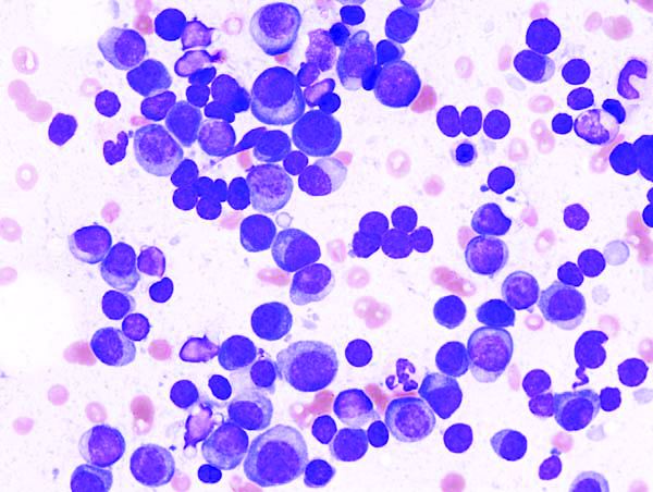
The advantages of levofloxacin prophylaxis appear to offset the potential risks in patients with newly diagnosed disease, explained Mark T. Drayson, MBChB, PhD, of the University of Birmingham (England) and colleagues. The study was published in the Lancet Oncology.
The randomized, placebo-controlled, phase 3 TEAMM study enrolled 977 patients with newly diagnosed myeloma. The effects of antimicrobial prophylaxis on infection risk and infection-related mortality were evaluated across 93 hospitals throughout the United Kingdom.
Study patients were randomly assigned to receive 500 mg of oral levofloxacin once daily or placebo for a total of 12 weeks. If applicable, dose adjustments were made based on estimated glomerular filtration rate.
At baseline, the team collected stool samples and nasal swabs, and follow-up assessment occurred every 4 weeks for up to 1 year. The primary endpoint was time to death (all causes) or first febrile event from the start of prophylactic therapy to 12 weeks.
After a median follow-up of 12 months, first febrile episodes or deaths were significantly lower for patients in the levofloxacin arm (19%), compared with the placebo arm (27%) for a hazard ratio for time to first event of 0.66 (95% confidence interval, 0.51-0.86; P = .0018).
With respect to safety, the rates of serious adverse events were similar between the study arms, with the exception of tendinitis in the levofloxacin group (1%). Among all patients, a total of 597 serious toxicities were observed from baseline to 16 weeks (52% in the levofloxacin arm vs. 48% in the placebo arm).
“To our knowledge, this is the first time that the use of prophylactic antibiotics has shown a survival benefit in patients with newly diagnosed myeloma,” the researchers reported.
One key limitation of the study was the younger patient population relative to the general population. As a result, differences in survival estimates could exist between the trial and real-world populations, they noted.
“Patients with newly diagnosed myeloma could benefit from levofloxacin prophylaxis, although local antibiotic resistance proportions must be considered,” the researchers cautioned.
The study was funded by the National Institute for Health Research in the United Kingdom. The authors reported financial affiliations with Actelion, Astellas, Celgene, Gilead, Janssen, Pfizer, Takeda, and other companies.
SOURCE: Drayson MT et al. Lancet Oncol. 2019 Oct 23. doi: 10.1016/S1470-2045(19)30506-6.
Adding levofloxacin to antimyeloma therapy improved survival and reduced infections in patients with newly diagnosed myeloma, findings from a phase 3 trial suggest.
The advantages of levofloxacin prophylaxis appear to offset the potential risks in patients with newly diagnosed disease, explained Mark T. Drayson, MBChB, PhD, of the University of Birmingham (England) and colleagues. The study was published in the Lancet Oncology.
The randomized, placebo-controlled, phase 3 TEAMM study enrolled 977 patients with newly diagnosed myeloma. The effects of antimicrobial prophylaxis on infection risk and infection-related mortality were evaluated across 93 hospitals throughout the United Kingdom.
Study patients were randomly assigned to receive 500 mg of oral levofloxacin once daily or placebo for a total of 12 weeks. If applicable, dose adjustments were made based on estimated glomerular filtration rate.
At baseline, the team collected stool samples and nasal swabs, and follow-up assessment occurred every 4 weeks for up to 1 year. The primary endpoint was time to death (all causes) or first febrile event from the start of prophylactic therapy to 12 weeks.
After a median follow-up of 12 months, first febrile episodes or deaths were significantly lower for patients in the levofloxacin arm (19%), compared with the placebo arm (27%) for a hazard ratio for time to first event of 0.66 (95% confidence interval, 0.51-0.86; P = .0018).
With respect to safety, the rates of serious adverse events were similar between the study arms, with the exception of tendinitis in the levofloxacin group (1%). Among all patients, a total of 597 serious toxicities were observed from baseline to 16 weeks (52% in the levofloxacin arm vs. 48% in the placebo arm).
“To our knowledge, this is the first time that the use of prophylactic antibiotics has shown a survival benefit in patients with newly diagnosed myeloma,” the researchers reported.
One key limitation of the study was the younger patient population relative to the general population. As a result, differences in survival estimates could exist between the trial and real-world populations, they noted.
“Patients with newly diagnosed myeloma could benefit from levofloxacin prophylaxis, although local antibiotic resistance proportions must be considered,” the researchers cautioned.
The study was funded by the National Institute for Health Research in the United Kingdom. The authors reported financial affiliations with Actelion, Astellas, Celgene, Gilead, Janssen, Pfizer, Takeda, and other companies.
SOURCE: Drayson MT et al. Lancet Oncol. 2019 Oct 23. doi: 10.1016/S1470-2045(19)30506-6.
FROM LANCET ONCOLOGY