User login
Idiopathic pulmonary fibrosis a ‘robust diagnosis’
LIVERPOOL, ENGLAND – Very few patients with idiopathic pulmonary fibrosis have connective tissue disease antibodies, suggesting that IPF is a “robust diagnosis” when made on the basis of standard diagnostic tests, it was reported at the British Society for Rheumatology annual conference.
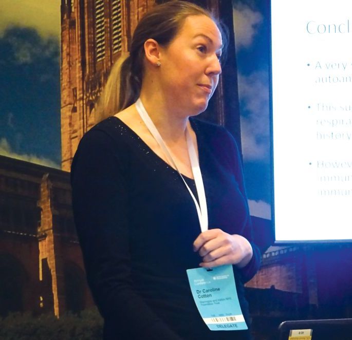
“The results were perhaps not what we’d expected,” said Caroline V. Cotton, PhD, of the Institute of Ageing and Chronic Disease at the University of Liverpool, England.
This means that chest physicians are getting the diagnosis right in the majority of cases, based on currently available methods, such as patients’ clinical history and examination, the results of high resolution–computed tomography, and widely available serology. “Which is good news,” Dr. Cotton observed.
Interstitial lung disease (ILD) comprises a huge spectrum of disorders. The main groups of ILDs are idiopathic, granulomatous, connective tissue disease–associated environmental, or medication exposure–associated; and the rare causes of ILD, each of which contain multiple subgroups of which IPF is one.
Sometimes it is obvious to respiratory physicians what the cause is, such as environmental exposure to asbestos or sarcoidosis for the granulomatous ILD, Dr. Cotton noted. Identifying connective tissue disease (CTD)–associated ILD can be more diagnostically challenging, however, and there are a large number of rheumatic conditions associated with CTD-associated ILD, including rheumatoid arthritis, systemic sclerosis, and Sjögren’s syndrome, to name a few.
One of the problems is that signs and symptoms of CTD may be absent at the time ILD starts to manifest and, even if signs are present, they may too subtle to be picked up in a general chest clinic. There also is a large number of antibodies for CTDs, but not all are widely available.
Dr. Cotton and her associates, therefore, wondered if there was a chance that patients being diagnosed with IPF actually could have covert CTD-associated ILD; this is an important distinction to make because the treatment differs for the two conditions. While ILD associated with CTD has a strong inflammatory component and is treated with corticosteroids and immunosuppressants, steroids can be harmful and increase mortality in IPF-ILD. The latter is treated with antifibrotic medications, such as pirfenidone and nintedanib.
For the study, serum samples from 250 patients with a definite diagnosis of IPF who were participating in the UK-BILD study were obtained and screened for known CTD antibodies using immunoprecipitation. Antibodies could be detected in just five (2%) patients – these included one patient each with anti-KS and anti-OJ antibodies, which are antisynthetase antibodies that are associated with myositis. Anti-Ku, another myositis-associated antibody, was identified in another patient, and one patient had an anti-RNA polymerase II antibody, which is associated with systemic sclerosis. Antimitochondrial autoantibodies were observed in one patient, and these are linked to primary biliary cirrhosis, which the patient was known to have.
There was nothing remarkable between the patients who did and did not have CTD antibodies in terms of their demographics, 76% and 80% were male, the mean ages were 73 and 70 years, respectively, and all were white.
However, 40% of patients did have unknown strong bands on immunoprecipitation, Dr. Cotton reported. This could suggest that there is an underlying immunological component to IPF, she added, but they had no recognized antibodies.
“A very small number of patients with IPF actually have the presence of autoantibodies strongly associated with CTDs. This suggests IPF is a very robust diagnosis; chest physicians are diagnosing it correctly most of the time, and they are really good at good at weeding out those who have got IPF and those who have potentially got connective tissue disease.” Dr. Cotton concluded.
Dr. Cotton had no conflicts of interest.
SOURCE: Cotton CV et al. Rheumatology. 2018;57[Suppl. 3]:key075.206.
LIVERPOOL, ENGLAND – Very few patients with idiopathic pulmonary fibrosis have connective tissue disease antibodies, suggesting that IPF is a “robust diagnosis” when made on the basis of standard diagnostic tests, it was reported at the British Society for Rheumatology annual conference.
“The results were perhaps not what we’d expected,” said Caroline V. Cotton, PhD, of the Institute of Ageing and Chronic Disease at the University of Liverpool, England.
This means that chest physicians are getting the diagnosis right in the majority of cases, based on currently available methods, such as patients’ clinical history and examination, the results of high resolution–computed tomography, and widely available serology. “Which is good news,” Dr. Cotton observed.
Interstitial lung disease (ILD) comprises a huge spectrum of disorders. The main groups of ILDs are idiopathic, granulomatous, connective tissue disease–associated environmental, or medication exposure–associated; and the rare causes of ILD, each of which contain multiple subgroups of which IPF is one.
Sometimes it is obvious to respiratory physicians what the cause is, such as environmental exposure to asbestos or sarcoidosis for the granulomatous ILD, Dr. Cotton noted. Identifying connective tissue disease (CTD)–associated ILD can be more diagnostically challenging, however, and there are a large number of rheumatic conditions associated with CTD-associated ILD, including rheumatoid arthritis, systemic sclerosis, and Sjögren’s syndrome, to name a few.
One of the problems is that signs and symptoms of CTD may be absent at the time ILD starts to manifest and, even if signs are present, they may too subtle to be picked up in a general chest clinic. There also is a large number of antibodies for CTDs, but not all are widely available.
Dr. Cotton and her associates, therefore, wondered if there was a chance that patients being diagnosed with IPF actually could have covert CTD-associated ILD; this is an important distinction to make because the treatment differs for the two conditions. While ILD associated with CTD has a strong inflammatory component and is treated with corticosteroids and immunosuppressants, steroids can be harmful and increase mortality in IPF-ILD. The latter is treated with antifibrotic medications, such as pirfenidone and nintedanib.
For the study, serum samples from 250 patients with a definite diagnosis of IPF who were participating in the UK-BILD study were obtained and screened for known CTD antibodies using immunoprecipitation. Antibodies could be detected in just five (2%) patients – these included one patient each with anti-KS and anti-OJ antibodies, which are antisynthetase antibodies that are associated with myositis. Anti-Ku, another myositis-associated antibody, was identified in another patient, and one patient had an anti-RNA polymerase II antibody, which is associated with systemic sclerosis. Antimitochondrial autoantibodies were observed in one patient, and these are linked to primary biliary cirrhosis, which the patient was known to have.
There was nothing remarkable between the patients who did and did not have CTD antibodies in terms of their demographics, 76% and 80% were male, the mean ages were 73 and 70 years, respectively, and all were white.
However, 40% of patients did have unknown strong bands on immunoprecipitation, Dr. Cotton reported. This could suggest that there is an underlying immunological component to IPF, she added, but they had no recognized antibodies.
“A very small number of patients with IPF actually have the presence of autoantibodies strongly associated with CTDs. This suggests IPF is a very robust diagnosis; chest physicians are diagnosing it correctly most of the time, and they are really good at good at weeding out those who have got IPF and those who have potentially got connective tissue disease.” Dr. Cotton concluded.
Dr. Cotton had no conflicts of interest.
SOURCE: Cotton CV et al. Rheumatology. 2018;57[Suppl. 3]:key075.206.
LIVERPOOL, ENGLAND – Very few patients with idiopathic pulmonary fibrosis have connective tissue disease antibodies, suggesting that IPF is a “robust diagnosis” when made on the basis of standard diagnostic tests, it was reported at the British Society for Rheumatology annual conference.
“The results were perhaps not what we’d expected,” said Caroline V. Cotton, PhD, of the Institute of Ageing and Chronic Disease at the University of Liverpool, England.
This means that chest physicians are getting the diagnosis right in the majority of cases, based on currently available methods, such as patients’ clinical history and examination, the results of high resolution–computed tomography, and widely available serology. “Which is good news,” Dr. Cotton observed.
Interstitial lung disease (ILD) comprises a huge spectrum of disorders. The main groups of ILDs are idiopathic, granulomatous, connective tissue disease–associated environmental, or medication exposure–associated; and the rare causes of ILD, each of which contain multiple subgroups of which IPF is one.
Sometimes it is obvious to respiratory physicians what the cause is, such as environmental exposure to asbestos or sarcoidosis for the granulomatous ILD, Dr. Cotton noted. Identifying connective tissue disease (CTD)–associated ILD can be more diagnostically challenging, however, and there are a large number of rheumatic conditions associated with CTD-associated ILD, including rheumatoid arthritis, systemic sclerosis, and Sjögren’s syndrome, to name a few.
One of the problems is that signs and symptoms of CTD may be absent at the time ILD starts to manifest and, even if signs are present, they may too subtle to be picked up in a general chest clinic. There also is a large number of antibodies for CTDs, but not all are widely available.
Dr. Cotton and her associates, therefore, wondered if there was a chance that patients being diagnosed with IPF actually could have covert CTD-associated ILD; this is an important distinction to make because the treatment differs for the two conditions. While ILD associated with CTD has a strong inflammatory component and is treated with corticosteroids and immunosuppressants, steroids can be harmful and increase mortality in IPF-ILD. The latter is treated with antifibrotic medications, such as pirfenidone and nintedanib.
For the study, serum samples from 250 patients with a definite diagnosis of IPF who were participating in the UK-BILD study were obtained and screened for known CTD antibodies using immunoprecipitation. Antibodies could be detected in just five (2%) patients – these included one patient each with anti-KS and anti-OJ antibodies, which are antisynthetase antibodies that are associated with myositis. Anti-Ku, another myositis-associated antibody, was identified in another patient, and one patient had an anti-RNA polymerase II antibody, which is associated with systemic sclerosis. Antimitochondrial autoantibodies were observed in one patient, and these are linked to primary biliary cirrhosis, which the patient was known to have.
There was nothing remarkable between the patients who did and did not have CTD antibodies in terms of their demographics, 76% and 80% were male, the mean ages were 73 and 70 years, respectively, and all were white.
However, 40% of patients did have unknown strong bands on immunoprecipitation, Dr. Cotton reported. This could suggest that there is an underlying immunological component to IPF, she added, but they had no recognized antibodies.
“A very small number of patients with IPF actually have the presence of autoantibodies strongly associated with CTDs. This suggests IPF is a very robust diagnosis; chest physicians are diagnosing it correctly most of the time, and they are really good at good at weeding out those who have got IPF and those who have potentially got connective tissue disease.” Dr. Cotton concluded.
Dr. Cotton had no conflicts of interest.
SOURCE: Cotton CV et al. Rheumatology. 2018;57[Suppl. 3]:key075.206.
REPORTING FROM RHEUMATOLOGY 2018
Key clinical point: Few patients diagnosed as having idiopathic pulmonary fibrosis (IPF) are likely to have connective tissue disorders (CTD).
Major finding: Only 2% of patients had a recognized CTD antibody present.
Study details: 250 patients with IPF participating in the UK-BILD multicenter study.
Disclosures: Dr. Cotton stated she had no conflicts of interest.
Source: Cotton CV et al. Rheumatology. 2018;57(Suppl. 3):key075.206.
Does Sleep Help Protect Against Amyloid Plaques?
A cardinal feature of Alzheimer disease is the way beta-amyloid—generally a metabolic waste product—clumps to form amyloid plaques. Now a National Institute on Alcohol Abuse and Alcoholism (NIAAA) study indicates that sleep may be an important link in that process. The researchers found that losing just 1 night of sleep led to an immediate increase in beta-amyloid.
Researchers used positron emission tomography to scan the brains of 20 healthy volunteers, aged 22 to 72 years, after a night of rested sleep and after being awake for 31 hours. They found beta-amyloid increases of about 5% after the sleep deprivation in the thalamus and hippocampus, regions especially vulnerable to damage in the early stages of Alzheimer disease, the researchers say. The study participants with larger increases also reported worse mood after sleep deprivation.
It is important to note, the researchers add, that the link between sleep disorders and Alzheimer risk is considered by many scientists to be bidirectional, since elevated beta-amyloid also may cause sleep disturbance.
It is unknown, the researchers say, whether the increase in beta-amyloid in the study participants would subside after a night of rest.
A cardinal feature of Alzheimer disease is the way beta-amyloid—generally a metabolic waste product—clumps to form amyloid plaques. Now a National Institute on Alcohol Abuse and Alcoholism (NIAAA) study indicates that sleep may be an important link in that process. The researchers found that losing just 1 night of sleep led to an immediate increase in beta-amyloid.
Researchers used positron emission tomography to scan the brains of 20 healthy volunteers, aged 22 to 72 years, after a night of rested sleep and after being awake for 31 hours. They found beta-amyloid increases of about 5% after the sleep deprivation in the thalamus and hippocampus, regions especially vulnerable to damage in the early stages of Alzheimer disease, the researchers say. The study participants with larger increases also reported worse mood after sleep deprivation.
It is important to note, the researchers add, that the link between sleep disorders and Alzheimer risk is considered by many scientists to be bidirectional, since elevated beta-amyloid also may cause sleep disturbance.
It is unknown, the researchers say, whether the increase in beta-amyloid in the study participants would subside after a night of rest.
A cardinal feature of Alzheimer disease is the way beta-amyloid—generally a metabolic waste product—clumps to form amyloid plaques. Now a National Institute on Alcohol Abuse and Alcoholism (NIAAA) study indicates that sleep may be an important link in that process. The researchers found that losing just 1 night of sleep led to an immediate increase in beta-amyloid.
Researchers used positron emission tomography to scan the brains of 20 healthy volunteers, aged 22 to 72 years, after a night of rested sleep and after being awake for 31 hours. They found beta-amyloid increases of about 5% after the sleep deprivation in the thalamus and hippocampus, regions especially vulnerable to damage in the early stages of Alzheimer disease, the researchers say. The study participants with larger increases also reported worse mood after sleep deprivation.
It is important to note, the researchers add, that the link between sleep disorders and Alzheimer risk is considered by many scientists to be bidirectional, since elevated beta-amyloid also may cause sleep disturbance.
It is unknown, the researchers say, whether the increase in beta-amyloid in the study participants would subside after a night of rest.
Trio of blood biomarkers elevated in children with LRTIs
TORONTO – While C-reactive protein, procalcitonin, and proadrenomedullin are associated with development of severe clinical outcomes in children with lower respiratory tract infections, proadrenomedullin is most strongly associated with disease severity, preliminary results from a prospective cohort study showed.
“Despite the fact that pneumonia guidelines call the site of care decision the most important decision in the management of pediatric pneumonia, no validated risk stratification tools exist for pediatric lower respiratory tract infections (LRTI),” lead study author Todd A. Florin, MD, said at the annual Pediatric Academic Societies meeting. “Biomarkers offer an objective means of classifying disease severity and clinical outcomes.”

PCT is a precursor of calcitonin secreted by the thyroid, lung, and intestine in response to bacterial infections. It also has been shown to be associated with adverse outcomes and mortality in adults, with results generally suggesting that it is a stronger predictor of severity than CRP. “There is limited data on the association of CRP or PCT with severe outcomes in children with LRTIs,” Dr. Florin noted. “One recent U.S. study of 532 children did demonstrate an association of elevated PCT with ICU admission, chest drainage, and hospital length of stay in children with [community-acquired pneumonia] CAP.”
ProADM, meanwhile, is a vasodilatory peptide with antimicrobial and anti-inflammatory functions synthesized during severe infections. It has a half-life of several hours and has been shown to be associated with disease severity in adults with LRTI. Recent studies have shown that it has improved prognostication over WBC, CRP, and PCT. “In two small studies of children with pneumonia, proADM levels were significantly elevated in children with complicated pneumonia, compared to those with uncomplicated pneumonia,” Dr. Florin said. “Although all three of these markers demonstrate promise in predicting severe outcomes in adults with LRTIs, very few studies have examined their association with disease severity in pediatric disease. Therefore, the aim of the current analysis was to determine the association between blood biomarkers and disease severity in children who present to the ED with lower respiratory tract infections.”
In a study known as Catalyzing Ambulatory Research in Pneumonia Etiology and Diagnostic Innovations in Emergency Medicine (CARPE DIEM), he and his associates performed a prospective cohort analysis of children with suspected CAP who were admitted to the Cincinnati Children’s Hospital ED between July 2012 and December 2017. They limited the analysis to children aged 3 months to 18 years with signs and symptoms of an LRTI, and all eligible patients were required to have a chest radiograph ordered for suspicion of CAP. They excluded children hospitalized within 14 days prior to the index ED visit, immunodeficient or immunosuppressed children, those with a history of aspiration or aspiration pneumonia, and those who weighed less than 5 kg because of blood drawing maximums. Biomarkers were measured only in children with focal findings on chest x-ray in the ED. The primary outcome was disease severity: mild (defined as discharged home), moderate (defined as hospitalized, but not severe) and severe (defined as having an ICU length of stay of greater than 48 hours, chest drainage, severe sepsis, noninvasive positive pressure ventilation, intubation, vasoactive infusions, or death). Biomarkers were obtained at the time of presentation to the ED, prior to the occurrence of clinical outcomes.
Over a period of 4.5 years, the researchers enrolled 1,142 patients. Of these, 478 had focal findings on chest x-ray and blood obtained. The median age of these 478 children was 4.4 years, 52% were male, and 82% had all three biomarkers performed. Specifically, 456 had CRP and PCT performed, while 358 had proADM performed. “Not every child had every marker performed due to challenges in obtaining sufficient blood for all three biomarkers in some children,” Dr. Florin explained.
Preliminary data that Dr. Florin presented at PAS found that the median CRP, PCT, and proADM did not differ by gender, race, ethnicity, or insurance status. “In addition, there were not significant differences in the distribution of disease severity by biomarker performed, with approximately 27% of patients being classified as mild, 66% as moderate, and 7% as severe,” he said.
The median CRP was 2.4 ng/mL in those with mild disease, 2.5 ng/mL in those with moderate disease, and 6.25 ng/mL in those with severe disease, with the difference between the two subclasses of nonsevere disease and moderate disease and severe disease reaching statistical significance (P = .002). The median PCT was 0.16 ng/mL in those with mild disease, 0.26 ng/mL in those with moderate disease, and 0.49 ng/mL in those with severe disease, with the difference between the two subclasses of nonsevere disease and moderate disease and severe disease reaching statistical significance (P = .047). Meanwhile, the median proADM was 0.53 ng/mL in those with mild disease, 0.59 ng/mL in those with moderate disease, and 0.81 ng/mL in those with severe disease, with the difference between the two subclasses of nonsevere disease and moderate disease and severe disease also reaching statistical significance (P less than .0001).
Next, the researchers performed logistic regression of each biomarker individually and in combination. They found that and had the best ability to discriminate those developing severe vs. nonsevere disease (area under the receiving operating curve of 0.72, vs. 0.67 and 0.60, respectively). When CRP and PCT markers were combined with proADM, they were no longer associated with severe disease, while a strong association with proADM remained significant.
Dr. Florin acknowledged certain limitations of the study, including the fact that requiring collection of blood samples may have resulted in an enrollment bias toward patients receiving phlebotomy or IV line placement in the ED. “In addition, the children in the moderate-severity group are likely more heterogeneous than the other two severity groups,” he said. “Finally, given that this is a single-center study, we had a relatively small number of outcomes for some of the individual severity measures, which may have limited power and precision.”
He concluded his presentation by saying that he is “cautiously optimistic” about the study results. “As is the case in many biomarker studies, I do not anticipate that any single biomarker will be the magic bullet for predicting disease severity in pediatric CAP,” Dr. Florin said. “It will likely be a combination of clinical factors and several biomarkers that will achieve optimal prognostic ability. That said, our results suggest that similar to adult studies, proADM appears to have the strongest association with severe disease, compared with CRP and PCT. Combinations of biomarkers did not perform better than proADM alone. With the advent of rapid point-of-care diagnostics, these markers may have a role in management and site-of-care decisions for children with LRTI.”
The study received funding support from the Gerber Foundation, the National Institute of Allergy and Infectious Diseases, and Cincinnati Children’s Hospital Medical Center. Dr. Florin reported having no financial disclosures.
TORONTO – While C-reactive protein, procalcitonin, and proadrenomedullin are associated with development of severe clinical outcomes in children with lower respiratory tract infections, proadrenomedullin is most strongly associated with disease severity, preliminary results from a prospective cohort study showed.
“Despite the fact that pneumonia guidelines call the site of care decision the most important decision in the management of pediatric pneumonia, no validated risk stratification tools exist for pediatric lower respiratory tract infections (LRTI),” lead study author Todd A. Florin, MD, said at the annual Pediatric Academic Societies meeting. “Biomarkers offer an objective means of classifying disease severity and clinical outcomes.”
PCT is a precursor of calcitonin secreted by the thyroid, lung, and intestine in response to bacterial infections. It also has been shown to be associated with adverse outcomes and mortality in adults, with results generally suggesting that it is a stronger predictor of severity than CRP. “There is limited data on the association of CRP or PCT with severe outcomes in children with LRTIs,” Dr. Florin noted. “One recent U.S. study of 532 children did demonstrate an association of elevated PCT with ICU admission, chest drainage, and hospital length of stay in children with [community-acquired pneumonia] CAP.”
ProADM, meanwhile, is a vasodilatory peptide with antimicrobial and anti-inflammatory functions synthesized during severe infections. It has a half-life of several hours and has been shown to be associated with disease severity in adults with LRTI. Recent studies have shown that it has improved prognostication over WBC, CRP, and PCT. “In two small studies of children with pneumonia, proADM levels were significantly elevated in children with complicated pneumonia, compared to those with uncomplicated pneumonia,” Dr. Florin said. “Although all three of these markers demonstrate promise in predicting severe outcomes in adults with LRTIs, very few studies have examined their association with disease severity in pediatric disease. Therefore, the aim of the current analysis was to determine the association between blood biomarkers and disease severity in children who present to the ED with lower respiratory tract infections.”
In a study known as Catalyzing Ambulatory Research in Pneumonia Etiology and Diagnostic Innovations in Emergency Medicine (CARPE DIEM), he and his associates performed a prospective cohort analysis of children with suspected CAP who were admitted to the Cincinnati Children’s Hospital ED between July 2012 and December 2017. They limited the analysis to children aged 3 months to 18 years with signs and symptoms of an LRTI, and all eligible patients were required to have a chest radiograph ordered for suspicion of CAP. They excluded children hospitalized within 14 days prior to the index ED visit, immunodeficient or immunosuppressed children, those with a history of aspiration or aspiration pneumonia, and those who weighed less than 5 kg because of blood drawing maximums. Biomarkers were measured only in children with focal findings on chest x-ray in the ED. The primary outcome was disease severity: mild (defined as discharged home), moderate (defined as hospitalized, but not severe) and severe (defined as having an ICU length of stay of greater than 48 hours, chest drainage, severe sepsis, noninvasive positive pressure ventilation, intubation, vasoactive infusions, or death). Biomarkers were obtained at the time of presentation to the ED, prior to the occurrence of clinical outcomes.
Over a period of 4.5 years, the researchers enrolled 1,142 patients. Of these, 478 had focal findings on chest x-ray and blood obtained. The median age of these 478 children was 4.4 years, 52% were male, and 82% had all three biomarkers performed. Specifically, 456 had CRP and PCT performed, while 358 had proADM performed. “Not every child had every marker performed due to challenges in obtaining sufficient blood for all three biomarkers in some children,” Dr. Florin explained.
Preliminary data that Dr. Florin presented at PAS found that the median CRP, PCT, and proADM did not differ by gender, race, ethnicity, or insurance status. “In addition, there were not significant differences in the distribution of disease severity by biomarker performed, with approximately 27% of patients being classified as mild, 66% as moderate, and 7% as severe,” he said.
The median CRP was 2.4 ng/mL in those with mild disease, 2.5 ng/mL in those with moderate disease, and 6.25 ng/mL in those with severe disease, with the difference between the two subclasses of nonsevere disease and moderate disease and severe disease reaching statistical significance (P = .002). The median PCT was 0.16 ng/mL in those with mild disease, 0.26 ng/mL in those with moderate disease, and 0.49 ng/mL in those with severe disease, with the difference between the two subclasses of nonsevere disease and moderate disease and severe disease reaching statistical significance (P = .047). Meanwhile, the median proADM was 0.53 ng/mL in those with mild disease, 0.59 ng/mL in those with moderate disease, and 0.81 ng/mL in those with severe disease, with the difference between the two subclasses of nonsevere disease and moderate disease and severe disease also reaching statistical significance (P less than .0001).
Next, the researchers performed logistic regression of each biomarker individually and in combination. They found that and had the best ability to discriminate those developing severe vs. nonsevere disease (area under the receiving operating curve of 0.72, vs. 0.67 and 0.60, respectively). When CRP and PCT markers were combined with proADM, they were no longer associated with severe disease, while a strong association with proADM remained significant.
Dr. Florin acknowledged certain limitations of the study, including the fact that requiring collection of blood samples may have resulted in an enrollment bias toward patients receiving phlebotomy or IV line placement in the ED. “In addition, the children in the moderate-severity group are likely more heterogeneous than the other two severity groups,” he said. “Finally, given that this is a single-center study, we had a relatively small number of outcomes for some of the individual severity measures, which may have limited power and precision.”
He concluded his presentation by saying that he is “cautiously optimistic” about the study results. “As is the case in many biomarker studies, I do not anticipate that any single biomarker will be the magic bullet for predicting disease severity in pediatric CAP,” Dr. Florin said. “It will likely be a combination of clinical factors and several biomarkers that will achieve optimal prognostic ability. That said, our results suggest that similar to adult studies, proADM appears to have the strongest association with severe disease, compared with CRP and PCT. Combinations of biomarkers did not perform better than proADM alone. With the advent of rapid point-of-care diagnostics, these markers may have a role in management and site-of-care decisions for children with LRTI.”
The study received funding support from the Gerber Foundation, the National Institute of Allergy and Infectious Diseases, and Cincinnati Children’s Hospital Medical Center. Dr. Florin reported having no financial disclosures.
TORONTO – While C-reactive protein, procalcitonin, and proadrenomedullin are associated with development of severe clinical outcomes in children with lower respiratory tract infections, proadrenomedullin is most strongly associated with disease severity, preliminary results from a prospective cohort study showed.
“Despite the fact that pneumonia guidelines call the site of care decision the most important decision in the management of pediatric pneumonia, no validated risk stratification tools exist for pediatric lower respiratory tract infections (LRTI),” lead study author Todd A. Florin, MD, said at the annual Pediatric Academic Societies meeting. “Biomarkers offer an objective means of classifying disease severity and clinical outcomes.”
PCT is a precursor of calcitonin secreted by the thyroid, lung, and intestine in response to bacterial infections. It also has been shown to be associated with adverse outcomes and mortality in adults, with results generally suggesting that it is a stronger predictor of severity than CRP. “There is limited data on the association of CRP or PCT with severe outcomes in children with LRTIs,” Dr. Florin noted. “One recent U.S. study of 532 children did demonstrate an association of elevated PCT with ICU admission, chest drainage, and hospital length of stay in children with [community-acquired pneumonia] CAP.”
ProADM, meanwhile, is a vasodilatory peptide with antimicrobial and anti-inflammatory functions synthesized during severe infections. It has a half-life of several hours and has been shown to be associated with disease severity in adults with LRTI. Recent studies have shown that it has improved prognostication over WBC, CRP, and PCT. “In two small studies of children with pneumonia, proADM levels were significantly elevated in children with complicated pneumonia, compared to those with uncomplicated pneumonia,” Dr. Florin said. “Although all three of these markers demonstrate promise in predicting severe outcomes in adults with LRTIs, very few studies have examined their association with disease severity in pediatric disease. Therefore, the aim of the current analysis was to determine the association between blood biomarkers and disease severity in children who present to the ED with lower respiratory tract infections.”
In a study known as Catalyzing Ambulatory Research in Pneumonia Etiology and Diagnostic Innovations in Emergency Medicine (CARPE DIEM), he and his associates performed a prospective cohort analysis of children with suspected CAP who were admitted to the Cincinnati Children’s Hospital ED between July 2012 and December 2017. They limited the analysis to children aged 3 months to 18 years with signs and symptoms of an LRTI, and all eligible patients were required to have a chest radiograph ordered for suspicion of CAP. They excluded children hospitalized within 14 days prior to the index ED visit, immunodeficient or immunosuppressed children, those with a history of aspiration or aspiration pneumonia, and those who weighed less than 5 kg because of blood drawing maximums. Biomarkers were measured only in children with focal findings on chest x-ray in the ED. The primary outcome was disease severity: mild (defined as discharged home), moderate (defined as hospitalized, but not severe) and severe (defined as having an ICU length of stay of greater than 48 hours, chest drainage, severe sepsis, noninvasive positive pressure ventilation, intubation, vasoactive infusions, or death). Biomarkers were obtained at the time of presentation to the ED, prior to the occurrence of clinical outcomes.
Over a period of 4.5 years, the researchers enrolled 1,142 patients. Of these, 478 had focal findings on chest x-ray and blood obtained. The median age of these 478 children was 4.4 years, 52% were male, and 82% had all three biomarkers performed. Specifically, 456 had CRP and PCT performed, while 358 had proADM performed. “Not every child had every marker performed due to challenges in obtaining sufficient blood for all three biomarkers in some children,” Dr. Florin explained.
Preliminary data that Dr. Florin presented at PAS found that the median CRP, PCT, and proADM did not differ by gender, race, ethnicity, or insurance status. “In addition, there were not significant differences in the distribution of disease severity by biomarker performed, with approximately 27% of patients being classified as mild, 66% as moderate, and 7% as severe,” he said.
The median CRP was 2.4 ng/mL in those with mild disease, 2.5 ng/mL in those with moderate disease, and 6.25 ng/mL in those with severe disease, with the difference between the two subclasses of nonsevere disease and moderate disease and severe disease reaching statistical significance (P = .002). The median PCT was 0.16 ng/mL in those with mild disease, 0.26 ng/mL in those with moderate disease, and 0.49 ng/mL in those with severe disease, with the difference between the two subclasses of nonsevere disease and moderate disease and severe disease reaching statistical significance (P = .047). Meanwhile, the median proADM was 0.53 ng/mL in those with mild disease, 0.59 ng/mL in those with moderate disease, and 0.81 ng/mL in those with severe disease, with the difference between the two subclasses of nonsevere disease and moderate disease and severe disease also reaching statistical significance (P less than .0001).
Next, the researchers performed logistic regression of each biomarker individually and in combination. They found that and had the best ability to discriminate those developing severe vs. nonsevere disease (area under the receiving operating curve of 0.72, vs. 0.67 and 0.60, respectively). When CRP and PCT markers were combined with proADM, they were no longer associated with severe disease, while a strong association with proADM remained significant.
Dr. Florin acknowledged certain limitations of the study, including the fact that requiring collection of blood samples may have resulted in an enrollment bias toward patients receiving phlebotomy or IV line placement in the ED. “In addition, the children in the moderate-severity group are likely more heterogeneous than the other two severity groups,” he said. “Finally, given that this is a single-center study, we had a relatively small number of outcomes for some of the individual severity measures, which may have limited power and precision.”
He concluded his presentation by saying that he is “cautiously optimistic” about the study results. “As is the case in many biomarker studies, I do not anticipate that any single biomarker will be the magic bullet for predicting disease severity in pediatric CAP,” Dr. Florin said. “It will likely be a combination of clinical factors and several biomarkers that will achieve optimal prognostic ability. That said, our results suggest that similar to adult studies, proADM appears to have the strongest association with severe disease, compared with CRP and PCT. Combinations of biomarkers did not perform better than proADM alone. With the advent of rapid point-of-care diagnostics, these markers may have a role in management and site-of-care decisions for children with LRTI.”
The study received funding support from the Gerber Foundation, the National Institute of Allergy and Infectious Diseases, and Cincinnati Children’s Hospital Medical Center. Dr. Florin reported having no financial disclosures.
AT PAS 18
Key clinical point: Blood biomarkers such as C-reactive protein (CRP), procalcitonin (PCT), and proadrenomedullin (proADM) may have a role in management and site-of-care decisions for children with LRTIs.
Major finding: The proADM alone was associated with the largest odds for severe disease (OR 13.1), compared with CRP alone (OR 1.6) and PCT alone (OR 1.4).
Study details: Preliminary results from prospective cohort analysis of 478 children with suspected community-acquired pneumonia who were admitted to the Cincinnati Children’s Hospital ED.
Disclosures: The study received funding support from the Gerber Foundation, the National Institute of Allergy and Infectious Diseases, and Cincinnati Children’s Hospital Medical Center. Dr. Florin reported having no financial disclosures.
AZD8871 delivered significant bronchodilation in two-week study
SAN DIEGO – , results from a phase 2a trial showed.
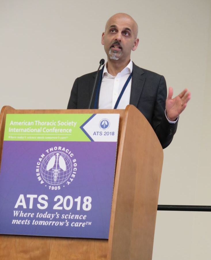
AZD8871 is a long-acting, bifunctional bronchodilator that combines a muscarinic antagonist and a beta-2 adrenoceptor agonist. “There are some interesting avenues that you can explore with such a molecule,” one of the study authors, Dave Singh, MD, said at an international conference of the American Thoracic Society. “First, theoretically, as a single molecule you will be able to deposit both the active ingredients to the same site in the lung. On a more practical note, if you want to add something else to a dual bronchodilator, which is essentially what AZD8871 is, this provides a platform. Perhaps that’s the most interesting use of this type of approach.”
Single doses of AZD8871 (400 mcg and 1,800 mcg) administered in COPD patients demonstrated sustained bronchodilation over 36 hours. In a study presented at the 2017 meeting of the European Respiratory Society, Dr. Singh and his associates found that AZD8871 1,800 mcg showed greater bronchodilation than both indacaterol and tiotropium for peak and trough FEV1.
For the current study, researchers at one site in the United Kingdom and one site in Germany conducted a phase 2 randomized, double-blind, placebo-controlled trial of AZD8871 in 42 patients aged 40-80 years with moderate to severe reversible COPD. Patients were randomized to receive repeated once-daily doses of AZD8871 100 mcg, 600 mcg, or placebo via a dry powder inhaler device for 14 days. Between-treatment washout periods were 28-35 days. “We keep the patients in-house on day one and day 14 of each treatment period, and we measure lung function over 24 hours,” said Dr. Singh, professor of clinical pharmacology and respiratory medicine at the University of Manchester, United Kingdom. “Patients were allowed to continue any pre-existing steroid therapy, but at the end of screening they had to withdraw any long-acting bronchodilator therapy.”
The primary efficacy endpoint was change from baseline trough FEV1 on day 15. Secondary endpoints included change from baseline in peak FEV1, total score of breathlessness, cough, sputum scale questionnaire, and rescue medication use.
At baseline, the mean age of the 42 patients was 64 years, and 67% were male. Their mean FEV1 was about 58% predicted, and their FEV1 absolute reversibility was a mean of 379 mL, “which is rather high,” he said.
Of the 42 randomized patients, 31 completed all three treatments. Both doses of AZD8871 had a positive, dose-dependent effect on FEV1, compared with placebo, and both doses demonstrated an onset of action within 15 minutes. On day 15, least square mean change from baseline differences in trough FEV1 for AZD8871 100 mcg and 600 mcg versus placebo were 161 mL and 260 mL, respectively.
A similar association was observed with peak FEV1, which between baseline and day 14 increased by 380 mL at the 100 mcg dose and by 420 mL at the 600 mcg dose, compared with placebo. Sustained bronchodilation was observed over 24 hours on both day 1 and day 14.
Statistically significant COPD symptom improvements, measured by breathlessness, cough and sputum scale (BCSS), were observed for AZD8871 600 mcg on day 8 (P=0.002) and day 14 (P less than 0.001), compared with placebo.
In addition, substantial symptomatic improvements were observed for AZD8871 600 mcg on D14 versus placebo (least square mean of -1.16). Similar results were observed for individual domains of the BCSS. “When you separate out the different components of the scale, most of this is driven by the change in breathlessness,” he said. “We were surprised that we could capture this in such a small number of patients.”
On days 1-8 and days 9-14, the researchers observed a statistically significant improvement in change from baseline rescue medication use for AZD8871 600 mcg (P less than 0.001) and 100 mcg (P=0.029 and P=0.012, respectively), compared with placebo.
The most common adverse events for patients in all three treatment groups were headache (21.4%) and worsening of COPD-related symptoms (14.3%). No dose-dependence was observed with any adverse event, including serious adverse events and/or those leading to discontinuation.
AstraZeneca, the developer of AZD8871, sponsored the study. Dr. Singh reported being a consultant to and receiving research support from AstraZeneca and numerous other pharmaceutical companies.
SOURCE: Singh, D., et al, Abstract 7708, ATS 2018.
SAN DIEGO – , results from a phase 2a trial showed.
AZD8871 is a long-acting, bifunctional bronchodilator that combines a muscarinic antagonist and a beta-2 adrenoceptor agonist. “There are some interesting avenues that you can explore with such a molecule,” one of the study authors, Dave Singh, MD, said at an international conference of the American Thoracic Society. “First, theoretically, as a single molecule you will be able to deposit both the active ingredients to the same site in the lung. On a more practical note, if you want to add something else to a dual bronchodilator, which is essentially what AZD8871 is, this provides a platform. Perhaps that’s the most interesting use of this type of approach.”
Single doses of AZD8871 (400 mcg and 1,800 mcg) administered in COPD patients demonstrated sustained bronchodilation over 36 hours. In a study presented at the 2017 meeting of the European Respiratory Society, Dr. Singh and his associates found that AZD8871 1,800 mcg showed greater bronchodilation than both indacaterol and tiotropium for peak and trough FEV1.
For the current study, researchers at one site in the United Kingdom and one site in Germany conducted a phase 2 randomized, double-blind, placebo-controlled trial of AZD8871 in 42 patients aged 40-80 years with moderate to severe reversible COPD. Patients were randomized to receive repeated once-daily doses of AZD8871 100 mcg, 600 mcg, or placebo via a dry powder inhaler device for 14 days. Between-treatment washout periods were 28-35 days. “We keep the patients in-house on day one and day 14 of each treatment period, and we measure lung function over 24 hours,” said Dr. Singh, professor of clinical pharmacology and respiratory medicine at the University of Manchester, United Kingdom. “Patients were allowed to continue any pre-existing steroid therapy, but at the end of screening they had to withdraw any long-acting bronchodilator therapy.”
The primary efficacy endpoint was change from baseline trough FEV1 on day 15. Secondary endpoints included change from baseline in peak FEV1, total score of breathlessness, cough, sputum scale questionnaire, and rescue medication use.
At baseline, the mean age of the 42 patients was 64 years, and 67% were male. Their mean FEV1 was about 58% predicted, and their FEV1 absolute reversibility was a mean of 379 mL, “which is rather high,” he said.
Of the 42 randomized patients, 31 completed all three treatments. Both doses of AZD8871 had a positive, dose-dependent effect on FEV1, compared with placebo, and both doses demonstrated an onset of action within 15 minutes. On day 15, least square mean change from baseline differences in trough FEV1 for AZD8871 100 mcg and 600 mcg versus placebo were 161 mL and 260 mL, respectively.
A similar association was observed with peak FEV1, which between baseline and day 14 increased by 380 mL at the 100 mcg dose and by 420 mL at the 600 mcg dose, compared with placebo. Sustained bronchodilation was observed over 24 hours on both day 1 and day 14.
Statistically significant COPD symptom improvements, measured by breathlessness, cough and sputum scale (BCSS), were observed for AZD8871 600 mcg on day 8 (P=0.002) and day 14 (P less than 0.001), compared with placebo.
In addition, substantial symptomatic improvements were observed for AZD8871 600 mcg on D14 versus placebo (least square mean of -1.16). Similar results were observed for individual domains of the BCSS. “When you separate out the different components of the scale, most of this is driven by the change in breathlessness,” he said. “We were surprised that we could capture this in such a small number of patients.”
On days 1-8 and days 9-14, the researchers observed a statistically significant improvement in change from baseline rescue medication use for AZD8871 600 mcg (P less than 0.001) and 100 mcg (P=0.029 and P=0.012, respectively), compared with placebo.
The most common adverse events for patients in all three treatment groups were headache (21.4%) and worsening of COPD-related symptoms (14.3%). No dose-dependence was observed with any adverse event, including serious adverse events and/or those leading to discontinuation.
AstraZeneca, the developer of AZD8871, sponsored the study. Dr. Singh reported being a consultant to and receiving research support from AstraZeneca and numerous other pharmaceutical companies.
SOURCE: Singh, D., et al, Abstract 7708, ATS 2018.
SAN DIEGO – , results from a phase 2a trial showed.
AZD8871 is a long-acting, bifunctional bronchodilator that combines a muscarinic antagonist and a beta-2 adrenoceptor agonist. “There are some interesting avenues that you can explore with such a molecule,” one of the study authors, Dave Singh, MD, said at an international conference of the American Thoracic Society. “First, theoretically, as a single molecule you will be able to deposit both the active ingredients to the same site in the lung. On a more practical note, if you want to add something else to a dual bronchodilator, which is essentially what AZD8871 is, this provides a platform. Perhaps that’s the most interesting use of this type of approach.”
Single doses of AZD8871 (400 mcg and 1,800 mcg) administered in COPD patients demonstrated sustained bronchodilation over 36 hours. In a study presented at the 2017 meeting of the European Respiratory Society, Dr. Singh and his associates found that AZD8871 1,800 mcg showed greater bronchodilation than both indacaterol and tiotropium for peak and trough FEV1.
For the current study, researchers at one site in the United Kingdom and one site in Germany conducted a phase 2 randomized, double-blind, placebo-controlled trial of AZD8871 in 42 patients aged 40-80 years with moderate to severe reversible COPD. Patients were randomized to receive repeated once-daily doses of AZD8871 100 mcg, 600 mcg, or placebo via a dry powder inhaler device for 14 days. Between-treatment washout periods were 28-35 days. “We keep the patients in-house on day one and day 14 of each treatment period, and we measure lung function over 24 hours,” said Dr. Singh, professor of clinical pharmacology and respiratory medicine at the University of Manchester, United Kingdom. “Patients were allowed to continue any pre-existing steroid therapy, but at the end of screening they had to withdraw any long-acting bronchodilator therapy.”
The primary efficacy endpoint was change from baseline trough FEV1 on day 15. Secondary endpoints included change from baseline in peak FEV1, total score of breathlessness, cough, sputum scale questionnaire, and rescue medication use.
At baseline, the mean age of the 42 patients was 64 years, and 67% were male. Their mean FEV1 was about 58% predicted, and their FEV1 absolute reversibility was a mean of 379 mL, “which is rather high,” he said.
Of the 42 randomized patients, 31 completed all three treatments. Both doses of AZD8871 had a positive, dose-dependent effect on FEV1, compared with placebo, and both doses demonstrated an onset of action within 15 minutes. On day 15, least square mean change from baseline differences in trough FEV1 for AZD8871 100 mcg and 600 mcg versus placebo were 161 mL and 260 mL, respectively.
A similar association was observed with peak FEV1, which between baseline and day 14 increased by 380 mL at the 100 mcg dose and by 420 mL at the 600 mcg dose, compared with placebo. Sustained bronchodilation was observed over 24 hours on both day 1 and day 14.
Statistically significant COPD symptom improvements, measured by breathlessness, cough and sputum scale (BCSS), were observed for AZD8871 600 mcg on day 8 (P=0.002) and day 14 (P less than 0.001), compared with placebo.
In addition, substantial symptomatic improvements were observed for AZD8871 600 mcg on D14 versus placebo (least square mean of -1.16). Similar results were observed for individual domains of the BCSS. “When you separate out the different components of the scale, most of this is driven by the change in breathlessness,” he said. “We were surprised that we could capture this in such a small number of patients.”
On days 1-8 and days 9-14, the researchers observed a statistically significant improvement in change from baseline rescue medication use for AZD8871 600 mcg (P less than 0.001) and 100 mcg (P=0.029 and P=0.012, respectively), compared with placebo.
The most common adverse events for patients in all three treatment groups were headache (21.4%) and worsening of COPD-related symptoms (14.3%). No dose-dependence was observed with any adverse event, including serious adverse events and/or those leading to discontinuation.
AstraZeneca, the developer of AZD8871, sponsored the study. Dr. Singh reported being a consultant to and receiving research support from AstraZeneca and numerous other pharmaceutical companies.
SOURCE: Singh, D., et al, Abstract 7708, ATS 2018.
REPORTING FROM ATS 2018
Key clinical point: Once daily doses of AZD8871 100 mcg and 600 mcg elicited significant and clinically relevant differences in trough FEV1, compared with placebo.
Major finding: On day 15, least square mean change from baseline differences in trough FEV1 for AZD8871 100 mcg and 600 mcg versus placebo were 161 mL and 260 mL, respectively.
Study details: A phase 2a trial of 42 patients aged 40-80 years with moderate to severe reversible COPD.
Disclosures: AstraZeneca sponsored the study. Dr. Singh reported financial affiliations with AstraZeneca and numerous other pharmaceutical companies.Source: Singh, D., et al. Abstract 7708, ATS 2018.
COPD patient subset gains no benefit from low-dose theophylline
, results from a large trial funded by the United Kingdom found.
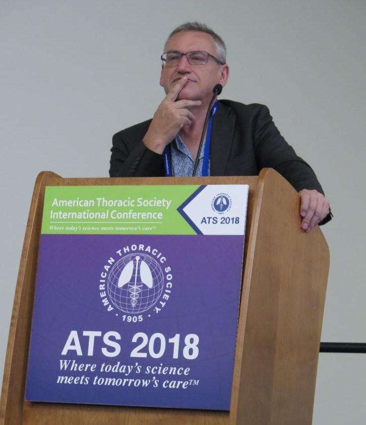
“Globally, theophylline was used for decades as a bronchodilator,” one of the study authors, David B. Price, MB BChir, said at an international conference of the American Thoracic Society. “The problem is theophylline has a narrow therapeutic index, it requires some blood monitoring, and it has been replaced by more effective inhaled bronchodilators. However, there has been a lot of discussion about whether low-dose theophylline has anti-inflammatory effects on its own and whether it increases sensitivity to inhaled steroids in COPD.”
According to the 2018 Global Initiative for Chronic Obstructive Lung Disease (GOLD) guidelines, there is “limited and contradictory evidence regarding the effect of low-dose theophylline on exacerbation rates,” and its clinical relevance has “not yet been fully established.” Dr. Price, a professor of primary care respiratory medicine at the University of Aberdeen, United Kingdom, and his associates hypothesized that the addition of low-dose theophylline to inhaled steroid therapy in COPD would reduce the risk of moderate to severe COPD exacerbations after one year of treatment. “If it worked, it would be wonderful; it would save the National Health Service a fortune,” he said.
In a government-funded trial known as Theophylline With Inhaled Corticosteroids (TWICS), people aged 40 years and older with COPD on a drug regimen including inhaled corticosteroids with a history of at least two exacerbations treated with antibiotics and/or oral corticosteroids in the previous year were recruited in 121 U.K. primary and secondary care sites from January 2014 through August 2016. They were randomized to receive low-dose theophylline or placebo for one year. Theophylline dose (200 mg once/twice a day) was determined by ideal body weight and smoking status. Primary outcome was the number of participant-reported exacerbations in the one year treatment period treated with antibiotics and/or oral corticosteroids. Participants were assessed six and 12 months after randomization. The study was powered to detect a 15% reduction in exacerbations and aimed to recruit 1,424 participants.
In all, 1,578 people were randomized: 791 to theophylline and 787 to placebo. Of these, primary outcome data were available for 98% of participants: 772 in the theophylline group and 764 in the placebo group, which amounted to 1,489 person-years of follow-up data. The mean age of patients was 68 years, 54% were male, 32% currently smoked, 80% were using inhaled corticosteroids/long-acting beta 2-agonists/long-acting muscarinic agents, and their mean FEV1 was 51.7% predicted.
Slightly more than one-quarter of study participants (26%) ceased study medication. Dr. Price said that this was balanced between the theophylline and placebo groups and mitigated by over-recruitment and a high rate of follow-up.
He reported that there were 3,430 moderate to severe exacerbations: 1,727 in the theophylline group and 1,703 in the placebo group. The mean number of exacerbations in participants allocated to theophylline and placebo groups were essentially the same: 2.24 vs. 2.23. However, there were a fewer number of exacerbations that required hospitalization in the theophylline group, compared with the placebo groups (0.17 vs. 0.24, for an adjusted rate ratio of 0.72). Dr. Price was quick to point out that this finding applied to a relatively small number of study participants, about 3% overall.
“How you interpret this, I don’t know,” he said. “Our conclusion is that in the broad population there is no benefit [of low-dose theophylline], but maybe someone might want to study its use in frequent exacerbation patients who are getting hospitalized.”
The study was funded by the National Institute for Health Research (NIHR), United Kingdom. Dr. Price reported having no financial disclosures.
SOURCE: Price, D., et al, Abstract 7709, ATS 2018.
, results from a large trial funded by the United Kingdom found.
“Globally, theophylline was used for decades as a bronchodilator,” one of the study authors, David B. Price, MB BChir, said at an international conference of the American Thoracic Society. “The problem is theophylline has a narrow therapeutic index, it requires some blood monitoring, and it has been replaced by more effective inhaled bronchodilators. However, there has been a lot of discussion about whether low-dose theophylline has anti-inflammatory effects on its own and whether it increases sensitivity to inhaled steroids in COPD.”
According to the 2018 Global Initiative for Chronic Obstructive Lung Disease (GOLD) guidelines, there is “limited and contradictory evidence regarding the effect of low-dose theophylline on exacerbation rates,” and its clinical relevance has “not yet been fully established.” Dr. Price, a professor of primary care respiratory medicine at the University of Aberdeen, United Kingdom, and his associates hypothesized that the addition of low-dose theophylline to inhaled steroid therapy in COPD would reduce the risk of moderate to severe COPD exacerbations after one year of treatment. “If it worked, it would be wonderful; it would save the National Health Service a fortune,” he said.
In a government-funded trial known as Theophylline With Inhaled Corticosteroids (TWICS), people aged 40 years and older with COPD on a drug regimen including inhaled corticosteroids with a history of at least two exacerbations treated with antibiotics and/or oral corticosteroids in the previous year were recruited in 121 U.K. primary and secondary care sites from January 2014 through August 2016. They were randomized to receive low-dose theophylline or placebo for one year. Theophylline dose (200 mg once/twice a day) was determined by ideal body weight and smoking status. Primary outcome was the number of participant-reported exacerbations in the one year treatment period treated with antibiotics and/or oral corticosteroids. Participants were assessed six and 12 months after randomization. The study was powered to detect a 15% reduction in exacerbations and aimed to recruit 1,424 participants.
In all, 1,578 people were randomized: 791 to theophylline and 787 to placebo. Of these, primary outcome data were available for 98% of participants: 772 in the theophylline group and 764 in the placebo group, which amounted to 1,489 person-years of follow-up data. The mean age of patients was 68 years, 54% were male, 32% currently smoked, 80% were using inhaled corticosteroids/long-acting beta 2-agonists/long-acting muscarinic agents, and their mean FEV1 was 51.7% predicted.
Slightly more than one-quarter of study participants (26%) ceased study medication. Dr. Price said that this was balanced between the theophylline and placebo groups and mitigated by over-recruitment and a high rate of follow-up.
He reported that there were 3,430 moderate to severe exacerbations: 1,727 in the theophylline group and 1,703 in the placebo group. The mean number of exacerbations in participants allocated to theophylline and placebo groups were essentially the same: 2.24 vs. 2.23. However, there were a fewer number of exacerbations that required hospitalization in the theophylline group, compared with the placebo groups (0.17 vs. 0.24, for an adjusted rate ratio of 0.72). Dr. Price was quick to point out that this finding applied to a relatively small number of study participants, about 3% overall.
“How you interpret this, I don’t know,” he said. “Our conclusion is that in the broad population there is no benefit [of low-dose theophylline], but maybe someone might want to study its use in frequent exacerbation patients who are getting hospitalized.”
The study was funded by the National Institute for Health Research (NIHR), United Kingdom. Dr. Price reported having no financial disclosures.
SOURCE: Price, D., et al, Abstract 7709, ATS 2018.
, results from a large trial funded by the United Kingdom found.
“Globally, theophylline was used for decades as a bronchodilator,” one of the study authors, David B. Price, MB BChir, said at an international conference of the American Thoracic Society. “The problem is theophylline has a narrow therapeutic index, it requires some blood monitoring, and it has been replaced by more effective inhaled bronchodilators. However, there has been a lot of discussion about whether low-dose theophylline has anti-inflammatory effects on its own and whether it increases sensitivity to inhaled steroids in COPD.”
According to the 2018 Global Initiative for Chronic Obstructive Lung Disease (GOLD) guidelines, there is “limited and contradictory evidence regarding the effect of low-dose theophylline on exacerbation rates,” and its clinical relevance has “not yet been fully established.” Dr. Price, a professor of primary care respiratory medicine at the University of Aberdeen, United Kingdom, and his associates hypothesized that the addition of low-dose theophylline to inhaled steroid therapy in COPD would reduce the risk of moderate to severe COPD exacerbations after one year of treatment. “If it worked, it would be wonderful; it would save the National Health Service a fortune,” he said.
In a government-funded trial known as Theophylline With Inhaled Corticosteroids (TWICS), people aged 40 years and older with COPD on a drug regimen including inhaled corticosteroids with a history of at least two exacerbations treated with antibiotics and/or oral corticosteroids in the previous year were recruited in 121 U.K. primary and secondary care sites from January 2014 through August 2016. They were randomized to receive low-dose theophylline or placebo for one year. Theophylline dose (200 mg once/twice a day) was determined by ideal body weight and smoking status. Primary outcome was the number of participant-reported exacerbations in the one year treatment period treated with antibiotics and/or oral corticosteroids. Participants were assessed six and 12 months after randomization. The study was powered to detect a 15% reduction in exacerbations and aimed to recruit 1,424 participants.
In all, 1,578 people were randomized: 791 to theophylline and 787 to placebo. Of these, primary outcome data were available for 98% of participants: 772 in the theophylline group and 764 in the placebo group, which amounted to 1,489 person-years of follow-up data. The mean age of patients was 68 years, 54% were male, 32% currently smoked, 80% were using inhaled corticosteroids/long-acting beta 2-agonists/long-acting muscarinic agents, and their mean FEV1 was 51.7% predicted.
Slightly more than one-quarter of study participants (26%) ceased study medication. Dr. Price said that this was balanced between the theophylline and placebo groups and mitigated by over-recruitment and a high rate of follow-up.
He reported that there were 3,430 moderate to severe exacerbations: 1,727 in the theophylline group and 1,703 in the placebo group. The mean number of exacerbations in participants allocated to theophylline and placebo groups were essentially the same: 2.24 vs. 2.23. However, there were a fewer number of exacerbations that required hospitalization in the theophylline group, compared with the placebo groups (0.17 vs. 0.24, for an adjusted rate ratio of 0.72). Dr. Price was quick to point out that this finding applied to a relatively small number of study participants, about 3% overall.
“How you interpret this, I don’t know,” he said. “Our conclusion is that in the broad population there is no benefit [of low-dose theophylline], but maybe someone might want to study its use in frequent exacerbation patients who are getting hospitalized.”
The study was funded by the National Institute for Health Research (NIHR), United Kingdom. Dr. Price reported having no financial disclosures.
SOURCE: Price, D., et al, Abstract 7709, ATS 2018.
AT ATS 2018
Key clinical point: Among COPD patients at high risk of exacerbation, adding low-dose oral theophylline to a drug regimen that includes an inhaled corticosteroid provides no overall clinical benefit.
Major finding: The number of exacerbations was 2.24 in participants allocated to theophylline and 2.23 for participants allocated to placebo.
Study details: A trial of 1,578 people with COPD and a history of at least two exacerbations in the previous year who were randomized to receive low-dose theophylline or placebo for one year.
Disclosures: The study was funded by National Institute for Health Research (NIHR), United Kingdom. Dr. Price reported having no financial disclosures.
Source: Price, D., et al. Abstract 7709, ATS 2018.
A blood test to detect lung cancer inches toward the clinic
The video associated with this article is no longer available on this site. Please view all of our videos on the MDedge YouTube channel
CHICAGO - Uptake of recommended low-dose CT for lung cancer screening has been dismal. Blood-based assays are an attractive alternative being explored by the Circulating Cell–Free Genome Atlas (CCGA) project. Interim results of a CCGA study of 561 individuals without cancer and 118 patients with lung cancers of all stages have found that a trio of assays searching for molecular signatures in plasma cell-free DNA achieved roughly 50% sensitivity for detection of early-stage (stage I-IIIA) lung cancers and 91% sensitivity for detection of late-stage (stage IIIB-IV) lung cancers.
In this video interview from the annual meeting of the American Society of Clinical Oncology, lead study author Geoffrey R. Oxnard, MD, of the Dana-Farber Cancer Institute, Boston, discusses the science behind these assays, how they may fill an unmet medical need, and ongoing work to bring them into the clinic.
The video associated with this article is no longer available on this site. Please view all of our videos on the MDedge YouTube channel
CHICAGO - Uptake of recommended low-dose CT for lung cancer screening has been dismal. Blood-based assays are an attractive alternative being explored by the Circulating Cell–Free Genome Atlas (CCGA) project. Interim results of a CCGA study of 561 individuals without cancer and 118 patients with lung cancers of all stages have found that a trio of assays searching for molecular signatures in plasma cell-free DNA achieved roughly 50% sensitivity for detection of early-stage (stage I-IIIA) lung cancers and 91% sensitivity for detection of late-stage (stage IIIB-IV) lung cancers.
In this video interview from the annual meeting of the American Society of Clinical Oncology, lead study author Geoffrey R. Oxnard, MD, of the Dana-Farber Cancer Institute, Boston, discusses the science behind these assays, how they may fill an unmet medical need, and ongoing work to bring them into the clinic.
The video associated with this article is no longer available on this site. Please view all of our videos on the MDedge YouTube channel
CHICAGO - Uptake of recommended low-dose CT for lung cancer screening has been dismal. Blood-based assays are an attractive alternative being explored by the Circulating Cell–Free Genome Atlas (CCGA) project. Interim results of a CCGA study of 561 individuals without cancer and 118 patients with lung cancers of all stages have found that a trio of assays searching for molecular signatures in plasma cell-free DNA achieved roughly 50% sensitivity for detection of early-stage (stage I-IIIA) lung cancers and 91% sensitivity for detection of late-stage (stage IIIB-IV) lung cancers.
In this video interview from the annual meeting of the American Society of Clinical Oncology, lead study author Geoffrey R. Oxnard, MD, of the Dana-Farber Cancer Institute, Boston, discusses the science behind these assays, how they may fill an unmet medical need, and ongoing work to bring them into the clinic.
REPORTING FROM ASCO 2018

A Path to Safer Opioids?
NIH researchers tested the common wisdom that opioids act only on the same surface receptors as endogenous opioids. They discovered that opioids, such as morphine and oxycodone, bind to receptors inside neurons—which are not the targets of naturally occurring opioids. The difference in the actions could help guide the design of pain relievers that do not produce addiction or other adverse opioid-related effects.
The researchers used a new type of antibody biosensor—a nanobody—that generates a fluorescent signal when certain proteins are activated. The signal allowed researchers to track chemicals as they move through cells and responded to stimuli.
The first discovery was that the opioid receptors were activated not only on the surface, but also in the endosome, where the mu-receptor remained activated over several minutes. The researchers also found that there are large differences across a range of opioid drugs in how strongly they induce receptor activation in the endosome. Yet another discovery was that the opioid drugs uniquely induce rapid nanobody signaling, within tens of seconds, in the Golgi apparatus. Therapeutic opioids also uniquely activated mu-opioid receptors in Golgi outposts, in the long, branched structures of neurons.
Based on those findings, the researchers hypothesize that current medically used opioids distort the normal time and spatial sequence of mu-opioid receptor activating and signaling. That distortion may provide the mechanistic link, they say, that explains the undesired adverse effects of opioid medicines.
NIH researchers tested the common wisdom that opioids act only on the same surface receptors as endogenous opioids. They discovered that opioids, such as morphine and oxycodone, bind to receptors inside neurons—which are not the targets of naturally occurring opioids. The difference in the actions could help guide the design of pain relievers that do not produce addiction or other adverse opioid-related effects.
The researchers used a new type of antibody biosensor—a nanobody—that generates a fluorescent signal when certain proteins are activated. The signal allowed researchers to track chemicals as they move through cells and responded to stimuli.
The first discovery was that the opioid receptors were activated not only on the surface, but also in the endosome, where the mu-receptor remained activated over several minutes. The researchers also found that there are large differences across a range of opioid drugs in how strongly they induce receptor activation in the endosome. Yet another discovery was that the opioid drugs uniquely induce rapid nanobody signaling, within tens of seconds, in the Golgi apparatus. Therapeutic opioids also uniquely activated mu-opioid receptors in Golgi outposts, in the long, branched structures of neurons.
Based on those findings, the researchers hypothesize that current medically used opioids distort the normal time and spatial sequence of mu-opioid receptor activating and signaling. That distortion may provide the mechanistic link, they say, that explains the undesired adverse effects of opioid medicines.
NIH researchers tested the common wisdom that opioids act only on the same surface receptors as endogenous opioids. They discovered that opioids, such as morphine and oxycodone, bind to receptors inside neurons—which are not the targets of naturally occurring opioids. The difference in the actions could help guide the design of pain relievers that do not produce addiction or other adverse opioid-related effects.
The researchers used a new type of antibody biosensor—a nanobody—that generates a fluorescent signal when certain proteins are activated. The signal allowed researchers to track chemicals as they move through cells and responded to stimuli.
The first discovery was that the opioid receptors were activated not only on the surface, but also in the endosome, where the mu-receptor remained activated over several minutes. The researchers also found that there are large differences across a range of opioid drugs in how strongly they induce receptor activation in the endosome. Yet another discovery was that the opioid drugs uniquely induce rapid nanobody signaling, within tens of seconds, in the Golgi apparatus. Therapeutic opioids also uniquely activated mu-opioid receptors in Golgi outposts, in the long, branched structures of neurons.
Based on those findings, the researchers hypothesize that current medically used opioids distort the normal time and spatial sequence of mu-opioid receptor activating and signaling. That distortion may provide the mechanistic link, they say, that explains the undesired adverse effects of opioid medicines.
Overcoming TP53 mutation proves difficult in MCL
, new findings suggest.
“TP53 mutated MCL remains a major challenge, and our results underline the importance of molecular profiling, including TP53 status, in future trials exploring novel agents,” wrote Christian Winther Eskelund, MD, of Rigshospitalet in Copenhagen, and his colleagues. The findings were published in Haematologica.
The researchers noted that the results will need validation in a larger cohort of patients.
They performed an analysis of 50 MCL patients who enrolled in the Nordic MCL4 trial between 2009 and 2013. Patients were either over age 65 years or were younger but unfit for autologous stem cell transplantation. Despite the addition of lenalidomide to the chemoimmunotherapy regimen, patients with TP53 mutations had worse overall and progression-free survival and were significantly quicker to experience relapse.
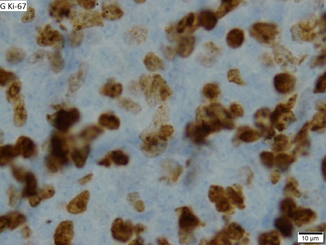
After a median follow up of 47 months, median overall survival was 25 months for patients with TP53 mutations, compared with 69 months for those without (P less than .0001). Similarly, median progression-free survival was 10 months in patients with the mutation, compared with 42 months in patients without it (P = .001). Time to relapse was a median of 10 months in these mutated patients, compared with 58 months for unmutated MCL patients (P less than .0001).
TP53 mutations were identified in six patients (14%), one of whom withdrew consent at day 28 of the study. Of the remaining patients with mutations, all of them either progressed or relapsed during the study and none were alive at the most recent follow-up. During the study, patients received an induction phase of six cycles of lenalidomide plus bendamustine-rituximab (weeks 1-24), followed by a maintenance phase of lenalidomide (weeks 25-56).
SOURCE: Eskelund CW et al. Haematologica. 2018 May 24. doi: 10.3324/haematol.2018.194399.
, new findings suggest.
“TP53 mutated MCL remains a major challenge, and our results underline the importance of molecular profiling, including TP53 status, in future trials exploring novel agents,” wrote Christian Winther Eskelund, MD, of Rigshospitalet in Copenhagen, and his colleagues. The findings were published in Haematologica.
The researchers noted that the results will need validation in a larger cohort of patients.
They performed an analysis of 50 MCL patients who enrolled in the Nordic MCL4 trial between 2009 and 2013. Patients were either over age 65 years or were younger but unfit for autologous stem cell transplantation. Despite the addition of lenalidomide to the chemoimmunotherapy regimen, patients with TP53 mutations had worse overall and progression-free survival and were significantly quicker to experience relapse.
After a median follow up of 47 months, median overall survival was 25 months for patients with TP53 mutations, compared with 69 months for those without (P less than .0001). Similarly, median progression-free survival was 10 months in patients with the mutation, compared with 42 months in patients without it (P = .001). Time to relapse was a median of 10 months in these mutated patients, compared with 58 months for unmutated MCL patients (P less than .0001).
TP53 mutations were identified in six patients (14%), one of whom withdrew consent at day 28 of the study. Of the remaining patients with mutations, all of them either progressed or relapsed during the study and none were alive at the most recent follow-up. During the study, patients received an induction phase of six cycles of lenalidomide plus bendamustine-rituximab (weeks 1-24), followed by a maintenance phase of lenalidomide (weeks 25-56).
SOURCE: Eskelund CW et al. Haematologica. 2018 May 24. doi: 10.3324/haematol.2018.194399.
, new findings suggest.
“TP53 mutated MCL remains a major challenge, and our results underline the importance of molecular profiling, including TP53 status, in future trials exploring novel agents,” wrote Christian Winther Eskelund, MD, of Rigshospitalet in Copenhagen, and his colleagues. The findings were published in Haematologica.
The researchers noted that the results will need validation in a larger cohort of patients.
They performed an analysis of 50 MCL patients who enrolled in the Nordic MCL4 trial between 2009 and 2013. Patients were either over age 65 years or were younger but unfit for autologous stem cell transplantation. Despite the addition of lenalidomide to the chemoimmunotherapy regimen, patients with TP53 mutations had worse overall and progression-free survival and were significantly quicker to experience relapse.
After a median follow up of 47 months, median overall survival was 25 months for patients with TP53 mutations, compared with 69 months for those without (P less than .0001). Similarly, median progression-free survival was 10 months in patients with the mutation, compared with 42 months in patients without it (P = .001). Time to relapse was a median of 10 months in these mutated patients, compared with 58 months for unmutated MCL patients (P less than .0001).
TP53 mutations were identified in six patients (14%), one of whom withdrew consent at day 28 of the study. Of the remaining patients with mutations, all of them either progressed or relapsed during the study and none were alive at the most recent follow-up. During the study, patients received an induction phase of six cycles of lenalidomide plus bendamustine-rituximab (weeks 1-24), followed by a maintenance phase of lenalidomide (weeks 25-56).
SOURCE: Eskelund CW et al. Haematologica. 2018 May 24. doi: 10.3324/haematol.2018.194399.
FROM HAEMATOLOGICA
Many hospitals had no mandatory flu vaccine requirements in 2017
Many U.S. hospitals still did not have influenza vaccination requirements for health care personnel as of summer 2017, suggested the results of a national survey.
Nearly two-thirds of hospitals had mandatory influenza vaccination in place in 2017, up from just one-third in 2013, according to survey responses submitted by infection preventionists working at Veterans Affairs (VA) and non-VA hospitals.
Despite recommendations to vaccinate health care personnel against influenza, there are several challenges and barriers to implementing the practice, the authors wrote in JAMA Network Open.
“Mandating influenza vaccination remains a controversial topic, with uncertainty of the effectiveness of health care personnel influenza vaccination in reducing patient morbidity and mortality, different conclusions regarding the grading of the evidence, and numerous legal and ethical precedents to be carefully considered,” they wrote.
Their study was based on 1,062 responses to a panel survey of infection preventionists conducted every 4 years. The survey asked providers about practices used in their hospitals to prevent health care–associated infections.
Compared with 2013, when only 37.1% of non-VA hospitals had mandatory influenza vaccination requirements, the 2017 survey showed a significant increase to 61.4% (P less than .001), Dr. Greene and his colleagues wrote in their report.
By contrast, the proportion of VA hospitals with such requirements increased only slightly, from 1.3% in 2013 to just 4.1% in 2017 (P = .29), the report showed.
Penalties for not complying with the policy were not universal in hospitals with mandates, they added. Only 74% said they had such penalties, and 13% allowed health care personnel to decline influenza vaccination without a specified reason.
After the survey responses were received, the VA issued a directive stating that all health care personnel should receive annual influenza vaccination and should wear masks during influenza season, Dr. Greene noted.
That directive is in line with recommendations from the Centers for Disease Control and Prevention Advisory Committee on Immunization Practices, which have stated that all health care personnel should receive influenza vaccination each year.
In addition, the U.S. Department of Health & Human Services has set a goal of 90% of health care personnel to be vaccinated by 2020, Dr. Green and his coauthors noted.
Mandating influenza vaccination is just one proven successful strategy for increasing coverage at hospitals, according to the study authors. Other approaches include influenza education, incentives, free and easy access to vaccination, and annual campaigns directed at health care personnel, as well as written policies describing the vaccination goal.
“Regardless of whether an organization has an official mandate for vaccinations, establishing a written policy that states the organizational commitment to increasing vaccination rates is among the recommended strategies for improving vaccination coverage among health care personnel,” they wrote.
Dr. Greene and his coauthors reported receiving grants from the Blue Cross Blue Shield of Michigan Foundation and the U.S. Department of Veterans Affairs Patient Safety Center of Inquiry during the conduct of the study. One study coauthor reported personal fees from Jvion and from Doximity outside the submitted work.
SOURCE: Greene MT et al. JAMA Network Open. 2018;1(2):e180143.
This study suggests a significant increase in use of mandatory influenza vaccination policies during 2013-2017, driven mainly by increases at non–Veterans Affairs (VA) hospitals and little change at VA facilities. However, there are some caveats to the findings that should be considered, Hilary M. Babcock, MD, MPH, wrote in an editorial referencing the study.
The sample for the 2013 and 2017 surveys included different facilities and different size facilities, so direct comparisons cannot be made, according to Dr. Babcock.
Moreover, the survey questions were worded somewhat differently in the two surveys, and it does not appear that “mandate” was defined by the study authors, she said in her editorial.
The VA recently issued a directive that all health care personnel should receive influenza vaccination and wear masks during influenza season. This new directive provides an “excellent opportunity” to address knowledge gaps regarding the effects of influenza vaccination of health care personnel on patient outcomes, according to Dr. Babcock.
“While the assumption that decreasing the risk of influenza in health care personnel will result in decreased risk of influenza in patients cared for by those health care personnel is common sense, for acute care settings, it is still largely an assumption,” Dr. Babcock wrote. “Hopefully, the Veterans Health Administration will combine this initiative with thoughtful, planned, patient outcome assessments to help define the anticipated benefit of these efforts.”
Dr. Babcock is with Washington University and the BJC HealthCare Infection Prevention & Epidemiology Consortium, both in St. Louis. These comments are derived from her editorial in JAMA Network Open (2018;1[2]:e180144). Dr. Babcock reported no conflict of interest disclosures related to her editorial.
This study suggests a significant increase in use of mandatory influenza vaccination policies during 2013-2017, driven mainly by increases at non–Veterans Affairs (VA) hospitals and little change at VA facilities. However, there are some caveats to the findings that should be considered, Hilary M. Babcock, MD, MPH, wrote in an editorial referencing the study.
The sample for the 2013 and 2017 surveys included different facilities and different size facilities, so direct comparisons cannot be made, according to Dr. Babcock.
Moreover, the survey questions were worded somewhat differently in the two surveys, and it does not appear that “mandate” was defined by the study authors, she said in her editorial.
The VA recently issued a directive that all health care personnel should receive influenza vaccination and wear masks during influenza season. This new directive provides an “excellent opportunity” to address knowledge gaps regarding the effects of influenza vaccination of health care personnel on patient outcomes, according to Dr. Babcock.
“While the assumption that decreasing the risk of influenza in health care personnel will result in decreased risk of influenza in patients cared for by those health care personnel is common sense, for acute care settings, it is still largely an assumption,” Dr. Babcock wrote. “Hopefully, the Veterans Health Administration will combine this initiative with thoughtful, planned, patient outcome assessments to help define the anticipated benefit of these efforts.”
Dr. Babcock is with Washington University and the BJC HealthCare Infection Prevention & Epidemiology Consortium, both in St. Louis. These comments are derived from her editorial in JAMA Network Open (2018;1[2]:e180144). Dr. Babcock reported no conflict of interest disclosures related to her editorial.
This study suggests a significant increase in use of mandatory influenza vaccination policies during 2013-2017, driven mainly by increases at non–Veterans Affairs (VA) hospitals and little change at VA facilities. However, there are some caveats to the findings that should be considered, Hilary M. Babcock, MD, MPH, wrote in an editorial referencing the study.
The sample for the 2013 and 2017 surveys included different facilities and different size facilities, so direct comparisons cannot be made, according to Dr. Babcock.
Moreover, the survey questions were worded somewhat differently in the two surveys, and it does not appear that “mandate” was defined by the study authors, she said in her editorial.
The VA recently issued a directive that all health care personnel should receive influenza vaccination and wear masks during influenza season. This new directive provides an “excellent opportunity” to address knowledge gaps regarding the effects of influenza vaccination of health care personnel on patient outcomes, according to Dr. Babcock.
“While the assumption that decreasing the risk of influenza in health care personnel will result in decreased risk of influenza in patients cared for by those health care personnel is common sense, for acute care settings, it is still largely an assumption,” Dr. Babcock wrote. “Hopefully, the Veterans Health Administration will combine this initiative with thoughtful, planned, patient outcome assessments to help define the anticipated benefit of these efforts.”
Dr. Babcock is with Washington University and the BJC HealthCare Infection Prevention & Epidemiology Consortium, both in St. Louis. These comments are derived from her editorial in JAMA Network Open (2018;1[2]:e180144). Dr. Babcock reported no conflict of interest disclosures related to her editorial.
Many U.S. hospitals still did not have influenza vaccination requirements for health care personnel as of summer 2017, suggested the results of a national survey.
Nearly two-thirds of hospitals had mandatory influenza vaccination in place in 2017, up from just one-third in 2013, according to survey responses submitted by infection preventionists working at Veterans Affairs (VA) and non-VA hospitals.
Despite recommendations to vaccinate health care personnel against influenza, there are several challenges and barriers to implementing the practice, the authors wrote in JAMA Network Open.
“Mandating influenza vaccination remains a controversial topic, with uncertainty of the effectiveness of health care personnel influenza vaccination in reducing patient morbidity and mortality, different conclusions regarding the grading of the evidence, and numerous legal and ethical precedents to be carefully considered,” they wrote.
Their study was based on 1,062 responses to a panel survey of infection preventionists conducted every 4 years. The survey asked providers about practices used in their hospitals to prevent health care–associated infections.
Compared with 2013, when only 37.1% of non-VA hospitals had mandatory influenza vaccination requirements, the 2017 survey showed a significant increase to 61.4% (P less than .001), Dr. Greene and his colleagues wrote in their report.
By contrast, the proportion of VA hospitals with such requirements increased only slightly, from 1.3% in 2013 to just 4.1% in 2017 (P = .29), the report showed.
Penalties for not complying with the policy were not universal in hospitals with mandates, they added. Only 74% said they had such penalties, and 13% allowed health care personnel to decline influenza vaccination without a specified reason.
After the survey responses were received, the VA issued a directive stating that all health care personnel should receive annual influenza vaccination and should wear masks during influenza season, Dr. Greene noted.
That directive is in line with recommendations from the Centers for Disease Control and Prevention Advisory Committee on Immunization Practices, which have stated that all health care personnel should receive influenza vaccination each year.
In addition, the U.S. Department of Health & Human Services has set a goal of 90% of health care personnel to be vaccinated by 2020, Dr. Green and his coauthors noted.
Mandating influenza vaccination is just one proven successful strategy for increasing coverage at hospitals, according to the study authors. Other approaches include influenza education, incentives, free and easy access to vaccination, and annual campaigns directed at health care personnel, as well as written policies describing the vaccination goal.
“Regardless of whether an organization has an official mandate for vaccinations, establishing a written policy that states the organizational commitment to increasing vaccination rates is among the recommended strategies for improving vaccination coverage among health care personnel,” they wrote.
Dr. Greene and his coauthors reported receiving grants from the Blue Cross Blue Shield of Michigan Foundation and the U.S. Department of Veterans Affairs Patient Safety Center of Inquiry during the conduct of the study. One study coauthor reported personal fees from Jvion and from Doximity outside the submitted work.
SOURCE: Greene MT et al. JAMA Network Open. 2018;1(2):e180143.
Many U.S. hospitals still did not have influenza vaccination requirements for health care personnel as of summer 2017, suggested the results of a national survey.
Nearly two-thirds of hospitals had mandatory influenza vaccination in place in 2017, up from just one-third in 2013, according to survey responses submitted by infection preventionists working at Veterans Affairs (VA) and non-VA hospitals.
Despite recommendations to vaccinate health care personnel against influenza, there are several challenges and barriers to implementing the practice, the authors wrote in JAMA Network Open.
“Mandating influenza vaccination remains a controversial topic, with uncertainty of the effectiveness of health care personnel influenza vaccination in reducing patient morbidity and mortality, different conclusions regarding the grading of the evidence, and numerous legal and ethical precedents to be carefully considered,” they wrote.
Their study was based on 1,062 responses to a panel survey of infection preventionists conducted every 4 years. The survey asked providers about practices used in their hospitals to prevent health care–associated infections.
Compared with 2013, when only 37.1% of non-VA hospitals had mandatory influenza vaccination requirements, the 2017 survey showed a significant increase to 61.4% (P less than .001), Dr. Greene and his colleagues wrote in their report.
By contrast, the proportion of VA hospitals with such requirements increased only slightly, from 1.3% in 2013 to just 4.1% in 2017 (P = .29), the report showed.
Penalties for not complying with the policy were not universal in hospitals with mandates, they added. Only 74% said they had such penalties, and 13% allowed health care personnel to decline influenza vaccination without a specified reason.
After the survey responses were received, the VA issued a directive stating that all health care personnel should receive annual influenza vaccination and should wear masks during influenza season, Dr. Greene noted.
That directive is in line with recommendations from the Centers for Disease Control and Prevention Advisory Committee on Immunization Practices, which have stated that all health care personnel should receive influenza vaccination each year.
In addition, the U.S. Department of Health & Human Services has set a goal of 90% of health care personnel to be vaccinated by 2020, Dr. Green and his coauthors noted.
Mandating influenza vaccination is just one proven successful strategy for increasing coverage at hospitals, according to the study authors. Other approaches include influenza education, incentives, free and easy access to vaccination, and annual campaigns directed at health care personnel, as well as written policies describing the vaccination goal.
“Regardless of whether an organization has an official mandate for vaccinations, establishing a written policy that states the organizational commitment to increasing vaccination rates is among the recommended strategies for improving vaccination coverage among health care personnel,” they wrote.
Dr. Greene and his coauthors reported receiving grants from the Blue Cross Blue Shield of Michigan Foundation and the U.S. Department of Veterans Affairs Patient Safety Center of Inquiry during the conduct of the study. One study coauthor reported personal fees from Jvion and from Doximity outside the submitted work.
SOURCE: Greene MT et al. JAMA Network Open. 2018;1(2):e180143.
FROM JAMA Network Open
Key clinical point: Despite a significant increase in influenza vaccination at non-VA hospitals, many VA and non-VA hospitals still do not have mandatory influenza vaccination requirements for health care personnel.
Major finding: During 2013-2017, the proportion of non-VA hospitals with requirements increased from 37.1% to 61.4% (P less than .001), contrasting with a rise from 1.3% to just 4.1% at VA hospitals (P = .29).
Study details: A study of survey responses from 1,062 infection preventionists at VA and non-VA hospitals in the United States submitted between 2013 and 2017.
Disclosures: Authors reported receiving grants from the Blue Cross Blue Shield of Michigan Foundation and the U.S. Department of Veterans Affairs Patient Safety Center of Inquiry during the conduct of the study. One study coauthor reported personal fees from Jvion and from Doximity outside the submitted work.
Source: Greene MT et al. JAMA Network Open. 2018;1(2):e180143.
FDA Commissioner Comments on Signing of the Right to Try Act
SILVER SPRING, MD—On May 30, 2018, President Donald Trump signed into law the Trickett Wendler, Frank Mongiello, Jordan McLinn, and Matthew Bellina Right to Try Act of 2017 (Right to Try Act). This new law amends the Federal Food, Drug, and Cosmetic Act to establish a new pathway aimed at increasing access to unapproved, investigational treatments for patients diagnosed with life-threatening diseases or conditions who have exhausted

“For patients with serious or immediately life-threatening diseases, the FDA remains committed to enhancing access to promising investigational medicines for those unable to access products through clinical trials,” said Dr. Gottlieb. “This is the mission of our expanded access program. The agency is dedicated to these purposes, and it has been for more than three decades.”
“We’ve taken many steps to improve our process through which patients can access promising investigational drugs,” said Dr. Gottlieb. “We understand that treatment decisions for those facing terminal illnesses are best made by patients and families, with the support of their treating physicians. When appropriate, those suffering from a terminal illness who’ve exhausted available options should be able to access promising treatments being studied in clinical trials, or products under active review by the FDA. The agency is faithfully committed to these goals, to protecting patients, and to making sure they’re able to make informed decisions.”
“We stand ready to implement this legislation in a way that achieves Congress’ intent to promote access and protect patients. The FDA is dedicated to achieving the goals that Congress set forth in this legislation, so that patients facing terminal conditions have an additional avenue to access promising investigational medicines.”
“The decisions we reach related to products that can serve as an effective treatment for a terminal illness, or that can arrest a devastating and debilitating condition, are among the most important and carefully considered judgments that we make,” said Dr. Gottlieb. “We recognize the important balance between making sure patients have the assurances Congress intends, while enabling timely access to promising treatments in these devastating circumstances. And we’ll implement this new law consistent with these longstanding values.”
SILVER SPRING, MD—On May 30, 2018, President Donald Trump signed into law the Trickett Wendler, Frank Mongiello, Jordan McLinn, and Matthew Bellina Right to Try Act of 2017 (Right to Try Act). This new law amends the Federal Food, Drug, and Cosmetic Act to establish a new pathway aimed at increasing access to unapproved, investigational treatments for patients diagnosed with life-threatening diseases or conditions who have exhausted
“For patients with serious or immediately life-threatening diseases, the FDA remains committed to enhancing access to promising investigational medicines for those unable to access products through clinical trials,” said Dr. Gottlieb. “This is the mission of our expanded access program. The agency is dedicated to these purposes, and it has been for more than three decades.”
“We’ve taken many steps to improve our process through which patients can access promising investigational drugs,” said Dr. Gottlieb. “We understand that treatment decisions for those facing terminal illnesses are best made by patients and families, with the support of their treating physicians. When appropriate, those suffering from a terminal illness who’ve exhausted available options should be able to access promising treatments being studied in clinical trials, or products under active review by the FDA. The agency is faithfully committed to these goals, to protecting patients, and to making sure they’re able to make informed decisions.”
“We stand ready to implement this legislation in a way that achieves Congress’ intent to promote access and protect patients. The FDA is dedicated to achieving the goals that Congress set forth in this legislation, so that patients facing terminal conditions have an additional avenue to access promising investigational medicines.”
“The decisions we reach related to products that can serve as an effective treatment for a terminal illness, or that can arrest a devastating and debilitating condition, are among the most important and carefully considered judgments that we make,” said Dr. Gottlieb. “We recognize the important balance between making sure patients have the assurances Congress intends, while enabling timely access to promising treatments in these devastating circumstances. And we’ll implement this new law consistent with these longstanding values.”
SILVER SPRING, MD—On May 30, 2018, President Donald Trump signed into law the Trickett Wendler, Frank Mongiello, Jordan McLinn, and Matthew Bellina Right to Try Act of 2017 (Right to Try Act). This new law amends the Federal Food, Drug, and Cosmetic Act to establish a new pathway aimed at increasing access to unapproved, investigational treatments for patients diagnosed with life-threatening diseases or conditions who have exhausted
“For patients with serious or immediately life-threatening diseases, the FDA remains committed to enhancing access to promising investigational medicines for those unable to access products through clinical trials,” said Dr. Gottlieb. “This is the mission of our expanded access program. The agency is dedicated to these purposes, and it has been for more than three decades.”
“We’ve taken many steps to improve our process through which patients can access promising investigational drugs,” said Dr. Gottlieb. “We understand that treatment decisions for those facing terminal illnesses are best made by patients and families, with the support of their treating physicians. When appropriate, those suffering from a terminal illness who’ve exhausted available options should be able to access promising treatments being studied in clinical trials, or products under active review by the FDA. The agency is faithfully committed to these goals, to protecting patients, and to making sure they’re able to make informed decisions.”
“We stand ready to implement this legislation in a way that achieves Congress’ intent to promote access and protect patients. The FDA is dedicated to achieving the goals that Congress set forth in this legislation, so that patients facing terminal conditions have an additional avenue to access promising investigational medicines.”
“The decisions we reach related to products that can serve as an effective treatment for a terminal illness, or that can arrest a devastating and debilitating condition, are among the most important and carefully considered judgments that we make,” said Dr. Gottlieb. “We recognize the important balance between making sure patients have the assurances Congress intends, while enabling timely access to promising treatments in these devastating circumstances. And we’ll implement this new law consistent with these longstanding values.”