User login
MDedge conference coverage features onsite reporting of the latest study results and expert perspectives from leading researchers.
Breakthrough COVID-19 milder in vaccinated patients with IBD
Vaccination against SARS-CoV-2 appears to protect people with inflammatory bowel disease (IBD) from the more serious consequences of breakthrough COVID-19 infections, but results may vary by which vaccine was received, results of a small study suggest.
In a study of patients with IBD who had completed a primary vaccine series but went on to develop COVID-19, there were trends toward worse outcomes for patients who received a non-mRNA vaccine, older patients, and those who were on combination therapy rather than monotherapy, reported Emily Spiera, a medical student at the Icahn School of Medicine at Mount Sinai, New York.
“Overall, we saw that vaccinated patients who subsequently developed COVID-19 had low rates of hospitalization, severe COVID, and death,” she said in an oral abstract at the annual Crohn’s & Colitis Congress®, a partnership of the Crohn’s & Colitis Foundation and the American Gastroenterological Association.
The study was conducted before the highly infectious Omicron variant of SARS-CoV-2 became dominant, however, and the sample size of 88 patients, combined with a low number of study events, was too small for statistical significance to emerge for most measures, Ms. Spiera acknowledged.
Nonetheless, the findings support the protective benefit of vaccines in this population, said Freddy Caldera, DO, associate professor of gastroenterology at the University of Wisconsin–Madison, who was not involved in the study.
“In my mind, when we think about COVID vaccines, the whole goal is to prevent severe disease,” he said.

Dr. Caldera and colleagues conducted an earlier study of humoral immunogenicity of mRNA COVID-19 vaccines in 122 patients with IBD and 60 healthy controls, and found that all controls and 97% of patients with IBD developed antibodies, although antibody concentrations were lower in patients with IBD, compared with controls (P < .001). Those who received the mRNA-1273 (Moderna) COVID-19 had significantly higher antibody concentrations than those who received the Pfizer-BNT vaccine series (P < .001).
They also found that patients on immune-modifying therapy had lower antibody concentrations, compared with those who were not on such therapy, or those who received aminosalicylates or vedolizumab (Entyvio; P = .003).
The protective effect of vaccines in this population became even more apparent after patients received an additional vaccine dose.
“We actually have a study in preprint of what happens after a third dose, where everyone made antibodies,” he said. “What we tell patients is that vaccines work.”
SECURE-IBD data
The investigators at Mount Sinai, the University of North Carolina at Chapel Hill, and Tel Aviv University analyzed data from the Surveillance Epidemiology of Coronavirus Under Research Exclusion in Inflammatory Bowel Disease (SECURE-IBD) database, an international web-based registry that includes reports from 74 countries, with data reported by 48 U.S. states.
The study sample consisted of patients enrolled from Dec. 12, 2020, to Oct. 1, 2021, who had completed a primary vaccination series with either mRNA vaccines (Pfizer or Moderna) adenoviral vector-based vaccines (AstraZeneca, Sputnik, CanSino, or Janssen/Johnson & Johnson), or an inactivated SARS-CoV-2 vaccine (Sinovac).
Of 2,477 patients with COVID-19 infections reported to SECURE-IBD, 160 reported being vaccinated. Of this group, 53 were excluded because they were only partially vaccinated, and 19 were excluded because of missing data on either vaccine type, number of doses, or COVID-19 outcomes, leaving 88 patients with completed primary vaccination series at the time of COVID-19 infection.
The median patient age was 40.1 years. Nearly two-thirds of the patients had a diagnosis of Crohn’s disease, and slightly more than one-third has a diagnosis of ulcerative colitis. The patients came from 18 countries, with 45.3% of the sample in the United States.
A total of 58% of patients were on biologic monotherapy, with either a tumor necrosis factor antagonist, integrin antagonist, or anti–interleukin-12/13. In addition, 3.4% were on immunomodulator monotherapy, 21.6% were on combination therapy, and 5.7% were receiving corticosteroids.
Lower severity
COVID-19 severity was numerically but not significantly lower among the 88 vaccinated patients, with a rate of 5.7%, compared with 9.3% among 2,317 patients with COVID-19 infections in the database who were not vaccinated.
COVID-19 severity defined as a composite of ICU admission, need for mechanical ventilation and/or death was actually slightly higher among the vaccinated patients, with a rate of 3.4% versus 1.9% for nonvaccinated patients, but this difference was not statistically significant.
There was 1 death among vaccinated patients (1.1%) versus 29 among the unvaccinated (1.2%).
There were trends toward fewer hospitalizations and less-frequent severe COVID-19 infection among patients who received a mRNA vaccine, compared with other vaccine types, but again these differences did not reach statistical significance.
As noted before, there was a higher frequency of severe COVID-19 among patients on combination therapy than on monotherapy, but this difference too was not statistically significant.
As seen with COVID-19 in the general population older patients tended to have worse outcomes, with a mean age of 53 for patients requiring hospitalization, compared with 39 years for patients who stayed out of the hospital (P = .04), and a mean age of 59 among patients with severe COVID-19 infections, compared with 39 for patients with moderate or mild infections (P = .03).
Ms. Spiera described the case of the single vaccinated patient who died. The 63-year-old woman had moderately active Crohn’s disease treated with corticosteroids, adalimumab (Humira) and azathioprine at the time of COVID-19 infection. She had received the AstraZeneca adenoviral-based vaccine more than 30 days prior to infection. She was hospitalized and intubated, and died from gastrointestinal bleeding.
Ms. Spiera noted that, although the sample size was small, and only patients known to have COVID-19 were included, it is one of the largest cohorts to date of vaccinated patients with IBD who developed COVID-19. She said that the study supports prior studies showing that combination therapy and tumor necrosis factor antagonists may result in reduced immunity, and that mRNA vaccines may offer better protection against severe illness in this population.
The study was supported by a Digestive Disease Research Foundation Fellowship. Ms. Spiera and Dr. Caldera reported no relevant disclosures.
Vaccination against SARS-CoV-2 appears to protect people with inflammatory bowel disease (IBD) from the more serious consequences of breakthrough COVID-19 infections, but results may vary by which vaccine was received, results of a small study suggest.
In a study of patients with IBD who had completed a primary vaccine series but went on to develop COVID-19, there were trends toward worse outcomes for patients who received a non-mRNA vaccine, older patients, and those who were on combination therapy rather than monotherapy, reported Emily Spiera, a medical student at the Icahn School of Medicine at Mount Sinai, New York.
“Overall, we saw that vaccinated patients who subsequently developed COVID-19 had low rates of hospitalization, severe COVID, and death,” she said in an oral abstract at the annual Crohn’s & Colitis Congress®, a partnership of the Crohn’s & Colitis Foundation and the American Gastroenterological Association.
The study was conducted before the highly infectious Omicron variant of SARS-CoV-2 became dominant, however, and the sample size of 88 patients, combined with a low number of study events, was too small for statistical significance to emerge for most measures, Ms. Spiera acknowledged.
Nonetheless, the findings support the protective benefit of vaccines in this population, said Freddy Caldera, DO, associate professor of gastroenterology at the University of Wisconsin–Madison, who was not involved in the study.
“In my mind, when we think about COVID vaccines, the whole goal is to prevent severe disease,” he said.
Dr. Caldera and colleagues conducted an earlier study of humoral immunogenicity of mRNA COVID-19 vaccines in 122 patients with IBD and 60 healthy controls, and found that all controls and 97% of patients with IBD developed antibodies, although antibody concentrations were lower in patients with IBD, compared with controls (P < .001). Those who received the mRNA-1273 (Moderna) COVID-19 had significantly higher antibody concentrations than those who received the Pfizer-BNT vaccine series (P < .001).
They also found that patients on immune-modifying therapy had lower antibody concentrations, compared with those who were not on such therapy, or those who received aminosalicylates or vedolizumab (Entyvio; P = .003).
The protective effect of vaccines in this population became even more apparent after patients received an additional vaccine dose.
“We actually have a study in preprint of what happens after a third dose, where everyone made antibodies,” he said. “What we tell patients is that vaccines work.”
SECURE-IBD data
The investigators at Mount Sinai, the University of North Carolina at Chapel Hill, and Tel Aviv University analyzed data from the Surveillance Epidemiology of Coronavirus Under Research Exclusion in Inflammatory Bowel Disease (SECURE-IBD) database, an international web-based registry that includes reports from 74 countries, with data reported by 48 U.S. states.
The study sample consisted of patients enrolled from Dec. 12, 2020, to Oct. 1, 2021, who had completed a primary vaccination series with either mRNA vaccines (Pfizer or Moderna) adenoviral vector-based vaccines (AstraZeneca, Sputnik, CanSino, or Janssen/Johnson & Johnson), or an inactivated SARS-CoV-2 vaccine (Sinovac).
Of 2,477 patients with COVID-19 infections reported to SECURE-IBD, 160 reported being vaccinated. Of this group, 53 were excluded because they were only partially vaccinated, and 19 were excluded because of missing data on either vaccine type, number of doses, or COVID-19 outcomes, leaving 88 patients with completed primary vaccination series at the time of COVID-19 infection.
The median patient age was 40.1 years. Nearly two-thirds of the patients had a diagnosis of Crohn’s disease, and slightly more than one-third has a diagnosis of ulcerative colitis. The patients came from 18 countries, with 45.3% of the sample in the United States.
A total of 58% of patients were on biologic monotherapy, with either a tumor necrosis factor antagonist, integrin antagonist, or anti–interleukin-12/13. In addition, 3.4% were on immunomodulator monotherapy, 21.6% were on combination therapy, and 5.7% were receiving corticosteroids.
Lower severity
COVID-19 severity was numerically but not significantly lower among the 88 vaccinated patients, with a rate of 5.7%, compared with 9.3% among 2,317 patients with COVID-19 infections in the database who were not vaccinated.
COVID-19 severity defined as a composite of ICU admission, need for mechanical ventilation and/or death was actually slightly higher among the vaccinated patients, with a rate of 3.4% versus 1.9% for nonvaccinated patients, but this difference was not statistically significant.
There was 1 death among vaccinated patients (1.1%) versus 29 among the unvaccinated (1.2%).
There were trends toward fewer hospitalizations and less-frequent severe COVID-19 infection among patients who received a mRNA vaccine, compared with other vaccine types, but again these differences did not reach statistical significance.
As noted before, there was a higher frequency of severe COVID-19 among patients on combination therapy than on monotherapy, but this difference too was not statistically significant.
As seen with COVID-19 in the general population older patients tended to have worse outcomes, with a mean age of 53 for patients requiring hospitalization, compared with 39 years for patients who stayed out of the hospital (P = .04), and a mean age of 59 among patients with severe COVID-19 infections, compared with 39 for patients with moderate or mild infections (P = .03).
Ms. Spiera described the case of the single vaccinated patient who died. The 63-year-old woman had moderately active Crohn’s disease treated with corticosteroids, adalimumab (Humira) and azathioprine at the time of COVID-19 infection. She had received the AstraZeneca adenoviral-based vaccine more than 30 days prior to infection. She was hospitalized and intubated, and died from gastrointestinal bleeding.
Ms. Spiera noted that, although the sample size was small, and only patients known to have COVID-19 were included, it is one of the largest cohorts to date of vaccinated patients with IBD who developed COVID-19. She said that the study supports prior studies showing that combination therapy and tumor necrosis factor antagonists may result in reduced immunity, and that mRNA vaccines may offer better protection against severe illness in this population.
The study was supported by a Digestive Disease Research Foundation Fellowship. Ms. Spiera and Dr. Caldera reported no relevant disclosures.
FROM THE CROHN’S & COLITIS CONGRESS
Medicaid expansion benefits some colorectal patients, others not so lucky
Two new studies suggest the expansion of Medicaid under the Patient Protection and Affordable Care Act in 2010 may be leading to more frequent diagnosis of colorectal cancer (CRC) among Hispanics.
The studies, presented at the 2022 Gastrointestinal Cancers Symposium, suggest that Medicaid expansion may have a diverse impact on various ethnic groups.

“The take-home message for both physicians and policy makers is that health policy has the capacity to shift health care delivery, yet we need to consider the effects of health policy might influence subgroups of patients differently. This is useful information for providers caring for a diverse group of patients. For policy makers, this study emphasizes the importance of evaluating the impact of health policy among different racial and ethnic subgroups to fully understand the impact of [policy] change,” said study lead author James D. Murphy, MD, MS, assistant vice chair of radiation medicine at the University of California San Diego.
Dr. Murphy and associates cautioned that other factors, not just Medicaid expansion, could be responsible for the uptick in colon cancer diagnoses.
“Our observations could potentially be influenced by other risk factors. Medicaid expansion was not a ‘randomized experiment,’ and states which opted to expand Medicaid might have fundamental differences which could impact colorectal cancer incidence,” he said.
His group’s analysis of the Surveillance, Epidemiology, and End Results database included 21 states where Medicaid was expanded and 16 states where expansion did not occur. Between 2010-2013 and 2014-2018, among patients under 65, overall colorectal cancer incidence rates did not differ by Medicaid expansion status. In nonexpansion states, there was a greater increase in CRC rates among Hispanics (5.4 vs. 1.6 increase per 100,000; P = .002) and Asian/Pacific Islanders (4.3 vs. 0.4 per 100,000; P = .02), but there was no difference among Black or non-Hispanic White individuals.
Early-onset colorectal cancer diagnoses increase under Medicaid expansion
In another study presented at the meeting, researchers examined early-onset CRC data from the National Cancer Database. Among Hispanics, the rate of change of incidence of newly diagnosed cases among patients age 40-49 in Medicaid expansion states increased from 4.3% per year between 2010 and 2014 and 9.8% between 2014 and 2017. That compares with the general background increase in incidence of about 2%. In nonexpansion states, the rate of change decreased from 6.4% to 1% (P = .03). There were no statistically significant differences in the change of incidence among Blacks or Whites between expansion and nonexpansion states.
The reduced rate of change among Hispanics in nonexpansion states was a surprise, and the researchers haven’t determined the reason, according to Sanjay Goel, MD, an oncologist with Montefiore Medical Center, New York, and lead author on the National Cancer Database study. Dr. Goel speculated that some people may have migrated from nonexpansion states to states that expanded Medicaid in order to gain health care coverage.
The apparent benefit seen in Hispanics, but not Black patients, may be caused by greater susceptibility to early-onset CRC among Hispanics, leading to a stronger effect on that population when Medicaid was expanded, Dr. Goel said.
“At this point, with our available data, we do not have the ability to understand the underlying sources of these disparities, though these are questions which deserve additional research,” Dr. Murphy said.
Regardless of the reason, the message is clear, Dr. Goel said. “The bottom we want to state is that politics aside, providing health care coverage to as many people as possible, ideally to everyone, is the right way of going forward.”
The implications of the findings extend beyond policy. “The general advice I give is that, especially if you treat a Hispanic person, regardless of age, with any symptom or sign that could be suggestive of a malignancy, do not take it lightly. Follow the patient closely. I’m not advocating that you refer everybody with lower abdominal pain or bleeding for a colonoscopy, but do factor it in mind. Call them back in a week or 2, or have them make a follow-up appointment in a month so that they don’t get neglected by the system.”
Dr. Murphy and Dr. Goel have no relevant financial disclosures. The Gastrointestinal Cancers Symposium is sponsored by the American Gastroenterological Association, the American Society for Clinical Oncology, the American Society for Radiation Oncology, and the Society of Surgical Oncology.
Two new studies suggest the expansion of Medicaid under the Patient Protection and Affordable Care Act in 2010 may be leading to more frequent diagnosis of colorectal cancer (CRC) among Hispanics.
The studies, presented at the 2022 Gastrointestinal Cancers Symposium, suggest that Medicaid expansion may have a diverse impact on various ethnic groups.
“The take-home message for both physicians and policy makers is that health policy has the capacity to shift health care delivery, yet we need to consider the effects of health policy might influence subgroups of patients differently. This is useful information for providers caring for a diverse group of patients. For policy makers, this study emphasizes the importance of evaluating the impact of health policy among different racial and ethnic subgroups to fully understand the impact of [policy] change,” said study lead author James D. Murphy, MD, MS, assistant vice chair of radiation medicine at the University of California San Diego.
Dr. Murphy and associates cautioned that other factors, not just Medicaid expansion, could be responsible for the uptick in colon cancer diagnoses.
“Our observations could potentially be influenced by other risk factors. Medicaid expansion was not a ‘randomized experiment,’ and states which opted to expand Medicaid might have fundamental differences which could impact colorectal cancer incidence,” he said.
His group’s analysis of the Surveillance, Epidemiology, and End Results database included 21 states where Medicaid was expanded and 16 states where expansion did not occur. Between 2010-2013 and 2014-2018, among patients under 65, overall colorectal cancer incidence rates did not differ by Medicaid expansion status. In nonexpansion states, there was a greater increase in CRC rates among Hispanics (5.4 vs. 1.6 increase per 100,000; P = .002) and Asian/Pacific Islanders (4.3 vs. 0.4 per 100,000; P = .02), but there was no difference among Black or non-Hispanic White individuals.
Early-onset colorectal cancer diagnoses increase under Medicaid expansion
In another study presented at the meeting, researchers examined early-onset CRC data from the National Cancer Database. Among Hispanics, the rate of change of incidence of newly diagnosed cases among patients age 40-49 in Medicaid expansion states increased from 4.3% per year between 2010 and 2014 and 9.8% between 2014 and 2017. That compares with the general background increase in incidence of about 2%. In nonexpansion states, the rate of change decreased from 6.4% to 1% (P = .03). There were no statistically significant differences in the change of incidence among Blacks or Whites between expansion and nonexpansion states.
The reduced rate of change among Hispanics in nonexpansion states was a surprise, and the researchers haven’t determined the reason, according to Sanjay Goel, MD, an oncologist with Montefiore Medical Center, New York, and lead author on the National Cancer Database study. Dr. Goel speculated that some people may have migrated from nonexpansion states to states that expanded Medicaid in order to gain health care coverage.
The apparent benefit seen in Hispanics, but not Black patients, may be caused by greater susceptibility to early-onset CRC among Hispanics, leading to a stronger effect on that population when Medicaid was expanded, Dr. Goel said.
“At this point, with our available data, we do not have the ability to understand the underlying sources of these disparities, though these are questions which deserve additional research,” Dr. Murphy said.
Regardless of the reason, the message is clear, Dr. Goel said. “The bottom we want to state is that politics aside, providing health care coverage to as many people as possible, ideally to everyone, is the right way of going forward.”
The implications of the findings extend beyond policy. “The general advice I give is that, especially if you treat a Hispanic person, regardless of age, with any symptom or sign that could be suggestive of a malignancy, do not take it lightly. Follow the patient closely. I’m not advocating that you refer everybody with lower abdominal pain or bleeding for a colonoscopy, but do factor it in mind. Call them back in a week or 2, or have them make a follow-up appointment in a month so that they don’t get neglected by the system.”
Dr. Murphy and Dr. Goel have no relevant financial disclosures. The Gastrointestinal Cancers Symposium is sponsored by the American Gastroenterological Association, the American Society for Clinical Oncology, the American Society for Radiation Oncology, and the Society of Surgical Oncology.
FROM THE GI CANCERS SYMPOSIUM 2022
Shorter courses of chemo treatment taking hold in colon cancer
In the wake of the pivotal IDEA TRIAL, oncologists have shifted towards shorter adjuvant chemotherapy regimens for stage III colon cancer and a greater reliance on CAPOX (capecitabine plus oxaliplatin) instead of FOLFOX (oxaliplatin, 5-fluorouracil, and leucovorin), according to a review of 366 patients.
The international IDEA trial showed that, across almost 13,000 subjects, 3 months of treatment with either regimen was not inferior to 6 months, which was standard at the time for low-risk disease and often led to significantly less grade 2, but more neuropathy.
In high-risk patients (T4, N2), 3-year disease-free survival was almost identical between 3 months of CAPOX (64.1%) and 6 months (64.0%), but 3 months of FOLFOX was inferior to 6 months of FOLFOX (61.5% vs. 64.7%).
Oncologists paid attention, according to the new review, which was presented at the 2022 Gastrointestinal Cancers Symposium.
Overall, 16.3% of patients were prescribed CAPOX in June 2016, but before the study was published in the New England Journal of Medicine, the number rose to 66.8% by June 2020.
“We are using a lot more CAPOX in this country now, and it’s interesting we are doing that because the data aren’t quite there” yet for patients in the United States, said lead investigator Daniel Walden, MD, a hematology/oncology fellow at Mayo Clinic Arizona, Phoenix.
IDEA pulled data from six trials, but only one included U.S. patients and it did not permit CAPOX, only FOLFOX. As a result, the growing use of CAPOX in the United States is based on outcomes elsewhere, primarily Europe and Japan.
The problem, Dr. Walden said, is that U.S. patients don’t tolerate capecitabine as well as people in other countries because of the high intake of dietary folic acid, which is added to grains in the United States and interferes with capecitabine clearance.
He and his team are now looking into outcomes, particularly with CAPOX, in their U.S. cohort, which was pulled from Mayo Clinic campuses in Arizona, Minnesota, and Florida, with additional subjects from Emory and Vanderbilt Universities. “Hopefully,” data to support the shift to adjuvant CAPOX in the United States “will be here soon. I feel more confident prescribing 3 months of CAPOX for high-risk patients, seeing that more people do it than I would have thought,” Dr. Walden said. His study found a 25.9% adoption in June 2020, which was up from 1.3% in June 2016.
Among other findings, 78.3% of patients received 6 months of FOLFOX in June 2016, which fell to 17.3% 4 years later. There was a corresponding shift in 3-month courses of CAPOX, up from 7.4% to 67.5% over the same period.
By June 2020, low-risk patients were far more likely to receive 3 months of CAPOX (67.9%) than any other regimen.
Among high-risk patients, the number who received 6 months of FOLFOX fell from 86.6% to 47.8%, while the number who received 3 months of FOLFOX increased from 0.9% to 3.9%. Use of CAPOX for 6 months in high-risk patients climbed from 11.2% of patients to 22.4%.
There was no funding for the work, and Dr. Walden didn’t have any disclosures.
FROM THE ASCO GI CANCERS SYMPOSIUM 2022
Does endovascular therapy benefit strokes with larger ischemic cores?
The RESCUE-JAPAN LIMIT trial was presented at the International Stroke Conference by Shinichi Yoshimura, MD, Hyogo College of Medicine, Nishinomiya, Japan. The study was also published online in the New England Journal of Medicine to coincide with its presentation at the ISC meeting.
The trial showed that among patients with acute stroke and a large ischemic brain region, functional outcomes at 90 days were better with endovascular therapy and medical care than with medical care alone.
Patients who received endovascular therapy were more than twice as likely to have a good functional outcome, defined as a modified Rankin scale (mRS) score of 0-3 at 90 days, than those who received medical care alone.
While the rate of intracranial hemorrhage increased with endovascular therapy, authors of the study and outside commentators suggested that the benefit appeared to outweigh the risk. “Our results provide strong evidence that endovascular therapy improves patient outcomes when the infarct area is large,” Dr. Yoshimura concluded at the meeting, presented by the American Stroke Association, a division of the American Heart Association.
Commenting on the study at an ISC press conference, Tudor Jovin, MD, chair of neurology at Cooper University Hospital, Cherry Hill, New Jersey, said: “The question of strokes with a large core stroke being an exclusion criteria for endovascular therapy is arguably one of the hottest topics the field is facing at this time. There are several randomized trials ongoing aiming to answer this question.”
“The RESCUE-JAPAN trial is the first of these trials to report and shed some light on this issue,” Dr. Jovin added. “The results appear to show that these patients with large core infarcts have just as much benefit from endovascular therapy as patients with smaller infarcts.”
Dr. Jovin described these findings as encouraging but also surprising. “When these large core randomized trials were planned, there was a belief that there would be benefit at some level. But what is surprising to me is that in this trial the benefit was similar to that seen in trials in patients with moderate or small core infarcts. This begs the question of whether we should care about the size of the infarct when we are considering taking these patients for thrombectomy,” he said.
Confirmation needed
On whether this will change practice, Dr. Jovin cautioned that this was just one study with a relatively small number of patients. “I think it is important that all the other randomized trials ongoing should continue so that we have a definitive answer to this question,” he said.
In his presentation, Dr. Yoshimura explained that current guidelines recommended endovascular therapy for patients with large cerebral vessel occlusion and a small or moderate infarct size – an ASPECTS score (Alberta Stroke Program Early Computed Tomographic Score) of 6 or higher. The ASPECTS score has a scale of 1-10, with lower values indicating larger infarction.
The RESCUE-JAPAN LIMIT study included 203 patients with occlusion of large cerebral vessels and sizable strokes on imaging, as indicated by an ASPECTS score of 3 to 5.
Patients were randomly assigned to receive endovascular therapy with medical care (endovascular-therapy group) or medical therapy alone (medical-care group) within 6 hours after they were last known to be well or within 24 hours if there was no early change on fluid-attenuated inversion recovery images indicating that the infarction was recent.
The percentage of patients with a good outcome as defined by an mRS score of 0 to 3 at 90 days, the primary outcome, was 31.0% in the endovascular-therapy group and 12.7% in the medical-care group (relative risk, 2.43; 95% confidence interval, 1.35 to 4.37; P = .002).
Secondary outcomes were mRS scores of 0 to 2 and 0 to 1, an ordinal shift across the range of mRS scores toward a better outcome at 90 days, and an improvement of at least 8 points in the National Institutes of Health Stroke Scale (NIHSS) score (range, 0 to 42, with higher scores indicating greater deficit) at 48 hours.
An mRS score of 0 to 2 was seen in 14% of patients in the endovascular-therapy group and 6.9% in the medical-care group (RR, 2.04; 95% CI, 0.86 to 4.84), and an mRS score of 0 to 1 was reported in 5% of the endovascular group versus 2.9% of the medical group (RR, 1.70; 95% CI, 0.42 to 6.93).
The ordinal shift across the range of mRS scores also favored endovascular therapy (common odds ratio, 2.42; 95% CI, 1.46 to 4.01).
An improvement of at least 8 points on the NIHSS score at 48 hours was observed in 31.0% of the patients in the endovascular-therapy group and 8.8% of those in the medical-care group (RR, 3.51; 95% CI, 1.76 to 7.00).
In terms of safety, any intracranial hemorrhage occurred in 58.0% of patients in the endovascular group and 31.4% of those in the medical therapy group (RR, 1.85; 95% CI, 1.33 to 2.58; P < .001).
There was also a trend toward an increase in symptomatic intracranial hemorrhage in the endovascular group (9% vs. 4.9%), but this did not reach significance (RR, 1.84; 95% CI, 0.64 to 5.29; P = .25).
In the NEJM paper, the authors pointed out that the ASPECTS value in most of the patients in this study was determined with the use of diffusion-weighted MRI, as MRI is widely used in Japan for the diagnosis of acute ischemic stroke. They noted that differences between ASPECTS values based on CT results and those based on diffusion-weighted MRI results should be considered in the interpretation of the results and that previous studies have suggested that an ASPECTS value determined with the use of diffusion-weighted MRI may be one level lower than that determined with the use of CT.
They also noted that there was a relatively low use of thrombolysis in the trial (27% to 29%), which may have altered the outcomes in both groups and disadvantaged the medical-care group. However, they add that most guidelines recommend against the use of thrombolysis when there is extensive ischemic change on imaging.
Risk/benefit trade-off
Commenting on the trade-off between benefits and risks in the study, Dr. Jovin said the increase in intracranial hemorrhage seen in the endovascular group was similar to that seen in other situations.
“This is not really any different from what is seen when giving tPA [tissue plasminogen activator] to stroke patients or when performing thrombectomy in small or moderate core strokes – we know that intracranial hemorrhage is the price to pay,” he stated.
“While the increase in symptomatic intracranial hemorrhage was nonsignificant, the trend is very clear, and I believe it is real,” Dr. Jovin said. “But I think what matters – and what matters to patients – is that there is a much higher chance of having a good outcome with endovascular therapy. I think most patients will accept the extra risk of intracranial hemorrhage if there is an even higher chance of having a better neurological outcome. This is no different to the approach that we take when we treat patients with IV tPA.”
Dr. Jovin pointed out that the RESCUE-JAPAN study did not include the largest core infarcts (ASPECTS score 0-1), but he added that these very large core infarcts are quite rare – especially in patients in the early time window.
He concluded that the study provided important information but cautioned that, with just 200 patients, the findings needed confirmation from other randomized trials that are ongoing.
Also speaking at the ISC press conference, Mitchell Elkind, MD, immediate past president of the American Heart Association/American Stroke Association, and professor of neurology at Columbia University, New York, said previous trials had established endovascular therapy for patients with large cerebral artery occlusions who have primarily preserved brain tissue and small infarct cores.
“We have picked off the low-lying fruit – the patients with small areas of infarcted brain. But perhaps most patients do not fit into this category and now we are seeing trials addressing these groups,” he said. “This initial study suggests that these patients with larger core infarcts can indeed still benefit from this therapy tremendously.”
The RESCUE-JAPAN LIMIT study was supported in part by the Mihara Cerebrovascular Disorder Research Promotion Fund and the Japanese Society for Neuroendovascular Therapy. There was no industry involvement. Dr. Yoshimura reported research grants from Stryker, Siemens Healthineers, Bristol-Myers Squibb, Sanofi, Eisai, Daiichi Sankyo, Teijin Pharma, Chugai Pharmaceutical, HEALIOS, Asahi Kasei Medical, Kowa, and CSL Behring; and lecturer fees from Stryker, Medtronic, Johnson & Johnson, Kaneka, Terumo, Biomedical Solutions, Boehringer-Ingelheim, Daiichi Sankyo, Bayer, and Bristol-Meyers Squibb.
A version of this article first appeared on Medscape.com.
FROM ISC 2022
Cost not a factor in radiotherapy type for breast cancer patients
A study comparing the cost of hypofractionated radiotherapy for early-stage breast cancer with the more expensive multidose conventional form, finds that physicians are increasingly opting for hypofractionated radiotherapy despite lower reimbursements rates for the procedure.
Hypofractionated radiotherapy is administered in fewer fractions requiring fewer hospital visits, which, in turn, should lead to less expensive procedures. According to previously reported randomized controlled trials of patients with early breast cancer, both procedures are equally efficacious. In 2011, the American Society of Radiation Oncology published guidelines recommending hypofractionated whole-breast irradiation for patients who have not undergone chemotherapy and who are at least 50 years old with a small primary tumor (T1-2).
In the new study, Loren Saulsberry, PhD, of the department of public health at the University of Chicago, and colleagues Chuanhong Liao and Dezheng Huo, hypothesized that a fee-for-service incentive structure in which doctors are paid by volume and quantity of services, would drive up use of conventional therapy among patients with commercial insurance. And, they hypothesized that, when presented with a smaller cost difference between the two procedures, physicians would recommend hypofractionated radiotherapy over the conventional form, but neither theory was proven true.
This was a retrospective study of private employer–sponsored health insurance claims processed between 2008 and 2017 for women with early-stage breast cancer who were treated with lumpectomy and whole-breast irradiation.
The study included 15,869 women who received hypofractionated radiotherapy and 59,328 who received the conventional form. Women who underwent hypofractionated radiotherapy received 15-24 fractions over 21-31 days. Those who received conventional radiotherapy received 25-40 fractions over 39-120 days. The primary outcomes and measures were the use of hypofractionated or conventional radiotherapy, costs incurred by insurers and out-of-pocket patient expenses.
Dr. Saulsberry and colleagues found the use of hypofractionated radiotherapy increased during this period. They found no association between the likelihood of receiving hypofractionated radiotherapy and insurance plan characteristics. At $23,286, conventional radiotherapy was $6,253 more expensive than hypofractionated radiotherapy which averaged $17,763.
After out-of-pocket expenses were paid (average of $502 for conventional and $363 for hypofractionated radiotherapy), insurers paid an average of $6,375 more for conventional therapy after adjustments.
“Hypofractionated radiotherapy represents significant savings to both the health care system and to individual patients. It may soon become the dominant form of radiation treatment in the U.S. if current trends continue,” Dr. Saulsberry said in an interview after she presented the study (Abstract P3-19-07) at the San Antonio Breast Cancer Symposium.
According to the National Cancer Institute, the cost of cancer care grew from $190.2 billion in 2015 to $208.9 billion in 2020.
Dr. Saulsberry declared no conflicts of interest.
FROM SABCS 2021
Limited benefits of early gestational diabetes screening
Screening pregnant women with obesity for gestational diabetes before 20 weeks of pregnancy did not lead to any improved maternal or neonatal outcomes compared with doing routine screening between 24 and 28 weeks, according to research presented Feb. 4 at the Pregnancy Meeting sponsored by the Society for Maternal-Fetal Medicine.
“There is increasing evidence that early screening does not reduce the risk of adverse perinatal outcomes,” Jennifer Thompson, MD, associate professor of ob.gyn. at Vanderbilt University, Nashville, Tenn., said in an interview. “The increasing number of studies that have demonstrated no benefit in reducing adverse perinatal outcomes leads to consideration to revise recommendations for early screening.”

However, she did note that early screening may be helpful in identifying patients with undiagnosed preexisting diabetes.
Michael Richley, MD, a maternal-fetal medicine fellow at the University of California, Los Angeles, said catching those previously undiagnosed cases is one of the goals of earlier screening with the expectation that earlier management will lead to better outcomes.
“If a patient then obtains the diagnosis of type 2 diabetes, introducing nutritional counseling and possible medical management early can lead to better outcomes,” said Dr. Richley, who attended the presentation but was not involved in the research. ”While catching undiagnosed type 2 diabetes is not common, it is becoming increasingly common lately.”
Obesity is a known risk factor for impaired glucose metabolism and for gestational diabetes, explained presenter Christopher A. Enakpene, MD, an ob.gyn. from Midland, Tex., who completed the study while completing his maternal-fetal medicine fellowship at the University of Illinois in Chicago. Dr. Enakpene reminded attendees that the American College of Obstetricians and Gynecologists (ACOG) currently recommends early screening for gestational diabetes in patients with certain risk factors, including obesity, a history of first-degree relatives with diabetes, or a history of gestational diabetes, impaired glucose tolerance, poor pregnancy outcomes, fetal demise, congenital abnormalities, or birth of an infant large for gestational age.
The researchers screened 7,126 patients for enrollment in the study from March 2017 through February 2019 and identified 600 who met the criteria: An adult with a singleton pregnancy and body mass index (BMI) of at least 30 kg/m2. Patients were excluded if they had preexisting diabetes, elevated blood glucose or impaired glucose tolerance, a history of gestational diabetes, any chromosomal anomalies or abnormalities in the pregnancy, or were past 20 weeks of pregnancy.
The prospective randomized controlled trial was open label and included 296 patients who were randomly assigned to early screening with a 1-hour glucose challenge test (GCT) and hemoglobin A1c before 20 weeks, followed by a 3-hour oral glucose tolerance test if the GCT result was between 140 and 200 mg/dL with an HbA1c of less than 6.5%. The other 304 patients were screened with a 1-hour GCT between 24 and 28 weeks but also had an HbA1c test before 20 weeks.
The primary outcome was macrosomia, defined as a birth weight at least 4,000 g, with various maternal and neonatal secondary outcomes. The only significant difference between the groups at baseline was a higher proportion of Hispanic participants in the early screening group (22.4%) compared to the routine screening group (13.7%).
The groups had no significant differences in birth weight or macrosomia, which occurred in 2.8% of the early screening group and 3.4% of the routine screening group (P = .7). There were no significant differences in gestational age at delivery, preeclampsia, polyhydramnios, shoulder dystocia, cesarean delivery, or NICU admission. However, the rate of gestational diabetes was significantly higher in the early screening group (22.5%) than in the routine screening group (15.7%; P < .05). In addition, more participants with gestational diabetes in the early screening group used insulin (34.4%) compared with those in the routine screening group (15.6%; P < .05).
Dr. Enakpene noted several reasons that the perinatal outcomes may have been similar between the groups, such as the increased rate of gestational diabetes requiring treatment in the early screening group or a higher proportion of participants using insulin in the early screening group.
“Hence, the similarity in adverse perinatal outcomes between the groups despite a higher proportion of gestational diabetes in the early group might be due to more utilization of insulin,” Dr. Enakpene said.
Dr. Richley was not surprised by the findings and hypothesized that the reason for not seeing a difference in outcomes might relate to using a 20-week cutoff for testing when type 2 diabetes would be evident at any stage of pregnancy.
“It would be interesting to have a study look at diabetes testing exclusively in the first trimester for high-risk patients that looks at neonatal outcomes and see if that would show a difference between the two groups,” Dr. Richley said.
Dr. Thompson was similarly interested in whether 20 weeks was an early enough time for early screening.
”I would also like to know the differences in management between the two groups and if the knowledge of early diagnosis impacted their management, such as timing of medication start, amount of medication required, and how that differed from the standard group,” Dr. Thompson said. ”Since patients with a hemoglobin A1c > 6.5% or glucose tolerance test > 200 [mg/dL] were excluded, I’m interested in the number of patients that were excluded since they likely have undiagnosed preexisting diabetes, which are the patients that may benefit most from early screening.”
Dr. Richley pointed out that the potential clinical implications of the study are limited right now.
“While their secondary outcomes of neonatal hypoglycemia, method of delivery, and other common obstetrical measures were not different, we cannot draw conclusions from this as the study was not powered to evaluate these findings,” Dr. Richley said. “I do still see a role in early screening for patients with risk factors but favor doing so at the first prenatal visit, whenever that is, as opposed to as late as mid-second trimester, though this is often when a patient’s first interaction with a health care system will be within their pregnancy.”
Dr. Enakpene, Dr. Thompson, and Dr. Richley reported no disclosures. External funding for the study was not noted.
Screening pregnant women with obesity for gestational diabetes before 20 weeks of pregnancy did not lead to any improved maternal or neonatal outcomes compared with doing routine screening between 24 and 28 weeks, according to research presented Feb. 4 at the Pregnancy Meeting sponsored by the Society for Maternal-Fetal Medicine.
“There is increasing evidence that early screening does not reduce the risk of adverse perinatal outcomes,” Jennifer Thompson, MD, associate professor of ob.gyn. at Vanderbilt University, Nashville, Tenn., said in an interview. “The increasing number of studies that have demonstrated no benefit in reducing adverse perinatal outcomes leads to consideration to revise recommendations for early screening.”
However, she did note that early screening may be helpful in identifying patients with undiagnosed preexisting diabetes.
Michael Richley, MD, a maternal-fetal medicine fellow at the University of California, Los Angeles, said catching those previously undiagnosed cases is one of the goals of earlier screening with the expectation that earlier management will lead to better outcomes.
“If a patient then obtains the diagnosis of type 2 diabetes, introducing nutritional counseling and possible medical management early can lead to better outcomes,” said Dr. Richley, who attended the presentation but was not involved in the research. ”While catching undiagnosed type 2 diabetes is not common, it is becoming increasingly common lately.”
Obesity is a known risk factor for impaired glucose metabolism and for gestational diabetes, explained presenter Christopher A. Enakpene, MD, an ob.gyn. from Midland, Tex., who completed the study while completing his maternal-fetal medicine fellowship at the University of Illinois in Chicago. Dr. Enakpene reminded attendees that the American College of Obstetricians and Gynecologists (ACOG) currently recommends early screening for gestational diabetes in patients with certain risk factors, including obesity, a history of first-degree relatives with diabetes, or a history of gestational diabetes, impaired glucose tolerance, poor pregnancy outcomes, fetal demise, congenital abnormalities, or birth of an infant large for gestational age.
The researchers screened 7,126 patients for enrollment in the study from March 2017 through February 2019 and identified 600 who met the criteria: An adult with a singleton pregnancy and body mass index (BMI) of at least 30 kg/m2. Patients were excluded if they had preexisting diabetes, elevated blood glucose or impaired glucose tolerance, a history of gestational diabetes, any chromosomal anomalies or abnormalities in the pregnancy, or were past 20 weeks of pregnancy.
The prospective randomized controlled trial was open label and included 296 patients who were randomly assigned to early screening with a 1-hour glucose challenge test (GCT) and hemoglobin A1c before 20 weeks, followed by a 3-hour oral glucose tolerance test if the GCT result was between 140 and 200 mg/dL with an HbA1c of less than 6.5%. The other 304 patients were screened with a 1-hour GCT between 24 and 28 weeks but also had an HbA1c test before 20 weeks.
The primary outcome was macrosomia, defined as a birth weight at least 4,000 g, with various maternal and neonatal secondary outcomes. The only significant difference between the groups at baseline was a higher proportion of Hispanic participants in the early screening group (22.4%) compared to the routine screening group (13.7%).
The groups had no significant differences in birth weight or macrosomia, which occurred in 2.8% of the early screening group and 3.4% of the routine screening group (P = .7). There were no significant differences in gestational age at delivery, preeclampsia, polyhydramnios, shoulder dystocia, cesarean delivery, or NICU admission. However, the rate of gestational diabetes was significantly higher in the early screening group (22.5%) than in the routine screening group (15.7%; P < .05). In addition, more participants with gestational diabetes in the early screening group used insulin (34.4%) compared with those in the routine screening group (15.6%; P < .05).
Dr. Enakpene noted several reasons that the perinatal outcomes may have been similar between the groups, such as the increased rate of gestational diabetes requiring treatment in the early screening group or a higher proportion of participants using insulin in the early screening group.
“Hence, the similarity in adverse perinatal outcomes between the groups despite a higher proportion of gestational diabetes in the early group might be due to more utilization of insulin,” Dr. Enakpene said.
Dr. Richley was not surprised by the findings and hypothesized that the reason for not seeing a difference in outcomes might relate to using a 20-week cutoff for testing when type 2 diabetes would be evident at any stage of pregnancy.
“It would be interesting to have a study look at diabetes testing exclusively in the first trimester for high-risk patients that looks at neonatal outcomes and see if that would show a difference between the two groups,” Dr. Richley said.
Dr. Thompson was similarly interested in whether 20 weeks was an early enough time for early screening.
”I would also like to know the differences in management between the two groups and if the knowledge of early diagnosis impacted their management, such as timing of medication start, amount of medication required, and how that differed from the standard group,” Dr. Thompson said. ”Since patients with a hemoglobin A1c > 6.5% or glucose tolerance test > 200 [mg/dL] were excluded, I’m interested in the number of patients that were excluded since they likely have undiagnosed preexisting diabetes, which are the patients that may benefit most from early screening.”
Dr. Richley pointed out that the potential clinical implications of the study are limited right now.
“While their secondary outcomes of neonatal hypoglycemia, method of delivery, and other common obstetrical measures were not different, we cannot draw conclusions from this as the study was not powered to evaluate these findings,” Dr. Richley said. “I do still see a role in early screening for patients with risk factors but favor doing so at the first prenatal visit, whenever that is, as opposed to as late as mid-second trimester, though this is often when a patient’s first interaction with a health care system will be within their pregnancy.”
Dr. Enakpene, Dr. Thompson, and Dr. Richley reported no disclosures. External funding for the study was not noted.
FROM THE PREGNANCY MEETING
Anesthesia care team may be quicker for GI endoscopy
Gastrointestinal endoscopy takes less time when an anesthesiologist oversees the sedation, researchers say.
“We have increased patient access to our GI unit by making these modifications,” said Adeel Faruki, MD, a senior instructor of anesthesiology and fellow in operations at the University of Colorado at Denver, Aurora.
The finding was presented at the American Society of Anesthesiologists’ ADVANCE 2022, the Anesthesiology Business Event.
Sedation for endoscopy in the United States generally follows one of two models, Dr. Faruki told this news organization: nurse-administered sedation (NAS) or monitored anesthesia care (MAC). During NAS, a GI proceduralist monitors a registered nurse who sedates patients using medications such as fentanyl, midazolam, and diphenhydramine. This was the approach at the researchers’ GI unit until July 1, 2021.
After that date, the GI unit switched to the MAC model, in which an anesthesiologist supervises a certified registered nurse anesthesiologist or an anesthesiology assistant who administers propofol. Propofol is faster acting than the drug combination the GI unit previously used and causes deeper sedation. But it can also cause respiratory or cardiovascular depression or low blood pressure, Dr. Faruki said, so most institutions require an anesthesiologist to oversee its use.
NAS versus MAC: Seeking the superior model
To see which approach was faster, Dr. Faruki and colleagues recorded times for endoscopic procedures from Aug. 1, 2021, to Oct. 31, 2021, and compared them with the data they had logged in electronic medical records from Jan. 1, 2021, to June 30, 2021. They excluded the month of July to allow for a transition period between the two approaches.
After comparing results from 4,606 patients undergoing endoscopy with NAS to 1,034 undergoing it with MAC, they observed that switching to the latter model reduced the time from sedation start to scope-in by 2-2.5 minutes.
Because recovery is faster with propofol, the patients also spent 7 minutes less in the postanesthesia care unit for upper GI endoscopies and 2 minutes less for lower GI endoscopies. Patients also told the researchers they felt less groggy.
At the same time the unit was transitioning from NAS to MAC, they also began requiring patients to sign consent forms for both the anesthesia and GI procedures in the preoperative room rather than in the procedure room. That saved about 19 minutes.
Putting all these changes together, the researchers calculated that they increased the capacity of their GI unit by 25%.
“With a pandemic raging and capacity crises continuing, it becomes very relevant to the care we can provide patients,” Dr. Faruki said. “That’s something we’re actually really proud of.”
The university is now instituting similar procedures at its other ambulatory surgical centers, he added.
How efficient is your endoscopy center?
“Other factors can also affect the efficiency of endoscopy,” said Joseph Vicari, MD, MBA, a partner at Rockford (Ill.) Gastroenterology Associates, who was not involved in this study.
For example, the unit has to have enough endoscopes and enough techs to clean them so they’re always available, he said in an interview. There have to be enough nurses and other staff to turn the rooms over efficiently. There also have to be enough pre- and postoperative beds, so that no one is waiting for either one.
Dr. Vicari recommended that GI endoscopy centers compare their times with those of benchmarks provided by professional societies and in published papers.
Having sorted out these factors, the MAC and NAS approaches both have their pros and cons, said Dr. Vicari.
“I think it’s a good idea for units that are struggling with efficiency, especially hospital-based units, to consider new ways to upload patient information and maybe have a dedicated anesthesia team to improve efficiency,” he said. “Procedure time can be reduced because you generally have a much steadier state of sedation with MAC, and then the recovery is much faster with propofol. Your patients wake up faster.”
But Rockford Gastroenterology continues to use the NAS approach in at least 90% of its endoscopies, because it is already so efficient that it doesn’t believe that MAC would make a significant difference.
“Academic centers tend to be less efficient,” he said. “Units like ours, an ambulatory endoscopy center, are different.”
NAS is also less expensive, Dr. Vicari said. “We have leveraged our lower-cost ambulatory endoscopy center by providing fentanyl and Versed [midazolam], turning it into an advantage in developing bundled contracts. Payers can significantly reduce expenses.”
The involvement of an anesthesiologist could increase the cost, Dr. Faruki acknowledged, and he said the researchers are analyzing that question. But he added that anesthesiologists can also oversee four rooms at once.
Dr. Faruki and Dr. Vicari reported no relevant financial relationships.
A version of this article first appeared on Medscape.com.
FROM ADVANCE 2022
Guselkumab controls axial involvement in PsA through 2 years
Guselkumab (Tremfya) received Food and Drug Administration approval for the treatment of psoriatic arthritis (PsA) almost 2 years ago on the basis of a phase 3 trial, but a new substudy from that trial has now demonstrated long-term benefit in the subgroup of patients who entered the trial with axial involvement, according to data presented at the annual meeting of the Canadian Rheumatology Association.
“The symptom relief was clinically meaningful and durable through week 100,” reported Dafna D. Gladman, MD, professor of medicine and director of the psoriatic arthritis program at the University of Toronto.
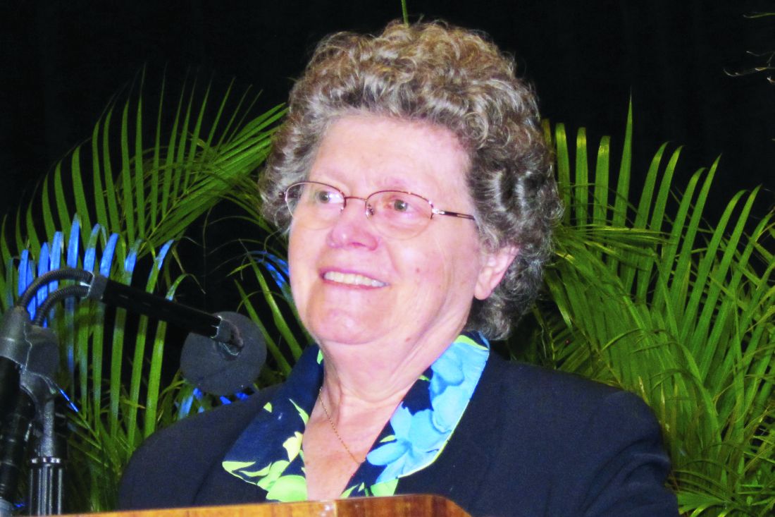
In the previously published double-blind, placebo-controlled DISCOVER-2 trial, two dosing regimens of the interleukin-23 (IL-23) inhibitor guselkumab were compared with placebo in biologic-naive patients. The active regimens were similarly effective relative to placebo for the primary endpoint of 20% improvement in American College of Rheumatology response criteria (ACR20) at week 24.
In this new long-term subgroup analysis, outcomes at 2 years were evaluated in the 246 patients with axial involvement (33.3% of the total number of 739 evaluable patients). Baseline characteristics across treatment groups in this subset of the DISCOVER-2 trial were similar.
Guselkumab exhibits nearly twofold advantage
At 24 weeks relative to baseline, the improvement on multiple axial involvement outcome measures was approximately twofold greater with active therapy than with placebo. For example, the mean Bath Ankylosing Spondylitis Disease Activity Index (BASDAI) score fell 2.6 points in both active treatment arms versus 1.4 on placebo.
The same relative advantage was observed when the BASDAI spinal pain subscore was isolated. There were also comparable responses on a modified BASDAI that excluded the peripheral pain response, and the Ankylosing Spondylitis Disease Activity Score (ASDAS).
When evaluated at week 52 and again at week 100, all outcomes employed to evaluate change in axial involvement were sustained. Many were further improved. In patients who initiated therapy on placebo, all of whom were switched to guselkumab at the end of the 24-week double-blind period, at least the same degree of axial symptom control relative to baseline was achieved at both time periods.
Incremental improvement observed over time
“For most measures there was further improvement at 2 years, and there was generally consistent response across patient groups stratified by HLA [human leucocyte antigen] status,” Dr. Gladman reported.
Relative to the 2.6-point reduction in the BASDAI score in the two guselkumab arms at week 24, the reductions reached 3.0, 3.1, and 3.3 at 100 weeks in the every-4-week guselkumab group, every-8-week guselkumab group, and the crossed-over placebo group, respectively. For ASDAS, the reductions at week 24 were 1.4, 1.5, and 0.7 points and reached 1.6, 1.7, and 1.6 points at 100 weeks in the every-4-week, every-8-week, and placebo crossover groups, respectively.
The sustained improvement is consistent with a previous post hoc analysis in which data from the phase 3 DISCOVER-1 trial were pooled with those from DISCOVER-2. This analysis focused on the 312 patients in these studies with axial disease documented by imaging. Again, the study showed improvement at week 24 was sustained at week 52 independent of HLA-B27 status.
Need for MRI confirmation seen
The potential problem with this new analysis as well as the previous analysis is the absence of MRI to provide objective evidence of axial involvement, according to Walter P. Maksymowych, MD, professor in the division of rheumatology at the University of Alberta, Edmonton.

Noting that previous studies have indicated that axial involvement assessed by investigators is not reliable even when performed with x-rays, Dr. Maksymowych said these data would be much more reassuring with MRI controls.
“We have seen little correlation between clinical symptoms and MRI manifestations of disease,” he said.
Dr. Gladman conceded this point. Baseline MRI was performed in some of the patients in this subgroup analysis, but it was not mandated. As a result, the data support benefit from guselkumab for symptomatic axial involvement, but she indicated that better evidence of a disease-modifying effect is expected from a more rigorous placebo-controlled trial of guselkumab called STAR.
This trial is requiring MRI at baseline and at week 24 and is using the Spondyloarthritis Research Consortium of Canada (SPARCC) score to assess change. Dr. Gladman said the trial is enrolling “as we speak.”
Overall, Dr. Gladman agreed with Dr. Maksymowych that objective biomarkers are important for demonstrating that treatments are improving long-term outcomes, not just relieving symptoms.
Guselkumab manufacturer Janssen supported the post hoc analysis. Dr. Gladman reported financial relationships with AbbVie, Amgen, Bristol-Myers Squibb, Eli Lilly, Galapagos, Gilead Janssen, Novartis, Pfizer, and UCB. Dr. Maksymowych reported financial relationships with AbbVie, Boehringer Ingelheim, Celgene, Eli Lilly, Galapagos, Gilead, Janssen, Novartis, Pfizer, and UCB.
Guselkumab (Tremfya) received Food and Drug Administration approval for the treatment of psoriatic arthritis (PsA) almost 2 years ago on the basis of a phase 3 trial, but a new substudy from that trial has now demonstrated long-term benefit in the subgroup of patients who entered the trial with axial involvement, according to data presented at the annual meeting of the Canadian Rheumatology Association.
“The symptom relief was clinically meaningful and durable through week 100,” reported Dafna D. Gladman, MD, professor of medicine and director of the psoriatic arthritis program at the University of Toronto.
In the previously published double-blind, placebo-controlled DISCOVER-2 trial, two dosing regimens of the interleukin-23 (IL-23) inhibitor guselkumab were compared with placebo in biologic-naive patients. The active regimens were similarly effective relative to placebo for the primary endpoint of 20% improvement in American College of Rheumatology response criteria (ACR20) at week 24.
In this new long-term subgroup analysis, outcomes at 2 years were evaluated in the 246 patients with axial involvement (33.3% of the total number of 739 evaluable patients). Baseline characteristics across treatment groups in this subset of the DISCOVER-2 trial were similar.
Guselkumab exhibits nearly twofold advantage
At 24 weeks relative to baseline, the improvement on multiple axial involvement outcome measures was approximately twofold greater with active therapy than with placebo. For example, the mean Bath Ankylosing Spondylitis Disease Activity Index (BASDAI) score fell 2.6 points in both active treatment arms versus 1.4 on placebo.
The same relative advantage was observed when the BASDAI spinal pain subscore was isolated. There were also comparable responses on a modified BASDAI that excluded the peripheral pain response, and the Ankylosing Spondylitis Disease Activity Score (ASDAS).
When evaluated at week 52 and again at week 100, all outcomes employed to evaluate change in axial involvement were sustained. Many were further improved. In patients who initiated therapy on placebo, all of whom were switched to guselkumab at the end of the 24-week double-blind period, at least the same degree of axial symptom control relative to baseline was achieved at both time periods.
Incremental improvement observed over time
“For most measures there was further improvement at 2 years, and there was generally consistent response across patient groups stratified by HLA [human leucocyte antigen] status,” Dr. Gladman reported.
Relative to the 2.6-point reduction in the BASDAI score in the two guselkumab arms at week 24, the reductions reached 3.0, 3.1, and 3.3 at 100 weeks in the every-4-week guselkumab group, every-8-week guselkumab group, and the crossed-over placebo group, respectively. For ASDAS, the reductions at week 24 were 1.4, 1.5, and 0.7 points and reached 1.6, 1.7, and 1.6 points at 100 weeks in the every-4-week, every-8-week, and placebo crossover groups, respectively.
The sustained improvement is consistent with a previous post hoc analysis in which data from the phase 3 DISCOVER-1 trial were pooled with those from DISCOVER-2. This analysis focused on the 312 patients in these studies with axial disease documented by imaging. Again, the study showed improvement at week 24 was sustained at week 52 independent of HLA-B27 status.
Need for MRI confirmation seen
The potential problem with this new analysis as well as the previous analysis is the absence of MRI to provide objective evidence of axial involvement, according to Walter P. Maksymowych, MD, professor in the division of rheumatology at the University of Alberta, Edmonton.
Noting that previous studies have indicated that axial involvement assessed by investigators is not reliable even when performed with x-rays, Dr. Maksymowych said these data would be much more reassuring with MRI controls.
“We have seen little correlation between clinical symptoms and MRI manifestations of disease,” he said.
Dr. Gladman conceded this point. Baseline MRI was performed in some of the patients in this subgroup analysis, but it was not mandated. As a result, the data support benefit from guselkumab for symptomatic axial involvement, but she indicated that better evidence of a disease-modifying effect is expected from a more rigorous placebo-controlled trial of guselkumab called STAR.
This trial is requiring MRI at baseline and at week 24 and is using the Spondyloarthritis Research Consortium of Canada (SPARCC) score to assess change. Dr. Gladman said the trial is enrolling “as we speak.”
Overall, Dr. Gladman agreed with Dr. Maksymowych that objective biomarkers are important for demonstrating that treatments are improving long-term outcomes, not just relieving symptoms.
Guselkumab manufacturer Janssen supported the post hoc analysis. Dr. Gladman reported financial relationships with AbbVie, Amgen, Bristol-Myers Squibb, Eli Lilly, Galapagos, Gilead Janssen, Novartis, Pfizer, and UCB. Dr. Maksymowych reported financial relationships with AbbVie, Boehringer Ingelheim, Celgene, Eli Lilly, Galapagos, Gilead, Janssen, Novartis, Pfizer, and UCB.
FROM THE ANNUAL MEETING OF THE CANADIAN RHEUMATOLOGY ASSOCIATION
Chronic marijuana use linked to recurrent stroke
, new observational research suggests. “Our analysis shows young marijuana users with a history of stroke or transient ischemic attack remain at significantly high risk for future strokes,” said lead study author Akhil Jain, MD, a resident physician at Mercy Fitzgerald Hospital in Darby, Pennsylvania.
“It’s essential to raise awareness among young adults about the impact of chronic habitual use of marijuana, especially if they have established cardiovascular risk factors or previous stroke.”
The study will be presented during the International Stroke Conference, presented by the American Stroke Association, a division of the American Heart Association.
An increasing number of jurisdictions are allowing marijuana use. To date, 18 states and the District of Columbia have legalized recreational cannabis use, the investigators noted.
Research suggests cannabis use disorder – defined as the chronic habitual use of cannabis – is more prevalent in the young adult population. But Dr. Jain said the population of marijuana users is “a changing dynamic.”
Cannabis use has been linked to an increased risk for first-time stroke or transient ischemic attack (TIA). Traditional stroke risk factors include hypertension, diabetes, and diseases related to blood vessels or blood circulation, including atherosclerosis.
Young adults might have additional stroke risk factors, such as behavioral habits like substance abuse, low physical activity, and smoking, oral contraceptives use among females, and brain infections, especially in the immunocompromised, said Dr. Jain.
Research from the American Heart Association shows stroke rates are increasing among adults 18 to 45 years of age. Each year, young adults account for up to 15% of strokes in the United States.
Prevalence and risk for recurrent stroke in patients with previous stroke or TIA in cannabis users have not been clearly established, the researchers pointed out.
A higher rate of recurrent stroke
For this new study, Dr. Jain and colleagues used data from the National Inpatient Sample from October 2015 to December 2017. They identified hospitalizations among young adults 18 to 45 years of age with a previous history of stroke or TIA.
They then grouped these patients into those with cannabis use disorder (4,690) and those without cannabis use disorder (156,700). The median age in both cohorts was 37 years.
The analysis did not include those who were considered in remission from cannabis use disorder.
Results showed that 6.9% of those with cannabis use disorder were hospitalized for a recurrent stroke, compared with 5.4% of those without cannabis use disorder (P < .001).
After adjustment for demographic factors (age, sex, race, household income), and pre-existing conditions, patients with cannabis use disorder were 48% more likely to be hospitalized for recurrent stroke than those without cannabis use disorder (odds ratio, 1.48; 95% confidence interval, 1.28-1.71; P < .001).
Compared with the group without cannabis use disorder, the cannabis use disorder group had more men (55.2% vs. 40.2%), more African American people (44.6% vs. 37.2%), and more use of tobacco (73.9% vs. 39.6%) and alcohol (16.5% vs. 3.6%). They also had a greater percentage of chronic obstructive pulmonary disease, depression, and psychoses.
But a smaller percentage of those with cannabis use disorder had hypertension (51.3% vs. 55.6%; P = .001) and diabetes (16.3% vs. 22.7%; P < .001), which is an “interesting” finding, said Dr. Jain.
“We observed that even with a lower rate of cardiovascular risk factors, after controlling for all the risk factors, we still found the cannabis users had a higher rate of recurrent stroke.”
He noted this was a retrospective study without a control group. “If both groups had comparable hypertension, then this risk might actually be more evident,” said Dr. Jain. “We need a prospective study with comparable groups.”
Living in low-income neighborhoods and in northeast and southern regions of the United States was also more common in the cannabis use disorder group.
Hypothesis-generating research
The study did not investigate the possible mechanisms by which marijuana use might increase stroke risk, but Dr. Jain speculated that these could include factors such as impaired blood vessel function, changes in blood supply, an increased tendency of blood clotting, impaired energy production in brain cells, and an imbalance between molecules that harm healthy tissue and the antioxidant defenses that neutralize them.
As cannabis use may pose a different risk for a new stroke, as opposed a previous stroke, Dr. Jain said it would be interesting to study the amount of “residual function deficit” experienced with the first stroke.
The new study represents “foundational research” upon which other research teams can build, said Dr. Jain. “Our study is hypothesis-generating research for a future prospective randomized controlled trial.”
A limitation of the study is that it did not consider the effect of various doses, duration, and forms of cannabis abuse, or use of medicinal cannabis or other drugs.
Robert L. Page II, PharmD, professor, departments of clinical pharmacy and physical medicine/rehabilitation, University of Colorado Skaggs School of Pharmacy and Pharmaceutical Sciences, Aurora, provided a comment on this new research.
A cannabis use disorder diagnosis provides “specific criteria” with regard to chronicity of use and reflects “more of a physical and psychological dependence upon cannabis,” said Dr. Page, who chaired the writing group for the AHA 2020 cannabis and cardiovascular disease scientific statement.
He explained what sets people with cannabis use disorder apart from “run-of-the-mill” recreational cannabis users is that “these are individuals who use a cannabis product, whether it’s smoking it, vaping it, or consuming it via an edible, and are using it on a regular basis, in a chronic fashion.”
The study received no outside funding. The authors report no relevant disclosures.
A version of this article first appeared on Medscape.com.
FROM ISC 2022
Expert shares workup pearls for children with severe atopic dermatitis
“Many patients who have failed topical steroids have never had adequate treatment,” Anna Yasmine Kirkorian, MD, chief of dermatology at National Children’s Hospital in Washington, said during the ODAC Dermatology, Aesthetic & Surgical Conference. “There is no lower age limit on the use of topical corticosteroids, and low potency corticosteroids are inadequate to treat severe eczema. The idea that only over-the-counter 2.5% hydrocortisone cream is necessary is not true,” she added.
“You also want to scrutinize the vehicle,” she said, noting that children are often prescribed cream formulations that hurt when applied, so parents stop applying them. “Ointments are generally the vehicles of choice in childhood,” she added.
It is generally not advised to use topical and oral antibiotics in children with AD, unless there are clear signs of infection. “If they’re just slightly oozy, don’t use them,” she continued. “Of course, every child or adult with eczema has Staph aureus on them, but most of the time, what you need to do is repair the barrier. We know that from data and common sense. When we repair their barrier, their rates of infection decrease.”
A focal area with pustules and pus should be cultured and treated, Dr. Kirkorian said. “Monotherapy with antibiotics is going to do nothing for you.” In cases of children with failure to thrive, she recommends referral to pediatric dermatology, allergy/immunology, GI, or genetics, as appropriate.
For children with severe AD, Dr. Kirkorian favors a rescue plan with a one-pound jar of triamcinolone ointment 0.1%. She recommends application of the ointment to all areas, including the face and scalp once nightly for 2 weeks, with a follow-up appointment at the end of that time. “If you just give people medicine and ask them to come back in 6 months, they are not able to comply with that and they don’t have faith that it’s going to work,” explained Dr. Kirkorian, associate professor of dermatology and pediatrics at George Washington University, Washington. At the end of 2 weeks, “the majority will have improved dramatically, and then you can implement maintenance therapy with topical calcineurin inhibitors, crisaborole, or possibly topical ruxolitinib.”
Some clinicians prescribe oral antihistamines for AD, but Dr. Kirkorian said that data supporting their use are limited and antihistamines are not approved for use in children younger than 6 months of age. Sedating antihistamines will induce sleep, “but do not provide durable night-long sleep,” and routine use may have an impact on learning and school performance. In addition, exposure to antihistamines in children under age 2 may be associated with development of ADHD at school age.
The interleukin-4 receptor alpha antagonist dupilumab (Dupixent) is approved by the Food and Drug Administration for moderate to severe AD in patients ages 6 and older. But obtaining it for patients can be tricky, she said, as this requires documented failure of corticosteroids, calcineurin inhibitors, crisaborole ointment, and phototherapy (if prescribed). Patients are often obligated to do step therapy with an off-label drug such as cyclosporine or methotrexate for 3 months, and they need to demonstrate responses with objective measures of severity such as the SCORAD (SCORing Atopic Dermatitis) and the validated Investigator Global Assessment.
“Most of my patients carry insurance that does not approve dupilumab without failure of a prior off-label systemic immunosuppressant medication,” Dr. Kirkorian said. Cyclosporine is her first choice for a systemic immunosuppressant “because it has a fast onset of action, it’s effective for treatment of atopic dermatitis, and safe for short-term use,” she said. “I don’t think that methotrexate works well for eczema. It can take weeks and weeks to work.”
She typically starts patients on a 5 mg/kg dose of cyclosporine. Baseline tests include CBC, CMP (comprehensive metabolic panel), lipids, and vitals. She repeats the labs at 1 month, and includes a blood pressure check. Potential adverse effects of cyclosporine include infections (including opportunistic infections), cytopenias, hypertension, nephrotoxicity, hepatotoxicity, neurotoxicity (including posterior reversible encephalopathy syndrome), electrolyte disturbance, lymphoma, and cutaneous malignancy.
“The good news is that we generally don’t see the adverse effects with short-term use,” Dr. Kirkorian said. “We will see some hypertrichosis and gingival hypertrophy, which resolves with cessation of therapy. There are serious side effects if you use it for long enough.”
As for methotrexate, “it is still a very important drug in pediatric dermatology, particularly in other conditions such as psoriasis,” she said. “The problem is that weekly dosing of methotrexate poses a greater risk of dosing errors. People aren’t really triggered to think of a once-weekly medication. If you do use it, give them a short supply to make sure that they come back, and that they don’t give it daily accidentally.”
Practical tips she offered for prescribing cyclosporine include supplying a patient handout with information on all adverse effects, dosing information, vaccination information, and pregnancy precautions, with contact information (a patient portal or on-call number) for the treating clinician in case a patient develops adverse effects. Administration of live vaccines while patients are on cyclosporine is not recommended.
When transitioning patients from cyclosporine or methotrexate to dupilumab, Dr. Kirkorian recommends tapering the immunosuppressant dose by half every 2 weeks to complete cessation by week 8 of treatment. For patients who experience a severe baseline flare once the immunosuppressant is tapered, despite the switch to dupilumab, she recommends restarting methotrexate at a full dose and then reducing the dose every 2 weeks until the lowest effective dose (2.5-5 mg weekly) is reached.
“Waning efficacy is real,” she said. “We can add methotrexate to recapture efficacy. Check for superimposed allergic contact dermatitis.”
With upadacitinib (Rinvoq), an oral Janus kinase (JAK) inhibitor recently approved for treating refractory, moderate to severe AD in patients 12 years of age and older, is the risk profile acceptable to parents and physicians? “I think the answer is yes,” Dr. Kirkorian said. “But we’re going to have to think through that very carefully. It’s going to be exciting to see how this drug changes management in our patients.”
Dr. Kirkorian disclosed that she is a member of the advisory board for Verrica Pharmaceuticals.
FROM ODAC 2022
