User login
MDedge conference coverage features onsite reporting of the latest study results and expert perspectives from leading researchers.
Patisiran benefits ATTR amyloidosis with cardiomyopathy: APOLLO-B
The RNA interference (RNAi) therapeutic, patisiran (Onpattro, Alnylam), showed a statistically significant and clinically meaningful benefit on functional capacity, as measured by the 6-minute walk test (6-MWT), compared with placebo, in the treatment of transthyretin-mediated amyloidosis with cardiomyopathy, in the APOLLO-B trial.
The study also met its first secondary endpoint, demonstrating a statistically significant and clinically meaningful benefit on health status and quality of life.
These positive results, their first formal presentation, were announced Sept. 8 at the 18th International Symposium on Amyloidosis. However, the company announced positive top-line results from the trial in early August.
Transthyretin-mediated (ATTR) amyloidosis is a rare, rapidly progressive, debilitating disease caused by misfolded transthyretin (TTR) proteins which accumulate as amyloid fibrils in multiple tissues including the nerves, heart, and gastrointestinal tract.
There are two different types of ATTR amyloidosis: hereditary ATTR (hATTR) amyloidosis, caused by a TTR gene variant, and wild-type ATTR (wtATTR) amyloidosis, which occurs without a TTR gene variant. hATTR amyloidosis affects approximately 50,000 people worldwide, whereas wtATTR amyloidosis is estimated to affect 200,000-300,000 people worldwide.
Patisiran is an intravenously administered RNAi therapeutic that is approved in the United States and Canada for the treatment of the polyneuropathy of hATTR amyloidosis in adults. It is also approved in the European Union, Switzerland, Brazil, and Japan for a similar indication. It is designed to target and silence TTR messenger RNA, thereby reducing the production of TTR protein before it is made. Reducing the pathogenic protein leads to a reduction in amyloid deposits in tissues.
“The results of the APOLLO-B phase 3 study are impressive, as I believe they underscore the potential for patisiran to provide a benefit on functional capacity and quality of life in patients living with ATTR amyloidosis with cardiomyopathy. Furthermore, these results were seen after only 12 months of treatment,” Mathew Maurer, MD, Arnold and Arlene Goldstein Professor of Cardiology at Columbia University Irving Medical Center, New York, said in an Alnylam press release.
“The cardiac manifestations associated with ATTR amyloidosis can have a devastating impact on patients’ lives and current treatment options are limited. With the rapidly progressive nature of the disease, there is a significant need for treatments like patisiran, which has the potential to be a new option for patients and physicians to treat the cardiomyopathy of ATTR amyloidosis,” Dr. Maurer added.
APOLLO-B is a phase 3, randomized, double-blind study evaluating the effects of patisiran on functional capacity and quality of life in patients with ATTR amyloidosis with cardiomyopathy. The study enrolled 360 adult patients with ATTR amyloidosis (hereditary or wild-type) with cardiomyopathy who were randomly assigned 1:1 to receive 0.3 mg/kg of patisiran or placebo intravenously administered every 3 weeks over a 12-month treatment period. After 12 months, all patients will receive patisiran in an open-label extension.
Results at 12 months, reported by Alnylam, found that the primary endpoint, the 6-MWT, showed a median change from baseline of –8.15 m for the patisiran group and –21.34 m for the placebo group, a significant difference favoring patisiran.
The first secondary endpoint was health status and quality of life, as measured by the Kansas City Cardiomyopathy Questionnaire Overall Summary score. This showed a mean change from baseline of +0.300 for the patisiran group and –3.408 for the placebo group, a significant difference favoring patisiran.
Secondary composite outcome endpoints did not achieve statistical significance.
A nonsignificant result (win ratio, 1.27; P = .0574) was found on the secondary composite endpoint of all-cause mortality, frequency of cardiovascular events, and change from baseline in 6-MWT over 12 months, compared with placebo.
The final two composite endpoints were not powered for statistical significance, given the sample size and short duration of the study – all-cause mortality and frequency of all-cause hospitalizations and urgent heart failure visits in patients not on tafamidis at baseline (hazard ratio, 0.997) and in the overall study population (HR, 0.883).
Patisiran achieved a rapid and sustained reduction in serum TTR levels, with a mean percent reduction from baseline in serum TTR reduction of 87% at month 12.
A beneficial effect on the exploratory endpoint, N-terminal of the prohormone brain natriuretic peptide, a measure of cardiac stress, was observed in the patisiran arm, with a 20% reduction in the adjusted geometric mean fold change from baseline, compared with placebo.
Patisiran also demonstrated an encouraging safety and tolerability profile, including no cardiac safety concerns relative to placebo, during the 12-month treatment period, Alnylam reported.
The majority of adverse events were mild or moderate in severity. Treatment emergent adverse events in the patisiran group included infusion-related reactions, arthralgia, and muscle spasms.
In the safety analysis, there were five deaths (2.8%) observed in patisiran-treated patients and eight deaths (4.5%) observed in the placebo group.
Pushkal Garg, MD, chief medical officer at Alnylam, said: “We believe these data validate the therapeutic hypothesis that TTR silencing by an RNAi therapeutic may be an effective approach to treating cardiomyopathy of both wild-type and hereditary ATTR amyloidosis.”
Alnylam plans to file a supplemental new drug application for patisiran as a potential treatment for ATTR amyloidosis with cardiomyopathy in the United States in late 2022.
A version of this article first appeared on Medscape.com.
Candy, desserts: A ‘gateway’ to unhealthy eating among teens
Certain ultraprocessed foods – especially candy, prepackaged pastries, and frozen desserts – could be “gateway foods” for adolescents, leading them to increase their intake of other unhealthy foods, a new study suggests.
“For teens, gateway ultraprocessed foods (candy, store pastries, frozen desserts) should be prioritized for preventive dietary interventions as they increase intake across all other UPFs,” lead researcher Maria Balhara said in an interview.
“The good news,” said Ms. Balhara, is that even small changes, such as reducing how often gateway foods are consumed, may reduce overall intake of unhealthy foods and have a “big impact” on overall health.
Ms. Balhara has a unique perspective on adolescent eating habits: She’s 16 years old, from Florida, and conducted the study while dual-enrolled at Broward College and Cooper City High School.
Her study was released Sept. 7 ahead of presentation at the American Heart Association Hypertension Scientific Sessions 2022 in San Diego.
Blame the pandemic?
Over the past 30 years, there’s been a steady increase in consumption of UPFs worldwide, coupled with mounting evidence that diets rich in UPFs raise the risk for several chronic diseases, including weight gain, hypertension, and increased risk for heart disease and premature death.
For her research, Ms. Balhara asked 315 teenagers (42% male) from 12 high schools in South Florida how often they consumed UPFs over two time periods – before COVID in 2019 and after COVID restrictions were eased in 2022 – using a survey that she developed called the Processed Intake Evaluation (PIE).
More than 2 in 5 teens (43%) increased their consumption of UPFs between 2019 and 2022.
During this time, increased consumption of frozen desserts was associated with an 11% increase in consumption of all other UPFs, whereas increased consumption of prepackaged pastries and candy was associated with a 12% and 31%, respectively, increase in consumption of all other UPFs, Ms. Balhara found.
Encouragingly, 57% of teens decreased their consumption of UPFs between 2019 and 2022.
During this time, decreased consumption of processed meats was associated with an 8% decrease in consumption of all other UPFs, whereas decreased consumption of white bread and biscuits was associated with a 9% and 10%, respectively, decrease in consumption of all other UPFs.
The results provide initial evidence for a new “gateway food model,” Ms. Balhara told this news organization, in which certain UPFs, when increased, drive overall consumption of all UPFs among teens.
Limitations of the study include the self-reported dietary data and the fact that the PIE survey has not been validated.
Not all UPFs are bad
“I commend Ms. Balhara for her project, which highlights the importance of establishing good dietary patterns early in life,” Donna K. Arnett, PhD, past president of the AHA, said in a news release.
“The relationship between poor dietary quality and cardiovascular risk factors is well-established. While this is a small, preliminary study, it’s an important topic to continue to investigate and help us understand ways we can influence dietary behaviors to promote optimal cardiovascular health for all ages,” said Dr. Arnett, executive vice president for academic affairs and provost at the University of South Carolina, Columbia.
Offering perspective on the study, Taylor C. Wallace, PhD, with the department of nutrition and food studies, George Mason University, Fairfax, Va., made the point that “food processing and ultraprocessed foods aren’t the problem. The problem is the types of ultraprocessed foods on the market that people consume.”
“Remember, non-fat, vitamin D fortified yogurt is also ‘ultra-processed,’ and it’s very healthy,” he told this news organization.
Dr. Wallace said that it’s no surprise that teens increased their intake of UPFs during the pandemic.
“Of course, people increased processed food intake during the pandemic. Processed foods are shelf stable at a time when grocery stores were running out of things and supply chains weren’t able to keep up. Also, many were depressed and use food to indulge,” he noted.
The study had no funding. Ms. Balhara has no relevant disclosures. Dr. Wallace is principal and CEO of Think Healthy Group; chief food and nutrition scientist with Produce for Better Health Foundation; editor, Journal of Dietary Supplements; deputy editor, Journal of the American College of Nutrition; nutrition section editor, Annals of Medicine; and an advisory board member with Forbes Health.
A version of this article first appeared on Medscape.com.
FROM HYPERTENSION 2022
Bimekizumab effective for axSpA with or without prior TNFi treatment
GHENT, BELGIUM – Patients with nonradiographic or radiographic axial spondyloarthritis (axSpA) experienced clinically relevant treatment responses to bimekizumab (Bimzelx) at similar rates that significantly exceeded placebo, regardless of prior experience with a tumor necrosis factor (TNF) inhibitor, according to results from two phase 3 trials presented at the 13th International Congress on Spondyloarthritides.
In addition, around half of patients with either nonradiographic or radiographic disease achieved complete remission of enthesitis by week 16 of treatment with bimekizumab. The drug, a humanized, monoclonal antibody dually inhibiting interleukins (IL) 17A and 17F, is approved in the European Union for treating adults with moderate to severe plaque psoriasis.

“Bimekizumab blockade works independently of axial spondyloarthritis pretreatment, which means this drug specifically blocks something that other drugs do not reach,” said Xenofon Baraliakos, MD, professor of internal medicine and rheumatology at Ruhr University Bochum (Germany). He presented 24-week data on the use of bimekizumab.
The BE MOBILE 1 trial involved 256 patients with nonradiographic axSpA, whereas BE MOBILE 2 involved 232 patients with radiographic axSpA. In both trials, bimekizumab 160 mg was administered subcutaneously every 4 weeks, and at week 16, all patients, including those who had received placebo, received open-label bimekizumab for another 8 weeks. This news organization previously reported results from BE MOBILE 2 that were presented at the European Alliance of Associations for Rheumatology (EULAR) 2022 annual meeting.
In Ghent, referring to the nonradiographic patients, Dr. Baraliakos said in an interview, “We saw a very clear response to the active drug even after 2 weeks. The curves separated out from placebo. The week 16 primary analysis showed patients on bimekizumab did significantly better, [and there was] a similar response in those who switched to [open-label] bimekizumab after placebo” at week 16.
In patients with nonradiographic disease at week 24, 52.3% on bimekizumab achieved the trial’s primary outcome of 40% improvement in Assessment in Spondyloarthritis International Society response criteria (ASAS 40), compared with 46.8% of patients who were receiving placebo and then switched to open-label bimekizumab at week 16, the latter rising from 21.4% at week 16. For comparison, 47.7% on bimekizumab achieved ASAS 40 at week 16.
At week 24 in BE MOBILE 2, 53.8% of patients with radiographic disease on continuous bimekizumab met ASAS 40 criteria, as did 56.8% of patients who switched from placebo to open-label bimekizumab, rising from 22.5% with placebo and 44.8% with bimekizumab at week 16.
Audience member Fabian Proft, MD, of Charité Medical University, Berlin, commented on the latest results as well as wider bimekizumab findings, including those relating to psoriasis. “When we compare this to drugs that are already approved and available, we can assume that bimekizumab is equally effective to existing ones,” he said, noting that “there is the additional option in patients with psoriasis, where it seems to be the most effective drug for this indication. If I had a patient with radiographic or nonradiographic axial SpA and who also had significant psoriasis, then bimekizumab would be my choice of treatment.”
Targeting IL-17A and IL-17F in one drug
In the BE MOBILE 1 study, Dr. Baraliakos and coinvestigators looked at whether inhibiting IL-17F as well as IL-17A “makes sense” in terms of clinical benefits in patients with axSpA.
“Previous experience with IL-17A inhibitors shows they work well but still miss some patients,” Dr. Baraliakos said, adding that, “the hope is that by blocking both IL-17A and IL-17F, the response will be a bit better in terms of both greater response and longevity of response than [with an] IL-17A [inhibitor] alone.”
Patients in BE MOBILE 1 were typical adult patients with nonradiographic axSpA who fulfilled ASAS classification criteria and had elevated C-reactive protein (CRP) and/or sacroiliitis on MRI. All patients were older than 18 years and had a mean age of 39 years. In each arm, 51%-57% were men. Overall, patients had a mean of 9 years of symptoms and a mean Ankylosing Spondylitis Disease Activity Score of 3.7 in both patient groups (placebo and bimekizumab).
All had active disease (Bath Ankylosing Spondylitis Disease Activity Index ≥ 4 and spinal pain ≥ 4) at baseline and demonstrated failure to respond to two different NSAIDs or had a history of intolerance to or contraindication to NSAIDs. Patients had previously received up to one TNF inhibitor (13.5% in the placebo group and 7.8% in the bimekizumab group).
The primary outcome compared rate of response to ASAS 40 criteria, which comprises patient global assessment of disease, spinal pain, function (as assessed by the Bath Ankylosing Spondylitis Functional Index [BASFI]), and inflammation (stiffness).
Early response seen regardless of previous TNF inhibitor experience
“We saw response to bimekizumab very early in our patients at 16 weeks. The amount of response was higher than that observed with IL-17A alone,” Dr. Baraliakos said in an interview. “It’s understood that IL-17A and IL-17F do not work together on the inflammatory cascade, but work separately, and this might explain the findings whereby this drug captures more inflammation.”
Dr. Baraliakos highlighted the unique response rates seen with bimekizumab regardless of past TNF inhibitor use. “The TNF inhibitor-experienced patients responded as well as the TNF inhibitor–naive ones. This is unusual because nonresponders to other drugs are usually more severely affected and have a lower chance of showing response to any drug. Also, we did not see this response in patients treated with IL-17A only.”
At 16 weeks, patients with nonradiographic disease without a past history of using a TNF inhibitor had ASAS 40 responses at rates of 46.6% with bimekizumab and 22.9% with placebo. These rates in patients with past TNF inhibitor use were 60% with bimekizumab and 11.8% with placebo.
Statistically significant differences between bimekizumab and placebo occurred for all primary and secondary outcomes. “This includes the MRI inflammation findings in bimekizumab-treated patients,” Dr. Baraliakos reported.
Complete resolution of enthesitis was also observed. By week 24, enthesitis completely resolved in 47.9% of patients with nonradiographic disease on continuous bimekizumab and 43.5% of those patients who switched from placebo to bimekizumab. In patients with radiographic disease, complete resolution occurred in 53% of those on continuous bimekizumab and 49.3% of patients who switched at week 16. “This was an excellent outcome,” Dr. Baraliakos said.
The safety profile at 24 weeks confirmed prior findings at 16 weeks in which the most common treatment-emergent adverse events with bimekizumab were nasopharyngitis (9.4%), upper respiratory tract infection (7%), and oral candidiasis (3.1%); fungal infections overall occurred in 7% taking bimekizumab.
“We saw slightly higher fungal infections, but this is because we block IL-17A and IL-17F, and [the risk for these infections] is linked to the mechanism of action. But we can deal with this,” Dr. Baraliakos said.
The trials were sponsored by UCB. Dr. Baraliakos disclosed serving on the speakers bureau and as a paid instructor and consultant for AbbVie, Bristol-Myers Squibb, Chugai, Eli Lilly, Galapagos, Gilead, Merck Sharp & Dohme, Novartis, Pfizer, and UCB. Dr. Proft disclosed serving on speakers bureaus for Amgen, AbbVie, Bristol-Myers Squibb, Celgene, Janssen, Merck Sharp & Dohme, Novartis, Pfizer, Roche, and UCB; being a consultant to Novartis; and receiving grant or research support from Novartis, UCB, and Lilly.
GHENT, BELGIUM – Patients with nonradiographic or radiographic axial spondyloarthritis (axSpA) experienced clinically relevant treatment responses to bimekizumab (Bimzelx) at similar rates that significantly exceeded placebo, regardless of prior experience with a tumor necrosis factor (TNF) inhibitor, according to results from two phase 3 trials presented at the 13th International Congress on Spondyloarthritides.
In addition, around half of patients with either nonradiographic or radiographic disease achieved complete remission of enthesitis by week 16 of treatment with bimekizumab. The drug, a humanized, monoclonal antibody dually inhibiting interleukins (IL) 17A and 17F, is approved in the European Union for treating adults with moderate to severe plaque psoriasis.
“Bimekizumab blockade works independently of axial spondyloarthritis pretreatment, which means this drug specifically blocks something that other drugs do not reach,” said Xenofon Baraliakos, MD, professor of internal medicine and rheumatology at Ruhr University Bochum (Germany). He presented 24-week data on the use of bimekizumab.
The BE MOBILE 1 trial involved 256 patients with nonradiographic axSpA, whereas BE MOBILE 2 involved 232 patients with radiographic axSpA. In both trials, bimekizumab 160 mg was administered subcutaneously every 4 weeks, and at week 16, all patients, including those who had received placebo, received open-label bimekizumab for another 8 weeks. This news organization previously reported results from BE MOBILE 2 that were presented at the European Alliance of Associations for Rheumatology (EULAR) 2022 annual meeting.
In Ghent, referring to the nonradiographic patients, Dr. Baraliakos said in an interview, “We saw a very clear response to the active drug even after 2 weeks. The curves separated out from placebo. The week 16 primary analysis showed patients on bimekizumab did significantly better, [and there was] a similar response in those who switched to [open-label] bimekizumab after placebo” at week 16.
In patients with nonradiographic disease at week 24, 52.3% on bimekizumab achieved the trial’s primary outcome of 40% improvement in Assessment in Spondyloarthritis International Society response criteria (ASAS 40), compared with 46.8% of patients who were receiving placebo and then switched to open-label bimekizumab at week 16, the latter rising from 21.4% at week 16. For comparison, 47.7% on bimekizumab achieved ASAS 40 at week 16.
At week 24 in BE MOBILE 2, 53.8% of patients with radiographic disease on continuous bimekizumab met ASAS 40 criteria, as did 56.8% of patients who switched from placebo to open-label bimekizumab, rising from 22.5% with placebo and 44.8% with bimekizumab at week 16.
Audience member Fabian Proft, MD, of Charité Medical University, Berlin, commented on the latest results as well as wider bimekizumab findings, including those relating to psoriasis. “When we compare this to drugs that are already approved and available, we can assume that bimekizumab is equally effective to existing ones,” he said, noting that “there is the additional option in patients with psoriasis, where it seems to be the most effective drug for this indication. If I had a patient with radiographic or nonradiographic axial SpA and who also had significant psoriasis, then bimekizumab would be my choice of treatment.”
Targeting IL-17A and IL-17F in one drug
In the BE MOBILE 1 study, Dr. Baraliakos and coinvestigators looked at whether inhibiting IL-17F as well as IL-17A “makes sense” in terms of clinical benefits in patients with axSpA.
“Previous experience with IL-17A inhibitors shows they work well but still miss some patients,” Dr. Baraliakos said, adding that, “the hope is that by blocking both IL-17A and IL-17F, the response will be a bit better in terms of both greater response and longevity of response than [with an] IL-17A [inhibitor] alone.”
Patients in BE MOBILE 1 were typical adult patients with nonradiographic axSpA who fulfilled ASAS classification criteria and had elevated C-reactive protein (CRP) and/or sacroiliitis on MRI. All patients were older than 18 years and had a mean age of 39 years. In each arm, 51%-57% were men. Overall, patients had a mean of 9 years of symptoms and a mean Ankylosing Spondylitis Disease Activity Score of 3.7 in both patient groups (placebo and bimekizumab).
All had active disease (Bath Ankylosing Spondylitis Disease Activity Index ≥ 4 and spinal pain ≥ 4) at baseline and demonstrated failure to respond to two different NSAIDs or had a history of intolerance to or contraindication to NSAIDs. Patients had previously received up to one TNF inhibitor (13.5% in the placebo group and 7.8% in the bimekizumab group).
The primary outcome compared rate of response to ASAS 40 criteria, which comprises patient global assessment of disease, spinal pain, function (as assessed by the Bath Ankylosing Spondylitis Functional Index [BASFI]), and inflammation (stiffness).
Early response seen regardless of previous TNF inhibitor experience
“We saw response to bimekizumab very early in our patients at 16 weeks. The amount of response was higher than that observed with IL-17A alone,” Dr. Baraliakos said in an interview. “It’s understood that IL-17A and IL-17F do not work together on the inflammatory cascade, but work separately, and this might explain the findings whereby this drug captures more inflammation.”
Dr. Baraliakos highlighted the unique response rates seen with bimekizumab regardless of past TNF inhibitor use. “The TNF inhibitor-experienced patients responded as well as the TNF inhibitor–naive ones. This is unusual because nonresponders to other drugs are usually more severely affected and have a lower chance of showing response to any drug. Also, we did not see this response in patients treated with IL-17A only.”
At 16 weeks, patients with nonradiographic disease without a past history of using a TNF inhibitor had ASAS 40 responses at rates of 46.6% with bimekizumab and 22.9% with placebo. These rates in patients with past TNF inhibitor use were 60% with bimekizumab and 11.8% with placebo.
Statistically significant differences between bimekizumab and placebo occurred for all primary and secondary outcomes. “This includes the MRI inflammation findings in bimekizumab-treated patients,” Dr. Baraliakos reported.
Complete resolution of enthesitis was also observed. By week 24, enthesitis completely resolved in 47.9% of patients with nonradiographic disease on continuous bimekizumab and 43.5% of those patients who switched from placebo to bimekizumab. In patients with radiographic disease, complete resolution occurred in 53% of those on continuous bimekizumab and 49.3% of patients who switched at week 16. “This was an excellent outcome,” Dr. Baraliakos said.
The safety profile at 24 weeks confirmed prior findings at 16 weeks in which the most common treatment-emergent adverse events with bimekizumab were nasopharyngitis (9.4%), upper respiratory tract infection (7%), and oral candidiasis (3.1%); fungal infections overall occurred in 7% taking bimekizumab.
“We saw slightly higher fungal infections, but this is because we block IL-17A and IL-17F, and [the risk for these infections] is linked to the mechanism of action. But we can deal with this,” Dr. Baraliakos said.
The trials were sponsored by UCB. Dr. Baraliakos disclosed serving on the speakers bureau and as a paid instructor and consultant for AbbVie, Bristol-Myers Squibb, Chugai, Eli Lilly, Galapagos, Gilead, Merck Sharp & Dohme, Novartis, Pfizer, and UCB. Dr. Proft disclosed serving on speakers bureaus for Amgen, AbbVie, Bristol-Myers Squibb, Celgene, Janssen, Merck Sharp & Dohme, Novartis, Pfizer, Roche, and UCB; being a consultant to Novartis; and receiving grant or research support from Novartis, UCB, and Lilly.
AT THE 2022 SPA CONGRESS
AI algorithm detects erosions, ankylosis with high accuracy in patients with sacroiliitis
Erosions and ankylosis in patients with sacroiliitis are detectable to a high degree of accuracy on CT images using an artificial intelligence (AI)–based algorithm, according to research presented at the 13th International Congress on Spondyloarthritides.
Lennart Jans, MD, head of clinics in musculoskeletal imaging in the department of radiology at Ghent (Belgium) University Hospital, shared data on the development and validation of the algorithm for automatic detection of erosion and ankylosis on CT images of the sacroiliac (SI) joints.
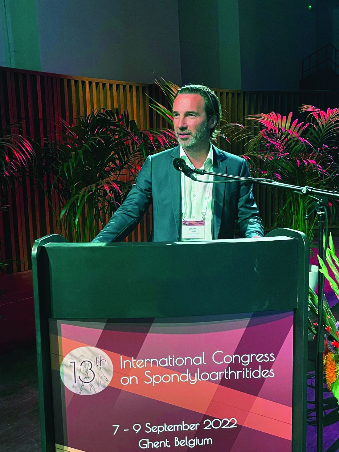
“Essentially, in terms of statistics, this AI algorithm has 95% sensitivity for picking up erosions in patients with clinical symptoms of sacroiliitis, and if this is further developed as a tool, it could aid detection in people with erosions that would otherwise go undetected and undiagnosed,” Dr. Jans said in an interview, stressing that the results were still preliminary.
“We want to move from reporting one patient at a time to a system that detects and helps to diagnose larger numbers of patients and makes a larger impact on patient outcomes.”
He stressed that, with thousands of images per patient, it is an impossible workload for any radiology department to read every image necessary to inform diagnoses, and this is only exacerbated by the shortage of rheumatologists, especially in the United States.
Denis Poddubnyy, MD, head of rheumatology at Charité University Hospital, Berlin, acknowledged that AI has potential to improve the recognition of changes indicative of spondyloarthritis (SpA) on imaging. “A standardized, valid, and reliable detection of those changes is relevant for both diagnosis, including differential diagnosis, and classification of SpA.”
Dr. Poddubnyy added that the AI-based algorithm developed by Dr. Jans and associates is designed to detect very specific SpA structural changes in the SI joints on CT. “CT is usually applied in the clinical practice after MRI ... normally in cases where MRI does not provide conclusive results,” he said. Since MRI scans have also been recently used to develop an AI-based algorithm for the detection of active inflammation – not captured by CT – and structural changes in SI joints, he noted that the “generated data on CT should be, therefore, seen in a broader context toward standardization of imaging findings detection.”
Proof-of-concept findings are due for scale-up
Dr. Jans acknowledged that the current data only establish proof of concept. Among the study’s 145 patients, 60% were used for training the AI algorithm and 40% for testing it. All patients who had clinical symptoms of sacroiliitis and had undergone a SI joint CT scan were included from two hospitals: Ghent University Hospital and the University of Alberta Hospital, Edmonton. The majority of patients were female (81 of 145). They had a mean age of 40 years, 84 had diagnosed axial SpA, 15 had mechanical back pain, and 46 did not have a final diagnosis.
CT images were examined by three independent and blinded radiologists who annotated erosions more than 1 mm and ankylosis more than 2 mm, while a type of AI algorithm known as a neural network pipeline was developed to segment the SI joints and detect structural lesions.
In the first instance, Dr. Jans explained, examination of CT images using the AI algorithm from patients who enter the hospital for other reasons, such as trauma, rheumatic diseases, kidney stones, or appendicitis, might lead to the detection of otherwise unknown erosions. “Often patients have complained of backache for years, seeing various physiotherapists and similar, but had no idea what might be causing it,” he said. “We just don’t have the time for examining all the thousands of images separately. We need some kind of aid here. We need an extra pair of eyes. This is what AI software does.”
Dr. Jans said rheumatologists who ultimately want to detect and diagnose patients with SI erosions want to reduce the false-negative findings. “They want the system to pick up all the patients who have erosions. Here, the most important parameter is sensitivity, and we find that our algorithm shows a very high sensitivity. Optimization of the AI algorithm to reduce false negatives resulted in a sensitivity of 95% for detection of erosions on CT of the sacroiliac joints on a patient level.”
While overall accuracy was over 90%, Dr. Jans acknowledged that the algorithm was run in a relatively select population of dedicated CT scans of the joints. He is also aware that a good AI algorithm needs to work well across locations and populations. “If you make something within your institution alone, it will not work in a hospital on the other side of the street.”
However, he added, the researchers used images from four different CT scanners and images from two different institutions – one in Canada and their own in Belgium, providing a case mix that makes their algorithm more refined.
Next step: Test in an unselected population
When asked to comment on the study, Mikael Boesen, MD, PhD, of Bispebjerg and Frederiksberg Hospital, Copenhagen, congratulated Dr. Jans on the work and remarked that he found the research potentially clinically useful.
“The next steps would be to test the performance of the model in an unselected population of patients who have CT scans of the abdomen for other reasons to test the model’s ability to flag potential SI joint disease to the reader, which is often overlooked, as well as [to see] how the model performs in larger datasets from other hospitals, vendors, and CT-reconstruction algorithms.”
Finally, Dr. Boesen pointed out that it would be interesting to see if the AI algorithm can detect different reasons for erosions. “Especially [for] separation between mechanical and inflammatory courses. This could potentially be done by automatically mapping the location of the erosions in the SI joints.”
Dr. Jans has now opened up the project to other radiologists to collaborate and provide images to train and test the algorithm further. “We now have 2.4 million images that have been enriched, and we will use these in the near future as we move beyond the proof-of-concept stage.
He is looking for as for as many partners as possible to help collect enriched images and develop this into a real tool for use in hospitals worldwide on clinical patients. “We have joined forces with several hospitals but continue looking for further collaborations.
“We need, just like self-driving cars, not just thousands, but tens of thousands or millions of images to develop this.”
Dr. Jans declared receiving speaker fees from UCB, AbbVie, Lilly, and Novartis, and that he is cofounder of a future spin-off of Ghent University RheumaFinder. Dr. Poddubnyy and Dr. Boesen declared no relevant disclosures.
Erosions and ankylosis in patients with sacroiliitis are detectable to a high degree of accuracy on CT images using an artificial intelligence (AI)–based algorithm, according to research presented at the 13th International Congress on Spondyloarthritides.
Lennart Jans, MD, head of clinics in musculoskeletal imaging in the department of radiology at Ghent (Belgium) University Hospital, shared data on the development and validation of the algorithm for automatic detection of erosion and ankylosis on CT images of the sacroiliac (SI) joints.
“Essentially, in terms of statistics, this AI algorithm has 95% sensitivity for picking up erosions in patients with clinical symptoms of sacroiliitis, and if this is further developed as a tool, it could aid detection in people with erosions that would otherwise go undetected and undiagnosed,” Dr. Jans said in an interview, stressing that the results were still preliminary.
“We want to move from reporting one patient at a time to a system that detects and helps to diagnose larger numbers of patients and makes a larger impact on patient outcomes.”
He stressed that, with thousands of images per patient, it is an impossible workload for any radiology department to read every image necessary to inform diagnoses, and this is only exacerbated by the shortage of rheumatologists, especially in the United States.
Denis Poddubnyy, MD, head of rheumatology at Charité University Hospital, Berlin, acknowledged that AI has potential to improve the recognition of changes indicative of spondyloarthritis (SpA) on imaging. “A standardized, valid, and reliable detection of those changes is relevant for both diagnosis, including differential diagnosis, and classification of SpA.”
Dr. Poddubnyy added that the AI-based algorithm developed by Dr. Jans and associates is designed to detect very specific SpA structural changes in the SI joints on CT. “CT is usually applied in the clinical practice after MRI ... normally in cases where MRI does not provide conclusive results,” he said. Since MRI scans have also been recently used to develop an AI-based algorithm for the detection of active inflammation – not captured by CT – and structural changes in SI joints, he noted that the “generated data on CT should be, therefore, seen in a broader context toward standardization of imaging findings detection.”
Proof-of-concept findings are due for scale-up
Dr. Jans acknowledged that the current data only establish proof of concept. Among the study’s 145 patients, 60% were used for training the AI algorithm and 40% for testing it. All patients who had clinical symptoms of sacroiliitis and had undergone a SI joint CT scan were included from two hospitals: Ghent University Hospital and the University of Alberta Hospital, Edmonton. The majority of patients were female (81 of 145). They had a mean age of 40 years, 84 had diagnosed axial SpA, 15 had mechanical back pain, and 46 did not have a final diagnosis.
CT images were examined by three independent and blinded radiologists who annotated erosions more than 1 mm and ankylosis more than 2 mm, while a type of AI algorithm known as a neural network pipeline was developed to segment the SI joints and detect structural lesions.
In the first instance, Dr. Jans explained, examination of CT images using the AI algorithm from patients who enter the hospital for other reasons, such as trauma, rheumatic diseases, kidney stones, or appendicitis, might lead to the detection of otherwise unknown erosions. “Often patients have complained of backache for years, seeing various physiotherapists and similar, but had no idea what might be causing it,” he said. “We just don’t have the time for examining all the thousands of images separately. We need some kind of aid here. We need an extra pair of eyes. This is what AI software does.”
Dr. Jans said rheumatologists who ultimately want to detect and diagnose patients with SI erosions want to reduce the false-negative findings. “They want the system to pick up all the patients who have erosions. Here, the most important parameter is sensitivity, and we find that our algorithm shows a very high sensitivity. Optimization of the AI algorithm to reduce false negatives resulted in a sensitivity of 95% for detection of erosions on CT of the sacroiliac joints on a patient level.”
While overall accuracy was over 90%, Dr. Jans acknowledged that the algorithm was run in a relatively select population of dedicated CT scans of the joints. He is also aware that a good AI algorithm needs to work well across locations and populations. “If you make something within your institution alone, it will not work in a hospital on the other side of the street.”
However, he added, the researchers used images from four different CT scanners and images from two different institutions – one in Canada and their own in Belgium, providing a case mix that makes their algorithm more refined.
Next step: Test in an unselected population
When asked to comment on the study, Mikael Boesen, MD, PhD, of Bispebjerg and Frederiksberg Hospital, Copenhagen, congratulated Dr. Jans on the work and remarked that he found the research potentially clinically useful.
“The next steps would be to test the performance of the model in an unselected population of patients who have CT scans of the abdomen for other reasons to test the model’s ability to flag potential SI joint disease to the reader, which is often overlooked, as well as [to see] how the model performs in larger datasets from other hospitals, vendors, and CT-reconstruction algorithms.”
Finally, Dr. Boesen pointed out that it would be interesting to see if the AI algorithm can detect different reasons for erosions. “Especially [for] separation between mechanical and inflammatory courses. This could potentially be done by automatically mapping the location of the erosions in the SI joints.”
Dr. Jans has now opened up the project to other radiologists to collaborate and provide images to train and test the algorithm further. “We now have 2.4 million images that have been enriched, and we will use these in the near future as we move beyond the proof-of-concept stage.
He is looking for as for as many partners as possible to help collect enriched images and develop this into a real tool for use in hospitals worldwide on clinical patients. “We have joined forces with several hospitals but continue looking for further collaborations.
“We need, just like self-driving cars, not just thousands, but tens of thousands or millions of images to develop this.”
Dr. Jans declared receiving speaker fees from UCB, AbbVie, Lilly, and Novartis, and that he is cofounder of a future spin-off of Ghent University RheumaFinder. Dr. Poddubnyy and Dr. Boesen declared no relevant disclosures.
FROM THE 2022 SPA CONGRESS
Biomarker-guided steroid therapy shown safe for COPD
Eosinophil-guided corticosteroid therapy for patients with chronic obstructive pulmonary disease (COPD) is equivalent in efficacy to standard of care therapy, but the eosinophil-guided therapy may help mitigate the harmful side effects associated with even short courses of corticosteroids, investigators said in a primary care–based randomized trial.
Among patients in 14 primary care practices in the United Kingdom who experienced COPD exacerbations, the proportion of patients who experienced treatment failure at day 28 was 27% for those who were randomized to receive prednisolone only when blood eosinophil counts on a point-of-care assay equaled or exceeded 2%, compared with 34% of all patients randomized to standard of care.
The relative risk for treatment failure using the eosinophil-guided approach was 0.82, which did not reach statistical significance, but indicated noninferiority for the biomarker-based dosing method, Mona Bafadhel, MD, of King’s College London, reported on behalf of colleagues in the Stratified Treatment to Reduce Risk in COPD (STARR2) trial.
This is the largest primary care multicenter trial, and probably adds another 20% to the literature base for exacerbations in COPD,” she said in an oral abstract presentation at the European Respiratory Society 2022 Congress.
“A personalized endotype-based treatment with oral prednisolone is possible in patients with COPD and I think should be now part of clinical guidelines,” she added.
Too much of a good thing
Although systemic corticosteroids are the universal treatment for COPD exacerbations, the drugs are also known to increase harm, with studies showing that cumulative doses of oral corticosteroids in COPD patients is associated with an increased risk for death. In addition, systemic corticosteroids are the third most common cause of adverse events leading to hospitalization, behind only chemotherapy and antibiotic use leading to Clostridioides difficile infections, Dr. Bafadhel said.
“And of course, corticosteroids are associated with significant harmful effects, including a five-times increased risk of sepsis, three-times increased risk of [venous thromboembolism], and a twice-increased risk of fracture,” she said.
Dr. Bafadhel and colleagues had previously shown in the single-center BEAT-COPD study that peripheral blood eosinophils at the time of a moderate COPD exacerbation could be used to safely direct oral corticosteroid therapy. She also pointed to a 2019 multicenter open-label study showing that eosinophil-guided care was noninferior to standard prescribing of oral corticosteroids for patients with severe exacerbations.
Primary care study
The investigators conducted the current study to test whether eosinophil-guided therapy at the point of care in a primary practice setting was efficacious, with the ultimate goal of encouraging changes in guidelines.
They recruited patients with COPD exacerbations from 14 general practices in Oxfordshire and Buckinghamshire in the Thames Valley.
The patients were randomly assigned to receive either standard of care or the biomarker-guided intervention for 14 days. In this arm, patients with eosinophil counts of 2% or greater received matched prednisolone, while patients with counts below 2% received placebo. The patients were blinded to the assigned drug.
A total of 203 exacerbations among 152 patients were evenly allocated to treatment or control groups. The mean patient age was 71. Of the 102 exacerbations allocated to eosinophil-guided therapy, 34 were treated with placebo.
As noted before, in the intention-to-treat analysis the primary outcome of the treatment failure rate, defined as any need for antibiotics and/or steroids at one month, was 27% in the biomarker-guided arm and 34% in the standard care arm.
“In the per-protocol analysis we also demonstrated that there was a suggestion that there is possible superiority of using blood eosinophil-directed oral corticosteroid prescriptions at the time of acute exacerbation using the point-of-care eosinophil test,” Dr. Bafadhel said.
There were no significant differences in the secondary outcomes of mean change in forced expiratory volume in 1 second (FEV1), COPD Assessment Test scores from exacerbation to follow-up, and symptoms according to a visual analog scale.
Invited discussant Dave Singh, MD, of the University of Manchester, England, asked Dr. Bafadhel how the data she presented supported her conclusions about the potential benefits of eosinophil-guided therapy, given that the P values were nonsignificant.
“The primary outcome was powered on noninferiority, and of course what we’ve shown is that it’s not any worse, it’s not any better, but of course it’s the effect of how many courses of steroids you can reduce in that population,” Dr. Bafadhel replied.
She noted that although the investigators have not performed an economic analysis to determine how many adverse events might be avoided using the biomarker-guided approach, “we do know that some of these patients who are given prednisolone, their comorbidities of diabetes worsened, for example.”
In the online Q&A for the presentation, Sohail Ansari, MD, from the Mid and South Essex NHS Foundation Trust in the United Kingdom, said that many patients in primary care practices receive “rescue packs” containing antibiotics and steroids, but may not be equipped to know when they should use the steroids and therefore may overuse them.
“Perhaps community-based, adequately resourced respiratory teams [may] be a way forward, but it will need adequate investment and commitment,” he wrote.
The trial was supported by the University of Oxford and National Institute for Health and Care Research, UK. Dr. Bafadhel reported grant and research support from the National Institute for Health and Care Research, Asthma & Lung UK, AstraZeneca, and Roche, and honoraria or fees from others. Dr. Singh reported speaking fees, honoraria, and research grants from multiple companies. Dr. Ansari reported no conflicts of interest.
A version of this article first appeared on Medscape.com.
FROM ERS 2022 CONGRESS
RETHINC takes air out of COPD-like therapy for smokers
Current or former smokers who have clinically significant respiratory symptoms but no spirometric evidence of airway obstruction are often treated with dual bronchodilators commonly prescribed for patients with chronic obstructive pulmonary disease (COPD).
But as results of the randomized RETHINC (Redefining Therapy In Early COPD for the Pulmonary Trials Cooperative) trial showed, bronchodilator therapy was no better than placebo at reducing respiratory symptoms in smokers, reported MeiLan K. Han, MD, from the University of Michigan, Ann Arbor, on behalf of colleagues in the RETHINC study group.
“Many tobacco-exposed symptomatic individuals are currently being treated. We don’t know if this is because physicians just aren’t doing spirometry and assuming COPD or they strongly believe that there’s a benefit, but the bottom line is that we really need to do spirometry to understand who benefits from bronchodilators, and we need further research to understand how to treat this specific group of patients because there truly is pathogenesis and disease burden,” Dr. Han said in an oral abstract presentation at the annual congress of the European Respiratory Society.
The study results were also published online in the New England Journal of Medicine to coincide with the presentation.
In an editorial accompanying the study, Don D. Sin, MD, MPH, from the University of British Columbia, Vancouver, commented that these medications should most likely be reserved for patients with COPD who have clinically significant airflow limitation,” and that “respiratory symptoms in tobacco-exposed persons are common but are highly variable over time.”
Dave Singh, MD, from the University of Manchester (England), the invited discussant, called it “a very important negative study.”
Not up to GOLD standard
Current or former smokers who are symptomatic, with COPD Assessment Test (CAT) scores of at least 10 despite having preserved function on spirometry, have been shown to have higher prospective rates of respiratory disease exacerbations and increased sputum total mucin concentrations. Approximately 43% of such patients are treated with bronchodilators, and 23% are treated with inhaled corticosteroids (ICS), Dr. Han noted.
Her group hypothesized that ever-smokers with spirometric values that fall within the normal range – that is, a postbronchodilator FEV1/FVC ratio of 70 or greater – would still derive benefit from long-acting bronchodilator therapy, even though these patients are currently excluded from Global Initiative for Chronic Obstructive Lung Disease (GOLD) recommendations.
To test this, they conducted a 12-week, multicenter, randomized, parallel-group study in which patients were assigned to receive either indacaterol (27.5 mcg) and glycopyrrolate (15.6 mcg) inhaled twice daily or placebo.
They enrolled adults aged 40-80 years with a minimum of 10 pack-years of smoking history, postbronchodilator FEV1/FVC ratio of 70 or greater, and CAT scores of 10 or greater. Patients with known concomitant lung disease, a primary diagnosis of asthma, or body mass index lower than 15 or higher than 40 and those being concomitantly treated with long-acting beta2-agonists or muscarinic antagonists or a short-acting combination were excluded, although patients were allowed to be on a short-acting beta-agonist.
A total of 535 participants were randomized, but COVID-19 pandemic–imposed obstacles resulted in a modified intention-to-treat population of 277 patients assigned to receive the active treatment and 244 assigned to receive placebo.
There was no difference between the groups for the primary outcome of an at least 4-point decrease in St. George’s Respiratory Questionnaire scores in patients who did not experience treatment failure, defined as an increase in respiratory symptoms requiring treatment with active long-acting bronchodilators or ICS.
The primary endpoint was seen in 56.4% of patients in the bronchodilator group, and 59% of controls.
Although there was greater improvement in pulmonary function from baseline in the treatment group, compared with the placebo group, the improvements did not correlate with similar improvements in symptoms, Dr. Han said.
There were 4 serious adverse events in the bronchodilator group and 11 in the placebo group, but none of the events were deemed to be related to the assigned treatments.
Dr. Han acknowledged limitations of the study, which may have included symptoms driven by other factors such as cardiac disease, suggesting that if such patients had been identified and excluded, a stronger effect might have been seen for the active treatment.
In addition, the study was underpowered to look at the subgroup of participants with chronic bronchitis, and the 12 weeks of the study may have been too short to see improvements in symptoms.
In his editorial, Dr. Sin noted that the study showed that cough and sputum production rather than exertion dyspnea are the primary symptoms among ever-smokers.
“Although bronchodilators are effective in ameliorating breathlessness and improving exercise tolerance, they are generally ineffective for cough,” he wrote. “Existing drugs for the treatment of COPD, such as inhaled glucocorticoids or phosphodiesterase-4 inhibitors, or new therapeutics such as P2X3 receptor antagonists may be more effective for the treatment of cough and sputum production related to smoking and could be considered for future evaluations in this patient population.”
The study was supported by the National Heart, Lung, and Blood Institute, the National Center for Advancing Translational Sciences, and Sunovion Pharmaceuticals. Novartis Pharmaceuticals donated the trial medication and placebo. Dr. Han disclosed grant/research support and honoraria or consulting fees from various companies. Dr. Singh reported speaking fees, honoraria, and research grants from multiple companies. Dr. Sin reported having no conflicts of interest to disclose.
A version of this article first appeared on Medscape.com.
FROM ERS 2022
Robots better than humans at detecting mental well-being issues in children
Robots can be better at detecting mental well-being issues in children than parent-reported or self-reported testing, say U.K. researchers.
The researchers behind a new study, presented at the 31st IEEE International Conference on Robot & Human Interactive Communication (RO-MAN) in Naples, Italy, have suggested that robots could be a useful addition to traditional methods of mental health assessment.
“There are times when traditional methods aren’t able to catch mental well-being lapses in children, as sometimes the changes are incredibly subtle,” said Nida Itrat Abbasi, a PhD student at Cambridge (England) Affective Computing and Robotics Group, University of Cambridge, and the study’s first author. “We wanted to see whether robots might be able to help with this process,” she explained.
The authors highlighted how, during the COVID-19 pandemic, home schooling, financial pressures, and isolation from peers and friends impacted the mental health of many children. Even before the pandemic however, anxiety and depression among children in the United Kingdom has been on the rise, but the resources and support to address mental well-being are severely limited.
Children engage with robots
For their study the research team – which comprised roboticists, computer scientists, and psychiatrists from the University of Cambridge – enrolled 28 participants between ages 8 and 13 years. While being observed from an adjacent room by a parent or guardian, along with members of the research team, the participants took part in a one-to-one 45-minute session with a Nao robot – a humanoid robot about 60 cm tall – that administered a series of standard psychological questionnaires to assess the mental well-being of each participant.
Participants interacted with the robot throughout the session by speaking with it or by touching sensors on the robot’s hands and feet. Additional sensors tracked participants’ heartbeat, head, and eye movements during the session.
Professor Hatice Gunes, affective intelligence and robotics laboratory, department of computer science, University of Cambridge, said: “Children are quite tactile, and they’re drawn to technology. If they’re using a screen-based tool, they’re withdrawn from the physical world,” she said. “But robots are perfect because they’re in the physical world – they’re more interactive, so the children are more engaged.”
Prior to each session the children and their parent or guardian completed standard online questionnaires to assess each child’s mental well-being.
During each session, the robot performed four different tasks:
- Asked open-ended questions about happy and sad memories over the last week.
- Administered the Short Mood and Feelings Questionnaire (SMFQ).
- Administered a picture task inspired by the Children’s Apperception Test (CAT), where children are asked to answer questions related to pictures shown.
- Administered the Revised Children’s Anxiety and Depression Scale (RCADS) for generalized anxiety, panic disorder, and low mood.
Following the SMFQ children were divided into three different groups according to how likely they were to be struggling with their mental well-being.
The researchers found that children with varying levels of well-being concerns interacted differently with the robot. For children that might not be experiencing mental well-being–related problems, the researchers found that interacting with the robot led to more positive response ratings to the questionnaires. However, for children that might be experiencing well-being–related concerns, the robot may have enabled them to divulge their true feelings and experiences, leading to more negative response ratings to the questionnaire.
Robots an addition not a replacement
“Since the robot we use is child-sized, and completely nonthreatening, children might see the robot as a confidant – they feel like they won’t get into trouble if they share secrets with it,” said Ms. Abbasi. “Other researchers have found that children are more likely to divulge private information – like that they’re being bullied, for example – to a robot than they would be to an adult,” she said.
Study participants all said they “enjoyed talking with the robot,” commented the authors, who added that, “the children were willing to confide in the robot, in some cases sharing information with the robot that they had not yet shared via the standard assessment method of online or in-person questionnaires.”
This is the first time that robots have been used to assess mental well-being in children, the researchers pointed out. “Robots could be a useful addition to traditional methods of mental health assessment,” they said, though they emphasized that robots are “not intended to be a substitute for professional mental health support.”
“We don’t have any intention of replacing psychologists or other mental health professionals with robots, since their expertise far surpasses anything a robot can do,” said Dr. Micol Spitale, affective computing and robotics laboratory, University of Cambridge, and study coauthor. “However, our work suggests that robots could be a useful tool in helping children to open up and share things they might not be comfortable sharing at first.”
The researchers say that they hope to expand their survey in future by including more participants and following them over time. They are also investigating whether similar results could be achieved if children interact with the robot via video chat.
A version of this article first appeared on Medscape UK.
Understanding GERD phenotypes
Approximately 30% of U.S. adults experience troublesome reflux symptoms of heartburn, regurgitation and noncardiac chest pain. Because the mechanisms driving symptoms vary across patients, phenotyping patients via a step-wise diagnostic framework effectively guides personalized management in GERD.
For instance, PPI trials are appropriate when esophageal symptoms are present, whereas up-front reflux monitoring rather than empiric PPI trials are recommended for evaluation of isolated extra-esophageal symptoms. All patients undergoing evaluation for GERD should receive counseling on weight management and lifestyle modifications as well as the brain-gut axis relationship. In the common scenario of inadequate symptom response to PPIs, upper GI endoscopy is recommended to assess for erosive reflux disease (which confirms a diagnosis of GERD) as well as the anti-reflux barrier integrity. For instance, the presence of a large hiatal hernia and/or grade III/IV gastro-esophageal flap valve may point to mechanical gastro-esophageal reflux as a driver of symptoms and lower the threshold for surgical referral. In the absence of erosive reflux disease the next recommended step is ambulatory reflux monitoring off PPI therapy, either as prolonged wireless telemetry (which can be done concurrently with index endoscopy as long as PPI was discontinued > 7 days) or 24-hour transnasal pH-impedance catheter-based testing. Studies suggest that 96-hour monitoring is optimal for diagnostic accuracy and to guide therapeutic strategies.

Patients without evidence of GERD on endoscopy or ambulatory reflux monitoring likely have a functional esophageal disorder for which therapy hinges on pharmacologic neuromodulation or behavioral interventions as well as PPI cessation.
Alternatively, management for GERD (erosive or nonerosive) aims to optimize lifestyle, PPI therapy and the individualized use of adjunctive therapy, which include H2-receptor antagonists, alginate antacids, GABA agonists, neuromodulation and/or behavioral interventions. Surgical or endoscopic antireflux interventions are also an option for refractory GERD. Prior to intervention, achalasia must be excluded (typically with esophageal manometry), and confirmation of PPI refractory GERD on pH-impedance monitoring on PPI is of value, particularly when the phenotype is unclear. Again, the choice of antireflux intervention (e.g., laparoscopic fundoplication, magnetic sphincter augmentation, transoral incisionless fundoplication, Roux-en-Y gastric bypass) should be individualized to the patient’s anatomy, physiology, and clinical profile.
A multitude of treatment options are available to manage GERD, including behavioral interventions, lifestyle modifications, pharmacotherapy, and endoscopic/surgical interventions. However, not every treatment strategy is appropriate for every patient. Data gathered from the step-down diagnostic approach, which starts with clinical presentation, then endoscopy, then reflux monitoring, then esophageal physiologic testing, helps determine the GERD phenotype and effectively guide therapy.
Dr. Yadlapati is associate professor of clinical medicine, and medical director, UCSD Center for Esophageal Diseases; director, GI Motility Lab, division of gastroenterology, University of California San Diego, La Jolla, Calif. She disclosed ties with Medtronic, Phathom Pharmaceuticals, StatLinkMD, Medscape, Ironwood Pharmaceuticals, and RJS Mediagnostix. These remarks were made during one of the AGA Postgraduate Course sessions held at DDW 2022.
Approximately 30% of U.S. adults experience troublesome reflux symptoms of heartburn, regurgitation and noncardiac chest pain. Because the mechanisms driving symptoms vary across patients, phenotyping patients via a step-wise diagnostic framework effectively guides personalized management in GERD.
For instance, PPI trials are appropriate when esophageal symptoms are present, whereas up-front reflux monitoring rather than empiric PPI trials are recommended for evaluation of isolated extra-esophageal symptoms. All patients undergoing evaluation for GERD should receive counseling on weight management and lifestyle modifications as well as the brain-gut axis relationship. In the common scenario of inadequate symptom response to PPIs, upper GI endoscopy is recommended to assess for erosive reflux disease (which confirms a diagnosis of GERD) as well as the anti-reflux barrier integrity. For instance, the presence of a large hiatal hernia and/or grade III/IV gastro-esophageal flap valve may point to mechanical gastro-esophageal reflux as a driver of symptoms and lower the threshold for surgical referral. In the absence of erosive reflux disease the next recommended step is ambulatory reflux monitoring off PPI therapy, either as prolonged wireless telemetry (which can be done concurrently with index endoscopy as long as PPI was discontinued > 7 days) or 24-hour transnasal pH-impedance catheter-based testing. Studies suggest that 96-hour monitoring is optimal for diagnostic accuracy and to guide therapeutic strategies.
Patients without evidence of GERD on endoscopy or ambulatory reflux monitoring likely have a functional esophageal disorder for which therapy hinges on pharmacologic neuromodulation or behavioral interventions as well as PPI cessation.
Alternatively, management for GERD (erosive or nonerosive) aims to optimize lifestyle, PPI therapy and the individualized use of adjunctive therapy, which include H2-receptor antagonists, alginate antacids, GABA agonists, neuromodulation and/or behavioral interventions. Surgical or endoscopic antireflux interventions are also an option for refractory GERD. Prior to intervention, achalasia must be excluded (typically with esophageal manometry), and confirmation of PPI refractory GERD on pH-impedance monitoring on PPI is of value, particularly when the phenotype is unclear. Again, the choice of antireflux intervention (e.g., laparoscopic fundoplication, magnetic sphincter augmentation, transoral incisionless fundoplication, Roux-en-Y gastric bypass) should be individualized to the patient’s anatomy, physiology, and clinical profile.
A multitude of treatment options are available to manage GERD, including behavioral interventions, lifestyle modifications, pharmacotherapy, and endoscopic/surgical interventions. However, not every treatment strategy is appropriate for every patient. Data gathered from the step-down diagnostic approach, which starts with clinical presentation, then endoscopy, then reflux monitoring, then esophageal physiologic testing, helps determine the GERD phenotype and effectively guide therapy.
Dr. Yadlapati is associate professor of clinical medicine, and medical director, UCSD Center for Esophageal Diseases; director, GI Motility Lab, division of gastroenterology, University of California San Diego, La Jolla, Calif. She disclosed ties with Medtronic, Phathom Pharmaceuticals, StatLinkMD, Medscape, Ironwood Pharmaceuticals, and RJS Mediagnostix. These remarks were made during one of the AGA Postgraduate Course sessions held at DDW 2022.
AT DDW 2022
EUS-guided gallbladder drainage for acute cholecystitis
Percutaneous transhepatic gallbladder drainage (PT-GBD) is the most common, nonoperative method for gallbladder decompression in patients unfit for cholecystectomy. However, drain-related complications (20%-75%), including tube changes, dyscosmesis, discomfort, and recurrent cholecystitis (up to 15%), limit its long-term use. Endoscopic transpapillary gallbladder drainage (ET-GBD) and now, endoscopic ultrasound–guided gallbladder drainage (EUS-GBD), have emerged as options.
ET-GBD is performed at endoscopic retrograde cholangiopancreatography (ERCP) by cannulating the cystic duct, allowing placement of a pigtail plastic stent into the gallbladder. However, obstructing pathology (stone, stricture, metal stent or mass) may result in lower technical and clinical success when compared with EUS-GBD (84% vs. 98% and 91% vs. 97%, respectively). Furthermore, it does not allow for treatment of gallstones, and may require stent exchanges.

EUS-GBD involves placing a stent from the duodenum/stomach into the gallbladder under EUS guidance. Initial use of pigtail plastic stents and biliary self-expandable metal stents were not ideal, because of their risk of leakage, longer length (contralateral wall injury, occlusions), and migration (lack of flanges). Lumen-apposing metal stents (LAMS) overcame these limitations because of their short length and large flanges, and their large diameters (up to 20 mm) aid passage of gallstones or cholecystoscopy. Several case series and comparative trials have been published on EUS-GBD including a randomized prospective trial of EUS-GBD vs. PT-GBD demonstrating its superiority. Adverse events are uncommon and include misdeployments, bleeding, perforation, bile leaks, occlusion (commonly with food, prompting some endoscopists to place pigtails stents through the LAMS and avoiding the stomach as a target), and migration.
EUS-GBD should be avoided in patients who have a perforated gallbladder, have large volume ascites, or are too sick to tolerate anesthesia. Although there are patients who have subsequently undergone cholecystectomy post EUS-GBD, a discussion with one’s surgeon must be had prior to choosing this approach over ET-GBD.
In conclusion, determining the ideal method for endoscopic GBD in high-surgical-risk patients requires consideration of comorbidities, anatomy (GB position, cystic duct characteristics), presence of ascites, future surgical candidacy, and local expertise. ET-GBD should be prioritized for patients requiring ERCP for alternative reasons, large volume ascites, and as a bridge to cholecystectomy. Conversely, EUS-GBD is preferred with indwelling metal biliary stents covering the cystic duct and/or high-volume cholelithiasis. LAMS can be left long term; however, in patients willing to undergo an additional procedure, exchanging the LAMS for plastic stents can be undertaken at 4-6 weeks. Ultimately, more randomized and prospective data are needed to compare ET- and EUS-GBD outcomes, including a formal cost analysis.
Dr. Irani is with Virginia Mason Medical Center, Seattle. He reports being a consultant for Boston Scientific and Gore, as well as remittance to his clinic. These remarks were made during one of the AGA Postgraduate Course sessions held at DDW 2022.
Percutaneous transhepatic gallbladder drainage (PT-GBD) is the most common, nonoperative method for gallbladder decompression in patients unfit for cholecystectomy. However, drain-related complications (20%-75%), including tube changes, dyscosmesis, discomfort, and recurrent cholecystitis (up to 15%), limit its long-term use. Endoscopic transpapillary gallbladder drainage (ET-GBD) and now, endoscopic ultrasound–guided gallbladder drainage (EUS-GBD), have emerged as options.
ET-GBD is performed at endoscopic retrograde cholangiopancreatography (ERCP) by cannulating the cystic duct, allowing placement of a pigtail plastic stent into the gallbladder. However, obstructing pathology (stone, stricture, metal stent or mass) may result in lower technical and clinical success when compared with EUS-GBD (84% vs. 98% and 91% vs. 97%, respectively). Furthermore, it does not allow for treatment of gallstones, and may require stent exchanges.
EUS-GBD involves placing a stent from the duodenum/stomach into the gallbladder under EUS guidance. Initial use of pigtail plastic stents and biliary self-expandable metal stents were not ideal, because of their risk of leakage, longer length (contralateral wall injury, occlusions), and migration (lack of flanges). Lumen-apposing metal stents (LAMS) overcame these limitations because of their short length and large flanges, and their large diameters (up to 20 mm) aid passage of gallstones or cholecystoscopy. Several case series and comparative trials have been published on EUS-GBD including a randomized prospective trial of EUS-GBD vs. PT-GBD demonstrating its superiority. Adverse events are uncommon and include misdeployments, bleeding, perforation, bile leaks, occlusion (commonly with food, prompting some endoscopists to place pigtails stents through the LAMS and avoiding the stomach as a target), and migration.
EUS-GBD should be avoided in patients who have a perforated gallbladder, have large volume ascites, or are too sick to tolerate anesthesia. Although there are patients who have subsequently undergone cholecystectomy post EUS-GBD, a discussion with one’s surgeon must be had prior to choosing this approach over ET-GBD.
In conclusion, determining the ideal method for endoscopic GBD in high-surgical-risk patients requires consideration of comorbidities, anatomy (GB position, cystic duct characteristics), presence of ascites, future surgical candidacy, and local expertise. ET-GBD should be prioritized for patients requiring ERCP for alternative reasons, large volume ascites, and as a bridge to cholecystectomy. Conversely, EUS-GBD is preferred with indwelling metal biliary stents covering the cystic duct and/or high-volume cholelithiasis. LAMS can be left long term; however, in patients willing to undergo an additional procedure, exchanging the LAMS for plastic stents can be undertaken at 4-6 weeks. Ultimately, more randomized and prospective data are needed to compare ET- and EUS-GBD outcomes, including a formal cost analysis.
Dr. Irani is with Virginia Mason Medical Center, Seattle. He reports being a consultant for Boston Scientific and Gore, as well as remittance to his clinic. These remarks were made during one of the AGA Postgraduate Course sessions held at DDW 2022.
AT DDW 2022
An approach to germline genetic testing in your practice
Traditionally, a hereditary colorectal cancer syndrome (HCCS) was suspected in individuals with an obvious personal and/or family cancer phenotype informed by a three-generation family cancer history. Family history is still required to inform cancer risk. Documentation of age at cancer diagnosis, age of relatives’ deaths, and key intestinal and extraintestinal features of a HCCS (for example, macrocephaly, café au lait spots, polyp number, size, and histology) are requisite. Historically, Sanger sequencing was used to determine the presence of a suspected single pathogenic germline variant (PGV). If no PGV was detected, another PGV would be sought. This old “single gene/single syndrome” testing was expensive, time consuming, and inefficient, and has been supplanted by multigene cancer panel testing (MGPT). MGPT-driven low-cost, high-throughput testing has widespread insurance coverage in eligible patients. Since considerable clinical phenotypic overlap exists between HCCSs, casting a broader net for determining PGV, compared with a more limited approach, allows for greater identification of carriers of PGV as well as variants of uncertain significance.
The frequency of PGV detection by MGPT in individuals with CRC is dependent on age at diagnosis and presence of DNA mismatch repair (MMR) deficiency in the tumor. According to one review, PGVs on MGPT are detected in approximately 10% and 34% of individuals aged more than 50 and more than 35 years, respectively.1 Pearlman and colleagues performed MGPT in 450 patients with CRC less than 50 years.2 PGV were found in 8% and 83.3% of cases with MMR-proficient and -deficient tumors, respectively. Overall, 33.3% of patients did not meet genetic testing criteria for the gene in which a PGV was detected, raising the impetus to consider MGPT in all patients with CRC. The Collaborative Group of the Americas on Inherited Gastrointestinal Cancer and National Comprehensive Cancer Network provide guidance on who warrants PGV testing.3,4
Germline testing outcomes and general approaches to patient management are provided in the graphic. HCCS are common and MGPT has broadened the identification of carriers of PGVs. In spite of advances in genetic testing technology, family history remains crucial to deploying risk-mitigation measures, regardless of the results of genetic testing.

Dr. Burke is in the department of gastroenterology, hepatology, and nutrition at the Cleveland Clinic. She disclosed ties to Janssen Pharma, Emtora Biosciences, Freenome, SLA Pharma, and Ambry Genetics. Dr. Burke is a member of the U.S. Multi-Society Task Force on Colorectal Cancer, National Comprehensive Cancer Network Guideline on Genetic/Familial High-Risk Assessment: Colorectal. These remarks were made during one of the AGA Postgraduate Course sessions held at DDW 2022.
References
1. Stoffel E and Murphy CC. Gastroenterology. 2020 Jan;158(2):341-353.
2. Pearlman R et al. JAMA Oncol. 2017 Apr 1;3(4):464-471.
3. Heald B et al. Fam Cancer. 2020 Jul;19(3):223-239.
4. National Comprehensive Cancer Network. NCCN Clinical Practice Guidelines in Oncology: Genetic/Familial High-Risk Assessment: Colorectal Version 1.2022. 2022 Jun 8.
Traditionally, a hereditary colorectal cancer syndrome (HCCS) was suspected in individuals with an obvious personal and/or family cancer phenotype informed by a three-generation family cancer history. Family history is still required to inform cancer risk. Documentation of age at cancer diagnosis, age of relatives’ deaths, and key intestinal and extraintestinal features of a HCCS (for example, macrocephaly, café au lait spots, polyp number, size, and histology) are requisite. Historically, Sanger sequencing was used to determine the presence of a suspected single pathogenic germline variant (PGV). If no PGV was detected, another PGV would be sought. This old “single gene/single syndrome” testing was expensive, time consuming, and inefficient, and has been supplanted by multigene cancer panel testing (MGPT). MGPT-driven low-cost, high-throughput testing has widespread insurance coverage in eligible patients. Since considerable clinical phenotypic overlap exists between HCCSs, casting a broader net for determining PGV, compared with a more limited approach, allows for greater identification of carriers of PGV as well as variants of uncertain significance.
The frequency of PGV detection by MGPT in individuals with CRC is dependent on age at diagnosis and presence of DNA mismatch repair (MMR) deficiency in the tumor. According to one review, PGVs on MGPT are detected in approximately 10% and 34% of individuals aged more than 50 and more than 35 years, respectively.1 Pearlman and colleagues performed MGPT in 450 patients with CRC less than 50 years.2 PGV were found in 8% and 83.3% of cases with MMR-proficient and -deficient tumors, respectively. Overall, 33.3% of patients did not meet genetic testing criteria for the gene in which a PGV was detected, raising the impetus to consider MGPT in all patients with CRC. The Collaborative Group of the Americas on Inherited Gastrointestinal Cancer and National Comprehensive Cancer Network provide guidance on who warrants PGV testing.3,4
Germline testing outcomes and general approaches to patient management are provided in the graphic. HCCS are common and MGPT has broadened the identification of carriers of PGVs. In spite of advances in genetic testing technology, family history remains crucial to deploying risk-mitigation measures, regardless of the results of genetic testing.
Dr. Burke is in the department of gastroenterology, hepatology, and nutrition at the Cleveland Clinic. She disclosed ties to Janssen Pharma, Emtora Biosciences, Freenome, SLA Pharma, and Ambry Genetics. Dr. Burke is a member of the U.S. Multi-Society Task Force on Colorectal Cancer, National Comprehensive Cancer Network Guideline on Genetic/Familial High-Risk Assessment: Colorectal. These remarks were made during one of the AGA Postgraduate Course sessions held at DDW 2022.
References
1. Stoffel E and Murphy CC. Gastroenterology. 2020 Jan;158(2):341-353.
2. Pearlman R et al. JAMA Oncol. 2017 Apr 1;3(4):464-471.
3. Heald B et al. Fam Cancer. 2020 Jul;19(3):223-239.
4. National Comprehensive Cancer Network. NCCN Clinical Practice Guidelines in Oncology: Genetic/Familial High-Risk Assessment: Colorectal Version 1.2022. 2022 Jun 8.
AT DDW 2022