User login
Bringing you the latest news, research and reviews, exclusive interviews, podcasts, quizzes, and more.
Powered by CHEST Physician, Clinician Reviews, MDedge Family Medicine, Internal Medicine News, and The Journal of Clinical Outcomes Management.
Coronary CT Angiography Reveals Vessel Involvement in Obstructive Sleep Apnea
CHICAGO – Patients with obstructive sleep apnea have more active atherosclerotic disease with a greater degree of vessel involvement and more vulnerable plaque than do patients without the disease, according to a retrospective matched cohort study presented at the annual meeting of the Radiological Society of North America.
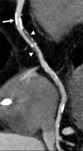
"We found that those folks who have obstructive sleep apnea have the more dangerous form of plaque," said Dr. U. Joseph Schoepf of the Medical University of South Carolina, Charleston, at a news conference.
"This is the first time that a noninvasive test, namely coronary CT angiography, has been applied to this, to actually demonstrate it. The clinical relevance is that we have a noninvasive tool at hand that allows us potentially to care better for our patients who have that disease."
Patients with obstructive sleep apnea (OSA) repeatedly stop breathing during sleep due to airway collapse. It is a disease of obesity, and 30% of Americans are obese, said Dr. Schoepf. Diminishing the oxygen supply exposes the cardiovascular system to oxidative stress, and may render vessel linings more vulnerable to the formation of atherosclerosis. OSA has been linked to coronary artery disease, he noted.
Computed tomography is the only noninvasive test that can directly evaluate cardiac vessel patency and analyze the composition of atherosclerotic lesions on vessel walls, said Dr. Schoepf. Therefore, it was used to study the association between OSA and coronary artery disease.
Investigators performed a retrospective search for patients who had undergone CT of the heart and polysomnogram assessment for suspected OSA. One observer measured coronary artery calcium, known informally as hard plaque. Two observers in consensus analyzed heart CT data for the presence and degree of coronary artery narrowing or blockages. The presence and extent of noncalcified or soft plaque, also known as vulnerable plaque, were also evaluated. Noncalcified plaque frequently causes acute cardiac events such as heart attack and unstable angina.
The retrospective search revealed 97 patients who had undergone both heart CT for atypical chest pain or prior equivocal test, as well as a polysomnogram. Two patients were excluded because their polysomnogram was performed more than 36 months before or after the date of their cardiac CT.
Of the 95 remaining patients, 49 (23 women, mean age 61 plus or minus 11 years, average body mass index 33 plus or minus 8 kg/m 2 ) had documented obstructive sleep apnea by polysomnogram. The other 46 patients (24 women, mean age 60 plus or minus 12 years, average BMI 30 plus or minus 6 kg/m 2 ) were found not to have obstructive sleep apnea by polysomnogram. The investigators found no significant difference in age, gender, BMI, or cardiovascular risk factors between the two groups.
Agatson calcium scores (hard plaque) were not significantly different between the two groups (mean score 272 plus or minus 422 for the group with obstructive sleep apnea versus 241 plus or minus 415 without, P = .5). The presence of any type of narrowing in the heart vessels was found to correlate with the presence of obstructive sleep apnea (P less than .0001). The total number of vessels with any narrowing was also found to correlate with the presence of obstructive sleep apnea (P = .0008). In addition, the degree of narrowing in the highest grade of lesion correlated with obstructive sleep apnea (P = .0013).
In conclusion, Dr. Schoepf said that any instance where noncalcified plaque (‘soft’ or ‘vulnerable’ plaque) was measured showed a significant correlation with obstructive sleep apnea.
If the study’s findings are sustained by larger prospective trials, cardiac CT could emerge as a useful noninvasive tool for investigating the relationship between OSA and coronary artery disease.
The study was not funded, and Dr. Schoepf made no disclosures relevant to the study. His general disclosures are that he has served on the speakers bureau of, and/or received research grants from, the Bracco Group, General Electric, and Bayer AG. He also has served on the Siemens AG Medical Advisory Board.
CHICAGO – Patients with obstructive sleep apnea have more active atherosclerotic disease with a greater degree of vessel involvement and more vulnerable plaque than do patients without the disease, according to a retrospective matched cohort study presented at the annual meeting of the Radiological Society of North America.
"We found that those folks who have obstructive sleep apnea have the more dangerous form of plaque," said Dr. U. Joseph Schoepf of the Medical University of South Carolina, Charleston, at a news conference.
"This is the first time that a noninvasive test, namely coronary CT angiography, has been applied to this, to actually demonstrate it. The clinical relevance is that we have a noninvasive tool at hand that allows us potentially to care better for our patients who have that disease."
Patients with obstructive sleep apnea (OSA) repeatedly stop breathing during sleep due to airway collapse. It is a disease of obesity, and 30% of Americans are obese, said Dr. Schoepf. Diminishing the oxygen supply exposes the cardiovascular system to oxidative stress, and may render vessel linings more vulnerable to the formation of atherosclerosis. OSA has been linked to coronary artery disease, he noted.
Computed tomography is the only noninvasive test that can directly evaluate cardiac vessel patency and analyze the composition of atherosclerotic lesions on vessel walls, said Dr. Schoepf. Therefore, it was used to study the association between OSA and coronary artery disease.
Investigators performed a retrospective search for patients who had undergone CT of the heart and polysomnogram assessment for suspected OSA. One observer measured coronary artery calcium, known informally as hard plaque. Two observers in consensus analyzed heart CT data for the presence and degree of coronary artery narrowing or blockages. The presence and extent of noncalcified or soft plaque, also known as vulnerable plaque, were also evaluated. Noncalcified plaque frequently causes acute cardiac events such as heart attack and unstable angina.
The retrospective search revealed 97 patients who had undergone both heart CT for atypical chest pain or prior equivocal test, as well as a polysomnogram. Two patients were excluded because their polysomnogram was performed more than 36 months before or after the date of their cardiac CT.
Of the 95 remaining patients, 49 (23 women, mean age 61 plus or minus 11 years, average body mass index 33 plus or minus 8 kg/m 2 ) had documented obstructive sleep apnea by polysomnogram. The other 46 patients (24 women, mean age 60 plus or minus 12 years, average BMI 30 plus or minus 6 kg/m 2 ) were found not to have obstructive sleep apnea by polysomnogram. The investigators found no significant difference in age, gender, BMI, or cardiovascular risk factors between the two groups.
Agatson calcium scores (hard plaque) were not significantly different between the two groups (mean score 272 plus or minus 422 for the group with obstructive sleep apnea versus 241 plus or minus 415 without, P = .5). The presence of any type of narrowing in the heart vessels was found to correlate with the presence of obstructive sleep apnea (P less than .0001). The total number of vessels with any narrowing was also found to correlate with the presence of obstructive sleep apnea (P = .0008). In addition, the degree of narrowing in the highest grade of lesion correlated with obstructive sleep apnea (P = .0013).
In conclusion, Dr. Schoepf said that any instance where noncalcified plaque (‘soft’ or ‘vulnerable’ plaque) was measured showed a significant correlation with obstructive sleep apnea.
If the study’s findings are sustained by larger prospective trials, cardiac CT could emerge as a useful noninvasive tool for investigating the relationship between OSA and coronary artery disease.
The study was not funded, and Dr. Schoepf made no disclosures relevant to the study. His general disclosures are that he has served on the speakers bureau of, and/or received research grants from, the Bracco Group, General Electric, and Bayer AG. He also has served on the Siemens AG Medical Advisory Board.
CHICAGO – Patients with obstructive sleep apnea have more active atherosclerotic disease with a greater degree of vessel involvement and more vulnerable plaque than do patients without the disease, according to a retrospective matched cohort study presented at the annual meeting of the Radiological Society of North America.
"We found that those folks who have obstructive sleep apnea have the more dangerous form of plaque," said Dr. U. Joseph Schoepf of the Medical University of South Carolina, Charleston, at a news conference.
"This is the first time that a noninvasive test, namely coronary CT angiography, has been applied to this, to actually demonstrate it. The clinical relevance is that we have a noninvasive tool at hand that allows us potentially to care better for our patients who have that disease."
Patients with obstructive sleep apnea (OSA) repeatedly stop breathing during sleep due to airway collapse. It is a disease of obesity, and 30% of Americans are obese, said Dr. Schoepf. Diminishing the oxygen supply exposes the cardiovascular system to oxidative stress, and may render vessel linings more vulnerable to the formation of atherosclerosis. OSA has been linked to coronary artery disease, he noted.
Computed tomography is the only noninvasive test that can directly evaluate cardiac vessel patency and analyze the composition of atherosclerotic lesions on vessel walls, said Dr. Schoepf. Therefore, it was used to study the association between OSA and coronary artery disease.
Investigators performed a retrospective search for patients who had undergone CT of the heart and polysomnogram assessment for suspected OSA. One observer measured coronary artery calcium, known informally as hard plaque. Two observers in consensus analyzed heart CT data for the presence and degree of coronary artery narrowing or blockages. The presence and extent of noncalcified or soft plaque, also known as vulnerable plaque, were also evaluated. Noncalcified plaque frequently causes acute cardiac events such as heart attack and unstable angina.
The retrospective search revealed 97 patients who had undergone both heart CT for atypical chest pain or prior equivocal test, as well as a polysomnogram. Two patients were excluded because their polysomnogram was performed more than 36 months before or after the date of their cardiac CT.
Of the 95 remaining patients, 49 (23 women, mean age 61 plus or minus 11 years, average body mass index 33 plus or minus 8 kg/m 2 ) had documented obstructive sleep apnea by polysomnogram. The other 46 patients (24 women, mean age 60 plus or minus 12 years, average BMI 30 plus or minus 6 kg/m 2 ) were found not to have obstructive sleep apnea by polysomnogram. The investigators found no significant difference in age, gender, BMI, or cardiovascular risk factors between the two groups.
Agatson calcium scores (hard plaque) were not significantly different between the two groups (mean score 272 plus or minus 422 for the group with obstructive sleep apnea versus 241 plus or minus 415 without, P = .5). The presence of any type of narrowing in the heart vessels was found to correlate with the presence of obstructive sleep apnea (P less than .0001). The total number of vessels with any narrowing was also found to correlate with the presence of obstructive sleep apnea (P = .0008). In addition, the degree of narrowing in the highest grade of lesion correlated with obstructive sleep apnea (P = .0013).
In conclusion, Dr. Schoepf said that any instance where noncalcified plaque (‘soft’ or ‘vulnerable’ plaque) was measured showed a significant correlation with obstructive sleep apnea.
If the study’s findings are sustained by larger prospective trials, cardiac CT could emerge as a useful noninvasive tool for investigating the relationship between OSA and coronary artery disease.
The study was not funded, and Dr. Schoepf made no disclosures relevant to the study. His general disclosures are that he has served on the speakers bureau of, and/or received research grants from, the Bracco Group, General Electric, and Bayer AG. He also has served on the Siemens AG Medical Advisory Board.
FROM THE ANNUAL MEETING OF THE RADIOLOGICAL SOCIETY OF NORTH AMERICA
Major Finding: Coronary CT angiography reveals a higher prevalence of stenotic coronary artery disease and more extensive vessel involvement in patients with obstructive sleep apnea than in those without it.
Data Source: A retrospective study of 95 obese patients, 49 with obstructive sleep apnea and 46 without.
Disclosures: The study was not funded, and Dr. Schoepf made no disclosures relevant to the study. His general disclosures are that he has served on the speakers bureau of, and/or received research grants from, the Bracco Group, General Electric, and Bayer AG. He also has served on the Siemens AG Medical Advisory Board.
Physicians Can Tip the Balance Toward Smoking Cessation
VANCOUVER, B.C. – Although physicians might be reluctant to bring up smoking cessation with their patients for many reasons, it is one of the most important preventive activities they can undertake, according to Dr. Arunabh Talwar.

Smokers are ambivalent about smoking, he said at the annual meeting of the American College of Chest Physicians. On any given day, their smoking status hangs in balance between one set of factors favoring quitting and another set favoring continuing (BMJ 2007;335:37-41).
"All we need to do is just [tip the balance]," said Dr. Talwar, who is a pulmonologist at North Shore University Hospital in Manhasset, N.Y.
Indeed, if self-reports are reliable, about 70% of smokers want to stop and 30% try each year. But only 2%-3% succeed.
Referring to the multistage model of behavioral change, he noted that making smokers aware of the link between smoking and end-organ damage is critical in starting the process. "That is the most important thing that physicians do – they move patients from a precontemplation to a contemplation stage and set the stage for the smoking cessation process to occur."
There is compelling evidence of the benefits of smoking cessation, he said. It has been identified as the single most effective step for lengthening and improving patients’ lives (BMJ 2004;328:947-9).
"Make no mistake, smoking cessation activity is very cost effective," he added. "I think it is the most cost-effective primary prevention action that a physician can take."
Brief advice to quit costs $338 per year of life saved – or less than 5% of the cost per year of life saved from giving pravastatin for primary prevention of cardiovascular disease, aspirin for secondary prevention of coronary heart disease, or simvastatin for secondary prevention of myocardial infarction (BMJ 2004;328:397-9).
Still, physicians cite numerous barriers to promoting smoking cessation with their patients, according to Dr. Talwar (J. Smok. Cessat. 2008;3:92-100). A common one is being too busy.
"But studies show us, a minimal intervention – as [little] as 3 minutes of a physician’s time – can move patients from ‘precontemplation’ to contemplation, can help improve quit status," he said. Furthermore, "as you increase the intervention, the success rate will improve."
For example, just 0.3% of smokers succeed in quitting long term on their own, but the value rises to 1.6% when physicians simply ask their smoking status, 3.3% when they ask and provide advice on quitting, and 5.1% when they ask, advise, and give a pamphlet (BMJ 1979;2:231-5).
Busy physicians can streamline efforts by using a team approach. "Some of it can be shared by other health care providers, whether they are nurses, nurse practitioners, physician assistants," Dr. Talwar explained. "We use our respiratory therapists and [pulmonary function test] lab technicians as well; that way, the load gets divided. But also, repeated messages to the patient will help move them along."
Physicians should also consider using telephone "quitlines" (now freely available in all states) and patient support groups in the behavioral modification part of cessation, he advised.
Another barrier physicians cite, lack of expertise, has a stronger negative influence on their smoking cessation activities than lack of interest, time, or materials (Eur. J. Public Health 2005;15:140-5).
Indeed, in a survey of New York City–area health care providers, Dr. Talwar and his colleagues found that only 20% believed their training had adequately prepared them to treat tobacco dependence. And less than 10% were familiar with treatment guidelines. "We are a little bit behind in this, but medical schools have made a change, and most medical schools now make an effort to make sure that standard curricula [on smoking cessation] are there," he said. In addition, comprehensive information is readily available in the ACCP’s Tobacco Dependence Treatment ToolKit.
Reassuringly, physicians who receive training in this area are 1.5 to 2.5 times more likely to perform smoking cessation tasks (Cochrane Database Syst. Rev. 2000;CD000214).
Physicians also report a lack of financial incentives to be a barrier. Dr. Talwar noted that two CPT codes – 99406 and 99407 – specifically pertain to cessation activities during visits. Physicians can usually bill for this counseling, in addition to routine office visits, four times annually.
Half of physicians still believe that reimbursement is insufficient. "But the situation is much better than 7 or 8 years ago, when it was much more difficult to get reimbursement for these activities," he commented.
Physicians also mention patients’ low likelihood of quitting as a barrier to broaching smoking cessation, according to Dr. Talwar. But the irony is that quit rates are influenced in large part by physicians’ efforts and the intensity of those efforts.
Discussing the so-called 5 A’s of smoking cessation – ask, advise, assess, assist, and arrange – he noted that physicians do fairly well on the first two, but not so well on the others.
For example, a study of 246 community-based primary care physicians found that 67% asked their patients about smoking status and 74% gave advice, but just 35% assisted with smoking cessation efforts and merely 8% arranged for follow-up (Prev. Med. 1998;27:720-9).
It is important to understand that relapses are part of the cessation process, Dr. Talwar stressed; in fact, smokers who succeed in quitting make five to seven attempts, on average, before succeeding. Hence, "you have just to have to be patient with them."
It might also be possible to improve the odds of successful quitting by approaching patients at teachable moments, he further noted. For instance, "admission [to the hospital] is an opportunity to interact, to make the change. Maybe that’s the time when you need to approach them."
His own 800-bed hospital generates a list each day of inpatients who smoke. A smoking cessation therapist then visits these patients and invites them to the smoking cessation clinic.
A final barrier is that some physicians themselves are smokers. "It’s been shown that physicians who smoke have very little faith in their own ability to promote smoking cessation," Dr. Talwar commented (Prev. Med. 2005;40:595-601).
On the other hand, this group has greater insight into the difficulties of quitting and might be able to draw on their own experiences to assist patients in this endeavor, he added.
Dr. Talwar did not report any conflicts of interest.
VANCOUVER, B.C. – Although physicians might be reluctant to bring up smoking cessation with their patients for many reasons, it is one of the most important preventive activities they can undertake, according to Dr. Arunabh Talwar.
Smokers are ambivalent about smoking, he said at the annual meeting of the American College of Chest Physicians. On any given day, their smoking status hangs in balance between one set of factors favoring quitting and another set favoring continuing (BMJ 2007;335:37-41).
"All we need to do is just [tip the balance]," said Dr. Talwar, who is a pulmonologist at North Shore University Hospital in Manhasset, N.Y.
Indeed, if self-reports are reliable, about 70% of smokers want to stop and 30% try each year. But only 2%-3% succeed.
Referring to the multistage model of behavioral change, he noted that making smokers aware of the link between smoking and end-organ damage is critical in starting the process. "That is the most important thing that physicians do – they move patients from a precontemplation to a contemplation stage and set the stage for the smoking cessation process to occur."
There is compelling evidence of the benefits of smoking cessation, he said. It has been identified as the single most effective step for lengthening and improving patients’ lives (BMJ 2004;328:947-9).
"Make no mistake, smoking cessation activity is very cost effective," he added. "I think it is the most cost-effective primary prevention action that a physician can take."
Brief advice to quit costs $338 per year of life saved – or less than 5% of the cost per year of life saved from giving pravastatin for primary prevention of cardiovascular disease, aspirin for secondary prevention of coronary heart disease, or simvastatin for secondary prevention of myocardial infarction (BMJ 2004;328:397-9).
Still, physicians cite numerous barriers to promoting smoking cessation with their patients, according to Dr. Talwar (J. Smok. Cessat. 2008;3:92-100). A common one is being too busy.
"But studies show us, a minimal intervention – as [little] as 3 minutes of a physician’s time – can move patients from ‘precontemplation’ to contemplation, can help improve quit status," he said. Furthermore, "as you increase the intervention, the success rate will improve."
For example, just 0.3% of smokers succeed in quitting long term on their own, but the value rises to 1.6% when physicians simply ask their smoking status, 3.3% when they ask and provide advice on quitting, and 5.1% when they ask, advise, and give a pamphlet (BMJ 1979;2:231-5).
Busy physicians can streamline efforts by using a team approach. "Some of it can be shared by other health care providers, whether they are nurses, nurse practitioners, physician assistants," Dr. Talwar explained. "We use our respiratory therapists and [pulmonary function test] lab technicians as well; that way, the load gets divided. But also, repeated messages to the patient will help move them along."
Physicians should also consider using telephone "quitlines" (now freely available in all states) and patient support groups in the behavioral modification part of cessation, he advised.
Another barrier physicians cite, lack of expertise, has a stronger negative influence on their smoking cessation activities than lack of interest, time, or materials (Eur. J. Public Health 2005;15:140-5).
Indeed, in a survey of New York City–area health care providers, Dr. Talwar and his colleagues found that only 20% believed their training had adequately prepared them to treat tobacco dependence. And less than 10% were familiar with treatment guidelines. "We are a little bit behind in this, but medical schools have made a change, and most medical schools now make an effort to make sure that standard curricula [on smoking cessation] are there," he said. In addition, comprehensive information is readily available in the ACCP’s Tobacco Dependence Treatment ToolKit.
Reassuringly, physicians who receive training in this area are 1.5 to 2.5 times more likely to perform smoking cessation tasks (Cochrane Database Syst. Rev. 2000;CD000214).
Physicians also report a lack of financial incentives to be a barrier. Dr. Talwar noted that two CPT codes – 99406 and 99407 – specifically pertain to cessation activities during visits. Physicians can usually bill for this counseling, in addition to routine office visits, four times annually.
Half of physicians still believe that reimbursement is insufficient. "But the situation is much better than 7 or 8 years ago, when it was much more difficult to get reimbursement for these activities," he commented.
Physicians also mention patients’ low likelihood of quitting as a barrier to broaching smoking cessation, according to Dr. Talwar. But the irony is that quit rates are influenced in large part by physicians’ efforts and the intensity of those efforts.
Discussing the so-called 5 A’s of smoking cessation – ask, advise, assess, assist, and arrange – he noted that physicians do fairly well on the first two, but not so well on the others.
For example, a study of 246 community-based primary care physicians found that 67% asked their patients about smoking status and 74% gave advice, but just 35% assisted with smoking cessation efforts and merely 8% arranged for follow-up (Prev. Med. 1998;27:720-9).
It is important to understand that relapses are part of the cessation process, Dr. Talwar stressed; in fact, smokers who succeed in quitting make five to seven attempts, on average, before succeeding. Hence, "you have just to have to be patient with them."
It might also be possible to improve the odds of successful quitting by approaching patients at teachable moments, he further noted. For instance, "admission [to the hospital] is an opportunity to interact, to make the change. Maybe that’s the time when you need to approach them."
His own 800-bed hospital generates a list each day of inpatients who smoke. A smoking cessation therapist then visits these patients and invites them to the smoking cessation clinic.
A final barrier is that some physicians themselves are smokers. "It’s been shown that physicians who smoke have very little faith in their own ability to promote smoking cessation," Dr. Talwar commented (Prev. Med. 2005;40:595-601).
On the other hand, this group has greater insight into the difficulties of quitting and might be able to draw on their own experiences to assist patients in this endeavor, he added.
Dr. Talwar did not report any conflicts of interest.
VANCOUVER, B.C. – Although physicians might be reluctant to bring up smoking cessation with their patients for many reasons, it is one of the most important preventive activities they can undertake, according to Dr. Arunabh Talwar.
Smokers are ambivalent about smoking, he said at the annual meeting of the American College of Chest Physicians. On any given day, their smoking status hangs in balance between one set of factors favoring quitting and another set favoring continuing (BMJ 2007;335:37-41).
"All we need to do is just [tip the balance]," said Dr. Talwar, who is a pulmonologist at North Shore University Hospital in Manhasset, N.Y.
Indeed, if self-reports are reliable, about 70% of smokers want to stop and 30% try each year. But only 2%-3% succeed.
Referring to the multistage model of behavioral change, he noted that making smokers aware of the link between smoking and end-organ damage is critical in starting the process. "That is the most important thing that physicians do – they move patients from a precontemplation to a contemplation stage and set the stage for the smoking cessation process to occur."
There is compelling evidence of the benefits of smoking cessation, he said. It has been identified as the single most effective step for lengthening and improving patients’ lives (BMJ 2004;328:947-9).
"Make no mistake, smoking cessation activity is very cost effective," he added. "I think it is the most cost-effective primary prevention action that a physician can take."
Brief advice to quit costs $338 per year of life saved – or less than 5% of the cost per year of life saved from giving pravastatin for primary prevention of cardiovascular disease, aspirin for secondary prevention of coronary heart disease, or simvastatin for secondary prevention of myocardial infarction (BMJ 2004;328:397-9).
Still, physicians cite numerous barriers to promoting smoking cessation with their patients, according to Dr. Talwar (J. Smok. Cessat. 2008;3:92-100). A common one is being too busy.
"But studies show us, a minimal intervention – as [little] as 3 minutes of a physician’s time – can move patients from ‘precontemplation’ to contemplation, can help improve quit status," he said. Furthermore, "as you increase the intervention, the success rate will improve."
For example, just 0.3% of smokers succeed in quitting long term on their own, but the value rises to 1.6% when physicians simply ask their smoking status, 3.3% when they ask and provide advice on quitting, and 5.1% when they ask, advise, and give a pamphlet (BMJ 1979;2:231-5).
Busy physicians can streamline efforts by using a team approach. "Some of it can be shared by other health care providers, whether they are nurses, nurse practitioners, physician assistants," Dr. Talwar explained. "We use our respiratory therapists and [pulmonary function test] lab technicians as well; that way, the load gets divided. But also, repeated messages to the patient will help move them along."
Physicians should also consider using telephone "quitlines" (now freely available in all states) and patient support groups in the behavioral modification part of cessation, he advised.
Another barrier physicians cite, lack of expertise, has a stronger negative influence on their smoking cessation activities than lack of interest, time, or materials (Eur. J. Public Health 2005;15:140-5).
Indeed, in a survey of New York City–area health care providers, Dr. Talwar and his colleagues found that only 20% believed their training had adequately prepared them to treat tobacco dependence. And less than 10% were familiar with treatment guidelines. "We are a little bit behind in this, but medical schools have made a change, and most medical schools now make an effort to make sure that standard curricula [on smoking cessation] are there," he said. In addition, comprehensive information is readily available in the ACCP’s Tobacco Dependence Treatment ToolKit.
Reassuringly, physicians who receive training in this area are 1.5 to 2.5 times more likely to perform smoking cessation tasks (Cochrane Database Syst. Rev. 2000;CD000214).
Physicians also report a lack of financial incentives to be a barrier. Dr. Talwar noted that two CPT codes – 99406 and 99407 – specifically pertain to cessation activities during visits. Physicians can usually bill for this counseling, in addition to routine office visits, four times annually.
Half of physicians still believe that reimbursement is insufficient. "But the situation is much better than 7 or 8 years ago, when it was much more difficult to get reimbursement for these activities," he commented.
Physicians also mention patients’ low likelihood of quitting as a barrier to broaching smoking cessation, according to Dr. Talwar. But the irony is that quit rates are influenced in large part by physicians’ efforts and the intensity of those efforts.
Discussing the so-called 5 A’s of smoking cessation – ask, advise, assess, assist, and arrange – he noted that physicians do fairly well on the first two, but not so well on the others.
For example, a study of 246 community-based primary care physicians found that 67% asked their patients about smoking status and 74% gave advice, but just 35% assisted with smoking cessation efforts and merely 8% arranged for follow-up (Prev. Med. 1998;27:720-9).
It is important to understand that relapses are part of the cessation process, Dr. Talwar stressed; in fact, smokers who succeed in quitting make five to seven attempts, on average, before succeeding. Hence, "you have just to have to be patient with them."
It might also be possible to improve the odds of successful quitting by approaching patients at teachable moments, he further noted. For instance, "admission [to the hospital] is an opportunity to interact, to make the change. Maybe that’s the time when you need to approach them."
His own 800-bed hospital generates a list each day of inpatients who smoke. A smoking cessation therapist then visits these patients and invites them to the smoking cessation clinic.
A final barrier is that some physicians themselves are smokers. "It’s been shown that physicians who smoke have very little faith in their own ability to promote smoking cessation," Dr. Talwar commented (Prev. Med. 2005;40:595-601).
On the other hand, this group has greater insight into the difficulties of quitting and might be able to draw on their own experiences to assist patients in this endeavor, he added.
Dr. Talwar did not report any conflicts of interest.
FROM THE ANNUAL MEETING OF THE AMERICAN COLLEGE OF CHEST PHYSICIANS
Postoperative Outcomes Worse in COPD Patients
VANCOUVER, B.C. – Patients with chronic obstructive pulmonary disease are more likely to die after surgery than are those without COPD, even after controlling for comorbidities and type of surgery, according to a cross-sectional study of nearly half a million patients undergoing surgery in the United States.

The researchers found that patients with COPD were 29% more likely to die and 35% were more likely to experience complications, compared with similar patients without the disease, said presenting investigator Dr. Prateek K. Gupta, a surgeon at Creighton University in Omaha, Neb.
In addition, hospital length of stay was four times longer for the COPD group.
"Knowledge of the increased risk associated with COPD may improve patient selection and the informed consent process," Dr. Gupta said at the annual meeting of the American College of Chest Physicians.
"Perioperative optimization of these patients may help in improving outcomes and health care costs, and there is a need to study such strategies in multicenter, randomized, prospective trials," he added. These strategies might include, for example, giving patients respiratory exercises and encouraging them to quit smoking, he said.
Dr. Gupta and his colleagues used the NSQIP (National Surgical Quality Improvement Program) database, which collects data from more than 250 hospitals nationwide, to identify patients who underwent surgery in 2007 and 2008.
They then compared 30-day postoperative outcomes between patients who did and did not have COPD, defined in the database as GOLD (Global Initiative for Chronic Obstructive Lung Disease) stage II, III, or IV or a prior hospitalization for COPD.
Analyses included 468,795 patients who underwent surgery. The types of surgery were typical of those seen in the general population, according to Dr. Gupta, with a predominance of cholecystectomy, appendectomy, hernia repair, and vascular and breast surgeries.
A total of 5% of the patients had COPD. Relative to their unaffected peers, patients with COPD had a higher mean body mass index (29 vs. 28 kg/m2) and an older median age (69 vs. 55 years); were more likely to be male (52% vs. 42%), white (82% vs. 72%), smokers (41% vs. 20%), and alcoholics (5% vs. 2%); and were more likely to be taking corticosteroids (10% vs. 3%).
The group with COPD also had higher prevalences of more than a dozen comorbidities, especially hypertension (74% vs. 44%), dependent functional status (20% vs. 6%), diabetes (25% vs. 14%), and an American Society of Anesthesiologists score of 3 or 4 (55% vs. 22%).
The median length of hospital stay was much longer for the patients with COPD than for their unaffected peers, at 4 days vs. 1 day (P less than .0001), Dr. Gupta reported. And the 30-day rate of postoperative mortality was higher, at 6.7% vs. 1.4% (P less than .0001).
After the investigators took into account more than 50 comorbidities and the type of surgery (including whether it was laparoscopic or open), patients with COPD still had higher risks of postoperative morbidity (odds ratio, 1.35; P less than .0001) and mortality (OR, 1.29; P less than .0001).
The odds of nine postoperative complications individually were also elevated for the COPD group, with the greatest increases seen for pneumonia (OR, 1.71), reintubation (OR, 1.54), and failure to wean from the ventilator within 48 hours (OR, 1.45) (all P less than .0001).
The study was limited by a lack of detailed information on therapies that patients were receiving, Dr. Gupta acknowledged. "We just know that they had this surgery [and] that they had COPD prior. We don’t know what medication or what preoperative optimization they underwent," he said.
In addition, the study did not specifically assess any influence of the urgency of the surgery (emergency vs. elective) and did not assess the potential impact of mild COPD.
GOLD stage II-IV COPD is "common among patients undergoing surgery and is associated with increased morbidity, mortality, and length of stay," Dr. Gupta concluded. Physicians may be able to use this information to help guide selection of appropriate surgical candidates, counsel patients about risks, and target interventions to improve outcomes, he said.
Dr. Gupta reported having no conflicts of interest related to the research.
VANCOUVER, B.C. – Patients with chronic obstructive pulmonary disease are more likely to die after surgery than are those without COPD, even after controlling for comorbidities and type of surgery, according to a cross-sectional study of nearly half a million patients undergoing surgery in the United States.
The researchers found that patients with COPD were 29% more likely to die and 35% were more likely to experience complications, compared with similar patients without the disease, said presenting investigator Dr. Prateek K. Gupta, a surgeon at Creighton University in Omaha, Neb.
In addition, hospital length of stay was four times longer for the COPD group.
"Knowledge of the increased risk associated with COPD may improve patient selection and the informed consent process," Dr. Gupta said at the annual meeting of the American College of Chest Physicians.
"Perioperative optimization of these patients may help in improving outcomes and health care costs, and there is a need to study such strategies in multicenter, randomized, prospective trials," he added. These strategies might include, for example, giving patients respiratory exercises and encouraging them to quit smoking, he said.
Dr. Gupta and his colleagues used the NSQIP (National Surgical Quality Improvement Program) database, which collects data from more than 250 hospitals nationwide, to identify patients who underwent surgery in 2007 and 2008.
They then compared 30-day postoperative outcomes between patients who did and did not have COPD, defined in the database as GOLD (Global Initiative for Chronic Obstructive Lung Disease) stage II, III, or IV or a prior hospitalization for COPD.
Analyses included 468,795 patients who underwent surgery. The types of surgery were typical of those seen in the general population, according to Dr. Gupta, with a predominance of cholecystectomy, appendectomy, hernia repair, and vascular and breast surgeries.
A total of 5% of the patients had COPD. Relative to their unaffected peers, patients with COPD had a higher mean body mass index (29 vs. 28 kg/m2) and an older median age (69 vs. 55 years); were more likely to be male (52% vs. 42%), white (82% vs. 72%), smokers (41% vs. 20%), and alcoholics (5% vs. 2%); and were more likely to be taking corticosteroids (10% vs. 3%).
The group with COPD also had higher prevalences of more than a dozen comorbidities, especially hypertension (74% vs. 44%), dependent functional status (20% vs. 6%), diabetes (25% vs. 14%), and an American Society of Anesthesiologists score of 3 or 4 (55% vs. 22%).
The median length of hospital stay was much longer for the patients with COPD than for their unaffected peers, at 4 days vs. 1 day (P less than .0001), Dr. Gupta reported. And the 30-day rate of postoperative mortality was higher, at 6.7% vs. 1.4% (P less than .0001).
After the investigators took into account more than 50 comorbidities and the type of surgery (including whether it was laparoscopic or open), patients with COPD still had higher risks of postoperative morbidity (odds ratio, 1.35; P less than .0001) and mortality (OR, 1.29; P less than .0001).
The odds of nine postoperative complications individually were also elevated for the COPD group, with the greatest increases seen for pneumonia (OR, 1.71), reintubation (OR, 1.54), and failure to wean from the ventilator within 48 hours (OR, 1.45) (all P less than .0001).
The study was limited by a lack of detailed information on therapies that patients were receiving, Dr. Gupta acknowledged. "We just know that they had this surgery [and] that they had COPD prior. We don’t know what medication or what preoperative optimization they underwent," he said.
In addition, the study did not specifically assess any influence of the urgency of the surgery (emergency vs. elective) and did not assess the potential impact of mild COPD.
GOLD stage II-IV COPD is "common among patients undergoing surgery and is associated with increased morbidity, mortality, and length of stay," Dr. Gupta concluded. Physicians may be able to use this information to help guide selection of appropriate surgical candidates, counsel patients about risks, and target interventions to improve outcomes, he said.
Dr. Gupta reported having no conflicts of interest related to the research.
VANCOUVER, B.C. – Patients with chronic obstructive pulmonary disease are more likely to die after surgery than are those without COPD, even after controlling for comorbidities and type of surgery, according to a cross-sectional study of nearly half a million patients undergoing surgery in the United States.
The researchers found that patients with COPD were 29% more likely to die and 35% were more likely to experience complications, compared with similar patients without the disease, said presenting investigator Dr. Prateek K. Gupta, a surgeon at Creighton University in Omaha, Neb.
In addition, hospital length of stay was four times longer for the COPD group.
"Knowledge of the increased risk associated with COPD may improve patient selection and the informed consent process," Dr. Gupta said at the annual meeting of the American College of Chest Physicians.
"Perioperative optimization of these patients may help in improving outcomes and health care costs, and there is a need to study such strategies in multicenter, randomized, prospective trials," he added. These strategies might include, for example, giving patients respiratory exercises and encouraging them to quit smoking, he said.
Dr. Gupta and his colleagues used the NSQIP (National Surgical Quality Improvement Program) database, which collects data from more than 250 hospitals nationwide, to identify patients who underwent surgery in 2007 and 2008.
They then compared 30-day postoperative outcomes between patients who did and did not have COPD, defined in the database as GOLD (Global Initiative for Chronic Obstructive Lung Disease) stage II, III, or IV or a prior hospitalization for COPD.
Analyses included 468,795 patients who underwent surgery. The types of surgery were typical of those seen in the general population, according to Dr. Gupta, with a predominance of cholecystectomy, appendectomy, hernia repair, and vascular and breast surgeries.
A total of 5% of the patients had COPD. Relative to their unaffected peers, patients with COPD had a higher mean body mass index (29 vs. 28 kg/m2) and an older median age (69 vs. 55 years); were more likely to be male (52% vs. 42%), white (82% vs. 72%), smokers (41% vs. 20%), and alcoholics (5% vs. 2%); and were more likely to be taking corticosteroids (10% vs. 3%).
The group with COPD also had higher prevalences of more than a dozen comorbidities, especially hypertension (74% vs. 44%), dependent functional status (20% vs. 6%), diabetes (25% vs. 14%), and an American Society of Anesthesiologists score of 3 or 4 (55% vs. 22%).
The median length of hospital stay was much longer for the patients with COPD than for their unaffected peers, at 4 days vs. 1 day (P less than .0001), Dr. Gupta reported. And the 30-day rate of postoperative mortality was higher, at 6.7% vs. 1.4% (P less than .0001).
After the investigators took into account more than 50 comorbidities and the type of surgery (including whether it was laparoscopic or open), patients with COPD still had higher risks of postoperative morbidity (odds ratio, 1.35; P less than .0001) and mortality (OR, 1.29; P less than .0001).
The odds of nine postoperative complications individually were also elevated for the COPD group, with the greatest increases seen for pneumonia (OR, 1.71), reintubation (OR, 1.54), and failure to wean from the ventilator within 48 hours (OR, 1.45) (all P less than .0001).
The study was limited by a lack of detailed information on therapies that patients were receiving, Dr. Gupta acknowledged. "We just know that they had this surgery [and] that they had COPD prior. We don’t know what medication or what preoperative optimization they underwent," he said.
In addition, the study did not specifically assess any influence of the urgency of the surgery (emergency vs. elective) and did not assess the potential impact of mild COPD.
GOLD stage II-IV COPD is "common among patients undergoing surgery and is associated with increased morbidity, mortality, and length of stay," Dr. Gupta concluded. Physicians may be able to use this information to help guide selection of appropriate surgical candidates, counsel patients about risks, and target interventions to improve outcomes, he said.
Dr. Gupta reported having no conflicts of interest related to the research.
Major Finding: Patients with COPD were 29% more likely to die and 35% more likely to experience complications after surgery.
Data Source: Cross-sectional NSQIP database study of 468,795 patients.
Disclosures: Dr. Gupta reported having no relevant conflicts of interest.
Fatigue, Sleep Issues Dominate Outpatient Cancer Care
CHICAGO - Nonpain symptoms, particularly fatigue, sleep disturbance, and drowsiness, were the most common moderate to severe symptoms among cancer patients undergoing treatment in a community ambulatory setting, according to new data from the Eastern Cooperative Oncology Group study of Symptom Outcomes and Practice Patterns.
The researchers had hypothesized that pain, fatigue, and emotional distress would surface as the most frequent problems in the sample of 3,123 patients (median age 61 years; 30% male) with solid breast (50%), colorectal (23%), lung (17%), and prostate tumors (10%).
The study also found that outpatients who were more ill and symptomatic at baseline were more likely to report benefiting from supportive oncology care at a 1-month follow-up visit, said Dr. Michael J. Fisch, director of the general medical oncology program at M.D. Anderson Cancer Center in Houston.
"It’s upside down of the way we think about treating cancer," Dr. Fisch said in a focus session on symptom management at the annual Chicago Supportive Oncology Conference. "The better off you are, the more likely you are to benefit from chemotherapy or aggressive therapy. But when it comes to symptom management, the sicker you are, the more likely you are to benefit. First of all, you have more room to improve ... but it probably also has something to do with the fact that the sicker you are, the more dedicated [physicians] are at making that problem better. We are not going to take aim at ... things that are more middle-of-the-road in severity."
The study was conducted at 7 academic and 32 community clinics to highlight the most prevalent and significant physical and psychological symptoms in the course of a visit and a follow-up visit in outpatient medical oncology practice, and to describe the pattern and magnitude of functional change in these symptoms.
"We weren’t just interested in patients who were highly symptomatic or patients who had advanced disease," he said. "We wanted to see how things really are as they flow through [the system]. We knew that solid tumor care dominates what really goes on in terms of the patterns and reflexes of care, so we restricted it to the four most common solid tumors. And we let people come as they were, whether they were there for their first visit, long-term follow-up, or third cycle chemotherapy."
Participants took the M.D. Anderson Symptom Inventory (MDASI), which asks patients to rate the severity of multiple symptoms over the previous 24 hours on a 0-10 scale. The inventory was administered again 1 month later. A 2-point change in numerical rating was used as the criterion for meaningful change in a symptom. Data were analyzed on 3,100 patients.
The most prevalent moderate to severe symptoms were fatigue (34%), disturbed sleep (27%) and drowsiness (22%). Other important symptoms in the moderate to severe range were pain, numbness/tingling, distress, dry mouth, and hair loss (19%), and anorexia/cachexia, depression, and cognitive disturbance (16-17%).
Less prevalent symptoms in the moderate to severe range were dyspnea (15%), constipation (13%), diarrhea (11%), coughing (10%), nausea (8%), rash/pruritis (6%), sore mouth (5%), and vomiting (3%).
"We’re always very interested in nausea and vomiting – it’s a big problem – but just in terms of the prevalence of moderate to severe nausea and vomiting in any given outpatient oncology day, it’s not as prevalent," said Dr. Fisch.
Overall, changes in the top symptoms at 1 month were not impressive. For example, fatigue improved in 20% of patients but worsened in 26% of patients after supportive care, and pain improved in 15% of patients but worsened in 18%.
These results are "a little bit humbling," said Dr. Fisch, "if you are comprehensively assessing people and then following up 4-5 weeks later" and are using patients’ ratings on the [MDASI] to plan care. "Overall, symptom burden remains substantial and difficult to resolve."
The largest improvements were seen in patients whose symptoms were moderate to severe. For example, vomiting improved in 78% of patients and worsened in only 2% of patients who reported moderate to severe problems with this symptom at baseline. Similarly, 52% of patients reported improvements in pain and 10% said their pain got worse.
"I had a sense that we would do better than that [with pain management] – that it might be more like 85%," Dr. Fisch said.
Colorectal cancer patients were the only group whose pain significantly improved. The improvement was significantly better than patients with lung cancer, even after adjustment for other variables. In addition, patients with poor performance status significantly improved their pain, and their improvement was significantly better than those with better performance status.
Dr. Fisch said he and his colleagues plan to make the data from this study available online so that clinicians can use it to plan care without having to wait for the results to be published.
Dr. Fisch reported having no disclosures.
CHICAGO - Nonpain symptoms, particularly fatigue, sleep disturbance, and drowsiness, were the most common moderate to severe symptoms among cancer patients undergoing treatment in a community ambulatory setting, according to new data from the Eastern Cooperative Oncology Group study of Symptom Outcomes and Practice Patterns.
The researchers had hypothesized that pain, fatigue, and emotional distress would surface as the most frequent problems in the sample of 3,123 patients (median age 61 years; 30% male) with solid breast (50%), colorectal (23%), lung (17%), and prostate tumors (10%).
The study also found that outpatients who were more ill and symptomatic at baseline were more likely to report benefiting from supportive oncology care at a 1-month follow-up visit, said Dr. Michael J. Fisch, director of the general medical oncology program at M.D. Anderson Cancer Center in Houston.
"It’s upside down of the way we think about treating cancer," Dr. Fisch said in a focus session on symptom management at the annual Chicago Supportive Oncology Conference. "The better off you are, the more likely you are to benefit from chemotherapy or aggressive therapy. But when it comes to symptom management, the sicker you are, the more likely you are to benefit. First of all, you have more room to improve ... but it probably also has something to do with the fact that the sicker you are, the more dedicated [physicians] are at making that problem better. We are not going to take aim at ... things that are more middle-of-the-road in severity."
The study was conducted at 7 academic and 32 community clinics to highlight the most prevalent and significant physical and psychological symptoms in the course of a visit and a follow-up visit in outpatient medical oncology practice, and to describe the pattern and magnitude of functional change in these symptoms.
"We weren’t just interested in patients who were highly symptomatic or patients who had advanced disease," he said. "We wanted to see how things really are as they flow through [the system]. We knew that solid tumor care dominates what really goes on in terms of the patterns and reflexes of care, so we restricted it to the four most common solid tumors. And we let people come as they were, whether they were there for their first visit, long-term follow-up, or third cycle chemotherapy."
Participants took the M.D. Anderson Symptom Inventory (MDASI), which asks patients to rate the severity of multiple symptoms over the previous 24 hours on a 0-10 scale. The inventory was administered again 1 month later. A 2-point change in numerical rating was used as the criterion for meaningful change in a symptom. Data were analyzed on 3,100 patients.
The most prevalent moderate to severe symptoms were fatigue (34%), disturbed sleep (27%) and drowsiness (22%). Other important symptoms in the moderate to severe range were pain, numbness/tingling, distress, dry mouth, and hair loss (19%), and anorexia/cachexia, depression, and cognitive disturbance (16-17%).
Less prevalent symptoms in the moderate to severe range were dyspnea (15%), constipation (13%), diarrhea (11%), coughing (10%), nausea (8%), rash/pruritis (6%), sore mouth (5%), and vomiting (3%).
"We’re always very interested in nausea and vomiting – it’s a big problem – but just in terms of the prevalence of moderate to severe nausea and vomiting in any given outpatient oncology day, it’s not as prevalent," said Dr. Fisch.
Overall, changes in the top symptoms at 1 month were not impressive. For example, fatigue improved in 20% of patients but worsened in 26% of patients after supportive care, and pain improved in 15% of patients but worsened in 18%.
These results are "a little bit humbling," said Dr. Fisch, "if you are comprehensively assessing people and then following up 4-5 weeks later" and are using patients’ ratings on the [MDASI] to plan care. "Overall, symptom burden remains substantial and difficult to resolve."
The largest improvements were seen in patients whose symptoms were moderate to severe. For example, vomiting improved in 78% of patients and worsened in only 2% of patients who reported moderate to severe problems with this symptom at baseline. Similarly, 52% of patients reported improvements in pain and 10% said their pain got worse.
"I had a sense that we would do better than that [with pain management] – that it might be more like 85%," Dr. Fisch said.
Colorectal cancer patients were the only group whose pain significantly improved. The improvement was significantly better than patients with lung cancer, even after adjustment for other variables. In addition, patients with poor performance status significantly improved their pain, and their improvement was significantly better than those with better performance status.
Dr. Fisch said he and his colleagues plan to make the data from this study available online so that clinicians can use it to plan care without having to wait for the results to be published.
Dr. Fisch reported having no disclosures.
CHICAGO - Nonpain symptoms, particularly fatigue, sleep disturbance, and drowsiness, were the most common moderate to severe symptoms among cancer patients undergoing treatment in a community ambulatory setting, according to new data from the Eastern Cooperative Oncology Group study of Symptom Outcomes and Practice Patterns.
The researchers had hypothesized that pain, fatigue, and emotional distress would surface as the most frequent problems in the sample of 3,123 patients (median age 61 years; 30% male) with solid breast (50%), colorectal (23%), lung (17%), and prostate tumors (10%).
The study also found that outpatients who were more ill and symptomatic at baseline were more likely to report benefiting from supportive oncology care at a 1-month follow-up visit, said Dr. Michael J. Fisch, director of the general medical oncology program at M.D. Anderson Cancer Center in Houston.
"It’s upside down of the way we think about treating cancer," Dr. Fisch said in a focus session on symptom management at the annual Chicago Supportive Oncology Conference. "The better off you are, the more likely you are to benefit from chemotherapy or aggressive therapy. But when it comes to symptom management, the sicker you are, the more likely you are to benefit. First of all, you have more room to improve ... but it probably also has something to do with the fact that the sicker you are, the more dedicated [physicians] are at making that problem better. We are not going to take aim at ... things that are more middle-of-the-road in severity."
The study was conducted at 7 academic and 32 community clinics to highlight the most prevalent and significant physical and psychological symptoms in the course of a visit and a follow-up visit in outpatient medical oncology practice, and to describe the pattern and magnitude of functional change in these symptoms.
"We weren’t just interested in patients who were highly symptomatic or patients who had advanced disease," he said. "We wanted to see how things really are as they flow through [the system]. We knew that solid tumor care dominates what really goes on in terms of the patterns and reflexes of care, so we restricted it to the four most common solid tumors. And we let people come as they were, whether they were there for their first visit, long-term follow-up, or third cycle chemotherapy."
Participants took the M.D. Anderson Symptom Inventory (MDASI), which asks patients to rate the severity of multiple symptoms over the previous 24 hours on a 0-10 scale. The inventory was administered again 1 month later. A 2-point change in numerical rating was used as the criterion for meaningful change in a symptom. Data were analyzed on 3,100 patients.
The most prevalent moderate to severe symptoms were fatigue (34%), disturbed sleep (27%) and drowsiness (22%). Other important symptoms in the moderate to severe range were pain, numbness/tingling, distress, dry mouth, and hair loss (19%), and anorexia/cachexia, depression, and cognitive disturbance (16-17%).
Less prevalent symptoms in the moderate to severe range were dyspnea (15%), constipation (13%), diarrhea (11%), coughing (10%), nausea (8%), rash/pruritis (6%), sore mouth (5%), and vomiting (3%).
"We’re always very interested in nausea and vomiting – it’s a big problem – but just in terms of the prevalence of moderate to severe nausea and vomiting in any given outpatient oncology day, it’s not as prevalent," said Dr. Fisch.
Overall, changes in the top symptoms at 1 month were not impressive. For example, fatigue improved in 20% of patients but worsened in 26% of patients after supportive care, and pain improved in 15% of patients but worsened in 18%.
These results are "a little bit humbling," said Dr. Fisch, "if you are comprehensively assessing people and then following up 4-5 weeks later" and are using patients’ ratings on the [MDASI] to plan care. "Overall, symptom burden remains substantial and difficult to resolve."
The largest improvements were seen in patients whose symptoms were moderate to severe. For example, vomiting improved in 78% of patients and worsened in only 2% of patients who reported moderate to severe problems with this symptom at baseline. Similarly, 52% of patients reported improvements in pain and 10% said their pain got worse.
"I had a sense that we would do better than that [with pain management] – that it might be more like 85%," Dr. Fisch said.
Colorectal cancer patients were the only group whose pain significantly improved. The improvement was significantly better than patients with lung cancer, even after adjustment for other variables. In addition, patients with poor performance status significantly improved their pain, and their improvement was significantly better than those with better performance status.
Dr. Fisch said he and his colleagues plan to make the data from this study available online so that clinicians can use it to plan care without having to wait for the results to be published.
Dr. Fisch reported having no disclosures.
FROM THE ANNUAL CHICAGO SUPPORTIVE ONCOLOGY CONFERENCE
Azathioprine Beats Mycophenolate Mofetil in Vasculitis Relapse Prevention
Azathioprine was more effective than was mycophenolate mofetil for the prevention of relapse in antineutrophil cytoplasmic antibody–associated vasculitis.
"Although mycophenolate mofetil may be considered in refractory cases, it should not be considered the first-line remission maintenance therapy in AAV," wrote Dr. Thomas F. Hiemstra and colleagues in an article published online Nov. 8 in JAMA (doi:10.1001/jama.2010.1658).
Dr. Hiemstra of the University of Cambridge, England, looked at 156 patients in an open-label, multicenter, randomized controlled trial known as IMPROVE (International Mycophenolate Mofetil Protocol to Reduce Outbreaks of Vasculitides).
Patients were aged at least 18 years with a new diagnosis of either Wegener granulomatosis or microscopic polyangiitis, recruited between April, 2002 and January, 2009. All patients had a positive indirect immunofluorescence or enzyme-linked immunosorbent assay test result for antineutrophil cytoplasmic antibodies, or ANCAs.
Initially, all study participants received cyclophosphamide and glucocorticoids for induction of remission. Once remission was established, patients were divided into two groups, to receive either azathioprine or mycophenolate mofetil.
A total of 80 patients in the azathioprine group were given 2 mg/kg per day of azathioprine, up to 200 mg. The dose was titrated to 1.5 mg/kg per day after 12 months of treatment, down to 1 mg/kg per day after 18 months, and discontinued after 42 months.
The remaining 76 patients received mycophenolate mofetil at 2,000 mg/day. The dose was reduced to 1,500 mg/day after 12 months, to 1,000 mg/day after 18 months and withdrawn after 42 months.
The median follow-up for both groups was 39 months. All participants were counted in an intent-to-treat analysis.
Dr. Hiemstra recorded 42 relapses in the mycophenolate mofetil group, out of 76 patients (55%), compared with the azathioprine group, which had 30 relapses out of 80 patients (38%).
That amounted to an unadjusted hazard ratio for relapse of 1.69 among mycophenolate mofetil users (95% confidence interval, 1.06-2.70; P = .03).
"After adjustment for the prespecified factors of age, sex, diagnostic subtype, route of cyclophosphamide administration [during initial treatment], and baseline creatinine level, the HR for relapses associated with mycophenolate mofetil use was 1.80 (95% CI, 1.10-2.93; P = .02)," added the researchers.
There were no significant adverse event rate differences between the groups. According to Dr. Hiemstra, there were 22 severe adverse events in 13 patients in the azathioprine group, and 8 events in 8 patients in the mycophenolate mofetil group.
These included severe infection (8 in the azathioprine group and 3 in the mycophenolate mofetil group); leukopenia (11 episodes in the azathioprine group and 5 in the mycophenolate mofetil group); and malignancies (2 bladder cancers and 3 skin cancers in the azathioprine group, versus 1 skin malignancy among mycophenolate mofetil users).
Intolerance led to discontinuation of therapy among six azathioprine and two mycophenolate mofetil users.
"Although mycophenolate mofetil is frequently regarded as a potent alternative to azathioprine, we found no evidence to support its use as the initial remission maintenance therapy for patients with AAV," they investigators said.
Moreover, "we found no significant advantages to the use of mycophenolate mofetil in terms of safety or tolerability," they added.
Dr. Hiemstra reported receiving honoraria from Amgen, and several coinvestigators disclosed financial relationships with other drug makers. The study was partially funded by Hoffmann-La Roche Ltd., which markets mycophenolate mofetil as CellCept.
Dr. Gary S. Hoffman of the Cleveland Clinic wrote that while there have been "major contributions" in the field of vasculitis over the last several decades, data on these rare diseases are still lacking.
"The difficulty inherent in the organization of this trial is implied by noting that recruitment of 175 newly diagnosed patients with [antineutrophil cytoplasmic antibody]-positive Wegener's granulomatosis and microscopic polyangiitis required 42 centers from 11 European countries over 7 years," he said.
Indeed, while the current trial "provides important and reliable new knowledge for the clinician," several issues remain unresolved, he added.
For one, "as in other trials involving patients with WG and MPA, severe infections and leukopenia continue to be a major concern," Dr. Hoffman pointed out.
Moreover, the authors only included ANCA-positive patients.
"Approximately 10% of patients with identical clinical phenotypes might be ANCA-negative and also respond to all anti-inflammatory or immunosuppressive therapies shown to be effective for those who are seropositive," he wrote.
Finally, given the side-effect profile, "the risk-benefit formulas of long-term maintenance therapy vs. discontinuation and treatment of relapses require further study."
"Although the therapeutic options have expanded, clinicians face difficult treatment decisions when patients in remission are unable to tolerate or have contraindications to maintenance agents such as methotrexate or azathioprine," he concluded.
Gary S. Hoffman, M.D., holds the Harold C. Schott Chair for Rheumatic and Immunologic Diseases at the Cleveland Clinic, and is director of the Center for Vasculitis Care and Research at the Cleveland Clinic Foundation. His comments were taken from an editorial accompanying the study (JAMA 2010 Nov. 8 [doi:10.1001/jama.2010.1676]). He reported having no financial disclosures.
Dr. Gary S. Hoffman of the Cleveland Clinic wrote that while there have been "major contributions" in the field of vasculitis over the last several decades, data on these rare diseases are still lacking.
"The difficulty inherent in the organization of this trial is implied by noting that recruitment of 175 newly diagnosed patients with [antineutrophil cytoplasmic antibody]-positive Wegener's granulomatosis and microscopic polyangiitis required 42 centers from 11 European countries over 7 years," he said.
Indeed, while the current trial "provides important and reliable new knowledge for the clinician," several issues remain unresolved, he added.
For one, "as in other trials involving patients with WG and MPA, severe infections and leukopenia continue to be a major concern," Dr. Hoffman pointed out.
Moreover, the authors only included ANCA-positive patients.
"Approximately 10% of patients with identical clinical phenotypes might be ANCA-negative and also respond to all anti-inflammatory or immunosuppressive therapies shown to be effective for those who are seropositive," he wrote.
Finally, given the side-effect profile, "the risk-benefit formulas of long-term maintenance therapy vs. discontinuation and treatment of relapses require further study."
"Although the therapeutic options have expanded, clinicians face difficult treatment decisions when patients in remission are unable to tolerate or have contraindications to maintenance agents such as methotrexate or azathioprine," he concluded.
Gary S. Hoffman, M.D., holds the Harold C. Schott Chair for Rheumatic and Immunologic Diseases at the Cleveland Clinic, and is director of the Center for Vasculitis Care and Research at the Cleveland Clinic Foundation. His comments were taken from an editorial accompanying the study (JAMA 2010 Nov. 8 [doi:10.1001/jama.2010.1676]). He reported having no financial disclosures.
Dr. Gary S. Hoffman of the Cleveland Clinic wrote that while there have been "major contributions" in the field of vasculitis over the last several decades, data on these rare diseases are still lacking.
"The difficulty inherent in the organization of this trial is implied by noting that recruitment of 175 newly diagnosed patients with [antineutrophil cytoplasmic antibody]-positive Wegener's granulomatosis and microscopic polyangiitis required 42 centers from 11 European countries over 7 years," he said.
Indeed, while the current trial "provides important and reliable new knowledge for the clinician," several issues remain unresolved, he added.
For one, "as in other trials involving patients with WG and MPA, severe infections and leukopenia continue to be a major concern," Dr. Hoffman pointed out.
Moreover, the authors only included ANCA-positive patients.
"Approximately 10% of patients with identical clinical phenotypes might be ANCA-negative and also respond to all anti-inflammatory or immunosuppressive therapies shown to be effective for those who are seropositive," he wrote.
Finally, given the side-effect profile, "the risk-benefit formulas of long-term maintenance therapy vs. discontinuation and treatment of relapses require further study."
"Although the therapeutic options have expanded, clinicians face difficult treatment decisions when patients in remission are unable to tolerate or have contraindications to maintenance agents such as methotrexate or azathioprine," he concluded.
Gary S. Hoffman, M.D., holds the Harold C. Schott Chair for Rheumatic and Immunologic Diseases at the Cleveland Clinic, and is director of the Center for Vasculitis Care and Research at the Cleveland Clinic Foundation. His comments were taken from an editorial accompanying the study (JAMA 2010 Nov. 8 [doi:10.1001/jama.2010.1676]). He reported having no financial disclosures.
Azathioprine was more effective than was mycophenolate mofetil for the prevention of relapse in antineutrophil cytoplasmic antibody–associated vasculitis.
"Although mycophenolate mofetil may be considered in refractory cases, it should not be considered the first-line remission maintenance therapy in AAV," wrote Dr. Thomas F. Hiemstra and colleagues in an article published online Nov. 8 in JAMA (doi:10.1001/jama.2010.1658).
Dr. Hiemstra of the University of Cambridge, England, looked at 156 patients in an open-label, multicenter, randomized controlled trial known as IMPROVE (International Mycophenolate Mofetil Protocol to Reduce Outbreaks of Vasculitides).
Patients were aged at least 18 years with a new diagnosis of either Wegener granulomatosis or microscopic polyangiitis, recruited between April, 2002 and January, 2009. All patients had a positive indirect immunofluorescence or enzyme-linked immunosorbent assay test result for antineutrophil cytoplasmic antibodies, or ANCAs.
Initially, all study participants received cyclophosphamide and glucocorticoids for induction of remission. Once remission was established, patients were divided into two groups, to receive either azathioprine or mycophenolate mofetil.
A total of 80 patients in the azathioprine group were given 2 mg/kg per day of azathioprine, up to 200 mg. The dose was titrated to 1.5 mg/kg per day after 12 months of treatment, down to 1 mg/kg per day after 18 months, and discontinued after 42 months.
The remaining 76 patients received mycophenolate mofetil at 2,000 mg/day. The dose was reduced to 1,500 mg/day after 12 months, to 1,000 mg/day after 18 months and withdrawn after 42 months.
The median follow-up for both groups was 39 months. All participants were counted in an intent-to-treat analysis.
Dr. Hiemstra recorded 42 relapses in the mycophenolate mofetil group, out of 76 patients (55%), compared with the azathioprine group, which had 30 relapses out of 80 patients (38%).
That amounted to an unadjusted hazard ratio for relapse of 1.69 among mycophenolate mofetil users (95% confidence interval, 1.06-2.70; P = .03).
"After adjustment for the prespecified factors of age, sex, diagnostic subtype, route of cyclophosphamide administration [during initial treatment], and baseline creatinine level, the HR for relapses associated with mycophenolate mofetil use was 1.80 (95% CI, 1.10-2.93; P = .02)," added the researchers.
There were no significant adverse event rate differences between the groups. According to Dr. Hiemstra, there were 22 severe adverse events in 13 patients in the azathioprine group, and 8 events in 8 patients in the mycophenolate mofetil group.
These included severe infection (8 in the azathioprine group and 3 in the mycophenolate mofetil group); leukopenia (11 episodes in the azathioprine group and 5 in the mycophenolate mofetil group); and malignancies (2 bladder cancers and 3 skin cancers in the azathioprine group, versus 1 skin malignancy among mycophenolate mofetil users).
Intolerance led to discontinuation of therapy among six azathioprine and two mycophenolate mofetil users.
"Although mycophenolate mofetil is frequently regarded as a potent alternative to azathioprine, we found no evidence to support its use as the initial remission maintenance therapy for patients with AAV," they investigators said.
Moreover, "we found no significant advantages to the use of mycophenolate mofetil in terms of safety or tolerability," they added.
Dr. Hiemstra reported receiving honoraria from Amgen, and several coinvestigators disclosed financial relationships with other drug makers. The study was partially funded by Hoffmann-La Roche Ltd., which markets mycophenolate mofetil as CellCept.
Azathioprine was more effective than was mycophenolate mofetil for the prevention of relapse in antineutrophil cytoplasmic antibody–associated vasculitis.
"Although mycophenolate mofetil may be considered in refractory cases, it should not be considered the first-line remission maintenance therapy in AAV," wrote Dr. Thomas F. Hiemstra and colleagues in an article published online Nov. 8 in JAMA (doi:10.1001/jama.2010.1658).
Dr. Hiemstra of the University of Cambridge, England, looked at 156 patients in an open-label, multicenter, randomized controlled trial known as IMPROVE (International Mycophenolate Mofetil Protocol to Reduce Outbreaks of Vasculitides).
Patients were aged at least 18 years with a new diagnosis of either Wegener granulomatosis or microscopic polyangiitis, recruited between April, 2002 and January, 2009. All patients had a positive indirect immunofluorescence or enzyme-linked immunosorbent assay test result for antineutrophil cytoplasmic antibodies, or ANCAs.
Initially, all study participants received cyclophosphamide and glucocorticoids for induction of remission. Once remission was established, patients were divided into two groups, to receive either azathioprine or mycophenolate mofetil.
A total of 80 patients in the azathioprine group were given 2 mg/kg per day of azathioprine, up to 200 mg. The dose was titrated to 1.5 mg/kg per day after 12 months of treatment, down to 1 mg/kg per day after 18 months, and discontinued after 42 months.
The remaining 76 patients received mycophenolate mofetil at 2,000 mg/day. The dose was reduced to 1,500 mg/day after 12 months, to 1,000 mg/day after 18 months and withdrawn after 42 months.
The median follow-up for both groups was 39 months. All participants were counted in an intent-to-treat analysis.
Dr. Hiemstra recorded 42 relapses in the mycophenolate mofetil group, out of 76 patients (55%), compared with the azathioprine group, which had 30 relapses out of 80 patients (38%).
That amounted to an unadjusted hazard ratio for relapse of 1.69 among mycophenolate mofetil users (95% confidence interval, 1.06-2.70; P = .03).
"After adjustment for the prespecified factors of age, sex, diagnostic subtype, route of cyclophosphamide administration [during initial treatment], and baseline creatinine level, the HR for relapses associated with mycophenolate mofetil use was 1.80 (95% CI, 1.10-2.93; P = .02)," added the researchers.
There were no significant adverse event rate differences between the groups. According to Dr. Hiemstra, there were 22 severe adverse events in 13 patients in the azathioprine group, and 8 events in 8 patients in the mycophenolate mofetil group.
These included severe infection (8 in the azathioprine group and 3 in the mycophenolate mofetil group); leukopenia (11 episodes in the azathioprine group and 5 in the mycophenolate mofetil group); and malignancies (2 bladder cancers and 3 skin cancers in the azathioprine group, versus 1 skin malignancy among mycophenolate mofetil users).
Intolerance led to discontinuation of therapy among six azathioprine and two mycophenolate mofetil users.
"Although mycophenolate mofetil is frequently regarded as a potent alternative to azathioprine, we found no evidence to support its use as the initial remission maintenance therapy for patients with AAV," they investigators said.
Moreover, "we found no significant advantages to the use of mycophenolate mofetil in terms of safety or tolerability," they added.
Dr. Hiemstra reported receiving honoraria from Amgen, and several coinvestigators disclosed financial relationships with other drug makers. The study was partially funded by Hoffmann-La Roche Ltd., which markets mycophenolate mofetil as CellCept.
FROM JAMA
Major Finding: Patients taking mycophenolate mofetil had a 1.80 adjusted hazard ratio for relapse of ANCA-associated vasculitis, compared with azathioprine patients.
Data Source: The International Mycophenolate Mofetil Protocol to Reduce Outbreaks of Vasculitides (IMPROVE) trial, an open-label, randomized controlled study.
Disclosures: Dr. Hiemstra reported receiving honoraria from Amgen, and several coinvestigators disclosed financial relationships with other drug makers. The study was partially funded by Hoffmann-La Roche Ltd., which markets mycophenolate mofetil as CellCept.
CT Scans Cut Lung Cancer Deaths by 20%
A large randomized national trial has provided the first evidence of a significant reduction in lung cancer deaths with a screening test.
The National Lung Screening Trial (NLST) reported a 20.3% reduction in lung cancer mortality among heavy smokers screened with low-dose helical computed tomography (CT), as compared with those given standard chest x-rays. The trial enrolled more than 53,000 older, high-risk individuals.

In addition, deaths from any cause, including lung cancer, were 7% lower among participants screened with low-dose helical CT, also known as spiral CT.
The initial results were released today by the study sponsor, the National Cancer Institute, after the study's independent data and safety monitoring board recommended halting the trial.
"The fact that low-dose helical CT provides a decided benefit is a result that will have implications for screening and management of lung cancer for many years to come," Dr. Christine Berg, project officer for the lung screening study at the NCI, said in a statement.
Beginning in 2002, the NLST recruited about 53,500 American men and women, aged 55-74 years, who were current or former smokers with a smoking history of at least 30 pack-years. It randomly assigned them to receive three annual screens with low-dose helical CT or chest x-ray. Helical CT uses x-rays to obtain a multiple-image scan of the entire chest during a 7- to 15-second breath-hold, whereas a standard chest x-ray produces only a single image of the chest from a sub-second breath-hold.
At the time of the Oct. 20, 2010 analysis, 354 deaths from lung cancer had occurred in the CT arm vs. 442 in the chest x-ray group. Approximately 25% of deaths in the NLST were due to lung cancer.
NCI director Dr. Harold E. Varmus said the well-designed study used rigorous scientific methods and that its findings could spare countless lives.
"Lung cancer is the leading cause of cancer mortality in the U.S. and throughout the world, so a validated approach that can reduce lung cancer mortality by even 20% has the potential to spare very significant numbers of people from the ravages of this disease," he said. "But these findings should in no way distract us from continued effort to curtail the use of tobacco, which will remain the major causative factor for lung cancer and several other diseases."
Like other screening strategies, the use of low-dose helical CT has disadvantages including the cumulative effects of radiation from multiple CT scans, complications among patients who need additional testing to make a definitive lung cancer diagnosis, and the anxiety and added cost associated with investigating incidental findings picked up on CT.
In 2009, investigators reported that low-dose CT screening was associated with twice the rate of false positives and more unneeded interventions, compared with chest x-ray, in a randomized feasibility trial that preceded the NLST. But low-dose CT also detected twice as many lung cancers as did chest x-ray screens in that study.
Although the NLST trial cohort was ethnically representative of the high-risk U.S. population, the researchers noted that participants were highly motivated and screened at major medical centers. Thus, the results may not accurately predict the effect of CT screening for other populations.
"What has happened here is that the technology shows you can cut down on lung cancer deaths, the leading cause of cancer mortality, and save nearly as many lives as the number of people who die from breast cancer per year. We as a medical community now need to figure out how to do this in a way that the cost is acceptable to the public," Dr. Bruce E. Johnson, an official with the American Society of Clinical Oncology and director of the Lowe Center for Thoracic Oncology at the Dana-Farber Cancer Institute in Boston, said in a statement.
A more detailed analysis of the NLST results is expected to be published in the coming months, although a paper describing its design and protocol was published Nov. 3 by the journal Radiology.
The trial was conducted at 33 sites across the country by the American College of Radiology Imaging Network and the Lung Screening Study group.
The study was sponsored by the National Cancer Institute.
A large randomized national trial has provided the first evidence of a significant reduction in lung cancer deaths with a screening test.
The National Lung Screening Trial (NLST) reported a 20.3% reduction in lung cancer mortality among heavy smokers screened with low-dose helical computed tomography (CT), as compared with those given standard chest x-rays. The trial enrolled more than 53,000 older, high-risk individuals.
In addition, deaths from any cause, including lung cancer, were 7% lower among participants screened with low-dose helical CT, also known as spiral CT.
The initial results were released today by the study sponsor, the National Cancer Institute, after the study's independent data and safety monitoring board recommended halting the trial.
"The fact that low-dose helical CT provides a decided benefit is a result that will have implications for screening and management of lung cancer for many years to come," Dr. Christine Berg, project officer for the lung screening study at the NCI, said in a statement.
Beginning in 2002, the NLST recruited about 53,500 American men and women, aged 55-74 years, who were current or former smokers with a smoking history of at least 30 pack-years. It randomly assigned them to receive three annual screens with low-dose helical CT or chest x-ray. Helical CT uses x-rays to obtain a multiple-image scan of the entire chest during a 7- to 15-second breath-hold, whereas a standard chest x-ray produces only a single image of the chest from a sub-second breath-hold.
At the time of the Oct. 20, 2010 analysis, 354 deaths from lung cancer had occurred in the CT arm vs. 442 in the chest x-ray group. Approximately 25% of deaths in the NLST were due to lung cancer.
NCI director Dr. Harold E. Varmus said the well-designed study used rigorous scientific methods and that its findings could spare countless lives.
"Lung cancer is the leading cause of cancer mortality in the U.S. and throughout the world, so a validated approach that can reduce lung cancer mortality by even 20% has the potential to spare very significant numbers of people from the ravages of this disease," he said. "But these findings should in no way distract us from continued effort to curtail the use of tobacco, which will remain the major causative factor for lung cancer and several other diseases."
Like other screening strategies, the use of low-dose helical CT has disadvantages including the cumulative effects of radiation from multiple CT scans, complications among patients who need additional testing to make a definitive lung cancer diagnosis, and the anxiety and added cost associated with investigating incidental findings picked up on CT.
In 2009, investigators reported that low-dose CT screening was associated with twice the rate of false positives and more unneeded interventions, compared with chest x-ray, in a randomized feasibility trial that preceded the NLST. But low-dose CT also detected twice as many lung cancers as did chest x-ray screens in that study.
Although the NLST trial cohort was ethnically representative of the high-risk U.S. population, the researchers noted that participants were highly motivated and screened at major medical centers. Thus, the results may not accurately predict the effect of CT screening for other populations.
"What has happened here is that the technology shows you can cut down on lung cancer deaths, the leading cause of cancer mortality, and save nearly as many lives as the number of people who die from breast cancer per year. We as a medical community now need to figure out how to do this in a way that the cost is acceptable to the public," Dr. Bruce E. Johnson, an official with the American Society of Clinical Oncology and director of the Lowe Center for Thoracic Oncology at the Dana-Farber Cancer Institute in Boston, said in a statement.
A more detailed analysis of the NLST results is expected to be published in the coming months, although a paper describing its design and protocol was published Nov. 3 by the journal Radiology.
The trial was conducted at 33 sites across the country by the American College of Radiology Imaging Network and the Lung Screening Study group.
The study was sponsored by the National Cancer Institute.
A large randomized national trial has provided the first evidence of a significant reduction in lung cancer deaths with a screening test.
The National Lung Screening Trial (NLST) reported a 20.3% reduction in lung cancer mortality among heavy smokers screened with low-dose helical computed tomography (CT), as compared with those given standard chest x-rays. The trial enrolled more than 53,000 older, high-risk individuals.
In addition, deaths from any cause, including lung cancer, were 7% lower among participants screened with low-dose helical CT, also known as spiral CT.
The initial results were released today by the study sponsor, the National Cancer Institute, after the study's independent data and safety monitoring board recommended halting the trial.
"The fact that low-dose helical CT provides a decided benefit is a result that will have implications for screening and management of lung cancer for many years to come," Dr. Christine Berg, project officer for the lung screening study at the NCI, said in a statement.
Beginning in 2002, the NLST recruited about 53,500 American men and women, aged 55-74 years, who were current or former smokers with a smoking history of at least 30 pack-years. It randomly assigned them to receive three annual screens with low-dose helical CT or chest x-ray. Helical CT uses x-rays to obtain a multiple-image scan of the entire chest during a 7- to 15-second breath-hold, whereas a standard chest x-ray produces only a single image of the chest from a sub-second breath-hold.
At the time of the Oct. 20, 2010 analysis, 354 deaths from lung cancer had occurred in the CT arm vs. 442 in the chest x-ray group. Approximately 25% of deaths in the NLST were due to lung cancer.
NCI director Dr. Harold E. Varmus said the well-designed study used rigorous scientific methods and that its findings could spare countless lives.
"Lung cancer is the leading cause of cancer mortality in the U.S. and throughout the world, so a validated approach that can reduce lung cancer mortality by even 20% has the potential to spare very significant numbers of people from the ravages of this disease," he said. "But these findings should in no way distract us from continued effort to curtail the use of tobacco, which will remain the major causative factor for lung cancer and several other diseases."
Like other screening strategies, the use of low-dose helical CT has disadvantages including the cumulative effects of radiation from multiple CT scans, complications among patients who need additional testing to make a definitive lung cancer diagnosis, and the anxiety and added cost associated with investigating incidental findings picked up on CT.
In 2009, investigators reported that low-dose CT screening was associated with twice the rate of false positives and more unneeded interventions, compared with chest x-ray, in a randomized feasibility trial that preceded the NLST. But low-dose CT also detected twice as many lung cancers as did chest x-ray screens in that study.
Although the NLST trial cohort was ethnically representative of the high-risk U.S. population, the researchers noted that participants were highly motivated and screened at major medical centers. Thus, the results may not accurately predict the effect of CT screening for other populations.
"What has happened here is that the technology shows you can cut down on lung cancer deaths, the leading cause of cancer mortality, and save nearly as many lives as the number of people who die from breast cancer per year. We as a medical community now need to figure out how to do this in a way that the cost is acceptable to the public," Dr. Bruce E. Johnson, an official with the American Society of Clinical Oncology and director of the Lowe Center for Thoracic Oncology at the Dana-Farber Cancer Institute in Boston, said in a statement.
A more detailed analysis of the NLST results is expected to be published in the coming months, although a paper describing its design and protocol was published Nov. 3 by the journal Radiology.
The trial was conducted at 33 sites across the country by the American College of Radiology Imaging Network and the Lung Screening Study group.
The study was sponsored by the National Cancer Institute.
The National Lung Screening Trial
Major Finding: Lung cancer deaths were reduced 20% among participants screened regularly with low-dose helical computed tomography vs. standard chest x-ray.
Data Source: The National Lung Cancer Screening Trial in about 53,500 current and former heavy smokers.
Disclosures: The study was sponsored by the National Cancer Institute.
Undiagnosed OSA Common in Hospitalized Patients, May Increase Risk of Complications
VANCOUVER, B.C. – A significant number of hospitalized patients are at high risk for obstructive sleep apnea, but few have been evaluated for OSA, Dr. Sunita Kumar reported at the annual meeting of the American College of Chest Physicians.
Because OSA has been shown to increase the risk of adverse outcomes such as stroke and heart failure, screening inpatients might help prevent complications. However, Dr. Kumar noted, the diagnosis and treatment of OSA in hospitalized patients have not been shown to affect outcomes.
Of 195 inpatients surveyed over a 24-hour period at Loyola University Medical Center in Maywood, Ill., 157 (81%) were found to be at high risk for OSA. Of those, 41 had undergone a previous sleep study, and of the 41 patients who had been evaluated, 31 were found to have OSA, said Dr. Kumar of the Division of Pulmonary and Critical Care Medicine at Loyola.
In comparison, 5% of the general population is estimated to have sleep apnea.
The patients had a mean age of 62 years, and 82% were older than 50 years. Their mean body mass index was 28 kg/m2, with 14% having a BMI over 35. More than half (59%) were men. Of the 31 patients with a previous diagnosis of OSA, 17 were using continuous positive airway pressure (CPAP), 1 had undergone surgery, and 13 were not receiving treatment, generally because of nontolerance of CPAP.
The patients were screened using the STOP-BANG questionnaire, which has high sensitivity for detecting a high risk of sleep apnea but is not very specific, Dr. Kumar reported. It has also not been validated in inpatients, she said, which was a limitation of the study. However, the STOP-BANG (snoring, tiredness, observed apnea, high blood pressure, body mass index, age, neck circumference, and gender) questionnaire has been reported to be of high quality for predicting OSA (Can. J. Anaesth. 2010;57:423-38). When these patients were evaluated using only the STOP portion of the survey, 65% were found to be at high risk.
The few previous studies that looked at OSA in hospitalized patients found the prevalence to be as high as 77% (J. Clin. Sleep Med. 2008;4:105-10; Sleep Breath. 2008;12:229-34).
The take-home point of the recent study might be that it is safer to make a presumptive diagnosis of OSA in inpatients, commented session moderator Dr. Rochelle Goldberg, president and chief medical officer of the American Sleep Apnea Association.
Also at the session, Dr. Dennis Auckley presented his study on the frequency of complications in 217 hospitalized patients divided into three groups: those with known OSA (36 patients, 17%), those determined to be at high risk using the STOP and Berlin questionnaires (106 patients, 49%), and those at low risk (75 patients, 35%) based on the questionnaires.
The patients’ mean age was 50 years. Those with known OSA had a mean BMI of 44, the high-risk patients had a mean BMI of 32, and the low-risk patients had a mean BMI of 28.
Dr. Auckley of Case Western Reserve University in Cleveland and his colleagues undertook their 4-month, prospective observational study to further explore earlier findings that patients with OSA experience more adverse outcomes in the perioperative setting (Chest 2008;133:1128-34).
In their study, 38% of those with diagnosed OSA, 22% of those at high risk, and 14% of the low-risk patients experienced complications. Hypoxemia was the most frequent complication. The difference in complication rate between the known OSA patients and the low-risk patients was significant, even after researchers controlled for age, diagnosis, and comorbidities.
Patients with sleep apnea more commonly experience complications, especially hypoxemia, while hospitalized, Dr. Auckley concluded. The questionnaires have not been validated in hospitalized patients, and the patients were not monitored by oximetry, which were two limitations of the study, he noted.
Dr. Kumar reported that she had no relevant financial conflicts. Dr. Auckley disclosed support from ResMed Corp., and Cephalon Inc., and has received equipment from Cleveland Medical Devices Inc. His current study received no funding.
VANCOUVER, B.C. – A significant number of hospitalized patients are at high risk for obstructive sleep apnea, but few have been evaluated for OSA, Dr. Sunita Kumar reported at the annual meeting of the American College of Chest Physicians.
Because OSA has been shown to increase the risk of adverse outcomes such as stroke and heart failure, screening inpatients might help prevent complications. However, Dr. Kumar noted, the diagnosis and treatment of OSA in hospitalized patients have not been shown to affect outcomes.
Of 195 inpatients surveyed over a 24-hour period at Loyola University Medical Center in Maywood, Ill., 157 (81%) were found to be at high risk for OSA. Of those, 41 had undergone a previous sleep study, and of the 41 patients who had been evaluated, 31 were found to have OSA, said Dr. Kumar of the Division of Pulmonary and Critical Care Medicine at Loyola.
In comparison, 5% of the general population is estimated to have sleep apnea.
The patients had a mean age of 62 years, and 82% were older than 50 years. Their mean body mass index was 28 kg/m2, with 14% having a BMI over 35. More than half (59%) were men. Of the 31 patients with a previous diagnosis of OSA, 17 were using continuous positive airway pressure (CPAP), 1 had undergone surgery, and 13 were not receiving treatment, generally because of nontolerance of CPAP.
The patients were screened using the STOP-BANG questionnaire, which has high sensitivity for detecting a high risk of sleep apnea but is not very specific, Dr. Kumar reported. It has also not been validated in inpatients, she said, which was a limitation of the study. However, the STOP-BANG (snoring, tiredness, observed apnea, high blood pressure, body mass index, age, neck circumference, and gender) questionnaire has been reported to be of high quality for predicting OSA (Can. J. Anaesth. 2010;57:423-38). When these patients were evaluated using only the STOP portion of the survey, 65% were found to be at high risk.
The few previous studies that looked at OSA in hospitalized patients found the prevalence to be as high as 77% (J. Clin. Sleep Med. 2008;4:105-10; Sleep Breath. 2008;12:229-34).
The take-home point of the recent study might be that it is safer to make a presumptive diagnosis of OSA in inpatients, commented session moderator Dr. Rochelle Goldberg, president and chief medical officer of the American Sleep Apnea Association.
Also at the session, Dr. Dennis Auckley presented his study on the frequency of complications in 217 hospitalized patients divided into three groups: those with known OSA (36 patients, 17%), those determined to be at high risk using the STOP and Berlin questionnaires (106 patients, 49%), and those at low risk (75 patients, 35%) based on the questionnaires.
The patients’ mean age was 50 years. Those with known OSA had a mean BMI of 44, the high-risk patients had a mean BMI of 32, and the low-risk patients had a mean BMI of 28.
Dr. Auckley of Case Western Reserve University in Cleveland and his colleagues undertook their 4-month, prospective observational study to further explore earlier findings that patients with OSA experience more adverse outcomes in the perioperative setting (Chest 2008;133:1128-34).
In their study, 38% of those with diagnosed OSA, 22% of those at high risk, and 14% of the low-risk patients experienced complications. Hypoxemia was the most frequent complication. The difference in complication rate between the known OSA patients and the low-risk patients was significant, even after researchers controlled for age, diagnosis, and comorbidities.
Patients with sleep apnea more commonly experience complications, especially hypoxemia, while hospitalized, Dr. Auckley concluded. The questionnaires have not been validated in hospitalized patients, and the patients were not monitored by oximetry, which were two limitations of the study, he noted.
Dr. Kumar reported that she had no relevant financial conflicts. Dr. Auckley disclosed support from ResMed Corp., and Cephalon Inc., and has received equipment from Cleveland Medical Devices Inc. His current study received no funding.
VANCOUVER, B.C. – A significant number of hospitalized patients are at high risk for obstructive sleep apnea, but few have been evaluated for OSA, Dr. Sunita Kumar reported at the annual meeting of the American College of Chest Physicians.
Because OSA has been shown to increase the risk of adverse outcomes such as stroke and heart failure, screening inpatients might help prevent complications. However, Dr. Kumar noted, the diagnosis and treatment of OSA in hospitalized patients have not been shown to affect outcomes.
Of 195 inpatients surveyed over a 24-hour period at Loyola University Medical Center in Maywood, Ill., 157 (81%) were found to be at high risk for OSA. Of those, 41 had undergone a previous sleep study, and of the 41 patients who had been evaluated, 31 were found to have OSA, said Dr. Kumar of the Division of Pulmonary and Critical Care Medicine at Loyola.
In comparison, 5% of the general population is estimated to have sleep apnea.
The patients had a mean age of 62 years, and 82% were older than 50 years. Their mean body mass index was 28 kg/m2, with 14% having a BMI over 35. More than half (59%) were men. Of the 31 patients with a previous diagnosis of OSA, 17 were using continuous positive airway pressure (CPAP), 1 had undergone surgery, and 13 were not receiving treatment, generally because of nontolerance of CPAP.
The patients were screened using the STOP-BANG questionnaire, which has high sensitivity for detecting a high risk of sleep apnea but is not very specific, Dr. Kumar reported. It has also not been validated in inpatients, she said, which was a limitation of the study. However, the STOP-BANG (snoring, tiredness, observed apnea, high blood pressure, body mass index, age, neck circumference, and gender) questionnaire has been reported to be of high quality for predicting OSA (Can. J. Anaesth. 2010;57:423-38). When these patients were evaluated using only the STOP portion of the survey, 65% were found to be at high risk.
The few previous studies that looked at OSA in hospitalized patients found the prevalence to be as high as 77% (J. Clin. Sleep Med. 2008;4:105-10; Sleep Breath. 2008;12:229-34).
The take-home point of the recent study might be that it is safer to make a presumptive diagnosis of OSA in inpatients, commented session moderator Dr. Rochelle Goldberg, president and chief medical officer of the American Sleep Apnea Association.
Also at the session, Dr. Dennis Auckley presented his study on the frequency of complications in 217 hospitalized patients divided into three groups: those with known OSA (36 patients, 17%), those determined to be at high risk using the STOP and Berlin questionnaires (106 patients, 49%), and those at low risk (75 patients, 35%) based on the questionnaires.
The patients’ mean age was 50 years. Those with known OSA had a mean BMI of 44, the high-risk patients had a mean BMI of 32, and the low-risk patients had a mean BMI of 28.
Dr. Auckley of Case Western Reserve University in Cleveland and his colleagues undertook their 4-month, prospective observational study to further explore earlier findings that patients with OSA experience more adverse outcomes in the perioperative setting (Chest 2008;133:1128-34).
In their study, 38% of those with diagnosed OSA, 22% of those at high risk, and 14% of the low-risk patients experienced complications. Hypoxemia was the most frequent complication. The difference in complication rate between the known OSA patients and the low-risk patients was significant, even after researchers controlled for age, diagnosis, and comorbidities.
Patients with sleep apnea more commonly experience complications, especially hypoxemia, while hospitalized, Dr. Auckley concluded. The questionnaires have not been validated in hospitalized patients, and the patients were not monitored by oximetry, which were two limitations of the study, he noted.
Dr. Kumar reported that she had no relevant financial conflicts. Dr. Auckley disclosed support from ResMed Corp., and Cephalon Inc., and has received equipment from Cleveland Medical Devices Inc. His current study received no funding.
FROM THE ANNUAL MEETING OF THE AMERICAN COLLEGE OF CHEST PHYSICIANS
Malpractice Chronicle
Reprinted with permission from Medical Malpractice Verdicts, Settlements and Experts, Lewis Laska, Editor, (800) 298-6288.
Hemorrhoid or Cancerous Mass?
In May 2000, a 35-year-old woman gave birth to her second child at a Massachusetts hospital. She sustained a second-degree vaginal tear. During repair of the tear, a large hemorrhoid was visualized by the physician, who instructed a nurse-midwife to have the hemorrhoid evaluated, with a possible gastroenterology consult to rule out a mass.
The next day, the patient was examined by another doctor and another nurse-midwife. It was agreed by the clinicians and the patient that she would defer a gastroenterology consult and follow up with her primary care provider in a few weeks. When she saw her primary care physician three weeks after the delivery, her examination was negative for hemorrhoids, and the patient was instructed to call back if she had a recurrence. Since she experienced no recurrence, she did not follow up with the primary care provider. Over the next four years, the woman received medical care from her gynecologist but at no time underwent a rectal examination.
In February 2005, the plaintiff went to her primary care physician with complaints of rectal bleeding during bowel movements. No external hemorrhoids were found, but a rectal mass was present.
The woman was referred for a gastroenterology consult and biopsy, through which an intramucosal adenocarcinoma was identified. Chest CT revealed a nodule in the patient’s right lower lung lobe, which was suspicious for metastasis. Abdominal CT and positron emission tomography showed likely liver metastases. A liver biopsy performed in mid-March 2005 confirmed adenocarcinoma of the liver.
The patient received chemotherapy and chemoradiation. In September 2005, she underwent an abdominal perineal resection, left lateral segmentectomy of the liver, cholecystectomy, and appendectomy. By the time of settlement, she was doing well and was no longer receiving treatment for cancer.
The plaintiff claimed that her primary care provider should have followed up on the initial rectal finding, which would have led to an earlier diagnosis and treatment of her cancer.
The defendant argued that the lesion noted at the time of the delivery was a simple hemorrhoid, which resolved after delivery. The defendant also contended that the absence of any symptoms for nearly five years indicated that the cancer could not have been present in 2000. The defendant further claimed that the cancer found at diagnosis and the hemorrhoid that was originally noted were in different locations.
A $1 million settlement was reached.
Diagnosis, Treatment Delayed by Suspicion of Abuse
A seven-week-old boy was taken to the pediatrician by his parents. They said he had been crying inconsolably all day, with decreased food intake, limited urinary output, and bruising. The pediatrician suspected meningitis and sent them to the emergency department (ED) at a children’s hospital in Georgia. The intake staff and emergency physician, Dr. C., were informed that the pediatrician suspected meningitis.
A blood culture, chest x-ray, and ultrasound were performed. The infant’s white blood cell count was normal, and his condition improved during the ED stay. He was afebrile, and the defendants maintained that he had no symptoms that would indicate meningitis.
Bruising was found on the child’s rib cage and one knee, and an ultrasound indicated that the bruising was due to trauma; thus, a mandatory report was filed with the authorities, and the physician made a diagnosis of abuse. The parents were forced to leave the child at the hospital and were told not to return because the child was in the hospital’s custody. The child was transferred out of the ED and the care of Dr. C.
Subsequently, the blood culture was reported as positive and showed gram-positive cocci. This report was returned about 26 hours after the child’s symptoms had begun at home. The report on the blood culture was relayed to one of the doctors, but no orders were given to evaluate the child. The child was not given antibiotics to treat the meningitis until the following day. The police dropped the report of abuse shortly after the investigation into the allegations began.
The child experienced a severe seizure with catastrophic brain damage, which the plaintiffs attributed to a delay in diagnosis and treatment for meningitis. The child was unable to roll over by age 2 years and would require extensive care and treatment for the remainder of his life.
The defendants claimed that the actions taken were proper and that there was no reason to suspect infection because the child did not have a fever and his condition improved while he was in the ED.
According to a published report, a defense verdict was returned.
Failure to Recognize Adrenal Crisis
A 26-year-old Massachusetts woman had a history of type 2 diabetes and newly diagnosed adrenal insufficiency, for which she was taking hydrocortisone. She presented to an emergency department (ED) complaining of cold symptoms and abdominal pain.
She was evaluated by an emergency physician, who noted moderate pain and tachycardia, a heart rate of 100 beats/min, and tenderness in the woman’s sinuses, neck, and left lower quadrant of the abdomen. Laboratory test results included a positive pregnancy test and an abnormal potassium level. Subsequent cardiac monitoring revealed atrial tachycardia. ECG revealed a rapid heart rate and diffuse, nonspecific abnormalities.
A second physician assumed care of the patient and treated her with potassium chloride, IV fluids of dextrose and sodium chloride, and ondansetron for nausea and vomiting. Neither physician had noted the medications being taken by the patient.
She returned to the ED two days after her initial admission with complaints of vomiting, chest pain, and abdominal pain. Her vital signs were abnormal. A different emergency physician evaluated the patient and noted tachycardia and vitiligo (consistent with adrenal insufficiency). Laboratory study results revealed metabolic abnormalities, and the woman was diagnosed with pregnancy-related nausea and vomiting and poorly controlled diabetes.
Over the following week, five additional physicians examined and/or treated the woman, one of whom was told that she had been taking hydrocortisone but stopped when she learned she was pregnant. The woman’s condition worsened, involving blindness, severe metabolic acidosis, and respiratory arrest. The family agreed to a transfer to a tertiary care facility.
At this time, during conversations between the patient’s family and her physicians, the physicians were made aware that she was taking hydrocortisone for adrenal insufficiency. The physicians immediately began treatment with methylprednisolone. The patient’s neurologic status continued to decline, however, and CT revealed findings consistent with a diffuse anoxic injury. She was placed on comfort measures only, and she died nine days after her original ED admission.
The plaintiffs alleged negligence in the ED staff’s failure to take a thorough medical history. The defendant claimed that the decedent’s symptoms were not consistent with adrenal crisis and that she had not fully disclosed her use of hydrocortisone.
According to a published account, a $3 million settlement was reached.
High-Dose Morphine After DNR Order
At age 79, a woman with chronic obstructive pulmonary disease was admitted to a hospital in Georgia with breathing difficulties. During her hospitalization, the patient experienced respiratory arrest. A code was called and the defendant, the critical care pulmonologist on duty, responded.
Once bag ventilation was implemented, the patient started to breathe and the code was stopped. After the incident, the attending physician, who had also responded to the code, initiated a discussion with the patient’s daughter about the plan of treatment and the patient’s prognosis. At the conclusion of this conversation, the patient’s daughter agreed to a “do-not-resuscitate” order. The attending physician ordered 2 mg morphine as needed to keep the patient comfortable.
Five minutes later, the pulmonologist overrode this order and ordered 20 mg morphine pushed. Shortly after the medication was administered, the patient, who was talking to her daughter and granddaughter, lost consciousness. She died about three hours later without regaining consciousness.
The plaintiff claimed that the decedent’s condition improved during her hospitalization until the night before her arrest, when she was not given her scheduled breathing treatments. The plaintiff also alleged that the defendant pulmonologist was negligent in ordering the 20-mg dose of morphine and that the hospital nurse was negligent in administering such a high dose.
The defendants claimed that no negligence occurred and that the woman would have died sooner than three hours after the morphine was administered, if that indeed was the cause of her death.
According to a published account, a $3 million verdict was returned.
Reprinted with permission from Medical Malpractice Verdicts, Settlements and Experts, Lewis Laska, Editor, (800) 298-6288.
Hemorrhoid or Cancerous Mass?
In May 2000, a 35-year-old woman gave birth to her second child at a Massachusetts hospital. She sustained a second-degree vaginal tear. During repair of the tear, a large hemorrhoid was visualized by the physician, who instructed a nurse-midwife to have the hemorrhoid evaluated, with a possible gastroenterology consult to rule out a mass.
The next day, the patient was examined by another doctor and another nurse-midwife. It was agreed by the clinicians and the patient that she would defer a gastroenterology consult and follow up with her primary care provider in a few weeks. When she saw her primary care physician three weeks after the delivery, her examination was negative for hemorrhoids, and the patient was instructed to call back if she had a recurrence. Since she experienced no recurrence, she did not follow up with the primary care provider. Over the next four years, the woman received medical care from her gynecologist but at no time underwent a rectal examination.
In February 2005, the plaintiff went to her primary care physician with complaints of rectal bleeding during bowel movements. No external hemorrhoids were found, but a rectal mass was present.
The woman was referred for a gastroenterology consult and biopsy, through which an intramucosal adenocarcinoma was identified. Chest CT revealed a nodule in the patient’s right lower lung lobe, which was suspicious for metastasis. Abdominal CT and positron emission tomography showed likely liver metastases. A liver biopsy performed in mid-March 2005 confirmed adenocarcinoma of the liver.
The patient received chemotherapy and chemoradiation. In September 2005, she underwent an abdominal perineal resection, left lateral segmentectomy of the liver, cholecystectomy, and appendectomy. By the time of settlement, she was doing well and was no longer receiving treatment for cancer.
The plaintiff claimed that her primary care provider should have followed up on the initial rectal finding, which would have led to an earlier diagnosis and treatment of her cancer.
The defendant argued that the lesion noted at the time of the delivery was a simple hemorrhoid, which resolved after delivery. The defendant also contended that the absence of any symptoms for nearly five years indicated that the cancer could not have been present in 2000. The defendant further claimed that the cancer found at diagnosis and the hemorrhoid that was originally noted were in different locations.
A $1 million settlement was reached.
Diagnosis, Treatment Delayed by Suspicion of Abuse
A seven-week-old boy was taken to the pediatrician by his parents. They said he had been crying inconsolably all day, with decreased food intake, limited urinary output, and bruising. The pediatrician suspected meningitis and sent them to the emergency department (ED) at a children’s hospital in Georgia. The intake staff and emergency physician, Dr. C., were informed that the pediatrician suspected meningitis.
A blood culture, chest x-ray, and ultrasound were performed. The infant’s white blood cell count was normal, and his condition improved during the ED stay. He was afebrile, and the defendants maintained that he had no symptoms that would indicate meningitis.
Bruising was found on the child’s rib cage and one knee, and an ultrasound indicated that the bruising was due to trauma; thus, a mandatory report was filed with the authorities, and the physician made a diagnosis of abuse. The parents were forced to leave the child at the hospital and were told not to return because the child was in the hospital’s custody. The child was transferred out of the ED and the care of Dr. C.
Subsequently, the blood culture was reported as positive and showed gram-positive cocci. This report was returned about 26 hours after the child’s symptoms had begun at home. The report on the blood culture was relayed to one of the doctors, but no orders were given to evaluate the child. The child was not given antibiotics to treat the meningitis until the following day. The police dropped the report of abuse shortly after the investigation into the allegations began.
The child experienced a severe seizure with catastrophic brain damage, which the plaintiffs attributed to a delay in diagnosis and treatment for meningitis. The child was unable to roll over by age 2 years and would require extensive care and treatment for the remainder of his life.
The defendants claimed that the actions taken were proper and that there was no reason to suspect infection because the child did not have a fever and his condition improved while he was in the ED.
According to a published report, a defense verdict was returned.
Failure to Recognize Adrenal Crisis
A 26-year-old Massachusetts woman had a history of type 2 diabetes and newly diagnosed adrenal insufficiency, for which she was taking hydrocortisone. She presented to an emergency department (ED) complaining of cold symptoms and abdominal pain.
She was evaluated by an emergency physician, who noted moderate pain and tachycardia, a heart rate of 100 beats/min, and tenderness in the woman’s sinuses, neck, and left lower quadrant of the abdomen. Laboratory test results included a positive pregnancy test and an abnormal potassium level. Subsequent cardiac monitoring revealed atrial tachycardia. ECG revealed a rapid heart rate and diffuse, nonspecific abnormalities.
A second physician assumed care of the patient and treated her with potassium chloride, IV fluids of dextrose and sodium chloride, and ondansetron for nausea and vomiting. Neither physician had noted the medications being taken by the patient.
She returned to the ED two days after her initial admission with complaints of vomiting, chest pain, and abdominal pain. Her vital signs were abnormal. A different emergency physician evaluated the patient and noted tachycardia and vitiligo (consistent with adrenal insufficiency). Laboratory study results revealed metabolic abnormalities, and the woman was diagnosed with pregnancy-related nausea and vomiting and poorly controlled diabetes.
Over the following week, five additional physicians examined and/or treated the woman, one of whom was told that she had been taking hydrocortisone but stopped when she learned she was pregnant. The woman’s condition worsened, involving blindness, severe metabolic acidosis, and respiratory arrest. The family agreed to a transfer to a tertiary care facility.
At this time, during conversations between the patient’s family and her physicians, the physicians were made aware that she was taking hydrocortisone for adrenal insufficiency. The physicians immediately began treatment with methylprednisolone. The patient’s neurologic status continued to decline, however, and CT revealed findings consistent with a diffuse anoxic injury. She was placed on comfort measures only, and she died nine days after her original ED admission.
The plaintiffs alleged negligence in the ED staff’s failure to take a thorough medical history. The defendant claimed that the decedent’s symptoms were not consistent with adrenal crisis and that she had not fully disclosed her use of hydrocortisone.
According to a published account, a $3 million settlement was reached.
High-Dose Morphine After DNR Order
At age 79, a woman with chronic obstructive pulmonary disease was admitted to a hospital in Georgia with breathing difficulties. During her hospitalization, the patient experienced respiratory arrest. A code was called and the defendant, the critical care pulmonologist on duty, responded.
Once bag ventilation was implemented, the patient started to breathe and the code was stopped. After the incident, the attending physician, who had also responded to the code, initiated a discussion with the patient’s daughter about the plan of treatment and the patient’s prognosis. At the conclusion of this conversation, the patient’s daughter agreed to a “do-not-resuscitate” order. The attending physician ordered 2 mg morphine as needed to keep the patient comfortable.
Five minutes later, the pulmonologist overrode this order and ordered 20 mg morphine pushed. Shortly after the medication was administered, the patient, who was talking to her daughter and granddaughter, lost consciousness. She died about three hours later without regaining consciousness.
The plaintiff claimed that the decedent’s condition improved during her hospitalization until the night before her arrest, when she was not given her scheduled breathing treatments. The plaintiff also alleged that the defendant pulmonologist was negligent in ordering the 20-mg dose of morphine and that the hospital nurse was negligent in administering such a high dose.
The defendants claimed that no negligence occurred and that the woman would have died sooner than three hours after the morphine was administered, if that indeed was the cause of her death.
According to a published account, a $3 million verdict was returned.
Reprinted with permission from Medical Malpractice Verdicts, Settlements and Experts, Lewis Laska, Editor, (800) 298-6288.
Hemorrhoid or Cancerous Mass?
In May 2000, a 35-year-old woman gave birth to her second child at a Massachusetts hospital. She sustained a second-degree vaginal tear. During repair of the tear, a large hemorrhoid was visualized by the physician, who instructed a nurse-midwife to have the hemorrhoid evaluated, with a possible gastroenterology consult to rule out a mass.
The next day, the patient was examined by another doctor and another nurse-midwife. It was agreed by the clinicians and the patient that she would defer a gastroenterology consult and follow up with her primary care provider in a few weeks. When she saw her primary care physician three weeks after the delivery, her examination was negative for hemorrhoids, and the patient was instructed to call back if she had a recurrence. Since she experienced no recurrence, she did not follow up with the primary care provider. Over the next four years, the woman received medical care from her gynecologist but at no time underwent a rectal examination.
In February 2005, the plaintiff went to her primary care physician with complaints of rectal bleeding during bowel movements. No external hemorrhoids were found, but a rectal mass was present.
The woman was referred for a gastroenterology consult and biopsy, through which an intramucosal adenocarcinoma was identified. Chest CT revealed a nodule in the patient’s right lower lung lobe, which was suspicious for metastasis. Abdominal CT and positron emission tomography showed likely liver metastases. A liver biopsy performed in mid-March 2005 confirmed adenocarcinoma of the liver.
The patient received chemotherapy and chemoradiation. In September 2005, she underwent an abdominal perineal resection, left lateral segmentectomy of the liver, cholecystectomy, and appendectomy. By the time of settlement, she was doing well and was no longer receiving treatment for cancer.
The plaintiff claimed that her primary care provider should have followed up on the initial rectal finding, which would have led to an earlier diagnosis and treatment of her cancer.
The defendant argued that the lesion noted at the time of the delivery was a simple hemorrhoid, which resolved after delivery. The defendant also contended that the absence of any symptoms for nearly five years indicated that the cancer could not have been present in 2000. The defendant further claimed that the cancer found at diagnosis and the hemorrhoid that was originally noted were in different locations.
A $1 million settlement was reached.
Diagnosis, Treatment Delayed by Suspicion of Abuse
A seven-week-old boy was taken to the pediatrician by his parents. They said he had been crying inconsolably all day, with decreased food intake, limited urinary output, and bruising. The pediatrician suspected meningitis and sent them to the emergency department (ED) at a children’s hospital in Georgia. The intake staff and emergency physician, Dr. C., were informed that the pediatrician suspected meningitis.
A blood culture, chest x-ray, and ultrasound were performed. The infant’s white blood cell count was normal, and his condition improved during the ED stay. He was afebrile, and the defendants maintained that he had no symptoms that would indicate meningitis.
Bruising was found on the child’s rib cage and one knee, and an ultrasound indicated that the bruising was due to trauma; thus, a mandatory report was filed with the authorities, and the physician made a diagnosis of abuse. The parents were forced to leave the child at the hospital and were told not to return because the child was in the hospital’s custody. The child was transferred out of the ED and the care of Dr. C.
Subsequently, the blood culture was reported as positive and showed gram-positive cocci. This report was returned about 26 hours after the child’s symptoms had begun at home. The report on the blood culture was relayed to one of the doctors, but no orders were given to evaluate the child. The child was not given antibiotics to treat the meningitis until the following day. The police dropped the report of abuse shortly after the investigation into the allegations began.
The child experienced a severe seizure with catastrophic brain damage, which the plaintiffs attributed to a delay in diagnosis and treatment for meningitis. The child was unable to roll over by age 2 years and would require extensive care and treatment for the remainder of his life.
The defendants claimed that the actions taken were proper and that there was no reason to suspect infection because the child did not have a fever and his condition improved while he was in the ED.
According to a published report, a defense verdict was returned.
Failure to Recognize Adrenal Crisis
A 26-year-old Massachusetts woman had a history of type 2 diabetes and newly diagnosed adrenal insufficiency, for which she was taking hydrocortisone. She presented to an emergency department (ED) complaining of cold symptoms and abdominal pain.
She was evaluated by an emergency physician, who noted moderate pain and tachycardia, a heart rate of 100 beats/min, and tenderness in the woman’s sinuses, neck, and left lower quadrant of the abdomen. Laboratory test results included a positive pregnancy test and an abnormal potassium level. Subsequent cardiac monitoring revealed atrial tachycardia. ECG revealed a rapid heart rate and diffuse, nonspecific abnormalities.
A second physician assumed care of the patient and treated her with potassium chloride, IV fluids of dextrose and sodium chloride, and ondansetron for nausea and vomiting. Neither physician had noted the medications being taken by the patient.
She returned to the ED two days after her initial admission with complaints of vomiting, chest pain, and abdominal pain. Her vital signs were abnormal. A different emergency physician evaluated the patient and noted tachycardia and vitiligo (consistent with adrenal insufficiency). Laboratory study results revealed metabolic abnormalities, and the woman was diagnosed with pregnancy-related nausea and vomiting and poorly controlled diabetes.
Over the following week, five additional physicians examined and/or treated the woman, one of whom was told that she had been taking hydrocortisone but stopped when she learned she was pregnant. The woman’s condition worsened, involving blindness, severe metabolic acidosis, and respiratory arrest. The family agreed to a transfer to a tertiary care facility.
At this time, during conversations between the patient’s family and her physicians, the physicians were made aware that she was taking hydrocortisone for adrenal insufficiency. The physicians immediately began treatment with methylprednisolone. The patient’s neurologic status continued to decline, however, and CT revealed findings consistent with a diffuse anoxic injury. She was placed on comfort measures only, and she died nine days after her original ED admission.
The plaintiffs alleged negligence in the ED staff’s failure to take a thorough medical history. The defendant claimed that the decedent’s symptoms were not consistent with adrenal crisis and that she had not fully disclosed her use of hydrocortisone.
According to a published account, a $3 million settlement was reached.
High-Dose Morphine After DNR Order
At age 79, a woman with chronic obstructive pulmonary disease was admitted to a hospital in Georgia with breathing difficulties. During her hospitalization, the patient experienced respiratory arrest. A code was called and the defendant, the critical care pulmonologist on duty, responded.
Once bag ventilation was implemented, the patient started to breathe and the code was stopped. After the incident, the attending physician, who had also responded to the code, initiated a discussion with the patient’s daughter about the plan of treatment and the patient’s prognosis. At the conclusion of this conversation, the patient’s daughter agreed to a “do-not-resuscitate” order. The attending physician ordered 2 mg morphine as needed to keep the patient comfortable.
Five minutes later, the pulmonologist overrode this order and ordered 20 mg morphine pushed. Shortly after the medication was administered, the patient, who was talking to her daughter and granddaughter, lost consciousness. She died about three hours later without regaining consciousness.
The plaintiff claimed that the decedent’s condition improved during her hospitalization until the night before her arrest, when she was not given her scheduled breathing treatments. The plaintiff also alleged that the defendant pulmonologist was negligent in ordering the 20-mg dose of morphine and that the hospital nurse was negligent in administering such a high dose.
The defendants claimed that no negligence occurred and that the woman would have died sooner than three hours after the morphine was administered, if that indeed was the cause of her death.
According to a published account, a $3 million verdict was returned.
Birth control prescription blamed for stroke...Removal of mole without follow-up leads to death...more
Birth control prescription blamed for stroke
A 29-YEAR-OLD WOMAN SUFFERED A BLOOD CLOT in her leg. Her family physician advised her to start taking aspirin, which she did, and counseled her to use birth control that didn’t contain estrogen. She was taking norgestimate/ ethinyl estradiol at the time of the clot.
The woman subsequently went to an obstetrician-gynecologist (ob-gyn), whom she said she told about her family physician’s advice to avoid estrogen-containing birth control medication. The ob-gyn prescribed and inserted an etonogestrel/ethinyl estradiol vaginal ring.
A few months later the patient was hospitalized with severe headaches. She had blood clots in her brain and had suffered a stroke, which affected her speech and executive functions.
PLAINTIFF’S CLAIM The ob-gyn was negligent in prescribing the vaginal ring.
THE DEFENSE The cause of the first clot was an injury; the vaginal ring didn’t cause the second clot and stroke.
VERDICT $523,000 Georgia verdict.
COMMENT A comprehensive history, and clear documentation of communicating the potential risks of therapy, might have prevented this judgment.
Elevated PSA without referral delays diagnosis
ROUTINE BLOOD WORK before orthopedic surgery revealed an elevated prostate-specific antigen (PSA) of 7.4 in a 53-year-old man. A medical assistant who was directed to refer the patient to a urologist didn’t do so. Widespread metastatic prostate cancer was diagnosed 18 months later.
PLAINTIFF’S CLAIM Diagnosing the cancer 18 months earlier would have given the patient a >50% chance of 5-year survival. Because of the delay, he was terminal. The clinic was negligent in having no written procedure or system for tracking adverse lab test results.
THE DEFENSE The patient already had metastatic disease when the PSA level was discovered and would have required the same treatment.
VERDICT $1 million Washington settlement.
COMMENT A clear system for tracking test results is imperative in today’s litigious society.
Removal of mole without follow-up leads to death
A MOLE ON THE UPPER BACK prompted a 26-year-old man to visit a dermatologist, who performed a complete excision. The pathologist who examined the excised tissue suggested that the patient return for follow-up. During the next 6 months, the patient saw the dermatologist twice but didn’t receive proper follow-up.
Two years later, the patient noticed a suspicious area on his back near the scar from the excision. A hospital biopsy resulted in a diagnosis of metastatic melanoma. A review of the slides from the original biopsy found “melanoma, superficial spreading type, invasive to a depth of a minimum of 1.0 mm anatomic level IV, extending to inked deep resection margin.”
The patient underwent a wide local excision and was given a diagnosis of stage III melanoma. The patient underwent neck and back radiation and high-dose treatment with alpha interferon, followed by high-dose interleukin-2 and chemotherapy. Nevertheless, the patient died.
PLAINTIFF’S CLAIM The dermatologist’s office had no system to contact the patient when he didn’t return. The chances for cure would have been between 73% and 94% if the melanoma had been diagnosed at the time of the original excision.
THE DEFENSE No information about the defense is available.
VERDICT $1.7 million Massachusetts settlement.
COMMENT Failure to follow up on abnormal results is a potentially preventable cause of malpractice. Do you have a mechanism to track such testing?
Suggestive symptoms, but no Dx until it was too late
A 42-YEAR-OLD WOMAN went to the hospital in February for chest pain, dizziness, and shortness of breath. The emergency room physician diagnosed sinusitis and bronchitis and discharged the patient in stable condition. In April, the woman visited her primary care physician complaining of fatigue and shortness of breath. She claimed that her physician knew about the February emergency room visit. Later in April, she again went to her physician with shortness of breath; in July, she reported an irregular heart rhythm.
In October, the patient was found unresponsive after suffering cardiorespiratory arrest, hypoxic ischemic brain injury, and static encephalopathy. She has since been in a vegetative state.
PLAINTIFF’S CLAIM The patient had gone to her primary care physician many times during the 2 years before her emergency room visit with complaints suggesting an underlying cardiac condition, including shortness of breath, dizziness, light-headedness, vertigo, chest tightness, fatigue, and an irregular heart rhythm. The defendants were negligent in failing to diagnose the patient’s condition and provide proper treatment, failing to order proper diagnostic testing, and failing to perform a cardiac workup.
THE DEFENSE No negligence occurred.
VERDICT $6.3 million Florida verdict.
COMMENT Comprehensive documentation, including your medical decision making, can help prevent multimillion dollar judgments.
A serendipitous finding—to no avail
A FALL ON THE ICE sent a 74-year-old woman to the hospital with a fractured ankle. A preoperative chest radiograph taken before open reduction and internal fixation to repair the fracture showed a 2-cm nodular opacity in the right upper hemithorax. The radiologist recommended a computed tomography scan to rule out lung cancer, but the treating internists didn’t order a scan or refer the patient for biopsy.
The nodule appeared again on a second radiograph taken 2 days later. The patient wasn’t informed, and the attending internist at the time didn’t order follow-up testing or refer the patient to a specialist. The attending physicians continued to treat the patient without further testing or referral for the nodule.
Two years after the fracture, the patient was admitted to the hospital with complaints of sweating and shortness of breath. A chest radiograph showed pneumonia and the previously noted nodule. The patient was diagnosed with metastatic, inoperable small-cell lung cancer. She died after receiving extensive chemotherapy and radiation.
PLAINTIFF’S CLAIM The doctors were negligent in failing to diagnose and treat the lung cancer in a timely manner.
THE DEFENSE No information about the defense is available.
VERDICT $325,000 Michigan settlement.
COMMENT Could this happen to you? How many times have you serendipitously noted an abnormal result that was not followed up adequately?
Birth control prescription blamed for stroke
A 29-YEAR-OLD WOMAN SUFFERED A BLOOD CLOT in her leg. Her family physician advised her to start taking aspirin, which she did, and counseled her to use birth control that didn’t contain estrogen. She was taking norgestimate/ ethinyl estradiol at the time of the clot.
The woman subsequently went to an obstetrician-gynecologist (ob-gyn), whom she said she told about her family physician’s advice to avoid estrogen-containing birth control medication. The ob-gyn prescribed and inserted an etonogestrel/ethinyl estradiol vaginal ring.
A few months later the patient was hospitalized with severe headaches. She had blood clots in her brain and had suffered a stroke, which affected her speech and executive functions.
PLAINTIFF’S CLAIM The ob-gyn was negligent in prescribing the vaginal ring.
THE DEFENSE The cause of the first clot was an injury; the vaginal ring didn’t cause the second clot and stroke.
VERDICT $523,000 Georgia verdict.
COMMENT A comprehensive history, and clear documentation of communicating the potential risks of therapy, might have prevented this judgment.
Elevated PSA without referral delays diagnosis
ROUTINE BLOOD WORK before orthopedic surgery revealed an elevated prostate-specific antigen (PSA) of 7.4 in a 53-year-old man. A medical assistant who was directed to refer the patient to a urologist didn’t do so. Widespread metastatic prostate cancer was diagnosed 18 months later.
PLAINTIFF’S CLAIM Diagnosing the cancer 18 months earlier would have given the patient a >50% chance of 5-year survival. Because of the delay, he was terminal. The clinic was negligent in having no written procedure or system for tracking adverse lab test results.
THE DEFENSE The patient already had metastatic disease when the PSA level was discovered and would have required the same treatment.
VERDICT $1 million Washington settlement.
COMMENT A clear system for tracking test results is imperative in today’s litigious society.
Removal of mole without follow-up leads to death
A MOLE ON THE UPPER BACK prompted a 26-year-old man to visit a dermatologist, who performed a complete excision. The pathologist who examined the excised tissue suggested that the patient return for follow-up. During the next 6 months, the patient saw the dermatologist twice but didn’t receive proper follow-up.
Two years later, the patient noticed a suspicious area on his back near the scar from the excision. A hospital biopsy resulted in a diagnosis of metastatic melanoma. A review of the slides from the original biopsy found “melanoma, superficial spreading type, invasive to a depth of a minimum of 1.0 mm anatomic level IV, extending to inked deep resection margin.”
The patient underwent a wide local excision and was given a diagnosis of stage III melanoma. The patient underwent neck and back radiation and high-dose treatment with alpha interferon, followed by high-dose interleukin-2 and chemotherapy. Nevertheless, the patient died.
PLAINTIFF’S CLAIM The dermatologist’s office had no system to contact the patient when he didn’t return. The chances for cure would have been between 73% and 94% if the melanoma had been diagnosed at the time of the original excision.
THE DEFENSE No information about the defense is available.
VERDICT $1.7 million Massachusetts settlement.
COMMENT Failure to follow up on abnormal results is a potentially preventable cause of malpractice. Do you have a mechanism to track such testing?
Suggestive symptoms, but no Dx until it was too late
A 42-YEAR-OLD WOMAN went to the hospital in February for chest pain, dizziness, and shortness of breath. The emergency room physician diagnosed sinusitis and bronchitis and discharged the patient in stable condition. In April, the woman visited her primary care physician complaining of fatigue and shortness of breath. She claimed that her physician knew about the February emergency room visit. Later in April, she again went to her physician with shortness of breath; in July, she reported an irregular heart rhythm.
In October, the patient was found unresponsive after suffering cardiorespiratory arrest, hypoxic ischemic brain injury, and static encephalopathy. She has since been in a vegetative state.
PLAINTIFF’S CLAIM The patient had gone to her primary care physician many times during the 2 years before her emergency room visit with complaints suggesting an underlying cardiac condition, including shortness of breath, dizziness, light-headedness, vertigo, chest tightness, fatigue, and an irregular heart rhythm. The defendants were negligent in failing to diagnose the patient’s condition and provide proper treatment, failing to order proper diagnostic testing, and failing to perform a cardiac workup.
THE DEFENSE No negligence occurred.
VERDICT $6.3 million Florida verdict.
COMMENT Comprehensive documentation, including your medical decision making, can help prevent multimillion dollar judgments.
A serendipitous finding—to no avail
A FALL ON THE ICE sent a 74-year-old woman to the hospital with a fractured ankle. A preoperative chest radiograph taken before open reduction and internal fixation to repair the fracture showed a 2-cm nodular opacity in the right upper hemithorax. The radiologist recommended a computed tomography scan to rule out lung cancer, but the treating internists didn’t order a scan or refer the patient for biopsy.
The nodule appeared again on a second radiograph taken 2 days later. The patient wasn’t informed, and the attending internist at the time didn’t order follow-up testing or refer the patient to a specialist. The attending physicians continued to treat the patient without further testing or referral for the nodule.
Two years after the fracture, the patient was admitted to the hospital with complaints of sweating and shortness of breath. A chest radiograph showed pneumonia and the previously noted nodule. The patient was diagnosed with metastatic, inoperable small-cell lung cancer. She died after receiving extensive chemotherapy and radiation.
PLAINTIFF’S CLAIM The doctors were negligent in failing to diagnose and treat the lung cancer in a timely manner.
THE DEFENSE No information about the defense is available.
VERDICT $325,000 Michigan settlement.
COMMENT Could this happen to you? How many times have you serendipitously noted an abnormal result that was not followed up adequately?
Birth control prescription blamed for stroke
A 29-YEAR-OLD WOMAN SUFFERED A BLOOD CLOT in her leg. Her family physician advised her to start taking aspirin, which she did, and counseled her to use birth control that didn’t contain estrogen. She was taking norgestimate/ ethinyl estradiol at the time of the clot.
The woman subsequently went to an obstetrician-gynecologist (ob-gyn), whom she said she told about her family physician’s advice to avoid estrogen-containing birth control medication. The ob-gyn prescribed and inserted an etonogestrel/ethinyl estradiol vaginal ring.
A few months later the patient was hospitalized with severe headaches. She had blood clots in her brain and had suffered a stroke, which affected her speech and executive functions.
PLAINTIFF’S CLAIM The ob-gyn was negligent in prescribing the vaginal ring.
THE DEFENSE The cause of the first clot was an injury; the vaginal ring didn’t cause the second clot and stroke.
VERDICT $523,000 Georgia verdict.
COMMENT A comprehensive history, and clear documentation of communicating the potential risks of therapy, might have prevented this judgment.
Elevated PSA without referral delays diagnosis
ROUTINE BLOOD WORK before orthopedic surgery revealed an elevated prostate-specific antigen (PSA) of 7.4 in a 53-year-old man. A medical assistant who was directed to refer the patient to a urologist didn’t do so. Widespread metastatic prostate cancer was diagnosed 18 months later.
PLAINTIFF’S CLAIM Diagnosing the cancer 18 months earlier would have given the patient a >50% chance of 5-year survival. Because of the delay, he was terminal. The clinic was negligent in having no written procedure or system for tracking adverse lab test results.
THE DEFENSE The patient already had metastatic disease when the PSA level was discovered and would have required the same treatment.
VERDICT $1 million Washington settlement.
COMMENT A clear system for tracking test results is imperative in today’s litigious society.
Removal of mole without follow-up leads to death
A MOLE ON THE UPPER BACK prompted a 26-year-old man to visit a dermatologist, who performed a complete excision. The pathologist who examined the excised tissue suggested that the patient return for follow-up. During the next 6 months, the patient saw the dermatologist twice but didn’t receive proper follow-up.
Two years later, the patient noticed a suspicious area on his back near the scar from the excision. A hospital biopsy resulted in a diagnosis of metastatic melanoma. A review of the slides from the original biopsy found “melanoma, superficial spreading type, invasive to a depth of a minimum of 1.0 mm anatomic level IV, extending to inked deep resection margin.”
The patient underwent a wide local excision and was given a diagnosis of stage III melanoma. The patient underwent neck and back radiation and high-dose treatment with alpha interferon, followed by high-dose interleukin-2 and chemotherapy. Nevertheless, the patient died.
PLAINTIFF’S CLAIM The dermatologist’s office had no system to contact the patient when he didn’t return. The chances for cure would have been between 73% and 94% if the melanoma had been diagnosed at the time of the original excision.
THE DEFENSE No information about the defense is available.
VERDICT $1.7 million Massachusetts settlement.
COMMENT Failure to follow up on abnormal results is a potentially preventable cause of malpractice. Do you have a mechanism to track such testing?
Suggestive symptoms, but no Dx until it was too late
A 42-YEAR-OLD WOMAN went to the hospital in February for chest pain, dizziness, and shortness of breath. The emergency room physician diagnosed sinusitis and bronchitis and discharged the patient in stable condition. In April, the woman visited her primary care physician complaining of fatigue and shortness of breath. She claimed that her physician knew about the February emergency room visit. Later in April, she again went to her physician with shortness of breath; in July, she reported an irregular heart rhythm.
In October, the patient was found unresponsive after suffering cardiorespiratory arrest, hypoxic ischemic brain injury, and static encephalopathy. She has since been in a vegetative state.
PLAINTIFF’S CLAIM The patient had gone to her primary care physician many times during the 2 years before her emergency room visit with complaints suggesting an underlying cardiac condition, including shortness of breath, dizziness, light-headedness, vertigo, chest tightness, fatigue, and an irregular heart rhythm. The defendants were negligent in failing to diagnose the patient’s condition and provide proper treatment, failing to order proper diagnostic testing, and failing to perform a cardiac workup.
THE DEFENSE No negligence occurred.
VERDICT $6.3 million Florida verdict.
COMMENT Comprehensive documentation, including your medical decision making, can help prevent multimillion dollar judgments.
A serendipitous finding—to no avail
A FALL ON THE ICE sent a 74-year-old woman to the hospital with a fractured ankle. A preoperative chest radiograph taken before open reduction and internal fixation to repair the fracture showed a 2-cm nodular opacity in the right upper hemithorax. The radiologist recommended a computed tomography scan to rule out lung cancer, but the treating internists didn’t order a scan or refer the patient for biopsy.
The nodule appeared again on a second radiograph taken 2 days later. The patient wasn’t informed, and the attending internist at the time didn’t order follow-up testing or refer the patient to a specialist. The attending physicians continued to treat the patient without further testing or referral for the nodule.
Two years after the fracture, the patient was admitted to the hospital with complaints of sweating and shortness of breath. A chest radiograph showed pneumonia and the previously noted nodule. The patient was diagnosed with metastatic, inoperable small-cell lung cancer. She died after receiving extensive chemotherapy and radiation.
PLAINTIFF’S CLAIM The doctors were negligent in failing to diagnose and treat the lung cancer in a timely manner.
THE DEFENSE No information about the defense is available.
VERDICT $325,000 Michigan settlement.
COMMENT Could this happen to you? How many times have you serendipitously noted an abnormal result that was not followed up adequately?
Crizotinib Shows Promise Against Non-Small-Cell Lung Cancer
More than half of select patients with advanced non–small-cell lung cancers responded to treatment with crizotinib, according to findings of a phase I multicenter clinical trial reported in the Oct. 28 issue of the New England Journal of Medicine.
Crizotinib inhibits the anaplastic lymphoma kinase (ALK) gene’s receptor, tyrosine kinase, which has been linked to several types of cancer.
In a group of 82 patients, many of whom had undergone numerous anticancer therapies for advanced ALK-positive tumors, the overall partial and complete response rate was 57%, and disease stabilized in an additional 33%. These results are "impressive" compared with the approximately 10% response rate seen in similar cancers treated with second-line multiagent chemotherapy, said Dr. Eunice L. Kwak of Massachusetts General Hospital Cancer Center, Boston, and her associates.
The probability of 6-month progression-free survival was estimated to be 72% with crizotinib therapy, compared with a rate of 27% for similar tumors treated with second-line chemotherapy.
The dose of oral crizotinib was escalated from 50 mg once daily to 300 mg twice daily. Dose-limiting fatigue was noted at this level, so the maximal dose was cut back to 250 mg twice daily.
A total of 46 patients met RECIST criteria for a partial response and 1 for a complete response to the drug, for an overall response rate of 57%. An additional 27 patients met criteria for stable disease.
Treatment response was quite rapid, with a disease-control rate of 87% at 8 weeks.
The rapid response to crizotinib "suggests that ALK-positive tumors constitute a second genetically defined subgroup of oncogene-driven lung cancer that is highly susceptible to targeted therapy," the investigators noted.
Nausea and diarrhea were the most common adverse effects. Thirty-four patients (41%) reported mild visual disturbances, but no abnormalities were detected on ophthalmologic examination.
Four patients showed elevated alanine aminotransferase (ALT) levels and 5 showed elevated aspartate amino transferase (AST) levels, all of which reverted to normal when crizotinib was discontinued. Four of these patients were able to resume the treatment at a lower dose without recurrence of this toxicity.
A total of 63 patients (77%) continued to receive crizotinib after the conclusion of the study and continue to be followed.
These findings demonstrate the importance and feasibility of genotyping to individualize treatment. They also show that non–small-cell lung tumors with ALK rearrangements – which occurred in approximately 5% of the patients screened for participation in this trial – are highly sensitive to ALK kinase inhibition, Dr. Kwak and her colleagues said (N. Engl. J. Med. 2010;363:1693-703).
Two separate case reports published in the same issue of the journal further delineated outcomes with crizotinib therapy.
In the first, a 28-year-old man with large tumor nodules in one lung, multiple enlarged lymph nodes in the mediastinum, atelectasis, and massive effusion in the right pleural cavity showed a marked initial response to the crizotinib within 1 week. However, after 5 months of treatment his tumor "abruptly started to grow again, resulting in a rapid expansion of the pleural effusion and the development of tumors in both lungs," said Dr. Young Lim Choi of Jichi Medical University, Tochigi, Japan, and associates.
Suspecting that the cancer may have acquired genetic changes that conferred resistance to crizotinib, the researchers identified in sputum and effusion specimens two de novo mutations within the kinase domain of the ALK gene. "We do not know whether the resistant clones were present initially or developed secondarily, during treatment," they noted (N. Engl. J. Med. 2010;363:1734-9).
It is likely that an as-yet unidentified mechanism is present in oncogenic tyrosine kinases, which facilitates the development of further mutations that confer resistance to many ALK inhibitors. Further research should shed light on this process and lead to the development of next-generation ALK inhibitors that address the mutations and the resistance they confer, Dr. Choi and colleagues added.
In the second case study, two patients with another type of cancer – inflammatory myofibroblastic tumors (IMTs) – were given crizotinib empirically. It was hoped that the drug would show activity in these patients because approximately half of IMTs carry rearrangements of the ALK gene, said Dr. James E. Butrynski of the Dana-Farber Cancer Institute, Boston, and his associates.
The first patient was a 44-year-old man whose extensive peritoneal and mesenteric cancer had recurred after surgical excision; peritoneal perfusion with combined cisplatin, doxorubicin, and mitomycin C; further treatment with doxorubicin and ifosfamide; and maintenance therapy with imatinib. The patient responded to daily crizotinib beginning in December 2008 and has maintained complete radiographic remission until press time.
The second patient, a 21-year-old man with IMT involving the stomach, large intestine, gall bladder, and spleen, did not respond to crizotinib, instead showing continued disease progression.
Further analysis showed that the tumor in patient 1 had ALK rearrangements while that in patient 2 did not. Together, these two cases indicate that crizotinib’s mechanism of action is to disrupt mutations in ALK signaling pathways that certain cancers require for continued growth. The drug is effective only in those IMTs with ALK rearrangements (N. Engl. J. Med. 2010;363:1727-33).
"Patient 1 continues to have an excellent performance status and only mild side effects, supporting the tolerability of the long-term administration of crizotinib," Dr. Butrynski and his colleagues added.
Dr. Kwak’s study was supported by Pfizer, Massachusetts General Hospital Cancer Center, the Aid for Cancer Research Foundation, National Cancer Institute, Dana-Farber Cancer Institute, Beth Israel Deaconess Medical Center, National Institutes of Health, American Society for Clinical Oncology Cancer Foundation, Memorial Sloan-Kettering Cancer Center, and the University of Colorado Cancer Center. Dr. Kwak and her associates reported numerous financial ties to 67 drug, device, and technology companies.
Dr. Choi’s study was supported in part by the Ministry of Health, Labor, and Welfare of Japan, the Ministry of Education, Culture, Sports, Science, and Technology of Japan, and the Japan Society for the Promotion of Science. Dr. Choi reported ties to Astellas Pharmaceuticals, and associates reported ties to 15 drug, device, and technology companies.
Dr. Butrynski’s study was supported by Pfizer, the National Institutes of Health, the National Cancer Institute–American Society of Clinical Oncology Cancer Foundation, and Cycle for Survival. His associates reported ties to 29 drug, device, and technology companies.
"Together, these three studies provide an optimistic view of the successful treatment of ALK-positive cancers. ... Clearly, in groups of patients with cancers in which ALK is implicated, a standard genotyping approach will be important for a more personalized therapeutic protocol," said Bengt Hallberg, Ph.D., and Ruth H. Palmer, Ph.D.
Given that approximately 5% of patients with non–small-cell lung cancer have tumors with ALK rearrangements, the number of potential recipients of crizotinib with that disease alone approaches 10,000 every year in the United States.
The case report by Dr. Butrynski and associates showed that at least one other malignancy, inflammatory myofibroblastic tumor, will respond to crizotinib, and Dr. Kwak and colleagues note that mutations or translocations of the ALK gene also have been implicated in anaplastic large-cell lymphoma and neuroblastoma. The latter, a devastating childhood cancer in which ALK mutations have been reported in approximately 10% of cases, makes a particularly attractive target for crizotinib, especially in view of the drug’s tolerability during long-term use in these phase I studies.
Dr. Choi and associates demonstrated that mutations conferring resistance to crizotinib are likely to emerge, much like resistance to other tyrosine kinase inhibitors. This "familiar story line" of emerging resistance "highlights the need for basic scientists and clinicians to work together to plan a step ahead of the evolving tumor.
"It is encouraging that some progress in this area has already been made, and a number of such drugs are in the pipeline, including a new ALK inhibitor," they noted.
Dr. Hallberg and Dr. Palmer are in the department of molecular biology at Umea (Sweden) University. Dr. Hallberg reported receiving support from the Swedish Cancer Society and the Swedish Research Council. These comments were summarized from their editorial accompanying the three reports (N. Engl. J. Med. 2010;363:1760-2).
"Together, these three studies provide an optimistic view of the successful treatment of ALK-positive cancers. ... Clearly, in groups of patients with cancers in which ALK is implicated, a standard genotyping approach will be important for a more personalized therapeutic protocol," said Bengt Hallberg, Ph.D., and Ruth H. Palmer, Ph.D.
Given that approximately 5% of patients with non–small-cell lung cancer have tumors with ALK rearrangements, the number of potential recipients of crizotinib with that disease alone approaches 10,000 every year in the United States.
The case report by Dr. Butrynski and associates showed that at least one other malignancy, inflammatory myofibroblastic tumor, will respond to crizotinib, and Dr. Kwak and colleagues note that mutations or translocations of the ALK gene also have been implicated in anaplastic large-cell lymphoma and neuroblastoma. The latter, a devastating childhood cancer in which ALK mutations have been reported in approximately 10% of cases, makes a particularly attractive target for crizotinib, especially in view of the drug’s tolerability during long-term use in these phase I studies.
Dr. Choi and associates demonstrated that mutations conferring resistance to crizotinib are likely to emerge, much like resistance to other tyrosine kinase inhibitors. This "familiar story line" of emerging resistance "highlights the need for basic scientists and clinicians to work together to plan a step ahead of the evolving tumor.
"It is encouraging that some progress in this area has already been made, and a number of such drugs are in the pipeline, including a new ALK inhibitor," they noted.
Dr. Hallberg and Dr. Palmer are in the department of molecular biology at Umea (Sweden) University. Dr. Hallberg reported receiving support from the Swedish Cancer Society and the Swedish Research Council. These comments were summarized from their editorial accompanying the three reports (N. Engl. J. Med. 2010;363:1760-2).
"Together, these three studies provide an optimistic view of the successful treatment of ALK-positive cancers. ... Clearly, in groups of patients with cancers in which ALK is implicated, a standard genotyping approach will be important for a more personalized therapeutic protocol," said Bengt Hallberg, Ph.D., and Ruth H. Palmer, Ph.D.
Given that approximately 5% of patients with non–small-cell lung cancer have tumors with ALK rearrangements, the number of potential recipients of crizotinib with that disease alone approaches 10,000 every year in the United States.
The case report by Dr. Butrynski and associates showed that at least one other malignancy, inflammatory myofibroblastic tumor, will respond to crizotinib, and Dr. Kwak and colleagues note that mutations or translocations of the ALK gene also have been implicated in anaplastic large-cell lymphoma and neuroblastoma. The latter, a devastating childhood cancer in which ALK mutations have been reported in approximately 10% of cases, makes a particularly attractive target for crizotinib, especially in view of the drug’s tolerability during long-term use in these phase I studies.
Dr. Choi and associates demonstrated that mutations conferring resistance to crizotinib are likely to emerge, much like resistance to other tyrosine kinase inhibitors. This "familiar story line" of emerging resistance "highlights the need for basic scientists and clinicians to work together to plan a step ahead of the evolving tumor.
"It is encouraging that some progress in this area has already been made, and a number of such drugs are in the pipeline, including a new ALK inhibitor," they noted.
Dr. Hallberg and Dr. Palmer are in the department of molecular biology at Umea (Sweden) University. Dr. Hallberg reported receiving support from the Swedish Cancer Society and the Swedish Research Council. These comments were summarized from their editorial accompanying the three reports (N. Engl. J. Med. 2010;363:1760-2).
More than half of select patients with advanced non–small-cell lung cancers responded to treatment with crizotinib, according to findings of a phase I multicenter clinical trial reported in the Oct. 28 issue of the New England Journal of Medicine.
Crizotinib inhibits the anaplastic lymphoma kinase (ALK) gene’s receptor, tyrosine kinase, which has been linked to several types of cancer.
In a group of 82 patients, many of whom had undergone numerous anticancer therapies for advanced ALK-positive tumors, the overall partial and complete response rate was 57%, and disease stabilized in an additional 33%. These results are "impressive" compared with the approximately 10% response rate seen in similar cancers treated with second-line multiagent chemotherapy, said Dr. Eunice L. Kwak of Massachusetts General Hospital Cancer Center, Boston, and her associates.
The probability of 6-month progression-free survival was estimated to be 72% with crizotinib therapy, compared with a rate of 27% for similar tumors treated with second-line chemotherapy.
The dose of oral crizotinib was escalated from 50 mg once daily to 300 mg twice daily. Dose-limiting fatigue was noted at this level, so the maximal dose was cut back to 250 mg twice daily.
A total of 46 patients met RECIST criteria for a partial response and 1 for a complete response to the drug, for an overall response rate of 57%. An additional 27 patients met criteria for stable disease.
Treatment response was quite rapid, with a disease-control rate of 87% at 8 weeks.
The rapid response to crizotinib "suggests that ALK-positive tumors constitute a second genetically defined subgroup of oncogene-driven lung cancer that is highly susceptible to targeted therapy," the investigators noted.
Nausea and diarrhea were the most common adverse effects. Thirty-four patients (41%) reported mild visual disturbances, but no abnormalities were detected on ophthalmologic examination.
Four patients showed elevated alanine aminotransferase (ALT) levels and 5 showed elevated aspartate amino transferase (AST) levels, all of which reverted to normal when crizotinib was discontinued. Four of these patients were able to resume the treatment at a lower dose without recurrence of this toxicity.
A total of 63 patients (77%) continued to receive crizotinib after the conclusion of the study and continue to be followed.
These findings demonstrate the importance and feasibility of genotyping to individualize treatment. They also show that non–small-cell lung tumors with ALK rearrangements – which occurred in approximately 5% of the patients screened for participation in this trial – are highly sensitive to ALK kinase inhibition, Dr. Kwak and her colleagues said (N. Engl. J. Med. 2010;363:1693-703).
Two separate case reports published in the same issue of the journal further delineated outcomes with crizotinib therapy.
In the first, a 28-year-old man with large tumor nodules in one lung, multiple enlarged lymph nodes in the mediastinum, atelectasis, and massive effusion in the right pleural cavity showed a marked initial response to the crizotinib within 1 week. However, after 5 months of treatment his tumor "abruptly started to grow again, resulting in a rapid expansion of the pleural effusion and the development of tumors in both lungs," said Dr. Young Lim Choi of Jichi Medical University, Tochigi, Japan, and associates.
Suspecting that the cancer may have acquired genetic changes that conferred resistance to crizotinib, the researchers identified in sputum and effusion specimens two de novo mutations within the kinase domain of the ALK gene. "We do not know whether the resistant clones were present initially or developed secondarily, during treatment," they noted (N. Engl. J. Med. 2010;363:1734-9).
It is likely that an as-yet unidentified mechanism is present in oncogenic tyrosine kinases, which facilitates the development of further mutations that confer resistance to many ALK inhibitors. Further research should shed light on this process and lead to the development of next-generation ALK inhibitors that address the mutations and the resistance they confer, Dr. Choi and colleagues added.
In the second case study, two patients with another type of cancer – inflammatory myofibroblastic tumors (IMTs) – were given crizotinib empirically. It was hoped that the drug would show activity in these patients because approximately half of IMTs carry rearrangements of the ALK gene, said Dr. James E. Butrynski of the Dana-Farber Cancer Institute, Boston, and his associates.
The first patient was a 44-year-old man whose extensive peritoneal and mesenteric cancer had recurred after surgical excision; peritoneal perfusion with combined cisplatin, doxorubicin, and mitomycin C; further treatment with doxorubicin and ifosfamide; and maintenance therapy with imatinib. The patient responded to daily crizotinib beginning in December 2008 and has maintained complete radiographic remission until press time.
The second patient, a 21-year-old man with IMT involving the stomach, large intestine, gall bladder, and spleen, did not respond to crizotinib, instead showing continued disease progression.
Further analysis showed that the tumor in patient 1 had ALK rearrangements while that in patient 2 did not. Together, these two cases indicate that crizotinib’s mechanism of action is to disrupt mutations in ALK signaling pathways that certain cancers require for continued growth. The drug is effective only in those IMTs with ALK rearrangements (N. Engl. J. Med. 2010;363:1727-33).
"Patient 1 continues to have an excellent performance status and only mild side effects, supporting the tolerability of the long-term administration of crizotinib," Dr. Butrynski and his colleagues added.
Dr. Kwak’s study was supported by Pfizer, Massachusetts General Hospital Cancer Center, the Aid for Cancer Research Foundation, National Cancer Institute, Dana-Farber Cancer Institute, Beth Israel Deaconess Medical Center, National Institutes of Health, American Society for Clinical Oncology Cancer Foundation, Memorial Sloan-Kettering Cancer Center, and the University of Colorado Cancer Center. Dr. Kwak and her associates reported numerous financial ties to 67 drug, device, and technology companies.
Dr. Choi’s study was supported in part by the Ministry of Health, Labor, and Welfare of Japan, the Ministry of Education, Culture, Sports, Science, and Technology of Japan, and the Japan Society for the Promotion of Science. Dr. Choi reported ties to Astellas Pharmaceuticals, and associates reported ties to 15 drug, device, and technology companies.
Dr. Butrynski’s study was supported by Pfizer, the National Institutes of Health, the National Cancer Institute–American Society of Clinical Oncology Cancer Foundation, and Cycle for Survival. His associates reported ties to 29 drug, device, and technology companies.
More than half of select patients with advanced non–small-cell lung cancers responded to treatment with crizotinib, according to findings of a phase I multicenter clinical trial reported in the Oct. 28 issue of the New England Journal of Medicine.
Crizotinib inhibits the anaplastic lymphoma kinase (ALK) gene’s receptor, tyrosine kinase, which has been linked to several types of cancer.
In a group of 82 patients, many of whom had undergone numerous anticancer therapies for advanced ALK-positive tumors, the overall partial and complete response rate was 57%, and disease stabilized in an additional 33%. These results are "impressive" compared with the approximately 10% response rate seen in similar cancers treated with second-line multiagent chemotherapy, said Dr. Eunice L. Kwak of Massachusetts General Hospital Cancer Center, Boston, and her associates.
The probability of 6-month progression-free survival was estimated to be 72% with crizotinib therapy, compared with a rate of 27% for similar tumors treated with second-line chemotherapy.
The dose of oral crizotinib was escalated from 50 mg once daily to 300 mg twice daily. Dose-limiting fatigue was noted at this level, so the maximal dose was cut back to 250 mg twice daily.
A total of 46 patients met RECIST criteria for a partial response and 1 for a complete response to the drug, for an overall response rate of 57%. An additional 27 patients met criteria for stable disease.
Treatment response was quite rapid, with a disease-control rate of 87% at 8 weeks.
The rapid response to crizotinib "suggests that ALK-positive tumors constitute a second genetically defined subgroup of oncogene-driven lung cancer that is highly susceptible to targeted therapy," the investigators noted.
Nausea and diarrhea were the most common adverse effects. Thirty-four patients (41%) reported mild visual disturbances, but no abnormalities were detected on ophthalmologic examination.
Four patients showed elevated alanine aminotransferase (ALT) levels and 5 showed elevated aspartate amino transferase (AST) levels, all of which reverted to normal when crizotinib was discontinued. Four of these patients were able to resume the treatment at a lower dose without recurrence of this toxicity.
A total of 63 patients (77%) continued to receive crizotinib after the conclusion of the study and continue to be followed.
These findings demonstrate the importance and feasibility of genotyping to individualize treatment. They also show that non–small-cell lung tumors with ALK rearrangements – which occurred in approximately 5% of the patients screened for participation in this trial – are highly sensitive to ALK kinase inhibition, Dr. Kwak and her colleagues said (N. Engl. J. Med. 2010;363:1693-703).
Two separate case reports published in the same issue of the journal further delineated outcomes with crizotinib therapy.
In the first, a 28-year-old man with large tumor nodules in one lung, multiple enlarged lymph nodes in the mediastinum, atelectasis, and massive effusion in the right pleural cavity showed a marked initial response to the crizotinib within 1 week. However, after 5 months of treatment his tumor "abruptly started to grow again, resulting in a rapid expansion of the pleural effusion and the development of tumors in both lungs," said Dr. Young Lim Choi of Jichi Medical University, Tochigi, Japan, and associates.
Suspecting that the cancer may have acquired genetic changes that conferred resistance to crizotinib, the researchers identified in sputum and effusion specimens two de novo mutations within the kinase domain of the ALK gene. "We do not know whether the resistant clones were present initially or developed secondarily, during treatment," they noted (N. Engl. J. Med. 2010;363:1734-9).
It is likely that an as-yet unidentified mechanism is present in oncogenic tyrosine kinases, which facilitates the development of further mutations that confer resistance to many ALK inhibitors. Further research should shed light on this process and lead to the development of next-generation ALK inhibitors that address the mutations and the resistance they confer, Dr. Choi and colleagues added.
In the second case study, two patients with another type of cancer – inflammatory myofibroblastic tumors (IMTs) – were given crizotinib empirically. It was hoped that the drug would show activity in these patients because approximately half of IMTs carry rearrangements of the ALK gene, said Dr. James E. Butrynski of the Dana-Farber Cancer Institute, Boston, and his associates.
The first patient was a 44-year-old man whose extensive peritoneal and mesenteric cancer had recurred after surgical excision; peritoneal perfusion with combined cisplatin, doxorubicin, and mitomycin C; further treatment with doxorubicin and ifosfamide; and maintenance therapy with imatinib. The patient responded to daily crizotinib beginning in December 2008 and has maintained complete radiographic remission until press time.
The second patient, a 21-year-old man with IMT involving the stomach, large intestine, gall bladder, and spleen, did not respond to crizotinib, instead showing continued disease progression.
Further analysis showed that the tumor in patient 1 had ALK rearrangements while that in patient 2 did not. Together, these two cases indicate that crizotinib’s mechanism of action is to disrupt mutations in ALK signaling pathways that certain cancers require for continued growth. The drug is effective only in those IMTs with ALK rearrangements (N. Engl. J. Med. 2010;363:1727-33).
"Patient 1 continues to have an excellent performance status and only mild side effects, supporting the tolerability of the long-term administration of crizotinib," Dr. Butrynski and his colleagues added.
Dr. Kwak’s study was supported by Pfizer, Massachusetts General Hospital Cancer Center, the Aid for Cancer Research Foundation, National Cancer Institute, Dana-Farber Cancer Institute, Beth Israel Deaconess Medical Center, National Institutes of Health, American Society for Clinical Oncology Cancer Foundation, Memorial Sloan-Kettering Cancer Center, and the University of Colorado Cancer Center. Dr. Kwak and her associates reported numerous financial ties to 67 drug, device, and technology companies.
Dr. Choi’s study was supported in part by the Ministry of Health, Labor, and Welfare of Japan, the Ministry of Education, Culture, Sports, Science, and Technology of Japan, and the Japan Society for the Promotion of Science. Dr. Choi reported ties to Astellas Pharmaceuticals, and associates reported ties to 15 drug, device, and technology companies.
Dr. Butrynski’s study was supported by Pfizer, the National Institutes of Health, the National Cancer Institute–American Society of Clinical Oncology Cancer Foundation, and Cycle for Survival. His associates reported ties to 29 drug, device, and technology companies.
FROM THE NEW ENGLAND JOURNAL OF MEDICINE
Major Finding: In 82 patients with non–small-cell lung cancers that showed rearrangements in the ALK gene, crizotinib produced an overall response rate of 57% and stabilized disease in another 33% of patients; 6-month progression-free survival was estimated to be 72%.
Data Source: A multicenter, open-label phase I clinical trial.
Disclosures: Dr. Kwak’s study was supported by Pfizer, Massachusetts General Hospital Cancer Center, the Aid for Cancer Research Foundation, National Cancer Institute, Dana-Farber Cancer Institute, Beth Israel Deaconess Medical Center, National Institutes of Health, American Society for Clinical Oncology Cancer Foundation, Memorial Sloan-Kettering Cancer Center, and the University of Colorado Cancer Center. Dr. Kwak and her associates reported numerous financial ties to 67 drug, device, and technology companies. Dr. Choi's study was supported in part by the Ministry of Health, Labor, and Welfare of Japan, the Ministry of Education, Culture, Sports, Science, and Technology of Japan, and the Japan Society for the Promotion of Science. Dr. Choi reported ties to Astellas Pharmaceuticals, and associates reported ties to 15 drug, device, and technology companies. Dr. Butrynski's study was supported by Pfizer, the National Institutes of Health, the National Cancer Institute–American Society of Clinical Oncology Cancer Foundation, and Cycle for Survival. His associates reported ties to 29 drug, device, and technology companies.