User login
Bringing you the latest news, research and reviews, exclusive interviews, podcasts, quizzes, and more.
Powered by CHEST Physician, Clinician Reviews, MDedge Family Medicine, Internal Medicine News, and The Journal of Clinical Outcomes Management.
Small study: Nasal EPAP plus nonsupine sleep benefits OSA patients
BALTIMORE – A treatment regimen combining nasal expiratory positive airway pressure with sleeping on one’s side can help manage obstructive sleep apnea, even in patients with cardiovascular comorbidities who have failed prior therapies, according to Cleveland researchers.
In a small study of 42 adults who tried this dual therapy for 1 month, 81% achieved good to optimal control of obstructive sleep apnea (OSA) and 92% reported tolerability and compliance with the Provent expiratory positive airway pressure (EPAP) device.
"Generally, continuous positive airway pressure [CPAP] is prescribed as first-line therapy because of its proven effectiveness, however adherence remains low with CPAP, with anywhere from 46% to 83% of patients still nonadherent," said Dr. Mita Deoras of University Hospitals Case Medical Center. Because of this, physicians continue to search for alternate therapies, she said at the annual meeting of the Associated Professional Sleep Societies.
Dr. Deoras and her colleagues have recruited 42 adults with OSA from an independent sleep center starting in 2010. Twenty-five (60%) of the participants were men. The average age was 61 years and the average BMI was 30.9. The majority (76%) had known cardiovascular comorbidities, including diabetes, hypertension, hyperlipidemia, and/or coronary artery disease. Ninety percent (38) had had previous treatment for their OSA, mostly with CPAP but a few with oral appliances, uvulopalatopharyngoplasty, or some combination. Fifteen percent had mild OSA, 42.5% had moderate OSA, 37.5% had severe OSA, and 5% had primary snoring.
"This study has the ability to offer an effective therapeutic option for this very common patient population."
Researchers obtained a baseline apnea-hypopnea index (AHI) from patients’ medical records. After an initial visit, participants were told about EPAP and shown how to use it, then asked to use it for 1 month. They also were asked to sleep in positions other than their backs. After the month, if patients were compliant, they came for a validation polysomnogram in which they stayed overnight at the sleep center while wearing the EPAP device and a cannula to measure nasal flow, and were encouraged to sleep in positions other than their backs.
The majority of patients had an improvement in AHI, with a mean reduction of 22.4. No statistically significant differences in improvement were seen between genders or among varying BMI levels. Sixty percent met the study condition for optimal control of OSA, defined as an AHI less than 5, and 20% met the definition of good control of OSA, defined as an AHI of 10 or less. Patients also reported a reduction in daytime sleepiness; the average Epworth Sleepiness Scale rating declined from 9.8 at baseline to 7.6.
"While we know that nasal EPAP is FDA approved for the treatment of OSA, clinicians are still often cautious against using it in patients with severe OSA or significant comorbidities," she said. And while sleep repositioning "can be as effective as CPAP in patients with position-dependent OSA, it isn’t often recommended as a first-line therapy.... This study has the ability to offer an effective therapeutic option for this very common patient population."
Study contributors were from MetroHealth Medical Center, the Cleveland Clinic Foundation, and Northcoast Clinical Trials Sleep Center. The study was funded by University Hospitals Case Medical Center. The researchers reported having no financial conflicts.
BALTIMORE – A treatment regimen combining nasal expiratory positive airway pressure with sleeping on one’s side can help manage obstructive sleep apnea, even in patients with cardiovascular comorbidities who have failed prior therapies, according to Cleveland researchers.
In a small study of 42 adults who tried this dual therapy for 1 month, 81% achieved good to optimal control of obstructive sleep apnea (OSA) and 92% reported tolerability and compliance with the Provent expiratory positive airway pressure (EPAP) device.
"Generally, continuous positive airway pressure [CPAP] is prescribed as first-line therapy because of its proven effectiveness, however adherence remains low with CPAP, with anywhere from 46% to 83% of patients still nonadherent," said Dr. Mita Deoras of University Hospitals Case Medical Center. Because of this, physicians continue to search for alternate therapies, she said at the annual meeting of the Associated Professional Sleep Societies.
Dr. Deoras and her colleagues have recruited 42 adults with OSA from an independent sleep center starting in 2010. Twenty-five (60%) of the participants were men. The average age was 61 years and the average BMI was 30.9. The majority (76%) had known cardiovascular comorbidities, including diabetes, hypertension, hyperlipidemia, and/or coronary artery disease. Ninety percent (38) had had previous treatment for their OSA, mostly with CPAP but a few with oral appliances, uvulopalatopharyngoplasty, or some combination. Fifteen percent had mild OSA, 42.5% had moderate OSA, 37.5% had severe OSA, and 5% had primary snoring.
"This study has the ability to offer an effective therapeutic option for this very common patient population."
Researchers obtained a baseline apnea-hypopnea index (AHI) from patients’ medical records. After an initial visit, participants were told about EPAP and shown how to use it, then asked to use it for 1 month. They also were asked to sleep in positions other than their backs. After the month, if patients were compliant, they came for a validation polysomnogram in which they stayed overnight at the sleep center while wearing the EPAP device and a cannula to measure nasal flow, and were encouraged to sleep in positions other than their backs.
The majority of patients had an improvement in AHI, with a mean reduction of 22.4. No statistically significant differences in improvement were seen between genders or among varying BMI levels. Sixty percent met the study condition for optimal control of OSA, defined as an AHI less than 5, and 20% met the definition of good control of OSA, defined as an AHI of 10 or less. Patients also reported a reduction in daytime sleepiness; the average Epworth Sleepiness Scale rating declined from 9.8 at baseline to 7.6.
"While we know that nasal EPAP is FDA approved for the treatment of OSA, clinicians are still often cautious against using it in patients with severe OSA or significant comorbidities," she said. And while sleep repositioning "can be as effective as CPAP in patients with position-dependent OSA, it isn’t often recommended as a first-line therapy.... This study has the ability to offer an effective therapeutic option for this very common patient population."
Study contributors were from MetroHealth Medical Center, the Cleveland Clinic Foundation, and Northcoast Clinical Trials Sleep Center. The study was funded by University Hospitals Case Medical Center. The researchers reported having no financial conflicts.
BALTIMORE – A treatment regimen combining nasal expiratory positive airway pressure with sleeping on one’s side can help manage obstructive sleep apnea, even in patients with cardiovascular comorbidities who have failed prior therapies, according to Cleveland researchers.
In a small study of 42 adults who tried this dual therapy for 1 month, 81% achieved good to optimal control of obstructive sleep apnea (OSA) and 92% reported tolerability and compliance with the Provent expiratory positive airway pressure (EPAP) device.
"Generally, continuous positive airway pressure [CPAP] is prescribed as first-line therapy because of its proven effectiveness, however adherence remains low with CPAP, with anywhere from 46% to 83% of patients still nonadherent," said Dr. Mita Deoras of University Hospitals Case Medical Center. Because of this, physicians continue to search for alternate therapies, she said at the annual meeting of the Associated Professional Sleep Societies.
Dr. Deoras and her colleagues have recruited 42 adults with OSA from an independent sleep center starting in 2010. Twenty-five (60%) of the participants were men. The average age was 61 years and the average BMI was 30.9. The majority (76%) had known cardiovascular comorbidities, including diabetes, hypertension, hyperlipidemia, and/or coronary artery disease. Ninety percent (38) had had previous treatment for their OSA, mostly with CPAP but a few with oral appliances, uvulopalatopharyngoplasty, or some combination. Fifteen percent had mild OSA, 42.5% had moderate OSA, 37.5% had severe OSA, and 5% had primary snoring.
"This study has the ability to offer an effective therapeutic option for this very common patient population."
Researchers obtained a baseline apnea-hypopnea index (AHI) from patients’ medical records. After an initial visit, participants were told about EPAP and shown how to use it, then asked to use it for 1 month. They also were asked to sleep in positions other than their backs. After the month, if patients were compliant, they came for a validation polysomnogram in which they stayed overnight at the sleep center while wearing the EPAP device and a cannula to measure nasal flow, and were encouraged to sleep in positions other than their backs.
The majority of patients had an improvement in AHI, with a mean reduction of 22.4. No statistically significant differences in improvement were seen between genders or among varying BMI levels. Sixty percent met the study condition for optimal control of OSA, defined as an AHI less than 5, and 20% met the definition of good control of OSA, defined as an AHI of 10 or less. Patients also reported a reduction in daytime sleepiness; the average Epworth Sleepiness Scale rating declined from 9.8 at baseline to 7.6.
"While we know that nasal EPAP is FDA approved for the treatment of OSA, clinicians are still often cautious against using it in patients with severe OSA or significant comorbidities," she said. And while sleep repositioning "can be as effective as CPAP in patients with position-dependent OSA, it isn’t often recommended as a first-line therapy.... This study has the ability to offer an effective therapeutic option for this very common patient population."
Study contributors were from MetroHealth Medical Center, the Cleveland Clinic Foundation, and Northcoast Clinical Trials Sleep Center. The study was funded by University Hospitals Case Medical Center. The researchers reported having no financial conflicts.
AT SLEEP 2013
Major finding: 81% percent of patients achieved good to optimal control of OSA within a month with EPAP plus side sleeping.
Data source: Study of 42 adults with OSA who tried the dual therapy.
Disclosures: The researchers reported having no financial conflicts.
Melatonin receptor agonist resets blind patients’ internal clocks
SAN FRANCISCO – Tasimelteon, an experimental melatonin receptor agonist, reset the internal clock of blind patients to a 24-hour cycle in a randomized trial by Vanda Pharmaceuticals, its maker.
The 84 totally blind subjects had non-24-hour sleep-wake disorder, meaning that their circadian rhythms were out of synch with the external world, causing sleep and daytime performance problems. That’s not uncommon in blindness; without the regulating effects of perceived light, internal clocks revert to their intrinsic rhythm of about 24.5 hours.
Forty-two patients were randomized to 20 mg of tasimelteon 1 hour before bedtime for 6 months, and 42 others to placebo. Entrainment to a 24-hour cycle was gauged by the timing of peak urinary excretions of cortisol and 6-sulfatoxymelatonin, a melatonin metabolite.
Eight patients on tasimelteon synched up to the 24-hour clock, and 10 both entrained and had improvements in various sleep and function measures. One placebo patient entrained during the trial, and none had improvements in sleep or function.
In a follow-up study, 10 entrained tasimelteon patients were randomized to continue the drug, and 10 others to switch to placebo. One tasimelteon patient, but eight placebo patients, reverted to a non-24-hour cycle. "Maintenance is required to maintain entrainment and clinical benefit," said lead investigator Steven Lockley, Ph.D., a neuroscientist in the division of sleep medicine at Brigham and Women’s Hospital in Boston.
"The medication is able to replace the time cue usually provided by light and synchronize the circadian clock in totally blind people. None of the traditional medications used to treat sleep disorders or sleepiness have this ability," he said at the Endocrine Society’s annual meeting.
Vanda filed with the Food and Drug Administration in May for an indication to treat non-24-hour sleep-wake disorder in totally blind patients. The drug might also prove useful for other circadian problems, Dr. Lockley said.
Side effects included nausea, headache, and sleepiness, but were uncommon and not significantly more frequent than with placebo. Subjects were aged 21-84 years, and 34 were women.
Dr. Lockley is an investigator for Vanda Pharmaceuticals, which funded the work. Another investigator is a consultant to the company, and the remaining six are employees.
SAN FRANCISCO – Tasimelteon, an experimental melatonin receptor agonist, reset the internal clock of blind patients to a 24-hour cycle in a randomized trial by Vanda Pharmaceuticals, its maker.
The 84 totally blind subjects had non-24-hour sleep-wake disorder, meaning that their circadian rhythms were out of synch with the external world, causing sleep and daytime performance problems. That’s not uncommon in blindness; without the regulating effects of perceived light, internal clocks revert to their intrinsic rhythm of about 24.5 hours.
Forty-two patients were randomized to 20 mg of tasimelteon 1 hour before bedtime for 6 months, and 42 others to placebo. Entrainment to a 24-hour cycle was gauged by the timing of peak urinary excretions of cortisol and 6-sulfatoxymelatonin, a melatonin metabolite.
Eight patients on tasimelteon synched up to the 24-hour clock, and 10 both entrained and had improvements in various sleep and function measures. One placebo patient entrained during the trial, and none had improvements in sleep or function.
In a follow-up study, 10 entrained tasimelteon patients were randomized to continue the drug, and 10 others to switch to placebo. One tasimelteon patient, but eight placebo patients, reverted to a non-24-hour cycle. "Maintenance is required to maintain entrainment and clinical benefit," said lead investigator Steven Lockley, Ph.D., a neuroscientist in the division of sleep medicine at Brigham and Women’s Hospital in Boston.
"The medication is able to replace the time cue usually provided by light and synchronize the circadian clock in totally blind people. None of the traditional medications used to treat sleep disorders or sleepiness have this ability," he said at the Endocrine Society’s annual meeting.
Vanda filed with the Food and Drug Administration in May for an indication to treat non-24-hour sleep-wake disorder in totally blind patients. The drug might also prove useful for other circadian problems, Dr. Lockley said.
Side effects included nausea, headache, and sleepiness, but were uncommon and not significantly more frequent than with placebo. Subjects were aged 21-84 years, and 34 were women.
Dr. Lockley is an investigator for Vanda Pharmaceuticals, which funded the work. Another investigator is a consultant to the company, and the remaining six are employees.
SAN FRANCISCO – Tasimelteon, an experimental melatonin receptor agonist, reset the internal clock of blind patients to a 24-hour cycle in a randomized trial by Vanda Pharmaceuticals, its maker.
The 84 totally blind subjects had non-24-hour sleep-wake disorder, meaning that their circadian rhythms were out of synch with the external world, causing sleep and daytime performance problems. That’s not uncommon in blindness; without the regulating effects of perceived light, internal clocks revert to their intrinsic rhythm of about 24.5 hours.
Forty-two patients were randomized to 20 mg of tasimelteon 1 hour before bedtime for 6 months, and 42 others to placebo. Entrainment to a 24-hour cycle was gauged by the timing of peak urinary excretions of cortisol and 6-sulfatoxymelatonin, a melatonin metabolite.
Eight patients on tasimelteon synched up to the 24-hour clock, and 10 both entrained and had improvements in various sleep and function measures. One placebo patient entrained during the trial, and none had improvements in sleep or function.
In a follow-up study, 10 entrained tasimelteon patients were randomized to continue the drug, and 10 others to switch to placebo. One tasimelteon patient, but eight placebo patients, reverted to a non-24-hour cycle. "Maintenance is required to maintain entrainment and clinical benefit," said lead investigator Steven Lockley, Ph.D., a neuroscientist in the division of sleep medicine at Brigham and Women’s Hospital in Boston.
"The medication is able to replace the time cue usually provided by light and synchronize the circadian clock in totally blind people. None of the traditional medications used to treat sleep disorders or sleepiness have this ability," he said at the Endocrine Society’s annual meeting.
Vanda filed with the Food and Drug Administration in May for an indication to treat non-24-hour sleep-wake disorder in totally blind patients. The drug might also prove useful for other circadian problems, Dr. Lockley said.
Side effects included nausea, headache, and sleepiness, but were uncommon and not significantly more frequent than with placebo. Subjects were aged 21-84 years, and 34 were women.
Dr. Lockley is an investigator for Vanda Pharmaceuticals, which funded the work. Another investigator is a consultant to the company, and the remaining six are employees.
AT ENDO 2013
Major finding: Tasimelteon, an experimental melatonin receptor agonist, entrains about half of totally blind patients to a 24-hour circadian rhythm.
Data Source: Randomized, blinded, placebo-controlled trial.
Disclosures: The lead researcher is an investigator for Vanda Pharmaceuticals, which makes the drug and funded the study. Another investigator is a consultant to the company, and the remaining six are employees.
Single CBT session helps cure insomnia for some
BALTIMORE – A single session of cognitive-behavioral therapy for insomnia, given in a large group format, may effectively improve sleep conditions for many healthy adults, California researchers have found.
Nearly 90% of 363 insomnia patients who attended the session reported improvements in their sleep patterns, of whom nearly a third said their insomnia had resolved, said Dr. Dennis Hwang, director of the Sleep Disorders Center at Kaiser Permanente Fontana (Calif.) Medical Center. He reported results of a retrospective analysis from his center at the annual meeting of the Associated Professional Sleep Societies.
To offer cognitive-behavioral therapy (CBT) efficiently, Dr. Hwang and his colleagues created a 2.5-hour CBT session, taught by a physician assistant (PA) to groups of 20 patients at a time. The first 2 hours of the class discuss proper sleep hygiene, sleep beliefs, relaxation techniques, and sleep restriction, and offer patients a chance to create an individual plan of action and sleep diary. The next 15 minutes of the class, taught by a PA or pharmacist, goes over basic insomnia medication education, including how to wean off insomnia medications, and the last 15-minute section of the class, taught by a PA or physical therapist, demonstrates optimal sleep positions.
"We teach patients how to sleep better by positioning pillows in certain areas and really try to get them into a neutral spine position," Dr. Hwang said. Many patients report that doing that alone helps their insomnia, he said.
Individual telephone follow-up calls are scheduled between the PA and the participants as needed, until there is an improvement in sleep patterns or the patient declines further participation.
Dr. Hwang’s group reviewed responses from patients participating between December 2010 and December 2011. There were 230 women and 133 men with an average age of 56 years. Among them, 117 had obstructive sleep apnea, 20 had restless legs syndrome, and 20 were night-shift workers; 134 took medications for insomnia and 102 took medications for depression or anxiety.
After completing the program, 321 (88%) patients said they had at least some improvement in their insomnia, and 110 (30%) said their insomnia had resolved. Twenty-five patients said they had no improvement.
Statistically significant improvements were seen in the following sleep parameters before and after the program: sleep latency (57 vs. 26 minutes); awakenings (3 vs. 1.4); and total sleep time (5 hours vs. 6.5 hours). The patients taking sleep medications decreased their use from 6.1 to 3.9 nights/week). And there was a decrease in primary care office visits in the year following the program, compared with the year before the program, from 4.3 to 3.5, an adjusted average of 1 full visit.
Most patients completed the program within 2.5 months, needing only one follow-up telephone call, Dr. Hwang said, indicating that "the class itself is effective even without follow-up." He cautioned that he could not find a good control group to match to those in the CBT program and that the office had a 30%-40% no-show rate for sessions.
An online program and weekly/biweekly interactive voice response questionnaire have been added to the therapy program since the study was completed, he said, allowing providers to single out those in need of additional follow-up.
The study was funded by his Kaiser Permanente.
BALTIMORE – A single session of cognitive-behavioral therapy for insomnia, given in a large group format, may effectively improve sleep conditions for many healthy adults, California researchers have found.
Nearly 90% of 363 insomnia patients who attended the session reported improvements in their sleep patterns, of whom nearly a third said their insomnia had resolved, said Dr. Dennis Hwang, director of the Sleep Disorders Center at Kaiser Permanente Fontana (Calif.) Medical Center. He reported results of a retrospective analysis from his center at the annual meeting of the Associated Professional Sleep Societies.
To offer cognitive-behavioral therapy (CBT) efficiently, Dr. Hwang and his colleagues created a 2.5-hour CBT session, taught by a physician assistant (PA) to groups of 20 patients at a time. The first 2 hours of the class discuss proper sleep hygiene, sleep beliefs, relaxation techniques, and sleep restriction, and offer patients a chance to create an individual plan of action and sleep diary. The next 15 minutes of the class, taught by a PA or pharmacist, goes over basic insomnia medication education, including how to wean off insomnia medications, and the last 15-minute section of the class, taught by a PA or physical therapist, demonstrates optimal sleep positions.
"We teach patients how to sleep better by positioning pillows in certain areas and really try to get them into a neutral spine position," Dr. Hwang said. Many patients report that doing that alone helps their insomnia, he said.
Individual telephone follow-up calls are scheduled between the PA and the participants as needed, until there is an improvement in sleep patterns or the patient declines further participation.
Dr. Hwang’s group reviewed responses from patients participating between December 2010 and December 2011. There were 230 women and 133 men with an average age of 56 years. Among them, 117 had obstructive sleep apnea, 20 had restless legs syndrome, and 20 were night-shift workers; 134 took medications for insomnia and 102 took medications for depression or anxiety.
After completing the program, 321 (88%) patients said they had at least some improvement in their insomnia, and 110 (30%) said their insomnia had resolved. Twenty-five patients said they had no improvement.
Statistically significant improvements were seen in the following sleep parameters before and after the program: sleep latency (57 vs. 26 minutes); awakenings (3 vs. 1.4); and total sleep time (5 hours vs. 6.5 hours). The patients taking sleep medications decreased their use from 6.1 to 3.9 nights/week). And there was a decrease in primary care office visits in the year following the program, compared with the year before the program, from 4.3 to 3.5, an adjusted average of 1 full visit.
Most patients completed the program within 2.5 months, needing only one follow-up telephone call, Dr. Hwang said, indicating that "the class itself is effective even without follow-up." He cautioned that he could not find a good control group to match to those in the CBT program and that the office had a 30%-40% no-show rate for sessions.
An online program and weekly/biweekly interactive voice response questionnaire have been added to the therapy program since the study was completed, he said, allowing providers to single out those in need of additional follow-up.
The study was funded by his Kaiser Permanente.
BALTIMORE – A single session of cognitive-behavioral therapy for insomnia, given in a large group format, may effectively improve sleep conditions for many healthy adults, California researchers have found.
Nearly 90% of 363 insomnia patients who attended the session reported improvements in their sleep patterns, of whom nearly a third said their insomnia had resolved, said Dr. Dennis Hwang, director of the Sleep Disorders Center at Kaiser Permanente Fontana (Calif.) Medical Center. He reported results of a retrospective analysis from his center at the annual meeting of the Associated Professional Sleep Societies.
To offer cognitive-behavioral therapy (CBT) efficiently, Dr. Hwang and his colleagues created a 2.5-hour CBT session, taught by a physician assistant (PA) to groups of 20 patients at a time. The first 2 hours of the class discuss proper sleep hygiene, sleep beliefs, relaxation techniques, and sleep restriction, and offer patients a chance to create an individual plan of action and sleep diary. The next 15 minutes of the class, taught by a PA or pharmacist, goes over basic insomnia medication education, including how to wean off insomnia medications, and the last 15-minute section of the class, taught by a PA or physical therapist, demonstrates optimal sleep positions.
"We teach patients how to sleep better by positioning pillows in certain areas and really try to get them into a neutral spine position," Dr. Hwang said. Many patients report that doing that alone helps their insomnia, he said.
Individual telephone follow-up calls are scheduled between the PA and the participants as needed, until there is an improvement in sleep patterns or the patient declines further participation.
Dr. Hwang’s group reviewed responses from patients participating between December 2010 and December 2011. There were 230 women and 133 men with an average age of 56 years. Among them, 117 had obstructive sleep apnea, 20 had restless legs syndrome, and 20 were night-shift workers; 134 took medications for insomnia and 102 took medications for depression or anxiety.
After completing the program, 321 (88%) patients said they had at least some improvement in their insomnia, and 110 (30%) said their insomnia had resolved. Twenty-five patients said they had no improvement.
Statistically significant improvements were seen in the following sleep parameters before and after the program: sleep latency (57 vs. 26 minutes); awakenings (3 vs. 1.4); and total sleep time (5 hours vs. 6.5 hours). The patients taking sleep medications decreased their use from 6.1 to 3.9 nights/week). And there was a decrease in primary care office visits in the year following the program, compared with the year before the program, from 4.3 to 3.5, an adjusted average of 1 full visit.
Most patients completed the program within 2.5 months, needing only one follow-up telephone call, Dr. Hwang said, indicating that "the class itself is effective even without follow-up." He cautioned that he could not find a good control group to match to those in the CBT program and that the office had a 30%-40% no-show rate for sessions.
An online program and weekly/biweekly interactive voice response questionnaire have been added to the therapy program since the study was completed, he said, allowing providers to single out those in need of additional follow-up.
The study was funded by his Kaiser Permanente.
AT SLEEP 2013
Major finding: A single session of cognitive-behavioral therapy for insomnia was enough to help improve sleep conditions for the majority (90%) of 363 patients enrolling in the program. A total of 30% said their insomnia resolved.
Data source: Retrospective analysis of patients participating in the CBT program.
Disclosures: None; the study was funded by the medical center.
Evidence-based medical marijuana for MS symptoms
ORLANDO – The randomized trials evidence supporting a positive clinical effect for medical marijuana in relieving symptoms of multiple sclerosis is strongest for improvement of pain, patient-reported spasticity, and sleep disturbances.
That’s the clear message from an analysis of the 19 published, reasonable-quality randomized trials of cannabis use in patients with MS, Dr. Allen C. Bowling reported at the fifth Cooperative Meeting of the Consortium of Multiple Sclerosis Centers and the Americas Committee for Treatment and Research in Multiple Sclerosis.
"That’s consistent with my personal observation in a state where we’ve had medical marijuana for 12 years and where recreational marijuana was approved in last November’s election. The most consistent effect my patients report is on nocturnal pain and/or spasticity, which is consistent with the results of the clinical trials. The patients typically use a little bit at bedtime," according to Dr. Bowling, medical director of the MS service and director of the complementary and alternative medicine service at the Colorado Neurological Institute, Englewood.
The evidence that medical marijuana is of benefit for the bladder dysfunction associated with MS is weaker: Of 9 clinical trials that included bladder dysfunction as an endpoint, 4 were positive, 5 negative.
Occupying the bottom tier in terms of evidence-based therapy are objectively measured spasticity and tremor. Of 13 clinical trials that incorporated objective, clinician-assessed spasticity as an endpoint, only 2 were positive, with 11 negative. And none of the 5 decent-quality randomized trials that examined medical marijuana for relief of tremor in MS patients was positive.
In contrast, 9 of 11 studies that looked at pain as an endpoint reported a positive effect. Of 13 studies with patient-reported spasticity as an endpoint, 9 were positive, 4 negative. And of 6 randomized trials with sleep quality as an endpoint, 5 were positive, 1 negative, noted Dr. Bowling, who is clinical professor of neurology at the University of Colorado, Denver.
Medical marijuana is now legal in 18 states and the District of Columbia.
The largest and in Dr. Bowling’s view one of the best-conducted randomized trials in the field was the multicenter U.K. CAMS trial. The 15-week study included 630 MS patients who were randomized to oral tetrahydrocannabinol (Marinol); Cannador, an oral capsule containing a standardized amount of whole-plant extract; or placebo. The investigators found no significant difference between the cannabis-based therapies and placebo in improving spasticity as objectively measured using the Ashworth score, which was the primary study endpoint. However, the two active treatment arms did show significant benefit in terms of the secondary endpoints of improvement in pain, self-reported spasticity, and sleep (Lancet 2003;362:1517-26).
Another speaker at the session on prescribing medical marijuana underscored the beneficial effect on sleep disruption.
"In every single population – whether it’s Crohn’s disease, epilepsy, HIV, noncancer pain, cancer pain – improved sleep is always one of the most robust outcomes patients report. This is something that in clinical practice we tend to push to one side. We have some medications for pain management that we sometimes use for sleep, but cannabis use at nighttime seems to be a very powerful agent to improve sleep quality," observed Dr. Mark F. Ware, a family physician and anesthesiologist who is director of clinical research at the Alan Edwards Pain Management Unit of McGill University in Montreal.
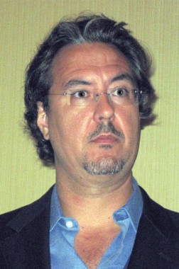
Dr. Bowling estimated that roughly 80% of his MS patients have little or no interest in medical marijuana. Those who are using it span a spectrum ranging from patients who were recreational users of pot before their MS diagnosis and who now seek beneficial MS effects or simply a license to use it recreationally, to elderly individuals with no history of marijuana use who have found cannabis is the only thing that effectively relieves some of their symptoms.
The neurologist noted two glaring paradoxes that emerged from his review of the clinical trials literature on medical marijuana for MS. One is that only 2 of the 19 studies utilized smoked marijuana, even though it’s far and away the most readily available form of the agent for U.S. patients. The most widely studied agent nabiximols (Sativex), an extract formulated as an oral spray – is approved as a prescription drug in Canada, the United Kingdom, and other countries, but not in the United States. And while Marinol and oral nabilone (Cesamet) are approved by the Food and Drug Administration, they’re not widely prescribed.
"I find it particularly challenging to apply the medical literature in this area to clinical situations. In our clinical practice, the way it seems to work out best is for patients to experiment on their own with marijuana as a prn add-on to their spasticity and/or pain medications, especially at bedtime or, if symptoms are worse, in the afternoon. But the dose, frequency, formulation, quality control ... it’s a real challenge. It’s like there are parallel universes of care, where we’re prescribing the conventional medications, and then we find out that the patients are also using marijuana," Dr. Bowling said.
The other paradox is the huge disconnect between the generally quite favorable subjective patient self-reports of improvement in spasticity and the largely negative objective results. The explanation may lie in part in the inadequacy of the Ashworth score as an outcome measure. It has come under frequent criticism, since some approved antispasticity medications have shown only modest or no improvement in Ashworth scores. But the limitations of the Ashworth score are probably only part of the story, in Dr. Bowling’s view.
"Patients’ assessments are 24/7; ours is a brief clinical visit at a prespecified time. Also, there is a pain component to spasticity, so for some patients who feel their spasticity is improving with the use of marijuana it may just be the pain aspect of the spasticity, or a euphoric view of the world that’s changing their perception of how severe the spasticity is. But for some patients, maybe it doesn’t matter whether it’s a subjective or objective assessment that’s improving, especially for those who don’t have high physical or cognitive demands during the day," he said.
Dr. Bowling reported having no conflicts of interest. Dr. Ware has received lecture fees from the Canadian Consortium for the Investigation of the Cannabinoids and a research grant and honoraria from Valeant for conducting a randomized trial of nabilone in fibromyalgia patients.
ORLANDO – The randomized trials evidence supporting a positive clinical effect for medical marijuana in relieving symptoms of multiple sclerosis is strongest for improvement of pain, patient-reported spasticity, and sleep disturbances.
That’s the clear message from an analysis of the 19 published, reasonable-quality randomized trials of cannabis use in patients with MS, Dr. Allen C. Bowling reported at the fifth Cooperative Meeting of the Consortium of Multiple Sclerosis Centers and the Americas Committee for Treatment and Research in Multiple Sclerosis.
"That’s consistent with my personal observation in a state where we’ve had medical marijuana for 12 years and where recreational marijuana was approved in last November’s election. The most consistent effect my patients report is on nocturnal pain and/or spasticity, which is consistent with the results of the clinical trials. The patients typically use a little bit at bedtime," according to Dr. Bowling, medical director of the MS service and director of the complementary and alternative medicine service at the Colorado Neurological Institute, Englewood.
The evidence that medical marijuana is of benefit for the bladder dysfunction associated with MS is weaker: Of 9 clinical trials that included bladder dysfunction as an endpoint, 4 were positive, 5 negative.
Occupying the bottom tier in terms of evidence-based therapy are objectively measured spasticity and tremor. Of 13 clinical trials that incorporated objective, clinician-assessed spasticity as an endpoint, only 2 were positive, with 11 negative. And none of the 5 decent-quality randomized trials that examined medical marijuana for relief of tremor in MS patients was positive.
In contrast, 9 of 11 studies that looked at pain as an endpoint reported a positive effect. Of 13 studies with patient-reported spasticity as an endpoint, 9 were positive, 4 negative. And of 6 randomized trials with sleep quality as an endpoint, 5 were positive, 1 negative, noted Dr. Bowling, who is clinical professor of neurology at the University of Colorado, Denver.
Medical marijuana is now legal in 18 states and the District of Columbia.
The largest and in Dr. Bowling’s view one of the best-conducted randomized trials in the field was the multicenter U.K. CAMS trial. The 15-week study included 630 MS patients who were randomized to oral tetrahydrocannabinol (Marinol); Cannador, an oral capsule containing a standardized amount of whole-plant extract; or placebo. The investigators found no significant difference between the cannabis-based therapies and placebo in improving spasticity as objectively measured using the Ashworth score, which was the primary study endpoint. However, the two active treatment arms did show significant benefit in terms of the secondary endpoints of improvement in pain, self-reported spasticity, and sleep (Lancet 2003;362:1517-26).
Another speaker at the session on prescribing medical marijuana underscored the beneficial effect on sleep disruption.
"In every single population – whether it’s Crohn’s disease, epilepsy, HIV, noncancer pain, cancer pain – improved sleep is always one of the most robust outcomes patients report. This is something that in clinical practice we tend to push to one side. We have some medications for pain management that we sometimes use for sleep, but cannabis use at nighttime seems to be a very powerful agent to improve sleep quality," observed Dr. Mark F. Ware, a family physician and anesthesiologist who is director of clinical research at the Alan Edwards Pain Management Unit of McGill University in Montreal.
Dr. Bowling estimated that roughly 80% of his MS patients have little or no interest in medical marijuana. Those who are using it span a spectrum ranging from patients who were recreational users of pot before their MS diagnosis and who now seek beneficial MS effects or simply a license to use it recreationally, to elderly individuals with no history of marijuana use who have found cannabis is the only thing that effectively relieves some of their symptoms.
The neurologist noted two glaring paradoxes that emerged from his review of the clinical trials literature on medical marijuana for MS. One is that only 2 of the 19 studies utilized smoked marijuana, even though it’s far and away the most readily available form of the agent for U.S. patients. The most widely studied agent nabiximols (Sativex), an extract formulated as an oral spray – is approved as a prescription drug in Canada, the United Kingdom, and other countries, but not in the United States. And while Marinol and oral nabilone (Cesamet) are approved by the Food and Drug Administration, they’re not widely prescribed.
"I find it particularly challenging to apply the medical literature in this area to clinical situations. In our clinical practice, the way it seems to work out best is for patients to experiment on their own with marijuana as a prn add-on to their spasticity and/or pain medications, especially at bedtime or, if symptoms are worse, in the afternoon. But the dose, frequency, formulation, quality control ... it’s a real challenge. It’s like there are parallel universes of care, where we’re prescribing the conventional medications, and then we find out that the patients are also using marijuana," Dr. Bowling said.
The other paradox is the huge disconnect between the generally quite favorable subjective patient self-reports of improvement in spasticity and the largely negative objective results. The explanation may lie in part in the inadequacy of the Ashworth score as an outcome measure. It has come under frequent criticism, since some approved antispasticity medications have shown only modest or no improvement in Ashworth scores. But the limitations of the Ashworth score are probably only part of the story, in Dr. Bowling’s view.
"Patients’ assessments are 24/7; ours is a brief clinical visit at a prespecified time. Also, there is a pain component to spasticity, so for some patients who feel their spasticity is improving with the use of marijuana it may just be the pain aspect of the spasticity, or a euphoric view of the world that’s changing their perception of how severe the spasticity is. But for some patients, maybe it doesn’t matter whether it’s a subjective or objective assessment that’s improving, especially for those who don’t have high physical or cognitive demands during the day," he said.
Dr. Bowling reported having no conflicts of interest. Dr. Ware has received lecture fees from the Canadian Consortium for the Investigation of the Cannabinoids and a research grant and honoraria from Valeant for conducting a randomized trial of nabilone in fibromyalgia patients.
ORLANDO – The randomized trials evidence supporting a positive clinical effect for medical marijuana in relieving symptoms of multiple sclerosis is strongest for improvement of pain, patient-reported spasticity, and sleep disturbances.
That’s the clear message from an analysis of the 19 published, reasonable-quality randomized trials of cannabis use in patients with MS, Dr. Allen C. Bowling reported at the fifth Cooperative Meeting of the Consortium of Multiple Sclerosis Centers and the Americas Committee for Treatment and Research in Multiple Sclerosis.
"That’s consistent with my personal observation in a state where we’ve had medical marijuana for 12 years and where recreational marijuana was approved in last November’s election. The most consistent effect my patients report is on nocturnal pain and/or spasticity, which is consistent with the results of the clinical trials. The patients typically use a little bit at bedtime," according to Dr. Bowling, medical director of the MS service and director of the complementary and alternative medicine service at the Colorado Neurological Institute, Englewood.
The evidence that medical marijuana is of benefit for the bladder dysfunction associated with MS is weaker: Of 9 clinical trials that included bladder dysfunction as an endpoint, 4 were positive, 5 negative.
Occupying the bottom tier in terms of evidence-based therapy are objectively measured spasticity and tremor. Of 13 clinical trials that incorporated objective, clinician-assessed spasticity as an endpoint, only 2 were positive, with 11 negative. And none of the 5 decent-quality randomized trials that examined medical marijuana for relief of tremor in MS patients was positive.
In contrast, 9 of 11 studies that looked at pain as an endpoint reported a positive effect. Of 13 studies with patient-reported spasticity as an endpoint, 9 were positive, 4 negative. And of 6 randomized trials with sleep quality as an endpoint, 5 were positive, 1 negative, noted Dr. Bowling, who is clinical professor of neurology at the University of Colorado, Denver.
Medical marijuana is now legal in 18 states and the District of Columbia.
The largest and in Dr. Bowling’s view one of the best-conducted randomized trials in the field was the multicenter U.K. CAMS trial. The 15-week study included 630 MS patients who were randomized to oral tetrahydrocannabinol (Marinol); Cannador, an oral capsule containing a standardized amount of whole-plant extract; or placebo. The investigators found no significant difference between the cannabis-based therapies and placebo in improving spasticity as objectively measured using the Ashworth score, which was the primary study endpoint. However, the two active treatment arms did show significant benefit in terms of the secondary endpoints of improvement in pain, self-reported spasticity, and sleep (Lancet 2003;362:1517-26).
Another speaker at the session on prescribing medical marijuana underscored the beneficial effect on sleep disruption.
"In every single population – whether it’s Crohn’s disease, epilepsy, HIV, noncancer pain, cancer pain – improved sleep is always one of the most robust outcomes patients report. This is something that in clinical practice we tend to push to one side. We have some medications for pain management that we sometimes use for sleep, but cannabis use at nighttime seems to be a very powerful agent to improve sleep quality," observed Dr. Mark F. Ware, a family physician and anesthesiologist who is director of clinical research at the Alan Edwards Pain Management Unit of McGill University in Montreal.
Dr. Bowling estimated that roughly 80% of his MS patients have little or no interest in medical marijuana. Those who are using it span a spectrum ranging from patients who were recreational users of pot before their MS diagnosis and who now seek beneficial MS effects or simply a license to use it recreationally, to elderly individuals with no history of marijuana use who have found cannabis is the only thing that effectively relieves some of their symptoms.
The neurologist noted two glaring paradoxes that emerged from his review of the clinical trials literature on medical marijuana for MS. One is that only 2 of the 19 studies utilized smoked marijuana, even though it’s far and away the most readily available form of the agent for U.S. patients. The most widely studied agent nabiximols (Sativex), an extract formulated as an oral spray – is approved as a prescription drug in Canada, the United Kingdom, and other countries, but not in the United States. And while Marinol and oral nabilone (Cesamet) are approved by the Food and Drug Administration, they’re not widely prescribed.
"I find it particularly challenging to apply the medical literature in this area to clinical situations. In our clinical practice, the way it seems to work out best is for patients to experiment on their own with marijuana as a prn add-on to their spasticity and/or pain medications, especially at bedtime or, if symptoms are worse, in the afternoon. But the dose, frequency, formulation, quality control ... it’s a real challenge. It’s like there are parallel universes of care, where we’re prescribing the conventional medications, and then we find out that the patients are also using marijuana," Dr. Bowling said.
The other paradox is the huge disconnect between the generally quite favorable subjective patient self-reports of improvement in spasticity and the largely negative objective results. The explanation may lie in part in the inadequacy of the Ashworth score as an outcome measure. It has come under frequent criticism, since some approved antispasticity medications have shown only modest or no improvement in Ashworth scores. But the limitations of the Ashworth score are probably only part of the story, in Dr. Bowling’s view.
"Patients’ assessments are 24/7; ours is a brief clinical visit at a prespecified time. Also, there is a pain component to spasticity, so for some patients who feel their spasticity is improving with the use of marijuana it may just be the pain aspect of the spasticity, or a euphoric view of the world that’s changing their perception of how severe the spasticity is. But for some patients, maybe it doesn’t matter whether it’s a subjective or objective assessment that’s improving, especially for those who don’t have high physical or cognitive demands during the day," he said.
Dr. Bowling reported having no conflicts of interest. Dr. Ware has received lecture fees from the Canadian Consortium for the Investigation of the Cannabinoids and a research grant and honoraria from Valeant for conducting a randomized trial of nabilone in fibromyalgia patients.
EXPERT ANALYSIS FROM THE CMSC/ACTRIMS ANNUAL MEETING

RSV hospitalization risk greatest among children less than 1 month old
Being under a month old, living with children under 5 years old, and being born during peak respiratory syncytial virus season all increase the likelihood that young children will be hospitalized with the infection, according to a 5-year, prospective surveillance study of children up to 2 years old published online July 22 in Pediatrics.
Investigators found 559 (26%) RSV-infected children among the 2,149 hospitalized with acute respiratory infections during 2000-2005 in counties that included Nashville, Tenn.; Rochester, N.Y.; and Cincinnati. Most of the children had been previously healthy, and most were born full-term (Pediatrics 2013;132:e341–e348 [doi: 10.1542/peds.2013-0303]).
The findings suggest that preventive measures – including vaccines under development – should be aimed at all young children, not just premature infants and others traditionally thought of as being at high risk, according to Dr. Caroline Breese Hall of the University of Rochester (N.Y.) and her associates.

"These findings indicate that strategies for diminishing the health care burden from RSV infections should include appropriate prophylaxis and the development of vaccines that are effective in very young infants, even those within the first month of life. In addition, general infection control practices such as restrictions of visits from ill individuals and careful hand washing should be emphasized, especially during the peak months of RSV circulation," which were December and January in this study, the authors concluded.
The RSV hospitalization rate was 5.2 per 1,000 children less than 24 months old; infants in their first month of life had the highest rate at 25.9 per 1,000 children. Infection was confirmed in the study by reverse-transcriptase polymerase chain reaction.
Infants no older than 2 months had a hospitalization rate of 17.9 per 1,000 children and accounted for 44% of RSV hospitalizations.
Ten percent of hospitalized kids were born preterm, "but their risk of hospitalization was not significantly different from that for term infants." It was a different story with very premature infants, that is, those born at less than 30 weeks’ gestation. They accounted for only 3% of RSV cases but had an RSV hospitalization rate of 18.7 per 1,000 children, about three times that of term infants, the authors noted.
Previous attempts to characterize children hospitalized with RSV have tended to rely on retrospective data – often discharge diagnoses – and have stratified risk by 6-month blocks of time. To increase precision, the authors used birth certificates to quantify risk by months of age.
About 21% of the children less than 12 months old had a comorbid condition, most often cardiopulmonary disease; coexisting medical conditions were even more common among older children, occurring in 53% of those aged 12-23 months.
The proportion of hospitalized children less than 24 months old living with another child under 5 years old (57%) was more than twice that of children living with older children (19%).
Overall, gender and race did not affect hospitalization rates.
The team did not study the effects of palivizumab – a monoclonal antibody used to prevent RSV complications in premature infants and others generally thought to be at high risk – on hospitalization rates, which "was unlikely to be appreciable because only a small proportion (less than 5%) of our study population was eligible" to receive it, the team noted.
The Centers for Disease Control and prevention funded the work. Six of the 14 authors reported research funding from or speaking, advising, or consulting relationships with several companies, including GlaxoSmithKline, MedImmune, Merck, Sanofi Pasteur, Novartis, BD Diagnostics, Novavax, and Quidel. Some of those firms are involved with RSV vaccine development.
Being under a month old, living with children under 5 years old, and being born during peak respiratory syncytial virus season all increase the likelihood that young children will be hospitalized with the infection, according to a 5-year, prospective surveillance study of children up to 2 years old published online July 22 in Pediatrics.
Investigators found 559 (26%) RSV-infected children among the 2,149 hospitalized with acute respiratory infections during 2000-2005 in counties that included Nashville, Tenn.; Rochester, N.Y.; and Cincinnati. Most of the children had been previously healthy, and most were born full-term (Pediatrics 2013;132:e341–e348 [doi: 10.1542/peds.2013-0303]).
The findings suggest that preventive measures – including vaccines under development – should be aimed at all young children, not just premature infants and others traditionally thought of as being at high risk, according to Dr. Caroline Breese Hall of the University of Rochester (N.Y.) and her associates.
"These findings indicate that strategies for diminishing the health care burden from RSV infections should include appropriate prophylaxis and the development of vaccines that are effective in very young infants, even those within the first month of life. In addition, general infection control practices such as restrictions of visits from ill individuals and careful hand washing should be emphasized, especially during the peak months of RSV circulation," which were December and January in this study, the authors concluded.
The RSV hospitalization rate was 5.2 per 1,000 children less than 24 months old; infants in their first month of life had the highest rate at 25.9 per 1,000 children. Infection was confirmed in the study by reverse-transcriptase polymerase chain reaction.
Infants no older than 2 months had a hospitalization rate of 17.9 per 1,000 children and accounted for 44% of RSV hospitalizations.
Ten percent of hospitalized kids were born preterm, "but their risk of hospitalization was not significantly different from that for term infants." It was a different story with very premature infants, that is, those born at less than 30 weeks’ gestation. They accounted for only 3% of RSV cases but had an RSV hospitalization rate of 18.7 per 1,000 children, about three times that of term infants, the authors noted.
Previous attempts to characterize children hospitalized with RSV have tended to rely on retrospective data – often discharge diagnoses – and have stratified risk by 6-month blocks of time. To increase precision, the authors used birth certificates to quantify risk by months of age.
About 21% of the children less than 12 months old had a comorbid condition, most often cardiopulmonary disease; coexisting medical conditions were even more common among older children, occurring in 53% of those aged 12-23 months.
The proportion of hospitalized children less than 24 months old living with another child under 5 years old (57%) was more than twice that of children living with older children (19%).
Overall, gender and race did not affect hospitalization rates.
The team did not study the effects of palivizumab – a monoclonal antibody used to prevent RSV complications in premature infants and others generally thought to be at high risk – on hospitalization rates, which "was unlikely to be appreciable because only a small proportion (less than 5%) of our study population was eligible" to receive it, the team noted.
The Centers for Disease Control and prevention funded the work. Six of the 14 authors reported research funding from or speaking, advising, or consulting relationships with several companies, including GlaxoSmithKline, MedImmune, Merck, Sanofi Pasteur, Novartis, BD Diagnostics, Novavax, and Quidel. Some of those firms are involved with RSV vaccine development.
Being under a month old, living with children under 5 years old, and being born during peak respiratory syncytial virus season all increase the likelihood that young children will be hospitalized with the infection, according to a 5-year, prospective surveillance study of children up to 2 years old published online July 22 in Pediatrics.
Investigators found 559 (26%) RSV-infected children among the 2,149 hospitalized with acute respiratory infections during 2000-2005 in counties that included Nashville, Tenn.; Rochester, N.Y.; and Cincinnati. Most of the children had been previously healthy, and most were born full-term (Pediatrics 2013;132:e341–e348 [doi: 10.1542/peds.2013-0303]).
The findings suggest that preventive measures – including vaccines under development – should be aimed at all young children, not just premature infants and others traditionally thought of as being at high risk, according to Dr. Caroline Breese Hall of the University of Rochester (N.Y.) and her associates.
"These findings indicate that strategies for diminishing the health care burden from RSV infections should include appropriate prophylaxis and the development of vaccines that are effective in very young infants, even those within the first month of life. In addition, general infection control practices such as restrictions of visits from ill individuals and careful hand washing should be emphasized, especially during the peak months of RSV circulation," which were December and January in this study, the authors concluded.
The RSV hospitalization rate was 5.2 per 1,000 children less than 24 months old; infants in their first month of life had the highest rate at 25.9 per 1,000 children. Infection was confirmed in the study by reverse-transcriptase polymerase chain reaction.
Infants no older than 2 months had a hospitalization rate of 17.9 per 1,000 children and accounted for 44% of RSV hospitalizations.
Ten percent of hospitalized kids were born preterm, "but their risk of hospitalization was not significantly different from that for term infants." It was a different story with very premature infants, that is, those born at less than 30 weeks’ gestation. They accounted for only 3% of RSV cases but had an RSV hospitalization rate of 18.7 per 1,000 children, about three times that of term infants, the authors noted.
Previous attempts to characterize children hospitalized with RSV have tended to rely on retrospective data – often discharge diagnoses – and have stratified risk by 6-month blocks of time. To increase precision, the authors used birth certificates to quantify risk by months of age.
About 21% of the children less than 12 months old had a comorbid condition, most often cardiopulmonary disease; coexisting medical conditions were even more common among older children, occurring in 53% of those aged 12-23 months.
The proportion of hospitalized children less than 24 months old living with another child under 5 years old (57%) was more than twice that of children living with older children (19%).
Overall, gender and race did not affect hospitalization rates.
The team did not study the effects of palivizumab – a monoclonal antibody used to prevent RSV complications in premature infants and others generally thought to be at high risk – on hospitalization rates, which "was unlikely to be appreciable because only a small proportion (less than 5%) of our study population was eligible" to receive it, the team noted.
The Centers for Disease Control and prevention funded the work. Six of the 14 authors reported research funding from or speaking, advising, or consulting relationships with several companies, including GlaxoSmithKline, MedImmune, Merck, Sanofi Pasteur, Novartis, BD Diagnostics, Novavax, and Quidel. Some of those firms are involved with RSV vaccine development.
FROM PEDIATRICS
Major finding: The RSV hospitalization rate for children in their first month of life is 25.9 per 1,000.
Data source: Prospective study of children up to 2 years old hospitalized with acute respiratory infections during 2000-2005.
Disclosures: The Centers for Disease Control and prevention funded the work. Six of the 14 authors reported research funding from or speaking, advising, or consulting relationships with several companies, including GlaxoSmithKline, MedImmune, Merck, Sanofi Pasteur, Novartis, BD Diagnostics, Novavax, and Quidel. Some of those firms are involved with RSV vaccine development.
Medical marijuana: Tips from an expert
ORLANDO – An essential element in prescribing medical marijuana responsibly is to insist that the patient must demonstrate improved functional status to be allowed to continue with the therapy, one expert has advised.
"Functional status is really the major issue. If cannabis is going to be used as a medicine, we have to see improvement in function: return to work, improvement in daily activities, engagement in society. I tell patients, ‘If this drug is really helping you, then show me. Show me that you can come off this other medication or reduce the dose. Show me you can go out and do volunteer work or join a club. Prove to me that this is valuable to you, because I cannot continue to authorize access to a substance if you cannot show me that it’s actually helping you improve your quality of life.’ That’s the gauntlet I throw down to patients," Dr. Mark A. Ware said at the fifth Cooperative Meeting sponsored by the Consortium of Multiple Sclerosis Centers and the Americas Committee for Treatment and Research in Multiple Sclerosis.

Through this requirement, a physician can validate that a patient with chronic non-cancer pain, epilepsy, HIV, multiple sclerosis, or any of the various other conditions for which medical marijuana is often used is not merely using the prescription recreationally, sitting around the house in a fog watching "M*A*S*H" reruns on television all day, explained Dr. Ware, a family physician and anesthesiologist who is director of clinical research at the Alan Edwards Pain Management Unit at McGill University in Montreal.
Canada has had a federal program for medical marijuana in place for a dozen years. Dr. Ware has extensive experience in prescribing medical marijuana in the pain clinic, where many patients report improvement not only in pain, but in spasticity, sleep, and/or mood. In addition, he has led randomized clinical trials that demonstrated that smoked cannabis reduced pain intensity and improved sleep quality in patients with chronic neuropathic pain (CMAJ 2010;182:E694-701) and that oral nabilone (Cesamet), a synthetic cannabinoid, improved sleep and was well tolerated in patients with fibromyalgia (Anesth. Analg. 2010;110:604-10).
He offered these tips for physicians who have patients asking about medical marijuana:
The doses used are modest: A World Health Organization report estimated that the average joint contains 0.5 g of cannabis, and that the average dose in patients using marijuana medically is four joints per day, or roughly 2 g of cannabis. That equates to 20-50 mg/day of tetrahydrocannabinol, the active molecule, which is consistent with the results of clinical trials using standardized extracts.
"A watchful dose is 5 g of cannabis per day. I would be very, very cautious about anybody who’s asking for more than 5 g/day. The likelihood of diversion goes way up. There’s very little reason on pharmacologic grounds why a patient would need that much," Dr. Ware advised.
Not everyone responds to medical marijuana: As with any other medication, there are nonresponders. Because cannabis has been widely available recreationally for so long, an individual’s past recreational experience can be used as a rough predictor of the likelihood of response to medical marijuana.
"One of the tests I use when a patient with a chronic medical illness comes in asking if maybe a cannabis-based drug could be useful is I ask if they’ve ever used the drug before, say, in college or high school. If they say they did and got anxious and paranoid and hated it, that tells me they’re not cannabinoid responders. I have no scientific evidence for this, it’s just a clinical tool I use. Prior positive recreational responders, I suspect, are more likely to have a favorable effect," Dr. Ware continued.
Most patients are more concerned about medical marijuana’s safety than effectiveness: Medical cannabinoids are "overall quite safe," according to the family physician, who with coauthors has published a systemic review of the adverse effects (CMAJ 2008;178:1669-78). Cannabis has no associated toxicity even at extremely high doses. The prescription oral cannabinoids have no apparent abuse potential. While dependence is seen in some recreational marijuana smokers, it doesn’t seem to occur with clinical use.
And regarding the key safety concern for most patients and physicians – the question of smoked marijuana’s effects on the lung (see accompanying story) – a new analysis of the published literature by one of the world’s pre-eminent pulmonologists, Dr. Donald P. Tashkin, emeritus professor of medicine and medical director of the pulmonary function laboratory of the University of California, Los Angeles, concluded that "the accumulated weight of evidence" suggests regular smoking of marijuana alone doesn’t increase the risk of lung or upper airway cancer or [chronic obstructive pulmonary disease] , and the evidence is inconclusive regarding a possible associated risk of lower respiratory tract infection (Ann. Am. Thorac. Soc. 2013;10:239-47).
Medical marijuana is contraindicated in adolescents and patients with unstable ischemic heart disease or a personal or family history of psychosis: All of the clinical trials have screened for and excluded patients with a history of psychosis, either personally or in a first-degree relative. So there is no evidence supporting its safe use in such individuals.
A growing number of case reports suggest recreational cannabis use in young adolescents can trigger a latent psychotic episode in selected susceptible individuals. This is a major concern.
Cannabis is a powerful peripheral vasodilator. "The way to remember that is the red eyes of Bob Marley," Dr. Ware suggested. Peripheral vasodilation results in an increased heart rate, which could trigger an MI in a patient with unstable ischemic heart disease.
Always ask about legal issues: A surprisingly large number of patients inquiring about medical marijuana are under investigation for a crime and are seeking a stay-out-of-jail card. They won’t mention it if they’re not asked.
Consider the prescription alternatives to medical pot: Nabilone and Marinol (dronabinol, which is tetrahydrocannabinol), are approved for prescription use in the United States. The beneficial effects last longer than with smoked cannabis, and there is no uncertainty about the concentration, source, or possible contaminants.
Keep an eye out for improved technology: Smoking is a dirty delivery system for marijuana. While it’s not nearly as harmful to the lungs as smoking tobacco, as Dr. Tashkin recently concluded, cannabis smoke nevertheless does contain carcinogens and toxins. Vaporization devices are now commercially available as an alternative: a smokeless marijuana delivery system. This approach has recently been shown effective in a randomized, double-blind, placebo-controlled, crossover clinical trial conducted in patients with neuropathic pain (J. Pain 2013;14:136-48).
Dr. Ware has received lecture fees from the Canadian Consortium for the Investigation of the Cannabinoids and a research grant and honoraria from Valeant for conducting a randomized trial of nabilone in fibromyalgia patients.
ORLANDO – An essential element in prescribing medical marijuana responsibly is to insist that the patient must demonstrate improved functional status to be allowed to continue with the therapy, one expert has advised.
"Functional status is really the major issue. If cannabis is going to be used as a medicine, we have to see improvement in function: return to work, improvement in daily activities, engagement in society. I tell patients, ‘If this drug is really helping you, then show me. Show me that you can come off this other medication or reduce the dose. Show me you can go out and do volunteer work or join a club. Prove to me that this is valuable to you, because I cannot continue to authorize access to a substance if you cannot show me that it’s actually helping you improve your quality of life.’ That’s the gauntlet I throw down to patients," Dr. Mark A. Ware said at the fifth Cooperative Meeting sponsored by the Consortium of Multiple Sclerosis Centers and the Americas Committee for Treatment and Research in Multiple Sclerosis.
Through this requirement, a physician can validate that a patient with chronic non-cancer pain, epilepsy, HIV, multiple sclerosis, or any of the various other conditions for which medical marijuana is often used is not merely using the prescription recreationally, sitting around the house in a fog watching "M*A*S*H" reruns on television all day, explained Dr. Ware, a family physician and anesthesiologist who is director of clinical research at the Alan Edwards Pain Management Unit at McGill University in Montreal.
Canada has had a federal program for medical marijuana in place for a dozen years. Dr. Ware has extensive experience in prescribing medical marijuana in the pain clinic, where many patients report improvement not only in pain, but in spasticity, sleep, and/or mood. In addition, he has led randomized clinical trials that demonstrated that smoked cannabis reduced pain intensity and improved sleep quality in patients with chronic neuropathic pain (CMAJ 2010;182:E694-701) and that oral nabilone (Cesamet), a synthetic cannabinoid, improved sleep and was well tolerated in patients with fibromyalgia (Anesth. Analg. 2010;110:604-10).
He offered these tips for physicians who have patients asking about medical marijuana:
The doses used are modest: A World Health Organization report estimated that the average joint contains 0.5 g of cannabis, and that the average dose in patients using marijuana medically is four joints per day, or roughly 2 g of cannabis. That equates to 20-50 mg/day of tetrahydrocannabinol, the active molecule, which is consistent with the results of clinical trials using standardized extracts.
"A watchful dose is 5 g of cannabis per day. I would be very, very cautious about anybody who’s asking for more than 5 g/day. The likelihood of diversion goes way up. There’s very little reason on pharmacologic grounds why a patient would need that much," Dr. Ware advised.
Not everyone responds to medical marijuana: As with any other medication, there are nonresponders. Because cannabis has been widely available recreationally for so long, an individual’s past recreational experience can be used as a rough predictor of the likelihood of response to medical marijuana.
"One of the tests I use when a patient with a chronic medical illness comes in asking if maybe a cannabis-based drug could be useful is I ask if they’ve ever used the drug before, say, in college or high school. If they say they did and got anxious and paranoid and hated it, that tells me they’re not cannabinoid responders. I have no scientific evidence for this, it’s just a clinical tool I use. Prior positive recreational responders, I suspect, are more likely to have a favorable effect," Dr. Ware continued.
Most patients are more concerned about medical marijuana’s safety than effectiveness: Medical cannabinoids are "overall quite safe," according to the family physician, who with coauthors has published a systemic review of the adverse effects (CMAJ 2008;178:1669-78). Cannabis has no associated toxicity even at extremely high doses. The prescription oral cannabinoids have no apparent abuse potential. While dependence is seen in some recreational marijuana smokers, it doesn’t seem to occur with clinical use.
And regarding the key safety concern for most patients and physicians – the question of smoked marijuana’s effects on the lung (see accompanying story) – a new analysis of the published literature by one of the world’s pre-eminent pulmonologists, Dr. Donald P. Tashkin, emeritus professor of medicine and medical director of the pulmonary function laboratory of the University of California, Los Angeles, concluded that "the accumulated weight of evidence" suggests regular smoking of marijuana alone doesn’t increase the risk of lung or upper airway cancer or [chronic obstructive pulmonary disease] , and the evidence is inconclusive regarding a possible associated risk of lower respiratory tract infection (Ann. Am. Thorac. Soc. 2013;10:239-47).
Medical marijuana is contraindicated in adolescents and patients with unstable ischemic heart disease or a personal or family history of psychosis: All of the clinical trials have screened for and excluded patients with a history of psychosis, either personally or in a first-degree relative. So there is no evidence supporting its safe use in such individuals.
A growing number of case reports suggest recreational cannabis use in young adolescents can trigger a latent psychotic episode in selected susceptible individuals. This is a major concern.
Cannabis is a powerful peripheral vasodilator. "The way to remember that is the red eyes of Bob Marley," Dr. Ware suggested. Peripheral vasodilation results in an increased heart rate, which could trigger an MI in a patient with unstable ischemic heart disease.
Always ask about legal issues: A surprisingly large number of patients inquiring about medical marijuana are under investigation for a crime and are seeking a stay-out-of-jail card. They won’t mention it if they’re not asked.
Consider the prescription alternatives to medical pot: Nabilone and Marinol (dronabinol, which is tetrahydrocannabinol), are approved for prescription use in the United States. The beneficial effects last longer than with smoked cannabis, and there is no uncertainty about the concentration, source, or possible contaminants.
Keep an eye out for improved technology: Smoking is a dirty delivery system for marijuana. While it’s not nearly as harmful to the lungs as smoking tobacco, as Dr. Tashkin recently concluded, cannabis smoke nevertheless does contain carcinogens and toxins. Vaporization devices are now commercially available as an alternative: a smokeless marijuana delivery system. This approach has recently been shown effective in a randomized, double-blind, placebo-controlled, crossover clinical trial conducted in patients with neuropathic pain (J. Pain 2013;14:136-48).
Dr. Ware has received lecture fees from the Canadian Consortium for the Investigation of the Cannabinoids and a research grant and honoraria from Valeant for conducting a randomized trial of nabilone in fibromyalgia patients.
ORLANDO – An essential element in prescribing medical marijuana responsibly is to insist that the patient must demonstrate improved functional status to be allowed to continue with the therapy, one expert has advised.
"Functional status is really the major issue. If cannabis is going to be used as a medicine, we have to see improvement in function: return to work, improvement in daily activities, engagement in society. I tell patients, ‘If this drug is really helping you, then show me. Show me that you can come off this other medication or reduce the dose. Show me you can go out and do volunteer work or join a club. Prove to me that this is valuable to you, because I cannot continue to authorize access to a substance if you cannot show me that it’s actually helping you improve your quality of life.’ That’s the gauntlet I throw down to patients," Dr. Mark A. Ware said at the fifth Cooperative Meeting sponsored by the Consortium of Multiple Sclerosis Centers and the Americas Committee for Treatment and Research in Multiple Sclerosis.
Through this requirement, a physician can validate that a patient with chronic non-cancer pain, epilepsy, HIV, multiple sclerosis, or any of the various other conditions for which medical marijuana is often used is not merely using the prescription recreationally, sitting around the house in a fog watching "M*A*S*H" reruns on television all day, explained Dr. Ware, a family physician and anesthesiologist who is director of clinical research at the Alan Edwards Pain Management Unit at McGill University in Montreal.
Canada has had a federal program for medical marijuana in place for a dozen years. Dr. Ware has extensive experience in prescribing medical marijuana in the pain clinic, where many patients report improvement not only in pain, but in spasticity, sleep, and/or mood. In addition, he has led randomized clinical trials that demonstrated that smoked cannabis reduced pain intensity and improved sleep quality in patients with chronic neuropathic pain (CMAJ 2010;182:E694-701) and that oral nabilone (Cesamet), a synthetic cannabinoid, improved sleep and was well tolerated in patients with fibromyalgia (Anesth. Analg. 2010;110:604-10).
He offered these tips for physicians who have patients asking about medical marijuana:
The doses used are modest: A World Health Organization report estimated that the average joint contains 0.5 g of cannabis, and that the average dose in patients using marijuana medically is four joints per day, or roughly 2 g of cannabis. That equates to 20-50 mg/day of tetrahydrocannabinol, the active molecule, which is consistent with the results of clinical trials using standardized extracts.
"A watchful dose is 5 g of cannabis per day. I would be very, very cautious about anybody who’s asking for more than 5 g/day. The likelihood of diversion goes way up. There’s very little reason on pharmacologic grounds why a patient would need that much," Dr. Ware advised.
Not everyone responds to medical marijuana: As with any other medication, there are nonresponders. Because cannabis has been widely available recreationally for so long, an individual’s past recreational experience can be used as a rough predictor of the likelihood of response to medical marijuana.
"One of the tests I use when a patient with a chronic medical illness comes in asking if maybe a cannabis-based drug could be useful is I ask if they’ve ever used the drug before, say, in college or high school. If they say they did and got anxious and paranoid and hated it, that tells me they’re not cannabinoid responders. I have no scientific evidence for this, it’s just a clinical tool I use. Prior positive recreational responders, I suspect, are more likely to have a favorable effect," Dr. Ware continued.
Most patients are more concerned about medical marijuana’s safety than effectiveness: Medical cannabinoids are "overall quite safe," according to the family physician, who with coauthors has published a systemic review of the adverse effects (CMAJ 2008;178:1669-78). Cannabis has no associated toxicity even at extremely high doses. The prescription oral cannabinoids have no apparent abuse potential. While dependence is seen in some recreational marijuana smokers, it doesn’t seem to occur with clinical use.
And regarding the key safety concern for most patients and physicians – the question of smoked marijuana’s effects on the lung (see accompanying story) – a new analysis of the published literature by one of the world’s pre-eminent pulmonologists, Dr. Donald P. Tashkin, emeritus professor of medicine and medical director of the pulmonary function laboratory of the University of California, Los Angeles, concluded that "the accumulated weight of evidence" suggests regular smoking of marijuana alone doesn’t increase the risk of lung or upper airway cancer or [chronic obstructive pulmonary disease] , and the evidence is inconclusive regarding a possible associated risk of lower respiratory tract infection (Ann. Am. Thorac. Soc. 2013;10:239-47).
Medical marijuana is contraindicated in adolescents and patients with unstable ischemic heart disease or a personal or family history of psychosis: All of the clinical trials have screened for and excluded patients with a history of psychosis, either personally or in a first-degree relative. So there is no evidence supporting its safe use in such individuals.
A growing number of case reports suggest recreational cannabis use in young adolescents can trigger a latent psychotic episode in selected susceptible individuals. This is a major concern.
Cannabis is a powerful peripheral vasodilator. "The way to remember that is the red eyes of Bob Marley," Dr. Ware suggested. Peripheral vasodilation results in an increased heart rate, which could trigger an MI in a patient with unstable ischemic heart disease.
Always ask about legal issues: A surprisingly large number of patients inquiring about medical marijuana are under investigation for a crime and are seeking a stay-out-of-jail card. They won’t mention it if they’re not asked.
Consider the prescription alternatives to medical pot: Nabilone and Marinol (dronabinol, which is tetrahydrocannabinol), are approved for prescription use in the United States. The beneficial effects last longer than with smoked cannabis, and there is no uncertainty about the concentration, source, or possible contaminants.
Keep an eye out for improved technology: Smoking is a dirty delivery system for marijuana. While it’s not nearly as harmful to the lungs as smoking tobacco, as Dr. Tashkin recently concluded, cannabis smoke nevertheless does contain carcinogens and toxins. Vaporization devices are now commercially available as an alternative: a smokeless marijuana delivery system. This approach has recently been shown effective in a randomized, double-blind, placebo-controlled, crossover clinical trial conducted in patients with neuropathic pain (J. Pain 2013;14:136-48).
Dr. Ware has received lecture fees from the Canadian Consortium for the Investigation of the Cannabinoids and a research grant and honoraria from Valeant for conducting a randomized trial of nabilone in fibromyalgia patients.
AT THE CMSC/ACTRIMS ANNUAL MEETING

COPD may boost risk of cerebral microbleeds
Chronic obstructive pulmonary disease was associated with an increased risk of cerebral microbleeds – a marker of cerebral small vessel disease – independent of factors such as age, sex, smoking status, and cholesterol levels, according to a prospective, population-based cohort study.
"Depression, postural instability, cognitive and functional impairment are known consequences of cerebral small-vessel disease, and are frequently described extrapulmonary manifestations in patients with COPD," wrote Dr. Lies Lahousse from Ghent University Hospital, Belgium, and colleagues. "However, it is unclear whether COPD is associated with incident cerebral small-vessel disease."
Patients with COPD had a significantly higher prevalence of cerebral microbleeds, compared with patients with normal lung function, even after accounting for age, sex, smoking status, atherosclerotic macroangiopathy, antithrombotic use, total cholesterol, triglycerides, and serum creatinine (odds ratio, 1.7; 95% confidence interval, 1.15-2.47; P = .007).
Patients with COPD also had a more than threefold increase in the prevalence of microbleeds in deep or infratentorial locations (OR, 3.3; 95% CI, 1.97-5.53; P less than .001), according to data published online July 19 in the American Journal of Respiratory and Critical Care Medicine.
When a longitudinal analysis was conducted in patients without microbleed at baseline, COPD independently predicted a sevenfold increase in risk of deep or infratentorial microbleeds (OR, 7.1; 95% CI, 2.1-24.5; P = .002) over a median time interval of 3.42 years.
"Our results are in line with two previous cross-sectional studies that showed that patients with COPD had a significantly increased volume of cerebral white matter lesions, which is another marker of cerebral small vessel disease, and known to be associated with microbleeds in a deep or infratentorial region," Dr. Lahousse and associates said.
Although the increased incidence of microbleeds in patients with COPD was independent of smoking status, the researchers did find a significantly higher prevalence of cerebral microbleeds among individuals who had smoked.
The study involved 165 patients with COPD and 645 patients without COPD who were enrolled in the Rotterdam Scan Study, a population-based cohort study using MRI scan to examine age-related brain changes.
Previous research had shown an increased prevalence of atherosclerotic macroangiopathy in patients with COPD, but that was taken into account in the analysis.
The authors therefore suggested that COPD may affect large and small blood vessels, and that stiffening of arteries and arterioles may be the result of systemic inflammation in COPD as well as hypoxia caused by airflow limitation.
"Although severity of airflow limitation may not entirely reflect disease activity, our results suggest that cerebral small vessel disease is more present in COPD patients with more severe airflow limitation," the investigators noted.
Given the potential cognitive and functional consequences of microbleeds, the study authors suggested that the results could help with identification of vulnerable patient groups and point to the need for more research into preventive strategies.
The study was funded by the Fund for Scientific Research Flanders project and the Netherlands Organization for Scientific Research.
Chronic obstructive pulmonary disease was associated with an increased risk of cerebral microbleeds – a marker of cerebral small vessel disease – independent of factors such as age, sex, smoking status, and cholesterol levels, according to a prospective, population-based cohort study.
"Depression, postural instability, cognitive and functional impairment are known consequences of cerebral small-vessel disease, and are frequently described extrapulmonary manifestations in patients with COPD," wrote Dr. Lies Lahousse from Ghent University Hospital, Belgium, and colleagues. "However, it is unclear whether COPD is associated with incident cerebral small-vessel disease."
Patients with COPD had a significantly higher prevalence of cerebral microbleeds, compared with patients with normal lung function, even after accounting for age, sex, smoking status, atherosclerotic macroangiopathy, antithrombotic use, total cholesterol, triglycerides, and serum creatinine (odds ratio, 1.7; 95% confidence interval, 1.15-2.47; P = .007).
Patients with COPD also had a more than threefold increase in the prevalence of microbleeds in deep or infratentorial locations (OR, 3.3; 95% CI, 1.97-5.53; P less than .001), according to data published online July 19 in the American Journal of Respiratory and Critical Care Medicine.
When a longitudinal analysis was conducted in patients without microbleed at baseline, COPD independently predicted a sevenfold increase in risk of deep or infratentorial microbleeds (OR, 7.1; 95% CI, 2.1-24.5; P = .002) over a median time interval of 3.42 years.
"Our results are in line with two previous cross-sectional studies that showed that patients with COPD had a significantly increased volume of cerebral white matter lesions, which is another marker of cerebral small vessel disease, and known to be associated with microbleeds in a deep or infratentorial region," Dr. Lahousse and associates said.
Although the increased incidence of microbleeds in patients with COPD was independent of smoking status, the researchers did find a significantly higher prevalence of cerebral microbleeds among individuals who had smoked.
The study involved 165 patients with COPD and 645 patients without COPD who were enrolled in the Rotterdam Scan Study, a population-based cohort study using MRI scan to examine age-related brain changes.
Previous research had shown an increased prevalence of atherosclerotic macroangiopathy in patients with COPD, but that was taken into account in the analysis.
The authors therefore suggested that COPD may affect large and small blood vessels, and that stiffening of arteries and arterioles may be the result of systemic inflammation in COPD as well as hypoxia caused by airflow limitation.
"Although severity of airflow limitation may not entirely reflect disease activity, our results suggest that cerebral small vessel disease is more present in COPD patients with more severe airflow limitation," the investigators noted.
Given the potential cognitive and functional consequences of microbleeds, the study authors suggested that the results could help with identification of vulnerable patient groups and point to the need for more research into preventive strategies.
The study was funded by the Fund for Scientific Research Flanders project and the Netherlands Organization for Scientific Research.
Chronic obstructive pulmonary disease was associated with an increased risk of cerebral microbleeds – a marker of cerebral small vessel disease – independent of factors such as age, sex, smoking status, and cholesterol levels, according to a prospective, population-based cohort study.
"Depression, postural instability, cognitive and functional impairment are known consequences of cerebral small-vessel disease, and are frequently described extrapulmonary manifestations in patients with COPD," wrote Dr. Lies Lahousse from Ghent University Hospital, Belgium, and colleagues. "However, it is unclear whether COPD is associated with incident cerebral small-vessel disease."
Patients with COPD had a significantly higher prevalence of cerebral microbleeds, compared with patients with normal lung function, even after accounting for age, sex, smoking status, atherosclerotic macroangiopathy, antithrombotic use, total cholesterol, triglycerides, and serum creatinine (odds ratio, 1.7; 95% confidence interval, 1.15-2.47; P = .007).
Patients with COPD also had a more than threefold increase in the prevalence of microbleeds in deep or infratentorial locations (OR, 3.3; 95% CI, 1.97-5.53; P less than .001), according to data published online July 19 in the American Journal of Respiratory and Critical Care Medicine.
When a longitudinal analysis was conducted in patients without microbleed at baseline, COPD independently predicted a sevenfold increase in risk of deep or infratentorial microbleeds (OR, 7.1; 95% CI, 2.1-24.5; P = .002) over a median time interval of 3.42 years.
"Our results are in line with two previous cross-sectional studies that showed that patients with COPD had a significantly increased volume of cerebral white matter lesions, which is another marker of cerebral small vessel disease, and known to be associated with microbleeds in a deep or infratentorial region," Dr. Lahousse and associates said.
Although the increased incidence of microbleeds in patients with COPD was independent of smoking status, the researchers did find a significantly higher prevalence of cerebral microbleeds among individuals who had smoked.
The study involved 165 patients with COPD and 645 patients without COPD who were enrolled in the Rotterdam Scan Study, a population-based cohort study using MRI scan to examine age-related brain changes.
Previous research had shown an increased prevalence of atherosclerotic macroangiopathy in patients with COPD, but that was taken into account in the analysis.
The authors therefore suggested that COPD may affect large and small blood vessels, and that stiffening of arteries and arterioles may be the result of systemic inflammation in COPD as well as hypoxia caused by airflow limitation.
"Although severity of airflow limitation may not entirely reflect disease activity, our results suggest that cerebral small vessel disease is more present in COPD patients with more severe airflow limitation," the investigators noted.
Given the potential cognitive and functional consequences of microbleeds, the study authors suggested that the results could help with identification of vulnerable patient groups and point to the need for more research into preventive strategies.
The study was funded by the Fund for Scientific Research Flanders project and the Netherlands Organization for Scientific Research.
FROM AMERICAN JOURNAL OF RESPIRATORY AND CRITICAL CARE MEDICINE
Major finding: Patients with COPD had a significantly higher prevalence of cerebral microbleeds, compared with patients with normal lung function (odds ratio, 1.7).
Data source: Prospective, population-based cohort study involving 165 patients with COPD and 645 patients without COPD.
Disclosures: The study was funded by the Fund for Scientific Research Flanders project and the Netherlands Organization for Scientific Research.
High-risk smokers benefit most from CT screening for lung cancer
Low-dose computed tomography screening for lung cancer prevents the greatest number of deaths if it is reserved for smokers at highest risk of dying from the disease within 5 years, and it prevents very few deaths among those at lowest risk, according to a report published online July 18 in the New England Journal of Medicine.
When smokers were stratified into quintiles based on 5-year risk of death, 161 patients in the highest-risk quintile would need to be screened using low-dose CT to prevent 1 lung cancer death. In contrast, among the smokers in the lowest-risk quintile, 5,276 would need to be screened to prevent 1 lung cancer death, said Stephanie A. Kovalchik, Ph.D., of the National Cancer Institute and her associates.
These findings provide the empirical evidence that the medical community has been seeking to identify patients who would reap the most benefit from targeted screening – a benefit that would clearly outweigh the harm of the relatively frequent false-positive results with low-dose CT screening.
At present, screening guidelines all recommend low-dose CT screening for patients who meet the NLST (National Lung Screening Trial) entry criteria, but some experts argue that further refinement of screening criteria would be appropriate. The observations in this study "argue for the use of individualized risk assessment of lung cancer death instead of the NLST entry criteria, to increase the efficiency of low-dose CT screening," the investigators said.
Dr. Kovalchik and her colleagues examined whether the benefits and harms of low-dose CT scanning in the NLST differed according to the study subjects’ prescreening risk of lung cancer death, which was determined at enrollment using a validated prediction model. Risk factors included in this model were age, body mass index, family history of lung cancer, pack-years of smoking, years since smoking cessation (among patients who had quit), presence or absence of emphysema, sex, and race.
The NLST was a large randomized clinical trial that compared the efficacy of this screening technique against that of chest radiography in 53,454 smokers aged 55-74 years who had a minimum of 30 pack-years of smoking.
For their study, Dr. Kovalchik and her associates assessed outcomes in 26,604 NLST participants who had undergone three annual CT scans and 26,554 who had undergone three annual radiographs in the trial’s intention-to-treat population.
The primary end point was the rate of death from lung cancer during a median of 5.5 years of follow-up. This end point was reached by 354 subjects in the CT group, compared with 442 in the radiography group.
The rate of lung cancer deaths was 24.6 per 10,000 person-years among subjects screened by CT, compared with 30.9 per 10,000 person-years among those screened by radiography. This reflects a 20% relative reduction in lung cancer deaths with low-dose CT screening, the investigators reported (N. Engl. J. Med. 2013 July 18 [doi: 10.1056/NEJMoa1301851]).
The researchers developed an absolute risk-prediction model for lung-cancer mortality that accounted for a participant’s specific risk characteristics and life expectancy by incorporating Cox proportional-hazards models of death from lung cancer and competing causes of death. Predictors of lung-cancer death were selected from a set of previously identified demographic and clinical risk factors for lung cancer (including smoking history) using Lasso regression. The prediction model was externally validated with outcome data from 15,114 NLST-eligible and 22,649 NLST-ineligible smokers aged 55-74 years who were enrolled in the radiography group of the PLCO (Prostate, Lung, Colorectal, and Ovarian) Cancer Screening Trial. The participants were stratified into five quintiles for the predicted 5-year risk of death from lung cancer.
"Participants at highest risk for lung cancer death accounted for a disproportionate share of the benefits of low-dose CT screening. For example, 77 of 88 CT-prevented lung cancer deaths (88%) occurred among the 60% of participants with a 5-year risk of lung-cancer death of 0.85% or more, whereas only 1% of prevented lung-cancer deaths occurred among the 20% of participants at lowest risk," the researchers said.
Restricting screening to the 60% of participants at highest risk for death from lung cancer within 5 years (more than 0.85%), as compared with the entire CT group, captured 88% of CT-preventable lung-cancer deaths, reduced the number of participants who would need to be screened to prevent one lung-cancer death from 302 to 161, and reduced the number of false-positive results per CT-prevented lung cancer death from 108 to 65. In contrast, the 20% of participants at lowest risk for lung cancer death accounted for almost none of the CT-prevented lung cancer deaths, the researchers said.
"These observations argue for the use of individualized risk assessment of lung cancer death instead of the NLST entry criteria to increase the efficiency of low-dose CT screening. ... [T]ailoring of low-dose CT screening to a patient’s predicted risk of lung cancer death could narrow the NLST-eligible population without a loss in the potential public health benefits of screening or a disproportionate increase in the potential harms," they wrote.
Dr. Kovalchik and her colleagues noted that this screening technique was of limited efficacy in one important subgroup of smokers: those who had coexisting pulmonary disorders. Further study of the benefits and harms of low-dose CT screening in such patients is needed.
This study was funded by the National Cancer Institute. No relevant financial conflicts of interest were reported.
Low-dose computed tomography screening for lung cancer prevents the greatest number of deaths if it is reserved for smokers at highest risk of dying from the disease within 5 years, and it prevents very few deaths among those at lowest risk, according to a report published online July 18 in the New England Journal of Medicine.
When smokers were stratified into quintiles based on 5-year risk of death, 161 patients in the highest-risk quintile would need to be screened using low-dose CT to prevent 1 lung cancer death. In contrast, among the smokers in the lowest-risk quintile, 5,276 would need to be screened to prevent 1 lung cancer death, said Stephanie A. Kovalchik, Ph.D., of the National Cancer Institute and her associates.
These findings provide the empirical evidence that the medical community has been seeking to identify patients who would reap the most benefit from targeted screening – a benefit that would clearly outweigh the harm of the relatively frequent false-positive results with low-dose CT screening.
At present, screening guidelines all recommend low-dose CT screening for patients who meet the NLST (National Lung Screening Trial) entry criteria, but some experts argue that further refinement of screening criteria would be appropriate. The observations in this study "argue for the use of individualized risk assessment of lung cancer death instead of the NLST entry criteria, to increase the efficiency of low-dose CT screening," the investigators said.
Dr. Kovalchik and her colleagues examined whether the benefits and harms of low-dose CT scanning in the NLST differed according to the study subjects’ prescreening risk of lung cancer death, which was determined at enrollment using a validated prediction model. Risk factors included in this model were age, body mass index, family history of lung cancer, pack-years of smoking, years since smoking cessation (among patients who had quit), presence or absence of emphysema, sex, and race.
The NLST was a large randomized clinical trial that compared the efficacy of this screening technique against that of chest radiography in 53,454 smokers aged 55-74 years who had a minimum of 30 pack-years of smoking.
For their study, Dr. Kovalchik and her associates assessed outcomes in 26,604 NLST participants who had undergone three annual CT scans and 26,554 who had undergone three annual radiographs in the trial’s intention-to-treat population.
The primary end point was the rate of death from lung cancer during a median of 5.5 years of follow-up. This end point was reached by 354 subjects in the CT group, compared with 442 in the radiography group.
The rate of lung cancer deaths was 24.6 per 10,000 person-years among subjects screened by CT, compared with 30.9 per 10,000 person-years among those screened by radiography. This reflects a 20% relative reduction in lung cancer deaths with low-dose CT screening, the investigators reported (N. Engl. J. Med. 2013 July 18 [doi: 10.1056/NEJMoa1301851]).
The researchers developed an absolute risk-prediction model for lung-cancer mortality that accounted for a participant’s specific risk characteristics and life expectancy by incorporating Cox proportional-hazards models of death from lung cancer and competing causes of death. Predictors of lung-cancer death were selected from a set of previously identified demographic and clinical risk factors for lung cancer (including smoking history) using Lasso regression. The prediction model was externally validated with outcome data from 15,114 NLST-eligible and 22,649 NLST-ineligible smokers aged 55-74 years who were enrolled in the radiography group of the PLCO (Prostate, Lung, Colorectal, and Ovarian) Cancer Screening Trial. The participants were stratified into five quintiles for the predicted 5-year risk of death from lung cancer.
"Participants at highest risk for lung cancer death accounted for a disproportionate share of the benefits of low-dose CT screening. For example, 77 of 88 CT-prevented lung cancer deaths (88%) occurred among the 60% of participants with a 5-year risk of lung-cancer death of 0.85% or more, whereas only 1% of prevented lung-cancer deaths occurred among the 20% of participants at lowest risk," the researchers said.
Restricting screening to the 60% of participants at highest risk for death from lung cancer within 5 years (more than 0.85%), as compared with the entire CT group, captured 88% of CT-preventable lung-cancer deaths, reduced the number of participants who would need to be screened to prevent one lung-cancer death from 302 to 161, and reduced the number of false-positive results per CT-prevented lung cancer death from 108 to 65. In contrast, the 20% of participants at lowest risk for lung cancer death accounted for almost none of the CT-prevented lung cancer deaths, the researchers said.
"These observations argue for the use of individualized risk assessment of lung cancer death instead of the NLST entry criteria to increase the efficiency of low-dose CT screening. ... [T]ailoring of low-dose CT screening to a patient’s predicted risk of lung cancer death could narrow the NLST-eligible population without a loss in the potential public health benefits of screening or a disproportionate increase in the potential harms," they wrote.
Dr. Kovalchik and her colleagues noted that this screening technique was of limited efficacy in one important subgroup of smokers: those who had coexisting pulmonary disorders. Further study of the benefits and harms of low-dose CT screening in such patients is needed.
This study was funded by the National Cancer Institute. No relevant financial conflicts of interest were reported.
Low-dose computed tomography screening for lung cancer prevents the greatest number of deaths if it is reserved for smokers at highest risk of dying from the disease within 5 years, and it prevents very few deaths among those at lowest risk, according to a report published online July 18 in the New England Journal of Medicine.
When smokers were stratified into quintiles based on 5-year risk of death, 161 patients in the highest-risk quintile would need to be screened using low-dose CT to prevent 1 lung cancer death. In contrast, among the smokers in the lowest-risk quintile, 5,276 would need to be screened to prevent 1 lung cancer death, said Stephanie A. Kovalchik, Ph.D., of the National Cancer Institute and her associates.
These findings provide the empirical evidence that the medical community has been seeking to identify patients who would reap the most benefit from targeted screening – a benefit that would clearly outweigh the harm of the relatively frequent false-positive results with low-dose CT screening.
At present, screening guidelines all recommend low-dose CT screening for patients who meet the NLST (National Lung Screening Trial) entry criteria, but some experts argue that further refinement of screening criteria would be appropriate. The observations in this study "argue for the use of individualized risk assessment of lung cancer death instead of the NLST entry criteria, to increase the efficiency of low-dose CT screening," the investigators said.
Dr. Kovalchik and her colleagues examined whether the benefits and harms of low-dose CT scanning in the NLST differed according to the study subjects’ prescreening risk of lung cancer death, which was determined at enrollment using a validated prediction model. Risk factors included in this model were age, body mass index, family history of lung cancer, pack-years of smoking, years since smoking cessation (among patients who had quit), presence or absence of emphysema, sex, and race.
The NLST was a large randomized clinical trial that compared the efficacy of this screening technique against that of chest radiography in 53,454 smokers aged 55-74 years who had a minimum of 30 pack-years of smoking.
For their study, Dr. Kovalchik and her associates assessed outcomes in 26,604 NLST participants who had undergone three annual CT scans and 26,554 who had undergone three annual radiographs in the trial’s intention-to-treat population.
The primary end point was the rate of death from lung cancer during a median of 5.5 years of follow-up. This end point was reached by 354 subjects in the CT group, compared with 442 in the radiography group.
The rate of lung cancer deaths was 24.6 per 10,000 person-years among subjects screened by CT, compared with 30.9 per 10,000 person-years among those screened by radiography. This reflects a 20% relative reduction in lung cancer deaths with low-dose CT screening, the investigators reported (N. Engl. J. Med. 2013 July 18 [doi: 10.1056/NEJMoa1301851]).
The researchers developed an absolute risk-prediction model for lung-cancer mortality that accounted for a participant’s specific risk characteristics and life expectancy by incorporating Cox proportional-hazards models of death from lung cancer and competing causes of death. Predictors of lung-cancer death were selected from a set of previously identified demographic and clinical risk factors for lung cancer (including smoking history) using Lasso regression. The prediction model was externally validated with outcome data from 15,114 NLST-eligible and 22,649 NLST-ineligible smokers aged 55-74 years who were enrolled in the radiography group of the PLCO (Prostate, Lung, Colorectal, and Ovarian) Cancer Screening Trial. The participants were stratified into five quintiles for the predicted 5-year risk of death from lung cancer.
"Participants at highest risk for lung cancer death accounted for a disproportionate share of the benefits of low-dose CT screening. For example, 77 of 88 CT-prevented lung cancer deaths (88%) occurred among the 60% of participants with a 5-year risk of lung-cancer death of 0.85% or more, whereas only 1% of prevented lung-cancer deaths occurred among the 20% of participants at lowest risk," the researchers said.
Restricting screening to the 60% of participants at highest risk for death from lung cancer within 5 years (more than 0.85%), as compared with the entire CT group, captured 88% of CT-preventable lung-cancer deaths, reduced the number of participants who would need to be screened to prevent one lung-cancer death from 302 to 161, and reduced the number of false-positive results per CT-prevented lung cancer death from 108 to 65. In contrast, the 20% of participants at lowest risk for lung cancer death accounted for almost none of the CT-prevented lung cancer deaths, the researchers said.
"These observations argue for the use of individualized risk assessment of lung cancer death instead of the NLST entry criteria to increase the efficiency of low-dose CT screening. ... [T]ailoring of low-dose CT screening to a patient’s predicted risk of lung cancer death could narrow the NLST-eligible population without a loss in the potential public health benefits of screening or a disproportionate increase in the potential harms," they wrote.
Dr. Kovalchik and her colleagues noted that this screening technique was of limited efficacy in one important subgroup of smokers: those who had coexisting pulmonary disorders. Further study of the benefits and harms of low-dose CT screening in such patients is needed.
This study was funded by the National Cancer Institute. No relevant financial conflicts of interest were reported.
FROM THE NEW ENGLAND JOURNAL OF MEDICINE
Major finding: The rate of lung cancer deaths was 24.6 per 10,000 person-years among subjects screened by CT, compared with 30.9 per 10,000 person-years among those screened by radiography, which reflects a 20% relative reduction.
Data source: A secondary analysis of data on 26,604 NLST participants who had three annual low-dose lung CTs and 26,554 who had three chest radiographs to screen for lung cancer, all of whom were followed for 5 years.
Disclosures: This study was funded by the National Cancer Institute. No relevant financial conflicts of interest were reported.
Should you clear a child with a URI for surgery?
• Consult the anesthesiologist if a pediatric patient is about to undergo an elective surgical procedure and is febrile or coughing—especially if the child has significant comorbidities. These conditions may warrant postponing the procedure. A
• Avoid surgery in a child with cardiac disease who has inflammatory respiratory disease—especially if he or she has had palliative procedures for cyanotic lesions or has a hypoplastic right or left heart. A
Strength of recommendation (SOR)
A Good-quality patient-oriented evidence
B Inconsistent or limited-quality patient-oriented evidence
C Consensus, usual practice, opinion, disease-oriented evidence, case series
CASE You are seeing a 2-year-old boy with a runny nose in your busy clinic. He was sent to you by a local surgeon who plans to repair a hernia 3 days from now. Other than the upper respiratory tract infection, the child is healthy. The surgeon wants you to clear the boy for surgery to avoid the possibility of the anesthesiologist canceling on the day of the procedure.
What are your next steps?
In our experience, children are regularly brought to the ambulatory surgery suite after having seen their family physician or pediatrician. To better equip you for such visits, we’ve put together the following summary of the risks for a child with an upper respiratory infection (URI) who is about to undergo surgery. We’ve also detailed some of the reasoning and evidence behind the decisions that anesthesiologists make in cases like this.
Making decisions in the absence of consensus
While the American Society of Anesthesiologists has a consensus statement on preoperative fasting to reduce the risk of pulmonary aspiration,1 there is no consensus on how to manage a child scheduled for elective surgery who develops a URI.
Historically, any child with a current or recent URI would not be considered a candidate for elective surgery due to the potential for respiratory complications caused by airway irritability.2 These complications can include bronchospasm, laryngospasm, hypoxemia, croup, pulmonary shunting, atelectasis, postoperative apnea, negative pressure pulmonary edema, and airway or endotracheal tube obstruction from increased secretions.3
This concern has been based on the clinical observation that children with URI-related airway irritability are at a greater risk of having such events during the perioperative period.4 In fact, pulmonary function studies reveal an increase in airway irritability for as long as 6 weeks after a significant URI.
Many children with a URI will have airway edema and increased secretions in the upper nasopharynx and the posterior oropharynx down to the level of the vocal cords. In addition, patients with some viral infections—including respiratory syncytial virus—may experience increased edema in the larynx, trachea, and small and large bronchi. The presence of airway inflammation increases mucus production, which is normally coughed out in an awake patient.
The period between the awake state and surgical anesthesia—referred to as Stage 2— is the time of highest risk for the development of laryngospasm. Stage 2 occurs both during the induction of and the emergence from general anesthesia. Children who develop laryngospasm may be difficult to ventilate by mask, and tracheal intubation can be difficult through the closed glottis. In these clinically emergent situations, patients become hypoxemic rapidly. Ventilation may be possible only if the vocal cords are relaxed with agents such as succinylcholine.5
If the anesthesia team cannot quickly treat such laryngospasm, it can lead to postobstructive pulmonary edema. Negative pressure developed in the thorax during spontaneous ventilation against a closed glottis causes a pressure gradient across the alveolar-capillary membrane, leading to movement of fluid into the alveoli, characterized by a typically pink, frothy transudate. Hypoxia may ensue, and the chest x-ray will reveal pulmonary edema. Mild forms may respond to an increase in ambient oxygen alone, but severe cases may require intubation, ventilation, and diuretics to restore the child to a normal state.6
Certain anesthetic agents may be problematic
Unfortunately, airway irritability is only one of many problems to contend with. Inhalational anesthetic agents have an adverse effect on the mucociliary elevator, as well.7,8 Cilia on the surface of epithelial cells lining the trachea and bronchi act to move mucus from the distal to the proximal airway so that it can be coughed out. Failure of this mechanism in a child with an inflammatory condition in the airway increases the risk of atelectasis from thickened secretions and occasionally from pneumonia.
Most of the potent general anesthetic agents have significant bronchodilatory properties. But desflurane, a commonly used agent, causes bronchoconstriction when used in a patient with an irritated, infected airway.9This agent will produce predictable wheezing from bronchospasm, especially in patients who have confounding pulmonary disease such as asthma.
Talk to the anesthesiologist. With these concerns in mind, clinicians must consider the type of anesthetic and the nature of the surgical procedure and discuss these issues with the anesthesiologist in the preoperative period. Some anesthetic agents and techniques are less irritating to airways.2,3 Avoidance of both desflurane and endotracheal intubation, for instance, will minimize airway irritation.
Brief procedures that do not involve major body cavities (eg, abdominal, thoracic, and intracranial) may be done without instrumenting the trachea. Face masks and laryngeal mask airways have been shown to decrease the incidence of adverse reactions because these forms of airway management are less invasive and physiologically insulting than direct laryngoscopy and endotracheal intubation.
Clinical observations suggest that endotracheal intubation increases pulmonary risks for the child with a URI.10,11 Long procedures, a patient position that limits access to the airway, the anticipated need to use muscle relaxants, airway surgery, and surgery in major cavities all require intubation for airway management. In these circumstances, it’s best to plan the perioperative care of a child suffering from a URI with an anesthesiologist who is comfortable caring for pediatric patients.12
Proceeding with surgery despite the risks
During emergency procedures on infants and children, the anesthesiologist has to do the best possible job under less than ideal conditions. Bowel obstruction, an incarcerated inguinal hernia, or a foreign body in the airway can all be life-threatening. In these cases, the anesthesiologist will counsel the surgeon and parent on the risks of the anesthetic. They likely will proceed with the knowledge that the usual methods of anesthetizing a child may have to be altered to provide the safest possible conditions.
But even certain nonemergent procedures may require taking some risks. Anesthesiologists are likely to anesthetize a child for placement of pressure-equalizing (PE) tubes, for instance, even with a mild infection in the upper oropharynx. This is because the possibility is high that the patient will be infected throughout the winter season, and waiting for a URI-free period might mean that the child would not get the PE tubes at all. Furthermore, PE tube placement is performed very quickly, with no instrumentation of the airway necessary. The anesthesiologist performs a mask anesthetic, always has control and access to the airway, and the procedure can be aborted at any time, with no incision to close.
How long should you wait it a URI is serious?
As mentioned earlier, there is no consensus on how long to wait, but clinical studies have suggested delaying surgery for as long as 6 weeks after the acute episode.4 The thinking was that this long period allowed time for the inflammatory response to dissipate completely. Unfortunately, in the middle of the winter, it’s likely that the child will be exposed to another viral strain and develop yet another URI. Clinical judgment plays a pivotal role here; it is always best to establish a relationship with an anesthesiologist in your community and call him or her with questions about individual patients.
Before you sign off on surgery
There are several other circumstances to consider when approving a child with a URI for surgery.
Children with cardiac disease, especially those who have had palliative procedures for cyanotic lesions or who have a hypoplastic right or left heart, are characteristically unstable in the face of inflammatory respiratory disease. Unless the surgical procedure is an emergency, such patients should not be considered for general anesthesia if they have a URI.13 As an example, bronchiolitis plus cyanotic heart disease can be rapidly fatal, requiring prolonged ventilation or extracorporeal membrane oxygenation in order to save the patient.
Intensive care nursery “graduates” may present to your office for preoperative assessment. Many of these infants and children will have marginally compensated lung disease, some with substantial pulmonary hypertension. Their respiratory function will continue to improve, some until the age of 7 to 10 years. In the meantime, they, too, are at high risk for complications from general anesthesia if they have a URI, and the decision to take them to the operating room should be discussed with other care providers and the parents.
Children with fever, mucopurulent discharge, wheezing, lethargy, and cough are at high risk for complications during the perioperative period, regardless of any comorbidities. Many anesthesiologists would cancel surgery in these circumstances, even if the patient has been seen recently by his or her primary care physician and is taking antibiotics for coverage of a potential bacterial infection.
Other indicators of increased risk of pulmonary complications include a history of reactive airway disease, exposure to tobacco smoke, snoring, nasal congestion, the need for endotracheal intubation, and surgery on the airway.14
CASE You evaluate the 2-year-old and note that he has a history of mucopurulent nasal discharge and a productive cough. The child’s temperature in the clinic is 99.8°F and his chest x-ray is consistent with bronchitis. After talking with a local anesthesiologist and the surgeon, you all agree that the boy’s surgery should be postponed for a month.
1. American Society of Anesthesiologists Task Force on Preoperative Fasting. Practice guidelines for preoperative fasting and the use of pharmacologic agents to reduce the risk of pulmonary aspiration: application to healthy patients undergoing elective procedures. Anesthesiology. 1999;90:896-905.
2. Parnis SJ, Barker DS, Van Der Walt JH. Clinical predictors of anaesthetic complications in children with respiratory tract infections. Paediatr Anaesth. 2001;11:29-40.
3. Cote CJ. The upper respiratory tract infection URI dilemma: fear of a complication or litigation? Anesthesiology. 2001;95:283-285.
4. Nandwani N, Raphael JH, Langton JA. Effect of an upper respiratory tract infection on airway reactivity. Br J Anaesth. 1997;
78:352-355.
5. Hampson-Evans D, Morgan P, Farrar M. Pediatric laryngospasm. Pediatr Anesth. 2008;18:303-307.
6. Krodel DJ, Bittner BA, Abdulnour R, et al. Case scenario: acute negative pressure pulmonary edema. Anesthesiology. 2010;113: 200-207.
7. Forbes AR. Halothane depresses mucociliary flow in the trachea. Anesthesiology. 1976;45:59-63.
8. Dikmen Y, Eminoglu E, Salihoglu Z, et al. Pulmonary mechanics during isoflurane, sevoflurane, and desflurane anaesthesia.
Anaesthesia. 2003;58:745-748.
9. Forbes AR, Horrigan RW. Mucociliary flow in the trachea during anesthesia with enflurane, ether, nitrous oxide and morphine. Anesthesiology. 1977;46:319-321.
10. Tait AR, Pandit UA, Voepel-Lewis T, et al. Use of the laryngeal mask airway in children with upper respiratory infections: a comparison with endotracheal intubation. Anesth Analg. 1998;
86:701-711.
11. Tait AR, Malviya S, Voepel-Lewis T, et al. Risk factors for perioperative adverse respiratory events in children with upper respiratory tract infections. Anesthesiology. 2001;95:299-306.
12. Von Ungern-Sternberg BS, Boda K, Chambers NA, et al. Risk assessment for respiratory complications in pediatric anaesthesia: a prospective cohort study. Lancet. 2010;376:773-783.
13. Malviya S, Voepel-Lewis T, Siewert M, et al. Risk factors for adverse postoperative outcomes in children presenting for cardiac surgery with upper respiratory tract infections. Anesthesiology. 2003;98:628-632.
14. Tait AR, Malviya S. Anesthesia for the child with an upper respiratory infection: still a dilemma? Anesth Analg. 2005;100:59-65.
• Consult the anesthesiologist if a pediatric patient is about to undergo an elective surgical procedure and is febrile or coughing—especially if the child has significant comorbidities. These conditions may warrant postponing the procedure. A
• Avoid surgery in a child with cardiac disease who has inflammatory respiratory disease—especially if he or she has had palliative procedures for cyanotic lesions or has a hypoplastic right or left heart. A
Strength of recommendation (SOR)
A Good-quality patient-oriented evidence
B Inconsistent or limited-quality patient-oriented evidence
C Consensus, usual practice, opinion, disease-oriented evidence, case series
CASE You are seeing a 2-year-old boy with a runny nose in your busy clinic. He was sent to you by a local surgeon who plans to repair a hernia 3 days from now. Other than the upper respiratory tract infection, the child is healthy. The surgeon wants you to clear the boy for surgery to avoid the possibility of the anesthesiologist canceling on the day of the procedure.
What are your next steps?
In our experience, children are regularly brought to the ambulatory surgery suite after having seen their family physician or pediatrician. To better equip you for such visits, we’ve put together the following summary of the risks for a child with an upper respiratory infection (URI) who is about to undergo surgery. We’ve also detailed some of the reasoning and evidence behind the decisions that anesthesiologists make in cases like this.
Making decisions in the absence of consensus
While the American Society of Anesthesiologists has a consensus statement on preoperative fasting to reduce the risk of pulmonary aspiration,1 there is no consensus on how to manage a child scheduled for elective surgery who develops a URI.
Historically, any child with a current or recent URI would not be considered a candidate for elective surgery due to the potential for respiratory complications caused by airway irritability.2 These complications can include bronchospasm, laryngospasm, hypoxemia, croup, pulmonary shunting, atelectasis, postoperative apnea, negative pressure pulmonary edema, and airway or endotracheal tube obstruction from increased secretions.3
This concern has been based on the clinical observation that children with URI-related airway irritability are at a greater risk of having such events during the perioperative period.4 In fact, pulmonary function studies reveal an increase in airway irritability for as long as 6 weeks after a significant URI.
Many children with a URI will have airway edema and increased secretions in the upper nasopharynx and the posterior oropharynx down to the level of the vocal cords. In addition, patients with some viral infections—including respiratory syncytial virus—may experience increased edema in the larynx, trachea, and small and large bronchi. The presence of airway inflammation increases mucus production, which is normally coughed out in an awake patient.
The period between the awake state and surgical anesthesia—referred to as Stage 2— is the time of highest risk for the development of laryngospasm. Stage 2 occurs both during the induction of and the emergence from general anesthesia. Children who develop laryngospasm may be difficult to ventilate by mask, and tracheal intubation can be difficult through the closed glottis. In these clinically emergent situations, patients become hypoxemic rapidly. Ventilation may be possible only if the vocal cords are relaxed with agents such as succinylcholine.5
If the anesthesia team cannot quickly treat such laryngospasm, it can lead to postobstructive pulmonary edema. Negative pressure developed in the thorax during spontaneous ventilation against a closed glottis causes a pressure gradient across the alveolar-capillary membrane, leading to movement of fluid into the alveoli, characterized by a typically pink, frothy transudate. Hypoxia may ensue, and the chest x-ray will reveal pulmonary edema. Mild forms may respond to an increase in ambient oxygen alone, but severe cases may require intubation, ventilation, and diuretics to restore the child to a normal state.6
Certain anesthetic agents may be problematic
Unfortunately, airway irritability is only one of many problems to contend with. Inhalational anesthetic agents have an adverse effect on the mucociliary elevator, as well.7,8 Cilia on the surface of epithelial cells lining the trachea and bronchi act to move mucus from the distal to the proximal airway so that it can be coughed out. Failure of this mechanism in a child with an inflammatory condition in the airway increases the risk of atelectasis from thickened secretions and occasionally from pneumonia.
Most of the potent general anesthetic agents have significant bronchodilatory properties. But desflurane, a commonly used agent, causes bronchoconstriction when used in a patient with an irritated, infected airway.9This agent will produce predictable wheezing from bronchospasm, especially in patients who have confounding pulmonary disease such as asthma.
Talk to the anesthesiologist. With these concerns in mind, clinicians must consider the type of anesthetic and the nature of the surgical procedure and discuss these issues with the anesthesiologist in the preoperative period. Some anesthetic agents and techniques are less irritating to airways.2,3 Avoidance of both desflurane and endotracheal intubation, for instance, will minimize airway irritation.
Brief procedures that do not involve major body cavities (eg, abdominal, thoracic, and intracranial) may be done without instrumenting the trachea. Face masks and laryngeal mask airways have been shown to decrease the incidence of adverse reactions because these forms of airway management are less invasive and physiologically insulting than direct laryngoscopy and endotracheal intubation.
Clinical observations suggest that endotracheal intubation increases pulmonary risks for the child with a URI.10,11 Long procedures, a patient position that limits access to the airway, the anticipated need to use muscle relaxants, airway surgery, and surgery in major cavities all require intubation for airway management. In these circumstances, it’s best to plan the perioperative care of a child suffering from a URI with an anesthesiologist who is comfortable caring for pediatric patients.12
Proceeding with surgery despite the risks
During emergency procedures on infants and children, the anesthesiologist has to do the best possible job under less than ideal conditions. Bowel obstruction, an incarcerated inguinal hernia, or a foreign body in the airway can all be life-threatening. In these cases, the anesthesiologist will counsel the surgeon and parent on the risks of the anesthetic. They likely will proceed with the knowledge that the usual methods of anesthetizing a child may have to be altered to provide the safest possible conditions.
But even certain nonemergent procedures may require taking some risks. Anesthesiologists are likely to anesthetize a child for placement of pressure-equalizing (PE) tubes, for instance, even with a mild infection in the upper oropharynx. This is because the possibility is high that the patient will be infected throughout the winter season, and waiting for a URI-free period might mean that the child would not get the PE tubes at all. Furthermore, PE tube placement is performed very quickly, with no instrumentation of the airway necessary. The anesthesiologist performs a mask anesthetic, always has control and access to the airway, and the procedure can be aborted at any time, with no incision to close.
How long should you wait it a URI is serious?
As mentioned earlier, there is no consensus on how long to wait, but clinical studies have suggested delaying surgery for as long as 6 weeks after the acute episode.4 The thinking was that this long period allowed time for the inflammatory response to dissipate completely. Unfortunately, in the middle of the winter, it’s likely that the child will be exposed to another viral strain and develop yet another URI. Clinical judgment plays a pivotal role here; it is always best to establish a relationship with an anesthesiologist in your community and call him or her with questions about individual patients.
Before you sign off on surgery
There are several other circumstances to consider when approving a child with a URI for surgery.
Children with cardiac disease, especially those who have had palliative procedures for cyanotic lesions or who have a hypoplastic right or left heart, are characteristically unstable in the face of inflammatory respiratory disease. Unless the surgical procedure is an emergency, such patients should not be considered for general anesthesia if they have a URI.13 As an example, bronchiolitis plus cyanotic heart disease can be rapidly fatal, requiring prolonged ventilation or extracorporeal membrane oxygenation in order to save the patient.
Intensive care nursery “graduates” may present to your office for preoperative assessment. Many of these infants and children will have marginally compensated lung disease, some with substantial pulmonary hypertension. Their respiratory function will continue to improve, some until the age of 7 to 10 years. In the meantime, they, too, are at high risk for complications from general anesthesia if they have a URI, and the decision to take them to the operating room should be discussed with other care providers and the parents.
Children with fever, mucopurulent discharge, wheezing, lethargy, and cough are at high risk for complications during the perioperative period, regardless of any comorbidities. Many anesthesiologists would cancel surgery in these circumstances, even if the patient has been seen recently by his or her primary care physician and is taking antibiotics for coverage of a potential bacterial infection.
Other indicators of increased risk of pulmonary complications include a history of reactive airway disease, exposure to tobacco smoke, snoring, nasal congestion, the need for endotracheal intubation, and surgery on the airway.14
CASE You evaluate the 2-year-old and note that he has a history of mucopurulent nasal discharge and a productive cough. The child’s temperature in the clinic is 99.8°F and his chest x-ray is consistent with bronchitis. After talking with a local anesthesiologist and the surgeon, you all agree that the boy’s surgery should be postponed for a month.
• Consult the anesthesiologist if a pediatric patient is about to undergo an elective surgical procedure and is febrile or coughing—especially if the child has significant comorbidities. These conditions may warrant postponing the procedure. A
• Avoid surgery in a child with cardiac disease who has inflammatory respiratory disease—especially if he or she has had palliative procedures for cyanotic lesions or has a hypoplastic right or left heart. A
Strength of recommendation (SOR)
A Good-quality patient-oriented evidence
B Inconsistent or limited-quality patient-oriented evidence
C Consensus, usual practice, opinion, disease-oriented evidence, case series
CASE You are seeing a 2-year-old boy with a runny nose in your busy clinic. He was sent to you by a local surgeon who plans to repair a hernia 3 days from now. Other than the upper respiratory tract infection, the child is healthy. The surgeon wants you to clear the boy for surgery to avoid the possibility of the anesthesiologist canceling on the day of the procedure.
What are your next steps?
In our experience, children are regularly brought to the ambulatory surgery suite after having seen their family physician or pediatrician. To better equip you for such visits, we’ve put together the following summary of the risks for a child with an upper respiratory infection (URI) who is about to undergo surgery. We’ve also detailed some of the reasoning and evidence behind the decisions that anesthesiologists make in cases like this.
Making decisions in the absence of consensus
While the American Society of Anesthesiologists has a consensus statement on preoperative fasting to reduce the risk of pulmonary aspiration,1 there is no consensus on how to manage a child scheduled for elective surgery who develops a URI.
Historically, any child with a current or recent URI would not be considered a candidate for elective surgery due to the potential for respiratory complications caused by airway irritability.2 These complications can include bronchospasm, laryngospasm, hypoxemia, croup, pulmonary shunting, atelectasis, postoperative apnea, negative pressure pulmonary edema, and airway or endotracheal tube obstruction from increased secretions.3
This concern has been based on the clinical observation that children with URI-related airway irritability are at a greater risk of having such events during the perioperative period.4 In fact, pulmonary function studies reveal an increase in airway irritability for as long as 6 weeks after a significant URI.
Many children with a URI will have airway edema and increased secretions in the upper nasopharynx and the posterior oropharynx down to the level of the vocal cords. In addition, patients with some viral infections—including respiratory syncytial virus—may experience increased edema in the larynx, trachea, and small and large bronchi. The presence of airway inflammation increases mucus production, which is normally coughed out in an awake patient.
The period between the awake state and surgical anesthesia—referred to as Stage 2— is the time of highest risk for the development of laryngospasm. Stage 2 occurs both during the induction of and the emergence from general anesthesia. Children who develop laryngospasm may be difficult to ventilate by mask, and tracheal intubation can be difficult through the closed glottis. In these clinically emergent situations, patients become hypoxemic rapidly. Ventilation may be possible only if the vocal cords are relaxed with agents such as succinylcholine.5
If the anesthesia team cannot quickly treat such laryngospasm, it can lead to postobstructive pulmonary edema. Negative pressure developed in the thorax during spontaneous ventilation against a closed glottis causes a pressure gradient across the alveolar-capillary membrane, leading to movement of fluid into the alveoli, characterized by a typically pink, frothy transudate. Hypoxia may ensue, and the chest x-ray will reveal pulmonary edema. Mild forms may respond to an increase in ambient oxygen alone, but severe cases may require intubation, ventilation, and diuretics to restore the child to a normal state.6
Certain anesthetic agents may be problematic
Unfortunately, airway irritability is only one of many problems to contend with. Inhalational anesthetic agents have an adverse effect on the mucociliary elevator, as well.7,8 Cilia on the surface of epithelial cells lining the trachea and bronchi act to move mucus from the distal to the proximal airway so that it can be coughed out. Failure of this mechanism in a child with an inflammatory condition in the airway increases the risk of atelectasis from thickened secretions and occasionally from pneumonia.
Most of the potent general anesthetic agents have significant bronchodilatory properties. But desflurane, a commonly used agent, causes bronchoconstriction when used in a patient with an irritated, infected airway.9This agent will produce predictable wheezing from bronchospasm, especially in patients who have confounding pulmonary disease such as asthma.
Talk to the anesthesiologist. With these concerns in mind, clinicians must consider the type of anesthetic and the nature of the surgical procedure and discuss these issues with the anesthesiologist in the preoperative period. Some anesthetic agents and techniques are less irritating to airways.2,3 Avoidance of both desflurane and endotracheal intubation, for instance, will minimize airway irritation.
Brief procedures that do not involve major body cavities (eg, abdominal, thoracic, and intracranial) may be done without instrumenting the trachea. Face masks and laryngeal mask airways have been shown to decrease the incidence of adverse reactions because these forms of airway management are less invasive and physiologically insulting than direct laryngoscopy and endotracheal intubation.
Clinical observations suggest that endotracheal intubation increases pulmonary risks for the child with a URI.10,11 Long procedures, a patient position that limits access to the airway, the anticipated need to use muscle relaxants, airway surgery, and surgery in major cavities all require intubation for airway management. In these circumstances, it’s best to plan the perioperative care of a child suffering from a URI with an anesthesiologist who is comfortable caring for pediatric patients.12
Proceeding with surgery despite the risks
During emergency procedures on infants and children, the anesthesiologist has to do the best possible job under less than ideal conditions. Bowel obstruction, an incarcerated inguinal hernia, or a foreign body in the airway can all be life-threatening. In these cases, the anesthesiologist will counsel the surgeon and parent on the risks of the anesthetic. They likely will proceed with the knowledge that the usual methods of anesthetizing a child may have to be altered to provide the safest possible conditions.
But even certain nonemergent procedures may require taking some risks. Anesthesiologists are likely to anesthetize a child for placement of pressure-equalizing (PE) tubes, for instance, even with a mild infection in the upper oropharynx. This is because the possibility is high that the patient will be infected throughout the winter season, and waiting for a URI-free period might mean that the child would not get the PE tubes at all. Furthermore, PE tube placement is performed very quickly, with no instrumentation of the airway necessary. The anesthesiologist performs a mask anesthetic, always has control and access to the airway, and the procedure can be aborted at any time, with no incision to close.
How long should you wait it a URI is serious?
As mentioned earlier, there is no consensus on how long to wait, but clinical studies have suggested delaying surgery for as long as 6 weeks after the acute episode.4 The thinking was that this long period allowed time for the inflammatory response to dissipate completely. Unfortunately, in the middle of the winter, it’s likely that the child will be exposed to another viral strain and develop yet another URI. Clinical judgment plays a pivotal role here; it is always best to establish a relationship with an anesthesiologist in your community and call him or her with questions about individual patients.
Before you sign off on surgery
There are several other circumstances to consider when approving a child with a URI for surgery.
Children with cardiac disease, especially those who have had palliative procedures for cyanotic lesions or who have a hypoplastic right or left heart, are characteristically unstable in the face of inflammatory respiratory disease. Unless the surgical procedure is an emergency, such patients should not be considered for general anesthesia if they have a URI.13 As an example, bronchiolitis plus cyanotic heart disease can be rapidly fatal, requiring prolonged ventilation or extracorporeal membrane oxygenation in order to save the patient.
Intensive care nursery “graduates” may present to your office for preoperative assessment. Many of these infants and children will have marginally compensated lung disease, some with substantial pulmonary hypertension. Their respiratory function will continue to improve, some until the age of 7 to 10 years. In the meantime, they, too, are at high risk for complications from general anesthesia if they have a URI, and the decision to take them to the operating room should be discussed with other care providers and the parents.
Children with fever, mucopurulent discharge, wheezing, lethargy, and cough are at high risk for complications during the perioperative period, regardless of any comorbidities. Many anesthesiologists would cancel surgery in these circumstances, even if the patient has been seen recently by his or her primary care physician and is taking antibiotics for coverage of a potential bacterial infection.
Other indicators of increased risk of pulmonary complications include a history of reactive airway disease, exposure to tobacco smoke, snoring, nasal congestion, the need for endotracheal intubation, and surgery on the airway.14
CASE You evaluate the 2-year-old and note that he has a history of mucopurulent nasal discharge and a productive cough. The child’s temperature in the clinic is 99.8°F and his chest x-ray is consistent with bronchitis. After talking with a local anesthesiologist and the surgeon, you all agree that the boy’s surgery should be postponed for a month.
1. American Society of Anesthesiologists Task Force on Preoperative Fasting. Practice guidelines for preoperative fasting and the use of pharmacologic agents to reduce the risk of pulmonary aspiration: application to healthy patients undergoing elective procedures. Anesthesiology. 1999;90:896-905.
2. Parnis SJ, Barker DS, Van Der Walt JH. Clinical predictors of anaesthetic complications in children with respiratory tract infections. Paediatr Anaesth. 2001;11:29-40.
3. Cote CJ. The upper respiratory tract infection URI dilemma: fear of a complication or litigation? Anesthesiology. 2001;95:283-285.
4. Nandwani N, Raphael JH, Langton JA. Effect of an upper respiratory tract infection on airway reactivity. Br J Anaesth. 1997;
78:352-355.
5. Hampson-Evans D, Morgan P, Farrar M. Pediatric laryngospasm. Pediatr Anesth. 2008;18:303-307.
6. Krodel DJ, Bittner BA, Abdulnour R, et al. Case scenario: acute negative pressure pulmonary edema. Anesthesiology. 2010;113: 200-207.
7. Forbes AR. Halothane depresses mucociliary flow in the trachea. Anesthesiology. 1976;45:59-63.
8. Dikmen Y, Eminoglu E, Salihoglu Z, et al. Pulmonary mechanics during isoflurane, sevoflurane, and desflurane anaesthesia.
Anaesthesia. 2003;58:745-748.
9. Forbes AR, Horrigan RW. Mucociliary flow in the trachea during anesthesia with enflurane, ether, nitrous oxide and morphine. Anesthesiology. 1977;46:319-321.
10. Tait AR, Pandit UA, Voepel-Lewis T, et al. Use of the laryngeal mask airway in children with upper respiratory infections: a comparison with endotracheal intubation. Anesth Analg. 1998;
86:701-711.
11. Tait AR, Malviya S, Voepel-Lewis T, et al. Risk factors for perioperative adverse respiratory events in children with upper respiratory tract infections. Anesthesiology. 2001;95:299-306.
12. Von Ungern-Sternberg BS, Boda K, Chambers NA, et al. Risk assessment for respiratory complications in pediatric anaesthesia: a prospective cohort study. Lancet. 2010;376:773-783.
13. Malviya S, Voepel-Lewis T, Siewert M, et al. Risk factors for adverse postoperative outcomes in children presenting for cardiac surgery with upper respiratory tract infections. Anesthesiology. 2003;98:628-632.
14. Tait AR, Malviya S. Anesthesia for the child with an upper respiratory infection: still a dilemma? Anesth Analg. 2005;100:59-65.
1. American Society of Anesthesiologists Task Force on Preoperative Fasting. Practice guidelines for preoperative fasting and the use of pharmacologic agents to reduce the risk of pulmonary aspiration: application to healthy patients undergoing elective procedures. Anesthesiology. 1999;90:896-905.
2. Parnis SJ, Barker DS, Van Der Walt JH. Clinical predictors of anaesthetic complications in children with respiratory tract infections. Paediatr Anaesth. 2001;11:29-40.
3. Cote CJ. The upper respiratory tract infection URI dilemma: fear of a complication or litigation? Anesthesiology. 2001;95:283-285.
4. Nandwani N, Raphael JH, Langton JA. Effect of an upper respiratory tract infection on airway reactivity. Br J Anaesth. 1997;
78:352-355.
5. Hampson-Evans D, Morgan P, Farrar M. Pediatric laryngospasm. Pediatr Anesth. 2008;18:303-307.
6. Krodel DJ, Bittner BA, Abdulnour R, et al. Case scenario: acute negative pressure pulmonary edema. Anesthesiology. 2010;113: 200-207.
7. Forbes AR. Halothane depresses mucociliary flow in the trachea. Anesthesiology. 1976;45:59-63.
8. Dikmen Y, Eminoglu E, Salihoglu Z, et al. Pulmonary mechanics during isoflurane, sevoflurane, and desflurane anaesthesia.
Anaesthesia. 2003;58:745-748.
9. Forbes AR, Horrigan RW. Mucociliary flow in the trachea during anesthesia with enflurane, ether, nitrous oxide and morphine. Anesthesiology. 1977;46:319-321.
10. Tait AR, Pandit UA, Voepel-Lewis T, et al. Use of the laryngeal mask airway in children with upper respiratory infections: a comparison with endotracheal intubation. Anesth Analg. 1998;
86:701-711.
11. Tait AR, Malviya S, Voepel-Lewis T, et al. Risk factors for perioperative adverse respiratory events in children with upper respiratory tract infections. Anesthesiology. 2001;95:299-306.
12. Von Ungern-Sternberg BS, Boda K, Chambers NA, et al. Risk assessment for respiratory complications in pediatric anaesthesia: a prospective cohort study. Lancet. 2010;376:773-783.
13. Malviya S, Voepel-Lewis T, Siewert M, et al. Risk factors for adverse postoperative outcomes in children presenting for cardiac surgery with upper respiratory tract infections. Anesthesiology. 2003;98:628-632.
14. Tait AR, Malviya S. Anesthesia for the child with an upper respiratory infection: still a dilemma? Anesth Analg. 2005;100:59-65.
Allergy risk in obese children may be related to vitamin D deficiency
SAN FRANCISCO – Vitamin D deficiency may help explain why obese children are more at risk than others for allergies and asthma, according to an observational cross-sectional study.
"In our population, we found that the allergic profile was increased in obese adolescents," said lead investigator Dr. Candace Percival, a pediatric endocrinology fellow at the Uniformed Services University of the Health Sciences in Bethesda, Md.
Fifty-four boys and girls 10-18 years old who were at or above the 85th percentile for age- and sex-adjusted body mass index and 32 normal-weight controls participated in the project. The investigators tested for blood levels of vitamin D, immunoglobulin E (IgE), leptin, adiponectin, and cytokines that contribute to allergy and asthma.
As expected, based on previous obesity studies, higher BMIs correlated significantly with higher leptin and lower adiponectin levels, both of which have been linked to enhanced allergic response. Obesity correlated significantly with increased levels of IgE, IL (interleukin)-6 and IL-13 as well; the obese subjects were all also vitamin D deficient.
However, "when we ran a multivariate analysis controlling for vitamin D deficiency, we found that the statistical significance of correlations between cytokines and IgE with BMI disappeared. The relationship between BMI and ... markers of allergic disease seemed to depend on vitamin D deficiency; it was a dependent cofactor for adolescents having this allergy profile. It’s hard to say if there’s any sort of causal relationship, [but] this makes us wonder if vitamin D may be a mediator of the increased risk for allergy in the setting of obesity," Dr. Percival said at the Endocrine Society’s annual meeting.
It’s not an unreasonable thought. "Past research has shown that vitamin D is important for normal immune system function. It has both anti-inflammatory and antiallergic effects. Vitamin D deficiency is very common in obese individuals [and] is associated with an increased risk for allergy and asthma; it may skew the immune system towards an allergic response," she said.
The next step is to see if vitamin D supplements make a difference in biochemical markers and symptoms of allergic response in obese adolescents. Dr. Percival said she hopes to initiate that trial.
The authors said they had no relevant financial disclosures. The study was funded by Walter Reed National Military Medical Center and the Diabetes Action Research and Education Foundation in Washington, D.C.
SAN FRANCISCO – Vitamin D deficiency may help explain why obese children are more at risk than others for allergies and asthma, according to an observational cross-sectional study.
"In our population, we found that the allergic profile was increased in obese adolescents," said lead investigator Dr. Candace Percival, a pediatric endocrinology fellow at the Uniformed Services University of the Health Sciences in Bethesda, Md.
Fifty-four boys and girls 10-18 years old who were at or above the 85th percentile for age- and sex-adjusted body mass index and 32 normal-weight controls participated in the project. The investigators tested for blood levels of vitamin D, immunoglobulin E (IgE), leptin, adiponectin, and cytokines that contribute to allergy and asthma.
As expected, based on previous obesity studies, higher BMIs correlated significantly with higher leptin and lower adiponectin levels, both of which have been linked to enhanced allergic response. Obesity correlated significantly with increased levels of IgE, IL (interleukin)-6 and IL-13 as well; the obese subjects were all also vitamin D deficient.
However, "when we ran a multivariate analysis controlling for vitamin D deficiency, we found that the statistical significance of correlations between cytokines and IgE with BMI disappeared. The relationship between BMI and ... markers of allergic disease seemed to depend on vitamin D deficiency; it was a dependent cofactor for adolescents having this allergy profile. It’s hard to say if there’s any sort of causal relationship, [but] this makes us wonder if vitamin D may be a mediator of the increased risk for allergy in the setting of obesity," Dr. Percival said at the Endocrine Society’s annual meeting.
It’s not an unreasonable thought. "Past research has shown that vitamin D is important for normal immune system function. It has both anti-inflammatory and antiallergic effects. Vitamin D deficiency is very common in obese individuals [and] is associated with an increased risk for allergy and asthma; it may skew the immune system towards an allergic response," she said.
The next step is to see if vitamin D supplements make a difference in biochemical markers and symptoms of allergic response in obese adolescents. Dr. Percival said she hopes to initiate that trial.
The authors said they had no relevant financial disclosures. The study was funded by Walter Reed National Military Medical Center and the Diabetes Action Research and Education Foundation in Washington, D.C.
SAN FRANCISCO – Vitamin D deficiency may help explain why obese children are more at risk than others for allergies and asthma, according to an observational cross-sectional study.
"In our population, we found that the allergic profile was increased in obese adolescents," said lead investigator Dr. Candace Percival, a pediatric endocrinology fellow at the Uniformed Services University of the Health Sciences in Bethesda, Md.
Fifty-four boys and girls 10-18 years old who were at or above the 85th percentile for age- and sex-adjusted body mass index and 32 normal-weight controls participated in the project. The investigators tested for blood levels of vitamin D, immunoglobulin E (IgE), leptin, adiponectin, and cytokines that contribute to allergy and asthma.
As expected, based on previous obesity studies, higher BMIs correlated significantly with higher leptin and lower adiponectin levels, both of which have been linked to enhanced allergic response. Obesity correlated significantly with increased levels of IgE, IL (interleukin)-6 and IL-13 as well; the obese subjects were all also vitamin D deficient.
However, "when we ran a multivariate analysis controlling for vitamin D deficiency, we found that the statistical significance of correlations between cytokines and IgE with BMI disappeared. The relationship between BMI and ... markers of allergic disease seemed to depend on vitamin D deficiency; it was a dependent cofactor for adolescents having this allergy profile. It’s hard to say if there’s any sort of causal relationship, [but] this makes us wonder if vitamin D may be a mediator of the increased risk for allergy in the setting of obesity," Dr. Percival said at the Endocrine Society’s annual meeting.
It’s not an unreasonable thought. "Past research has shown that vitamin D is important for normal immune system function. It has both anti-inflammatory and antiallergic effects. Vitamin D deficiency is very common in obese individuals [and] is associated with an increased risk for allergy and asthma; it may skew the immune system towards an allergic response," she said.
The next step is to see if vitamin D supplements make a difference in biochemical markers and symptoms of allergic response in obese adolescents. Dr. Percival said she hopes to initiate that trial.
The authors said they had no relevant financial disclosures. The study was funded by Walter Reed National Military Medical Center and the Diabetes Action Research and Education Foundation in Washington, D.C.
AT ENDO 2013
Major finding: Vitamin D deficiency is a dependent cofactor for allergic biochemical profiles in obese adolescents.
Data Source: An observational cross-sectional study involving 54 overweight and 32 normal-weight adolescents.
Disclosures: The authors said they had no relevant financial disclosures. The study was funded by the Walter Reed National Military Medical Center and the Diabetes Action Research and Education Foundation in Washington, D.C.