User login
No added benefit of time-restricted eating in NAFLD
, according to the results of a randomized controlled trial conducted in China.
With the same calorie restrictions, an 8-hour time-restricted eating (TRE) pattern was no more effective in lowering intrahepatic triglyceride content or achieving resolution of NAFLD than daily calorie restriction (DCR) without time constraints (habitual meal timing).
TRE also did not provide additional benefits over DCR for reducing body fat or metabolic risk factors.
Calorie intake restriction seems to explain most of the beneficial effects of TRE and supports the importance of calorie restriction in a TRE regimen in adults with obesity and NAFLD, say the investigators, led by Xueyun Wei, MD, with Southern Medical University, Guangzhou (China).
The study “supports some other recent data that kind of disproves that intermittent fasting actually works that well and that it basically comes down to calorie restriction,” said Lisa Ganjhu, DO, who wasn’t involved in the research.
“It doesn’t matter when you are calorie restricting; it’s just that you are restricting calories to a certain amount. We know that works,” Dr. Ganjhu, a clinical associate professor in the division of gastroenterology and hepatology at NYU Grossman School of Medicine, told this news organization.
Results of the TREATY-FLD study were published online in JAMA Network Open.
Calorie reduction is key
NAFLD has become a major worldwide public health challenge, affecting roughly 20%-30% of adults in the general population and more than 70% of adults with obesity and diabetes.
Weight loss through lifestyle modifications has been shown to improve liver fat and metabolic disorders. TRE, a type of intermittent fasting, has garnered attention as a potential alternative to DCR for weight loss. “However, most of the reported benefits of TRE are either ‘untested or under tested’ and can’t isolate the effects of TRE itself,” Dr. Wei and colleagues note.
In the TREATY-FLD study, 88 adults (mean age, 32 years; 56% male) with obesity and NAFLD and similar baseline characteristics were randomly allocated to a TRE or DCR group.
All participants were instructed to maintain a diet of 1,500-1,800 kcal per day for men and 1,200-1,500 kcal per day for women for 12 months. The diets consisted of 40%-55% carbohydrate, 15%-20% protein, and 20%-30% fat. Participants were also given one protein shake per day for the first 6 months and received dietary counseling throughout the study.
Participants in the TRE group were told to eat only between 8 AM and 4 PM each day. Only noncaloric beverages were permitted outside of the daily eating window. Participants in the DCR group had no restrictions on when they could eat.
Investigators found no significant between-group differences in change in MRI-measured IHTG content from baseline to 6 or 12 months (the primary outcome).
At 6 months, IHTG content was reduced by 8.3% in the TRE group and by 8.1% in the DRC group. At 12 months, IHTG content was reduced by 6.9% and 7.9%, respectively. The net change in IHTG content was not significantly different between the groups at 6 months (percentage point difference: −0.2; P = .86) or 12 months (percentage point difference: 1; P = .45)
Liver stiffness was reduced by 2.1 kPa in the TRE group and 1.7 kPa in the DCR group at 12 months, with no significant difference between the groups (P = .33). A percentage of participants in the TRE and DCR groups had resolution of NAFLD (defined as IHTG content less than 5%) at 12 months (33% vs. 49%; P = .31).
During the 12-month intervention, body weight was significantly reduced by 8.4 kg in the TRE group and 7.8 kg in the DCR group, with no significant between-group differences (P = .69).
In addition, waist circumference, body fat percentage, fat mass, lean mass, total abdominal fat, subcutaneous fat, visceral fat, and visceral to subcutaneous fat ratio were all significantly and comparably reduced in the two groups.
Both groups also saw significant and comparable improvement over 12 months in metabolic risk factors, including systolic and diastolic blood pressure, pulse rate, and total cholesterol, triglyceride, high-density lipoprotein cholesterol, and low-density lipoprotein cholesterol levels.
However, TRE might be more effective in improving insulin sensitivity than DCR. Both diets significantly reduced fasting plasma glucose level, hemoglobin A1c, and homeostasis model assessment of insulin resistance (HOMA-IR) at 6 months. TRE significantly reduced HOMA-IR, compared with DCR at 12 months.
Both diets significantly reduced levels of liver enzymes, including serum alanine aminotransferase, aspartate aminotransferase, and gamma-glutamyl transferase, with no significant between-group differences.
Eat less, exercise more
Although the study found no additional benefit from TRE, it’s still good advice to skip snacking in the evening, Dr. Ganjhu said in an interview. “No one snacks on anything healthy at night. I mean, who’s chewing on celery?” she added.
Eating late at night can trigger reflux, so “not eating anything for several hours before bed or better yet going for a walk after dinner to kickstart your metabolism is good advice,” Dr. Ganjhu said.
For obesity and fatty liver disease, it really comes down to diet and exercise, she noted.
“For all the money that is going into pharmaceuticals, the long and the short of it is you just have to eat less and work out more and manage all the other factors like diabetes, high blood pressure, and metabolic syndrome. But getting people to follow that is tough,” Dr. Ganjhu said.
The study was supported by grants from the National Key Research and Development Project, Joint Funds of the National Natural Science Foundation of China, and Key-Area Clinical Research Program of Southern Medical University. Dr. Wei and Dr. Ganjhu report no relevant financial relationships.
A version of this article originally appeared on Medscape.com.
, according to the results of a randomized controlled trial conducted in China.
With the same calorie restrictions, an 8-hour time-restricted eating (TRE) pattern was no more effective in lowering intrahepatic triglyceride content or achieving resolution of NAFLD than daily calorie restriction (DCR) without time constraints (habitual meal timing).
TRE also did not provide additional benefits over DCR for reducing body fat or metabolic risk factors.
Calorie intake restriction seems to explain most of the beneficial effects of TRE and supports the importance of calorie restriction in a TRE regimen in adults with obesity and NAFLD, say the investigators, led by Xueyun Wei, MD, with Southern Medical University, Guangzhou (China).
The study “supports some other recent data that kind of disproves that intermittent fasting actually works that well and that it basically comes down to calorie restriction,” said Lisa Ganjhu, DO, who wasn’t involved in the research.
“It doesn’t matter when you are calorie restricting; it’s just that you are restricting calories to a certain amount. We know that works,” Dr. Ganjhu, a clinical associate professor in the division of gastroenterology and hepatology at NYU Grossman School of Medicine, told this news organization.
Results of the TREATY-FLD study were published online in JAMA Network Open.
Calorie reduction is key
NAFLD has become a major worldwide public health challenge, affecting roughly 20%-30% of adults in the general population and more than 70% of adults with obesity and diabetes.
Weight loss through lifestyle modifications has been shown to improve liver fat and metabolic disorders. TRE, a type of intermittent fasting, has garnered attention as a potential alternative to DCR for weight loss. “However, most of the reported benefits of TRE are either ‘untested or under tested’ and can’t isolate the effects of TRE itself,” Dr. Wei and colleagues note.
In the TREATY-FLD study, 88 adults (mean age, 32 years; 56% male) with obesity and NAFLD and similar baseline characteristics were randomly allocated to a TRE or DCR group.
All participants were instructed to maintain a diet of 1,500-1,800 kcal per day for men and 1,200-1,500 kcal per day for women for 12 months. The diets consisted of 40%-55% carbohydrate, 15%-20% protein, and 20%-30% fat. Participants were also given one protein shake per day for the first 6 months and received dietary counseling throughout the study.
Participants in the TRE group were told to eat only between 8 AM and 4 PM each day. Only noncaloric beverages were permitted outside of the daily eating window. Participants in the DCR group had no restrictions on when they could eat.
Investigators found no significant between-group differences in change in MRI-measured IHTG content from baseline to 6 or 12 months (the primary outcome).
At 6 months, IHTG content was reduced by 8.3% in the TRE group and by 8.1% in the DRC group. At 12 months, IHTG content was reduced by 6.9% and 7.9%, respectively. The net change in IHTG content was not significantly different between the groups at 6 months (percentage point difference: −0.2; P = .86) or 12 months (percentage point difference: 1; P = .45)
Liver stiffness was reduced by 2.1 kPa in the TRE group and 1.7 kPa in the DCR group at 12 months, with no significant difference between the groups (P = .33). A percentage of participants in the TRE and DCR groups had resolution of NAFLD (defined as IHTG content less than 5%) at 12 months (33% vs. 49%; P = .31).
During the 12-month intervention, body weight was significantly reduced by 8.4 kg in the TRE group and 7.8 kg in the DCR group, with no significant between-group differences (P = .69).
In addition, waist circumference, body fat percentage, fat mass, lean mass, total abdominal fat, subcutaneous fat, visceral fat, and visceral to subcutaneous fat ratio were all significantly and comparably reduced in the two groups.
Both groups also saw significant and comparable improvement over 12 months in metabolic risk factors, including systolic and diastolic blood pressure, pulse rate, and total cholesterol, triglyceride, high-density lipoprotein cholesterol, and low-density lipoprotein cholesterol levels.
However, TRE might be more effective in improving insulin sensitivity than DCR. Both diets significantly reduced fasting plasma glucose level, hemoglobin A1c, and homeostasis model assessment of insulin resistance (HOMA-IR) at 6 months. TRE significantly reduced HOMA-IR, compared with DCR at 12 months.
Both diets significantly reduced levels of liver enzymes, including serum alanine aminotransferase, aspartate aminotransferase, and gamma-glutamyl transferase, with no significant between-group differences.
Eat less, exercise more
Although the study found no additional benefit from TRE, it’s still good advice to skip snacking in the evening, Dr. Ganjhu said in an interview. “No one snacks on anything healthy at night. I mean, who’s chewing on celery?” she added.
Eating late at night can trigger reflux, so “not eating anything for several hours before bed or better yet going for a walk after dinner to kickstart your metabolism is good advice,” Dr. Ganjhu said.
For obesity and fatty liver disease, it really comes down to diet and exercise, she noted.
“For all the money that is going into pharmaceuticals, the long and the short of it is you just have to eat less and work out more and manage all the other factors like diabetes, high blood pressure, and metabolic syndrome. But getting people to follow that is tough,” Dr. Ganjhu said.
The study was supported by grants from the National Key Research and Development Project, Joint Funds of the National Natural Science Foundation of China, and Key-Area Clinical Research Program of Southern Medical University. Dr. Wei and Dr. Ganjhu report no relevant financial relationships.
A version of this article originally appeared on Medscape.com.
, according to the results of a randomized controlled trial conducted in China.
With the same calorie restrictions, an 8-hour time-restricted eating (TRE) pattern was no more effective in lowering intrahepatic triglyceride content or achieving resolution of NAFLD than daily calorie restriction (DCR) without time constraints (habitual meal timing).
TRE also did not provide additional benefits over DCR for reducing body fat or metabolic risk factors.
Calorie intake restriction seems to explain most of the beneficial effects of TRE and supports the importance of calorie restriction in a TRE regimen in adults with obesity and NAFLD, say the investigators, led by Xueyun Wei, MD, with Southern Medical University, Guangzhou (China).
The study “supports some other recent data that kind of disproves that intermittent fasting actually works that well and that it basically comes down to calorie restriction,” said Lisa Ganjhu, DO, who wasn’t involved in the research.
“It doesn’t matter when you are calorie restricting; it’s just that you are restricting calories to a certain amount. We know that works,” Dr. Ganjhu, a clinical associate professor in the division of gastroenterology and hepatology at NYU Grossman School of Medicine, told this news organization.
Results of the TREATY-FLD study were published online in JAMA Network Open.
Calorie reduction is key
NAFLD has become a major worldwide public health challenge, affecting roughly 20%-30% of adults in the general population and more than 70% of adults with obesity and diabetes.
Weight loss through lifestyle modifications has been shown to improve liver fat and metabolic disorders. TRE, a type of intermittent fasting, has garnered attention as a potential alternative to DCR for weight loss. “However, most of the reported benefits of TRE are either ‘untested or under tested’ and can’t isolate the effects of TRE itself,” Dr. Wei and colleagues note.
In the TREATY-FLD study, 88 adults (mean age, 32 years; 56% male) with obesity and NAFLD and similar baseline characteristics were randomly allocated to a TRE or DCR group.
All participants were instructed to maintain a diet of 1,500-1,800 kcal per day for men and 1,200-1,500 kcal per day for women for 12 months. The diets consisted of 40%-55% carbohydrate, 15%-20% protein, and 20%-30% fat. Participants were also given one protein shake per day for the first 6 months and received dietary counseling throughout the study.
Participants in the TRE group were told to eat only between 8 AM and 4 PM each day. Only noncaloric beverages were permitted outside of the daily eating window. Participants in the DCR group had no restrictions on when they could eat.
Investigators found no significant between-group differences in change in MRI-measured IHTG content from baseline to 6 or 12 months (the primary outcome).
At 6 months, IHTG content was reduced by 8.3% in the TRE group and by 8.1% in the DRC group. At 12 months, IHTG content was reduced by 6.9% and 7.9%, respectively. The net change in IHTG content was not significantly different between the groups at 6 months (percentage point difference: −0.2; P = .86) or 12 months (percentage point difference: 1; P = .45)
Liver stiffness was reduced by 2.1 kPa in the TRE group and 1.7 kPa in the DCR group at 12 months, with no significant difference between the groups (P = .33). A percentage of participants in the TRE and DCR groups had resolution of NAFLD (defined as IHTG content less than 5%) at 12 months (33% vs. 49%; P = .31).
During the 12-month intervention, body weight was significantly reduced by 8.4 kg in the TRE group and 7.8 kg in the DCR group, with no significant between-group differences (P = .69).
In addition, waist circumference, body fat percentage, fat mass, lean mass, total abdominal fat, subcutaneous fat, visceral fat, and visceral to subcutaneous fat ratio were all significantly and comparably reduced in the two groups.
Both groups also saw significant and comparable improvement over 12 months in metabolic risk factors, including systolic and diastolic blood pressure, pulse rate, and total cholesterol, triglyceride, high-density lipoprotein cholesterol, and low-density lipoprotein cholesterol levels.
However, TRE might be more effective in improving insulin sensitivity than DCR. Both diets significantly reduced fasting plasma glucose level, hemoglobin A1c, and homeostasis model assessment of insulin resistance (HOMA-IR) at 6 months. TRE significantly reduced HOMA-IR, compared with DCR at 12 months.
Both diets significantly reduced levels of liver enzymes, including serum alanine aminotransferase, aspartate aminotransferase, and gamma-glutamyl transferase, with no significant between-group differences.
Eat less, exercise more
Although the study found no additional benefit from TRE, it’s still good advice to skip snacking in the evening, Dr. Ganjhu said in an interview. “No one snacks on anything healthy at night. I mean, who’s chewing on celery?” she added.
Eating late at night can trigger reflux, so “not eating anything for several hours before bed or better yet going for a walk after dinner to kickstart your metabolism is good advice,” Dr. Ganjhu said.
For obesity and fatty liver disease, it really comes down to diet and exercise, she noted.
“For all the money that is going into pharmaceuticals, the long and the short of it is you just have to eat less and work out more and manage all the other factors like diabetes, high blood pressure, and metabolic syndrome. But getting people to follow that is tough,” Dr. Ganjhu said.
The study was supported by grants from the National Key Research and Development Project, Joint Funds of the National Natural Science Foundation of China, and Key-Area Clinical Research Program of Southern Medical University. Dr. Wei and Dr. Ganjhu report no relevant financial relationships.
A version of this article originally appeared on Medscape.com.
Vedolizumab appears effective for inducing remission in chronic pouchitis
after undergoing ileal pouch-anal anastomosis (IPAA) for ulcerative colitis, according to a phase 4 trial.
The incidence of modified Pouchitis Disease Activity Index (mPDAI)–defined remission after 14 weeks was 31% for vedolizumab, compared with 10% for placebo.
“Vedolizumab works in both ulcerative colitis and Crohn’s disease, so it appeared rational to test its efficacy in chronic, antibiotic-resistant pouchitis,” lead author Simon Travis, DPhil, professor of clinical gastroenterology at the University of Oxford’s Kennedy Institute of Rheumatology and Translational Gastroenterology Unit in the United Kingdom, said in an interview.
“Vedolizumab works for antibiotic-resistant pouchitis,” he said. “It is the first advanced therapy licensed for chronic pouchitis in Europe and can be a game changer for patients who develop pouchitis after experiencing ulcerative colitis severe enough to need colectomy who might have thought that surgery would be the ultimate solution.”
The study was published online in The New England Journal of Medicine.
Treating chronic pouchitis
About half of patients with ulcerative colitis who undergo restorative proctocolectomy with IPAA will develop pouchitis within 5 years, the authors write. Among those, about one-fifth will have chronic pouchitis, with symptoms that last longer than 4 weeks. Symptoms include increased stool frequency, abdominal pain, fecal urgency, and impaired quality of life.
Typically, antibiotics are recommended as first-line treatment for acute pouchitis, but antibiotic resistance is common. Previous studies have suggested that tumor necrosis factor antagonists and the monoclonal antibodies vedolizumab and ustekinumab may be effective in pouchitis that is refractory to antibiotics.
The U.S. Food and Drug Administration has approved vedolizumab as a treatment for moderate to severe ulcerative colitis and Crohn’s disease. In early 2022, the European Commission approved vedolizumab for adult patients with moderate to severe active chronic pouchitis who had undergone proctocolectomy with IPAA and had an inadequate response to antibiotic therapy. The approval was based on results from the EARNEST trial.
As part of the EARNEST trial, Dr. Travis and colleagues at 31 sites in North America and Europe conducted a phase 4, double-blind, randomized trial to evaluate vedolizumab for chronic pouchitis after IPAA for ulcerative colitis.
Between October 2016 and March 2020, researchers identified 102 adult patients who met the study criteria. They were eligible if they had undergone proctocolectomy at least 1 year before screening and had active chronic pouchitis, which was defined by an mPDAI score of 5 or more and a minimum subscore of 2 on the endoscopic domain.
After a 28-day screening period, patients were randomly assigned in a 1:1 ratio to receive 300 mg of intravenous vedolizumab or placebo on day 1 and at weeks 2, 6, 14, 22, and 30. All patients also received 500 mg of oral ciprofloxacin twice daily from weeks 1 to 4. Additional courses of antibiotics were allowed, as needed, for pouchitis flares that occurred after week 14.
The primary endpoint was mPDAI-defined remission, or an mPDAI score of 4 or less and a reduction of 2 or more points on the 12-point scale at week 14.
Other endpoints included mPDAI-defined remission at week 34, mPDAI-defined response (a reduction of 2 or more points) at weeks 14 and 34, and PDAI-defined remission (a PDAI score of 6 or less and a reduction of 3 or more points on the 18-point scale) at weeks 14 and 34. The mPDAI is based on clinical symptoms and endoscopic findings, whereas the PDAI is based on clinical symptoms, endoscopic findings, and histologic findings.
Overall, 36 patients (71%) in the vedolizumab group and 32 patients (63%) in the placebo group completed treatment and received all infusions through week 30. Eight patients in each group discontinued vedolizumab or placebo owing to a lack of efficacy. Demographic and clinical characteristics were similar in the two groups – about 84% of the patients were White, and the majority were men.
At the 14-week mark, 16 of 51 patients (31%) in the vedolizumab group and 5 of 51 patients (10%) in the placebo group achieved mPDAI-defined remission (a 21–percentage point difference; 95% CI, 5-38; P = .01). At week 34, 35% of the vedolizumab group and 18% of the placebo group reached remission. A post hoc analysis found that a high percentage of patients in the vedolizumab group reached remission regardless of whether concomitant antibiotics were used before week 14 or 34.
“Concomitant antibiotic use after week 4 was reported in a higher percentage of patients in the vedolizumab group than in the placebo group, a finding that was unexpected,” the authors write. “However, the use of additional antibiotics was not considered to be a treatment failure because antibiotics are the current standard of care for chronic pouchitis.”
Additional findings
Vedolizumab showed major differences in the other endpoints as well. The percentage of patients with PDAI-defined remission was 35% in the vedolizumab group versus 10% in the placebo group at week 14, and 37% versus 18% at week 34.
The percentage of patients with mPDAI-defined response at week 14 was 63% among the vedolizumab group and 33% among the placebo group. By week 34, the between-group difference was 51% versus 29%.
Vedolizumab also showed greater changes in total PDAI scores, including endoscopic and histologic subscores, as well as remission and response defined by the Inflammatory Bowel Disease Questionnaire (IBDQ). However, there were no significant differences in changes from baseline for the IBDQ or the Cleveland Global Quality of Life (CGQL) score.
The vedolizumab group had a higher percentage of patients with sustained mPDAI-defined remission (difference, 22 percentage points; 95% CI, 6-37) and sustained PDAI-defined remission (difference, 23 percentage points; 95% CI, 8-39).
Adverse events were reported in 47 patients (92%) in the vedolizumab group and 44 patients (86%) in the placebo group. Pouchitis was reported as an adverse event in 24 patients (47%) in the vedolizumab group and 20 patients (39%) in the placebo group. More patients in the vedolizumab group also reported upper respiratory tract infections and headaches.
Serious adverse events occurred in three patients (6%) in the vedolizumab group and four patients (8%) in the placebo group. One adverse event led to discontinuation of vedolizumab, and no serious adverse events were related to vedolizumab or led to discontinuation of vedolizumab.
‘Landmark study’
“This is a landmark study that shows us that a biologic that we have used for Crohn’s disease and ulcerative colitis may also be used to treat chronic pouchitis. This is a large unmet need for our patients and an important advancement for the field,” Miguel Regueiro, MD, chair of the Digestive Disease and Surgery Institute at the Cleveland Clinic, told this news organization.
The Cleveland Clinic has one of the highest referral rates in the country for IPAA, noted Dr. Regueiro, who wasn’t involved with this study. Colleagues are currently conducting studies to determine who may develop pouchitis and understand why certain patients develop pouchitis after the procedure, he said.
One question the EARNEST trial leaves unanswered is whether vedolizumab will be required as a sustained medicine to control pouchitis or could be stopped at some point, he said. “My sense is that, as is the case with any IBD, chronic treatment will be required,” he added.
The higher rate of ciprofloxacin use among patients who received vedolizumab is interesting, Dr. Regueiro said.
“[The researchers] note that ciprofloxacin was used for symptoms and do not know if there was active inflammation. It’s possible that bacterial overgrowth caused symptoms and the antibiotic treated that, and in a study this small, it is difficult to say anything more,” he said.
The study was sponsored by Takeda, the manufacturer of vedolizumab. Several authors reported speaking fees and consultant roles for numerous pharmaceutical companies, including Takeda. Three of the authors are employees of Takeda. Dr. Regueiro reported no relevant disclosures.
A version of this article first appeared on Medscape.com.
after undergoing ileal pouch-anal anastomosis (IPAA) for ulcerative colitis, according to a phase 4 trial.
The incidence of modified Pouchitis Disease Activity Index (mPDAI)–defined remission after 14 weeks was 31% for vedolizumab, compared with 10% for placebo.
“Vedolizumab works in both ulcerative colitis and Crohn’s disease, so it appeared rational to test its efficacy in chronic, antibiotic-resistant pouchitis,” lead author Simon Travis, DPhil, professor of clinical gastroenterology at the University of Oxford’s Kennedy Institute of Rheumatology and Translational Gastroenterology Unit in the United Kingdom, said in an interview.
“Vedolizumab works for antibiotic-resistant pouchitis,” he said. “It is the first advanced therapy licensed for chronic pouchitis in Europe and can be a game changer for patients who develop pouchitis after experiencing ulcerative colitis severe enough to need colectomy who might have thought that surgery would be the ultimate solution.”
The study was published online in The New England Journal of Medicine.
Treating chronic pouchitis
About half of patients with ulcerative colitis who undergo restorative proctocolectomy with IPAA will develop pouchitis within 5 years, the authors write. Among those, about one-fifth will have chronic pouchitis, with symptoms that last longer than 4 weeks. Symptoms include increased stool frequency, abdominal pain, fecal urgency, and impaired quality of life.
Typically, antibiotics are recommended as first-line treatment for acute pouchitis, but antibiotic resistance is common. Previous studies have suggested that tumor necrosis factor antagonists and the monoclonal antibodies vedolizumab and ustekinumab may be effective in pouchitis that is refractory to antibiotics.
The U.S. Food and Drug Administration has approved vedolizumab as a treatment for moderate to severe ulcerative colitis and Crohn’s disease. In early 2022, the European Commission approved vedolizumab for adult patients with moderate to severe active chronic pouchitis who had undergone proctocolectomy with IPAA and had an inadequate response to antibiotic therapy. The approval was based on results from the EARNEST trial.
As part of the EARNEST trial, Dr. Travis and colleagues at 31 sites in North America and Europe conducted a phase 4, double-blind, randomized trial to evaluate vedolizumab for chronic pouchitis after IPAA for ulcerative colitis.
Between October 2016 and March 2020, researchers identified 102 adult patients who met the study criteria. They were eligible if they had undergone proctocolectomy at least 1 year before screening and had active chronic pouchitis, which was defined by an mPDAI score of 5 or more and a minimum subscore of 2 on the endoscopic domain.
After a 28-day screening period, patients were randomly assigned in a 1:1 ratio to receive 300 mg of intravenous vedolizumab or placebo on day 1 and at weeks 2, 6, 14, 22, and 30. All patients also received 500 mg of oral ciprofloxacin twice daily from weeks 1 to 4. Additional courses of antibiotics were allowed, as needed, for pouchitis flares that occurred after week 14.
The primary endpoint was mPDAI-defined remission, or an mPDAI score of 4 or less and a reduction of 2 or more points on the 12-point scale at week 14.
Other endpoints included mPDAI-defined remission at week 34, mPDAI-defined response (a reduction of 2 or more points) at weeks 14 and 34, and PDAI-defined remission (a PDAI score of 6 or less and a reduction of 3 or more points on the 18-point scale) at weeks 14 and 34. The mPDAI is based on clinical symptoms and endoscopic findings, whereas the PDAI is based on clinical symptoms, endoscopic findings, and histologic findings.
Overall, 36 patients (71%) in the vedolizumab group and 32 patients (63%) in the placebo group completed treatment and received all infusions through week 30. Eight patients in each group discontinued vedolizumab or placebo owing to a lack of efficacy. Demographic and clinical characteristics were similar in the two groups – about 84% of the patients were White, and the majority were men.
At the 14-week mark, 16 of 51 patients (31%) in the vedolizumab group and 5 of 51 patients (10%) in the placebo group achieved mPDAI-defined remission (a 21–percentage point difference; 95% CI, 5-38; P = .01). At week 34, 35% of the vedolizumab group and 18% of the placebo group reached remission. A post hoc analysis found that a high percentage of patients in the vedolizumab group reached remission regardless of whether concomitant antibiotics were used before week 14 or 34.
“Concomitant antibiotic use after week 4 was reported in a higher percentage of patients in the vedolizumab group than in the placebo group, a finding that was unexpected,” the authors write. “However, the use of additional antibiotics was not considered to be a treatment failure because antibiotics are the current standard of care for chronic pouchitis.”
Additional findings
Vedolizumab showed major differences in the other endpoints as well. The percentage of patients with PDAI-defined remission was 35% in the vedolizumab group versus 10% in the placebo group at week 14, and 37% versus 18% at week 34.
The percentage of patients with mPDAI-defined response at week 14 was 63% among the vedolizumab group and 33% among the placebo group. By week 34, the between-group difference was 51% versus 29%.
Vedolizumab also showed greater changes in total PDAI scores, including endoscopic and histologic subscores, as well as remission and response defined by the Inflammatory Bowel Disease Questionnaire (IBDQ). However, there were no significant differences in changes from baseline for the IBDQ or the Cleveland Global Quality of Life (CGQL) score.
The vedolizumab group had a higher percentage of patients with sustained mPDAI-defined remission (difference, 22 percentage points; 95% CI, 6-37) and sustained PDAI-defined remission (difference, 23 percentage points; 95% CI, 8-39).
Adverse events were reported in 47 patients (92%) in the vedolizumab group and 44 patients (86%) in the placebo group. Pouchitis was reported as an adverse event in 24 patients (47%) in the vedolizumab group and 20 patients (39%) in the placebo group. More patients in the vedolizumab group also reported upper respiratory tract infections and headaches.
Serious adverse events occurred in three patients (6%) in the vedolizumab group and four patients (8%) in the placebo group. One adverse event led to discontinuation of vedolizumab, and no serious adverse events were related to vedolizumab or led to discontinuation of vedolizumab.
‘Landmark study’
“This is a landmark study that shows us that a biologic that we have used for Crohn’s disease and ulcerative colitis may also be used to treat chronic pouchitis. This is a large unmet need for our patients and an important advancement for the field,” Miguel Regueiro, MD, chair of the Digestive Disease and Surgery Institute at the Cleveland Clinic, told this news organization.
The Cleveland Clinic has one of the highest referral rates in the country for IPAA, noted Dr. Regueiro, who wasn’t involved with this study. Colleagues are currently conducting studies to determine who may develop pouchitis and understand why certain patients develop pouchitis after the procedure, he said.
One question the EARNEST trial leaves unanswered is whether vedolizumab will be required as a sustained medicine to control pouchitis or could be stopped at some point, he said. “My sense is that, as is the case with any IBD, chronic treatment will be required,” he added.
The higher rate of ciprofloxacin use among patients who received vedolizumab is interesting, Dr. Regueiro said.
“[The researchers] note that ciprofloxacin was used for symptoms and do not know if there was active inflammation. It’s possible that bacterial overgrowth caused symptoms and the antibiotic treated that, and in a study this small, it is difficult to say anything more,” he said.
The study was sponsored by Takeda, the manufacturer of vedolizumab. Several authors reported speaking fees and consultant roles for numerous pharmaceutical companies, including Takeda. Three of the authors are employees of Takeda. Dr. Regueiro reported no relevant disclosures.
A version of this article first appeared on Medscape.com.
after undergoing ileal pouch-anal anastomosis (IPAA) for ulcerative colitis, according to a phase 4 trial.
The incidence of modified Pouchitis Disease Activity Index (mPDAI)–defined remission after 14 weeks was 31% for vedolizumab, compared with 10% for placebo.
“Vedolizumab works in both ulcerative colitis and Crohn’s disease, so it appeared rational to test its efficacy in chronic, antibiotic-resistant pouchitis,” lead author Simon Travis, DPhil, professor of clinical gastroenterology at the University of Oxford’s Kennedy Institute of Rheumatology and Translational Gastroenterology Unit in the United Kingdom, said in an interview.
“Vedolizumab works for antibiotic-resistant pouchitis,” he said. “It is the first advanced therapy licensed for chronic pouchitis in Europe and can be a game changer for patients who develop pouchitis after experiencing ulcerative colitis severe enough to need colectomy who might have thought that surgery would be the ultimate solution.”
The study was published online in The New England Journal of Medicine.
Treating chronic pouchitis
About half of patients with ulcerative colitis who undergo restorative proctocolectomy with IPAA will develop pouchitis within 5 years, the authors write. Among those, about one-fifth will have chronic pouchitis, with symptoms that last longer than 4 weeks. Symptoms include increased stool frequency, abdominal pain, fecal urgency, and impaired quality of life.
Typically, antibiotics are recommended as first-line treatment for acute pouchitis, but antibiotic resistance is common. Previous studies have suggested that tumor necrosis factor antagonists and the monoclonal antibodies vedolizumab and ustekinumab may be effective in pouchitis that is refractory to antibiotics.
The U.S. Food and Drug Administration has approved vedolizumab as a treatment for moderate to severe ulcerative colitis and Crohn’s disease. In early 2022, the European Commission approved vedolizumab for adult patients with moderate to severe active chronic pouchitis who had undergone proctocolectomy with IPAA and had an inadequate response to antibiotic therapy. The approval was based on results from the EARNEST trial.
As part of the EARNEST trial, Dr. Travis and colleagues at 31 sites in North America and Europe conducted a phase 4, double-blind, randomized trial to evaluate vedolizumab for chronic pouchitis after IPAA for ulcerative colitis.
Between October 2016 and March 2020, researchers identified 102 adult patients who met the study criteria. They were eligible if they had undergone proctocolectomy at least 1 year before screening and had active chronic pouchitis, which was defined by an mPDAI score of 5 or more and a minimum subscore of 2 on the endoscopic domain.
After a 28-day screening period, patients were randomly assigned in a 1:1 ratio to receive 300 mg of intravenous vedolizumab or placebo on day 1 and at weeks 2, 6, 14, 22, and 30. All patients also received 500 mg of oral ciprofloxacin twice daily from weeks 1 to 4. Additional courses of antibiotics were allowed, as needed, for pouchitis flares that occurred after week 14.
The primary endpoint was mPDAI-defined remission, or an mPDAI score of 4 or less and a reduction of 2 or more points on the 12-point scale at week 14.
Other endpoints included mPDAI-defined remission at week 34, mPDAI-defined response (a reduction of 2 or more points) at weeks 14 and 34, and PDAI-defined remission (a PDAI score of 6 or less and a reduction of 3 or more points on the 18-point scale) at weeks 14 and 34. The mPDAI is based on clinical symptoms and endoscopic findings, whereas the PDAI is based on clinical symptoms, endoscopic findings, and histologic findings.
Overall, 36 patients (71%) in the vedolizumab group and 32 patients (63%) in the placebo group completed treatment and received all infusions through week 30. Eight patients in each group discontinued vedolizumab or placebo owing to a lack of efficacy. Demographic and clinical characteristics were similar in the two groups – about 84% of the patients were White, and the majority were men.
At the 14-week mark, 16 of 51 patients (31%) in the vedolizumab group and 5 of 51 patients (10%) in the placebo group achieved mPDAI-defined remission (a 21–percentage point difference; 95% CI, 5-38; P = .01). At week 34, 35% of the vedolizumab group and 18% of the placebo group reached remission. A post hoc analysis found that a high percentage of patients in the vedolizumab group reached remission regardless of whether concomitant antibiotics were used before week 14 or 34.
“Concomitant antibiotic use after week 4 was reported in a higher percentage of patients in the vedolizumab group than in the placebo group, a finding that was unexpected,” the authors write. “However, the use of additional antibiotics was not considered to be a treatment failure because antibiotics are the current standard of care for chronic pouchitis.”
Additional findings
Vedolizumab showed major differences in the other endpoints as well. The percentage of patients with PDAI-defined remission was 35% in the vedolizumab group versus 10% in the placebo group at week 14, and 37% versus 18% at week 34.
The percentage of patients with mPDAI-defined response at week 14 was 63% among the vedolizumab group and 33% among the placebo group. By week 34, the between-group difference was 51% versus 29%.
Vedolizumab also showed greater changes in total PDAI scores, including endoscopic and histologic subscores, as well as remission and response defined by the Inflammatory Bowel Disease Questionnaire (IBDQ). However, there were no significant differences in changes from baseline for the IBDQ or the Cleveland Global Quality of Life (CGQL) score.
The vedolizumab group had a higher percentage of patients with sustained mPDAI-defined remission (difference, 22 percentage points; 95% CI, 6-37) and sustained PDAI-defined remission (difference, 23 percentage points; 95% CI, 8-39).
Adverse events were reported in 47 patients (92%) in the vedolizumab group and 44 patients (86%) in the placebo group. Pouchitis was reported as an adverse event in 24 patients (47%) in the vedolizumab group and 20 patients (39%) in the placebo group. More patients in the vedolizumab group also reported upper respiratory tract infections and headaches.
Serious adverse events occurred in three patients (6%) in the vedolizumab group and four patients (8%) in the placebo group. One adverse event led to discontinuation of vedolizumab, and no serious adverse events were related to vedolizumab or led to discontinuation of vedolizumab.
‘Landmark study’
“This is a landmark study that shows us that a biologic that we have used for Crohn’s disease and ulcerative colitis may also be used to treat chronic pouchitis. This is a large unmet need for our patients and an important advancement for the field,” Miguel Regueiro, MD, chair of the Digestive Disease and Surgery Institute at the Cleveland Clinic, told this news organization.
The Cleveland Clinic has one of the highest referral rates in the country for IPAA, noted Dr. Regueiro, who wasn’t involved with this study. Colleagues are currently conducting studies to determine who may develop pouchitis and understand why certain patients develop pouchitis after the procedure, he said.
One question the EARNEST trial leaves unanswered is whether vedolizumab will be required as a sustained medicine to control pouchitis or could be stopped at some point, he said. “My sense is that, as is the case with any IBD, chronic treatment will be required,” he added.
The higher rate of ciprofloxacin use among patients who received vedolizumab is interesting, Dr. Regueiro said.
“[The researchers] note that ciprofloxacin was used for symptoms and do not know if there was active inflammation. It’s possible that bacterial overgrowth caused symptoms and the antibiotic treated that, and in a study this small, it is difficult to say anything more,” he said.
The study was sponsored by Takeda, the manufacturer of vedolizumab. Several authors reported speaking fees and consultant roles for numerous pharmaceutical companies, including Takeda. Three of the authors are employees of Takeda. Dr. Regueiro reported no relevant disclosures.
A version of this article first appeared on Medscape.com.
FROM THE NEW ENGLAND JOURNAL OF MEDICINE
High EHR burden for some GI providers
as well as for nonphysician providers (NPPs), according to a new study.
IBD and hepatology specialists spend more time per appointment using the EHR, including for clinical review and outside of regular hours, compared with other subspecialists, the study finds. Additionally, NPPs spend more time in EHRs than physicians.
“Given the often-complicated medical histories of patients with IBD as well as the role of IBD specialists as de facto primary care providers for many patients, these findings are not surprising,” wrote authors Aman Bali, MD, and colleagues from Mayo Clinic, Jacksonville, Fla.
The finding that hepatology specialists show similar overall EHR burden as IBD specialists suggests that management of chronic disease in these patient populations may be contributing to this increased workload, they added.
The study was published online in the American Journal of Gastroenterology.
EHR burden dissected
Widespread adoption of EHRs has been shown to have significant benefits, but it’s also been identified as a “key driver” of physician burnout. However, the EHR burden specific to GI providers had not been well explored.
To investigate, Dr. Bali and colleagues analyzed data collected through an analytics tool in their EHR system for 41 outpatient GI providers. The data covered 2,803 clinic days and 16,407 appointments over the 6-month period of January to June 2021.
They compared metrics across provider gender; the subspecialties of IBD, hepatology (HEP), esophagus (ESO), advanced endoscopy (AE), and motility/irritable bowel syndrome (IBS); and training (physicians vs. NPPs).
Overall, 76% of providers were physicians and 24% were NPPs; 44% were women, including all the NPPs.
Among the key findings: Female and male gastroenterology providers spent a similar amount of time in the EHR per appointment (22.2 minutes and 19.4 minutes, respectively).
- IBD and hepatology specialists spent more time in the EHR per appointment than other subspecialists (38.3 minutes and 34.6 minutes for IBD and HEP, respectively, vs. 10, 11.2, and 19, for ESO, AE, and motility/IBS, respectively), including more time in clinical review. They also spent more time using the EHR outside of regularly scheduled hours per appointment.
- IBD specialists also received more messages requesting medical advice per appointment than other providers (2.4 per appointment for IBD vs. fewer than 1-1.2 per appointment for the other specialties).
- Junior faculty spent more time outside of scheduled hours, including “pajama time” (5:30 p.m.–7:00 a.m. and weekends), than senior faculty (21.1 minutes vs. 14.8 minutes per appointment) and had a lower percentage of visits closed the same day (20.3% vs. 57.1%), though comparison was limited by small sample size.
- NPPs spent more overall time in the EHR per appointment than physicians (36.2 minutes vs. 20.1 minutes), owing in part to increased time in clinical review per appointment (10.2 minutes vs. 6.6 minutes).
- NPPs received a similar number of medical advice request messages per appointment as physicians (1.4 vs. 1) but spent more time per completed message (70.9 seconds vs. 43.3 seconds).
More research needed
The findings align with a recent study that found “similar evidence of high EHR burden” for gastroenterology providers, Dr. Bali and colleagues wrote. In that study, for each hour of scheduled time, providers spent an additional 45-50 minutes on EHR-related tasks, though no statistically significant differences were identified when comparing NPPs to physicians.
Dr. Bali and colleagues said the increased EHR burden of NPPs in their study may be explained by their institution’s practice model, in which NPPs spend a significant portion of time seeing patients for follow-up visits. This allows physicians time for other tasks, such as procedures and research.
The study did not assess provider burnout and was limited to the metrics provided by their EHR system, the researchers noted. Their findings are from a single tertiary care center that was using one EHR system; the findings may not be valid in different practice settings that use different EHRs.
More data across various practice settings encompassing more providers are needed to understand the true landscape of EHR burden in gastroenterology. That knowledge will be essential to create strategies to address the problem, the researchers wrote.
The study had no external funding. The authors disclosed no relevant financial relationships.
A version of this article originally appeared on Medscape.com.
as well as for nonphysician providers (NPPs), according to a new study.
IBD and hepatology specialists spend more time per appointment using the EHR, including for clinical review and outside of regular hours, compared with other subspecialists, the study finds. Additionally, NPPs spend more time in EHRs than physicians.
“Given the often-complicated medical histories of patients with IBD as well as the role of IBD specialists as de facto primary care providers for many patients, these findings are not surprising,” wrote authors Aman Bali, MD, and colleagues from Mayo Clinic, Jacksonville, Fla.
The finding that hepatology specialists show similar overall EHR burden as IBD specialists suggests that management of chronic disease in these patient populations may be contributing to this increased workload, they added.
The study was published online in the American Journal of Gastroenterology.
EHR burden dissected
Widespread adoption of EHRs has been shown to have significant benefits, but it’s also been identified as a “key driver” of physician burnout. However, the EHR burden specific to GI providers had not been well explored.
To investigate, Dr. Bali and colleagues analyzed data collected through an analytics tool in their EHR system for 41 outpatient GI providers. The data covered 2,803 clinic days and 16,407 appointments over the 6-month period of January to June 2021.
They compared metrics across provider gender; the subspecialties of IBD, hepatology (HEP), esophagus (ESO), advanced endoscopy (AE), and motility/irritable bowel syndrome (IBS); and training (physicians vs. NPPs).
Overall, 76% of providers were physicians and 24% were NPPs; 44% were women, including all the NPPs.
Among the key findings: Female and male gastroenterology providers spent a similar amount of time in the EHR per appointment (22.2 minutes and 19.4 minutes, respectively).
- IBD and hepatology specialists spent more time in the EHR per appointment than other subspecialists (38.3 minutes and 34.6 minutes for IBD and HEP, respectively, vs. 10, 11.2, and 19, for ESO, AE, and motility/IBS, respectively), including more time in clinical review. They also spent more time using the EHR outside of regularly scheduled hours per appointment.
- IBD specialists also received more messages requesting medical advice per appointment than other providers (2.4 per appointment for IBD vs. fewer than 1-1.2 per appointment for the other specialties).
- Junior faculty spent more time outside of scheduled hours, including “pajama time” (5:30 p.m.–7:00 a.m. and weekends), than senior faculty (21.1 minutes vs. 14.8 minutes per appointment) and had a lower percentage of visits closed the same day (20.3% vs. 57.1%), though comparison was limited by small sample size.
- NPPs spent more overall time in the EHR per appointment than physicians (36.2 minutes vs. 20.1 minutes), owing in part to increased time in clinical review per appointment (10.2 minutes vs. 6.6 minutes).
- NPPs received a similar number of medical advice request messages per appointment as physicians (1.4 vs. 1) but spent more time per completed message (70.9 seconds vs. 43.3 seconds).
More research needed
The findings align with a recent study that found “similar evidence of high EHR burden” for gastroenterology providers, Dr. Bali and colleagues wrote. In that study, for each hour of scheduled time, providers spent an additional 45-50 minutes on EHR-related tasks, though no statistically significant differences were identified when comparing NPPs to physicians.
Dr. Bali and colleagues said the increased EHR burden of NPPs in their study may be explained by their institution’s practice model, in which NPPs spend a significant portion of time seeing patients for follow-up visits. This allows physicians time for other tasks, such as procedures and research.
The study did not assess provider burnout and was limited to the metrics provided by their EHR system, the researchers noted. Their findings are from a single tertiary care center that was using one EHR system; the findings may not be valid in different practice settings that use different EHRs.
More data across various practice settings encompassing more providers are needed to understand the true landscape of EHR burden in gastroenterology. That knowledge will be essential to create strategies to address the problem, the researchers wrote.
The study had no external funding. The authors disclosed no relevant financial relationships.
A version of this article originally appeared on Medscape.com.
as well as for nonphysician providers (NPPs), according to a new study.
IBD and hepatology specialists spend more time per appointment using the EHR, including for clinical review and outside of regular hours, compared with other subspecialists, the study finds. Additionally, NPPs spend more time in EHRs than physicians.
“Given the often-complicated medical histories of patients with IBD as well as the role of IBD specialists as de facto primary care providers for many patients, these findings are not surprising,” wrote authors Aman Bali, MD, and colleagues from Mayo Clinic, Jacksonville, Fla.
The finding that hepatology specialists show similar overall EHR burden as IBD specialists suggests that management of chronic disease in these patient populations may be contributing to this increased workload, they added.
The study was published online in the American Journal of Gastroenterology.
EHR burden dissected
Widespread adoption of EHRs has been shown to have significant benefits, but it’s also been identified as a “key driver” of physician burnout. However, the EHR burden specific to GI providers had not been well explored.
To investigate, Dr. Bali and colleagues analyzed data collected through an analytics tool in their EHR system for 41 outpatient GI providers. The data covered 2,803 clinic days and 16,407 appointments over the 6-month period of January to June 2021.
They compared metrics across provider gender; the subspecialties of IBD, hepatology (HEP), esophagus (ESO), advanced endoscopy (AE), and motility/irritable bowel syndrome (IBS); and training (physicians vs. NPPs).
Overall, 76% of providers were physicians and 24% were NPPs; 44% were women, including all the NPPs.
Among the key findings: Female and male gastroenterology providers spent a similar amount of time in the EHR per appointment (22.2 minutes and 19.4 minutes, respectively).
- IBD and hepatology specialists spent more time in the EHR per appointment than other subspecialists (38.3 minutes and 34.6 minutes for IBD and HEP, respectively, vs. 10, 11.2, and 19, for ESO, AE, and motility/IBS, respectively), including more time in clinical review. They also spent more time using the EHR outside of regularly scheduled hours per appointment.
- IBD specialists also received more messages requesting medical advice per appointment than other providers (2.4 per appointment for IBD vs. fewer than 1-1.2 per appointment for the other specialties).
- Junior faculty spent more time outside of scheduled hours, including “pajama time” (5:30 p.m.–7:00 a.m. and weekends), than senior faculty (21.1 minutes vs. 14.8 minutes per appointment) and had a lower percentage of visits closed the same day (20.3% vs. 57.1%), though comparison was limited by small sample size.
- NPPs spent more overall time in the EHR per appointment than physicians (36.2 minutes vs. 20.1 minutes), owing in part to increased time in clinical review per appointment (10.2 minutes vs. 6.6 minutes).
- NPPs received a similar number of medical advice request messages per appointment as physicians (1.4 vs. 1) but spent more time per completed message (70.9 seconds vs. 43.3 seconds).
More research needed
The findings align with a recent study that found “similar evidence of high EHR burden” for gastroenterology providers, Dr. Bali and colleagues wrote. In that study, for each hour of scheduled time, providers spent an additional 45-50 minutes on EHR-related tasks, though no statistically significant differences were identified when comparing NPPs to physicians.
Dr. Bali and colleagues said the increased EHR burden of NPPs in their study may be explained by their institution’s practice model, in which NPPs spend a significant portion of time seeing patients for follow-up visits. This allows physicians time for other tasks, such as procedures and research.
The study did not assess provider burnout and was limited to the metrics provided by their EHR system, the researchers noted. Their findings are from a single tertiary care center that was using one EHR system; the findings may not be valid in different practice settings that use different EHRs.
More data across various practice settings encompassing more providers are needed to understand the true landscape of EHR burden in gastroenterology. That knowledge will be essential to create strategies to address the problem, the researchers wrote.
The study had no external funding. The authors disclosed no relevant financial relationships.
A version of this article originally appeared on Medscape.com.
FROM THE AMERICAN JOURNAL OF GASTROENTEROLOGY
Genetic analysis shows causal link of GERD, other comorbidities to IPF
Relationships between 22 unique comorbidities and idiopathic pulmonary fibrosis (IPF) were assessed by a bidirectional Mendelian randomization (MR) approach in a retrospective study. Three of the comorbidities that were examined appeared causally associated with an increased risk of IPF.
Researchers used summary statistics of large-scale genomewide association studies (GWAS) obtained from the IPF Genetics Consortium. For replication, they used data from the Global Biobank Meta-Analysis Initiative.
Pulmonary or extrapulmonary illnesses are regularly observed to be comorbidities associated with IPF. Although randomized control trials can provide strong deductive evidence of causal relationships between diseases, they are also often subject to inherent practical and ethical limitations. MR is an alternative approach that exploits genetic variants of genes with known function as a means to infer a causal effect of a modifiable exposure on disease and minimizes possible confounding issues from unrelated environmental factors and reverse causation. Bidirectional MR extends the exposure-outcome association analysis of MR to both directions, producing a higher level of evidence for causality, Jiahao Zhu, of the Department of Epidemiology and Health Statistics, Hangzhou, China, and colleagues wrote.
Study details
In a study published in the journal Chest, the researchers reported on direction and causal associations between IPF and comorbidities, as determined by bidirectional MR analysis of GWAS summary statistics from five studies included in the IPF Genetics Consortium (4,125 patients and 20,464 control participants). For replication, they extracted IPF GWAS summary statistics from the nine biobanks from the GBMI (6,257 patients and 947,616 control participants). All individuals were of European ancestry.
The 22 comorbidities examined for a relationship to IPF were identified through a combination of a PubMed database literature search limited to English-language articles concerning IPF as either an exposure or an outcome and having an available full GWAS summary statistic. The number of patients in these studies ranged from a minimum of 3,203 for osteoporosis to a maximum of 246,363 for major depressive disorder.
To estimate causal relationships, single-nucleotide polymorphism selection for IPF and each comorbidity genetic instrument were based on a genomewide significance value (P < 5x10⁻⁸) and were clumped by linkage disequilibrium (r2 < 0.001 within a 10,000 kB clumping distance). Evidence from analysis associating each comorbidity with IPF was categorized as either convincing, suggestive, or weak. Follow-up studies examined the causal effects of measured lung and thyroid variables on IPF and IPF effects on blood pressure variables.
Convincing evidence
The bidirectional MR and follow-up analysis revealed “convincing evidence” of causal relationships between IPF and 2 of the evaluated 22 comorbidities. A higher risk of IPF was associated with gastroesophageal reflux disease (GERD). Importantly, a multivariable MR analysis conditioning for smoking continued to show the causal linkage between GERD and a higher risk of IPF. In contrast, the genetic liability of COPD appeared to confer a protective role, as indicated by an associated decrease in risk for IPF. The researchers suggest that this negative relationship may be caused by their distinct genetic architecture.
Suggestive evidence
“Suggestive evidence” of underlying relationships between IPF and lung cancer or blood pressure phenotype comorbidities was also found with this study. The MR results give support to existing evidence that IPF has a causal effect for a higher lung cancer risk. In contrast, IPF appeared to have a protective effect on hypertension and BP phenotypes. This contrasted with VTE: Evidence suggestive that genetic liability to hypothyroidism could lead to IPF was also found (International IPF Genetics Consortium, P < .040; MR PRESSO method; and GBMI, P < .002; IVW method).
Limitations
The primary strengths of the study was the ability of MR design to enhance causal inference, particularly when large cohorts for perspective investigations would be inherently difficult to obtain. Several noted limitations include the fact that causal estimates may not be well matched to observational or interventional studies and there was a low number of single-nucleotide polymorphisms available as genetic instruments for some diseases. In addition, there was a potential bias because of some sample overlap among the utilized databases, and it is unknown whether the results are applicable to ethnicities other than those of European ancestry.
Conclusion
Overall, this study showed that a bidirectional MR analysis approach could leverage genetic information from large databases to reveal causative associations between IPF and several different comorbidities. This included an apparent causal relationship of GERD, venous thromboembolism, and hypothyroidism with IPF, according to the researchers. These provide the basis to obtain “a deeper understanding of the pathways underlying these diverse associations” that may have valuable “implications for enhanced prevention and treatment strategies for comorbidities.”
The authors disclosed no relevant financial relationships.
A version of this article originally appeared on Medscape.com.
Relationships between 22 unique comorbidities and idiopathic pulmonary fibrosis (IPF) were assessed by a bidirectional Mendelian randomization (MR) approach in a retrospective study. Three of the comorbidities that were examined appeared causally associated with an increased risk of IPF.
Researchers used summary statistics of large-scale genomewide association studies (GWAS) obtained from the IPF Genetics Consortium. For replication, they used data from the Global Biobank Meta-Analysis Initiative.
Pulmonary or extrapulmonary illnesses are regularly observed to be comorbidities associated with IPF. Although randomized control trials can provide strong deductive evidence of causal relationships between diseases, they are also often subject to inherent practical and ethical limitations. MR is an alternative approach that exploits genetic variants of genes with known function as a means to infer a causal effect of a modifiable exposure on disease and minimizes possible confounding issues from unrelated environmental factors and reverse causation. Bidirectional MR extends the exposure-outcome association analysis of MR to both directions, producing a higher level of evidence for causality, Jiahao Zhu, of the Department of Epidemiology and Health Statistics, Hangzhou, China, and colleagues wrote.
Study details
In a study published in the journal Chest, the researchers reported on direction and causal associations between IPF and comorbidities, as determined by bidirectional MR analysis of GWAS summary statistics from five studies included in the IPF Genetics Consortium (4,125 patients and 20,464 control participants). For replication, they extracted IPF GWAS summary statistics from the nine biobanks from the GBMI (6,257 patients and 947,616 control participants). All individuals were of European ancestry.
The 22 comorbidities examined for a relationship to IPF were identified through a combination of a PubMed database literature search limited to English-language articles concerning IPF as either an exposure or an outcome and having an available full GWAS summary statistic. The number of patients in these studies ranged from a minimum of 3,203 for osteoporosis to a maximum of 246,363 for major depressive disorder.
To estimate causal relationships, single-nucleotide polymorphism selection for IPF and each comorbidity genetic instrument were based on a genomewide significance value (P < 5x10⁻⁸) and were clumped by linkage disequilibrium (r2 < 0.001 within a 10,000 kB clumping distance). Evidence from analysis associating each comorbidity with IPF was categorized as either convincing, suggestive, or weak. Follow-up studies examined the causal effects of measured lung and thyroid variables on IPF and IPF effects on blood pressure variables.
Convincing evidence
The bidirectional MR and follow-up analysis revealed “convincing evidence” of causal relationships between IPF and 2 of the evaluated 22 comorbidities. A higher risk of IPF was associated with gastroesophageal reflux disease (GERD). Importantly, a multivariable MR analysis conditioning for smoking continued to show the causal linkage between GERD and a higher risk of IPF. In contrast, the genetic liability of COPD appeared to confer a protective role, as indicated by an associated decrease in risk for IPF. The researchers suggest that this negative relationship may be caused by their distinct genetic architecture.
Suggestive evidence
“Suggestive evidence” of underlying relationships between IPF and lung cancer or blood pressure phenotype comorbidities was also found with this study. The MR results give support to existing evidence that IPF has a causal effect for a higher lung cancer risk. In contrast, IPF appeared to have a protective effect on hypertension and BP phenotypes. This contrasted with VTE: Evidence suggestive that genetic liability to hypothyroidism could lead to IPF was also found (International IPF Genetics Consortium, P < .040; MR PRESSO method; and GBMI, P < .002; IVW method).
Limitations
The primary strengths of the study was the ability of MR design to enhance causal inference, particularly when large cohorts for perspective investigations would be inherently difficult to obtain. Several noted limitations include the fact that causal estimates may not be well matched to observational or interventional studies and there was a low number of single-nucleotide polymorphisms available as genetic instruments for some diseases. In addition, there was a potential bias because of some sample overlap among the utilized databases, and it is unknown whether the results are applicable to ethnicities other than those of European ancestry.
Conclusion
Overall, this study showed that a bidirectional MR analysis approach could leverage genetic information from large databases to reveal causative associations between IPF and several different comorbidities. This included an apparent causal relationship of GERD, venous thromboembolism, and hypothyroidism with IPF, according to the researchers. These provide the basis to obtain “a deeper understanding of the pathways underlying these diverse associations” that may have valuable “implications for enhanced prevention and treatment strategies for comorbidities.”
The authors disclosed no relevant financial relationships.
A version of this article originally appeared on Medscape.com.
Relationships between 22 unique comorbidities and idiopathic pulmonary fibrosis (IPF) were assessed by a bidirectional Mendelian randomization (MR) approach in a retrospective study. Three of the comorbidities that were examined appeared causally associated with an increased risk of IPF.
Researchers used summary statistics of large-scale genomewide association studies (GWAS) obtained from the IPF Genetics Consortium. For replication, they used data from the Global Biobank Meta-Analysis Initiative.
Pulmonary or extrapulmonary illnesses are regularly observed to be comorbidities associated with IPF. Although randomized control trials can provide strong deductive evidence of causal relationships between diseases, they are also often subject to inherent practical and ethical limitations. MR is an alternative approach that exploits genetic variants of genes with known function as a means to infer a causal effect of a modifiable exposure on disease and minimizes possible confounding issues from unrelated environmental factors and reverse causation. Bidirectional MR extends the exposure-outcome association analysis of MR to both directions, producing a higher level of evidence for causality, Jiahao Zhu, of the Department of Epidemiology and Health Statistics, Hangzhou, China, and colleagues wrote.
Study details
In a study published in the journal Chest, the researchers reported on direction and causal associations between IPF and comorbidities, as determined by bidirectional MR analysis of GWAS summary statistics from five studies included in the IPF Genetics Consortium (4,125 patients and 20,464 control participants). For replication, they extracted IPF GWAS summary statistics from the nine biobanks from the GBMI (6,257 patients and 947,616 control participants). All individuals were of European ancestry.
The 22 comorbidities examined for a relationship to IPF were identified through a combination of a PubMed database literature search limited to English-language articles concerning IPF as either an exposure or an outcome and having an available full GWAS summary statistic. The number of patients in these studies ranged from a minimum of 3,203 for osteoporosis to a maximum of 246,363 for major depressive disorder.
To estimate causal relationships, single-nucleotide polymorphism selection for IPF and each comorbidity genetic instrument were based on a genomewide significance value (P < 5x10⁻⁸) and were clumped by linkage disequilibrium (r2 < 0.001 within a 10,000 kB clumping distance). Evidence from analysis associating each comorbidity with IPF was categorized as either convincing, suggestive, or weak. Follow-up studies examined the causal effects of measured lung and thyroid variables on IPF and IPF effects on blood pressure variables.
Convincing evidence
The bidirectional MR and follow-up analysis revealed “convincing evidence” of causal relationships between IPF and 2 of the evaluated 22 comorbidities. A higher risk of IPF was associated with gastroesophageal reflux disease (GERD). Importantly, a multivariable MR analysis conditioning for smoking continued to show the causal linkage between GERD and a higher risk of IPF. In contrast, the genetic liability of COPD appeared to confer a protective role, as indicated by an associated decrease in risk for IPF. The researchers suggest that this negative relationship may be caused by their distinct genetic architecture.
Suggestive evidence
“Suggestive evidence” of underlying relationships between IPF and lung cancer or blood pressure phenotype comorbidities was also found with this study. The MR results give support to existing evidence that IPF has a causal effect for a higher lung cancer risk. In contrast, IPF appeared to have a protective effect on hypertension and BP phenotypes. This contrasted with VTE: Evidence suggestive that genetic liability to hypothyroidism could lead to IPF was also found (International IPF Genetics Consortium, P < .040; MR PRESSO method; and GBMI, P < .002; IVW method).
Limitations
The primary strengths of the study was the ability of MR design to enhance causal inference, particularly when large cohorts for perspective investigations would be inherently difficult to obtain. Several noted limitations include the fact that causal estimates may not be well matched to observational or interventional studies and there was a low number of single-nucleotide polymorphisms available as genetic instruments for some diseases. In addition, there was a potential bias because of some sample overlap among the utilized databases, and it is unknown whether the results are applicable to ethnicities other than those of European ancestry.
Conclusion
Overall, this study showed that a bidirectional MR analysis approach could leverage genetic information from large databases to reveal causative associations between IPF and several different comorbidities. This included an apparent causal relationship of GERD, venous thromboembolism, and hypothyroidism with IPF, according to the researchers. These provide the basis to obtain “a deeper understanding of the pathways underlying these diverse associations” that may have valuable “implications for enhanced prevention and treatment strategies for comorbidities.”
The authors disclosed no relevant financial relationships.
A version of this article originally appeared on Medscape.com.
FROM CHEST
Painful axillary lesions
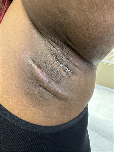
The patient’s recurrent indurated nodules under her arms and other intertriginous areas, often draining pus, are consistent with a diagnosis of hidradenitis suppurativa (HS).
HS is a chronic inflammatory and suppurative skin condition that primarily involves the sweat glands.1 The most commonly affected sites are intertriginous areas that include the axillae, groin, and perianal and inframammary regions.2 Prevalence of this disorder ranges from 0.05% to 4.1% of the population with an onset from puberty to adulthood, usually at around 40 years of age.3 Its incidence is twice as high in women as men and is more common in Black individuals.4,5
While its pathogenesis is not fully understood, it’s believed that excess proliferation of keratinocytes contributes to occlusion, leading to plugging of hair follicle ducts. Hormones, smoking, and obesity may contribute to and exacerbate HS. Intertriginous areas are prone to friction, leading to inflammation and further clogging.
The inflammation evolves into a chronic foreign body-type granulomatous inflammation with the potential for rupture, tunneling, and draining sinuses, which, although malodorous, are sterile, separating HS from an infected abscess.5 The result is thick, dense, scarred tissue.
The diagnosis is clinical in nature, with the history and physical exam distinguishing it from other skin disorders. In addition to the recurring physical pain, there is the emotional distress and self-consciousness about the drainage, odor, and scarring. This particular patient said that she avoided wearing sleeveless shirts due to the lesions’ appearance.
Treatment is multifactorial. Smoking and obesity are contributory factors, so smoking cessation and weight loss are recommended. For very mild HS, topical clindamycin 1% twice daily may suffice, but usually, due to the amount of inflammation, oral antibiotics are the initial therapy. (The use of antibiotics is for their anti-inflammatory component, as the nodules and unruptured tracts are sterile.)
Doxycycline 100 mg twice daily is the usual starting systemic antibiotic. In more severe or resistant cases, a combination of clindamycin and rifampin 300 mg each twice daily is used. (Worth noting: Rifampin interacts with oral contraceptives and many of these patients are women of reproductive age.) Treatment length is usually long (10 to 12 weeks) and recurrence is common.3
Spironolactone 100 mg daily and metformin 1000 mg extended release daily, which reduces insulin resistance, may be helpful. Intralesional injections of 10 mg/mL of triamcinolone in sterile saline can relieve the painful inflamed tracts. Referral for biologic agents, including infliximab, may be needed in severe cases that do not respond to other measures. Although invasive, wide debridement of the diseased tissue can reduce the disease burden.6
This particular patient said that she’d stopped smoking 3 years earlier and would work on losing weight. She was prescribed topical clindamycin 1% lotion twice daily along with oral clindamycin and rifampin dosed as above for 3 months. She declined metformin and intralesional injections. At a follow-up appointment 3 weeks later, she was pleased with the decrease in inflammation and had only 1 remaining tender area of fluctuance. She again declined injections and planned to continue on her oral and topical antibiotics.
Photo courtesy of Daniel Stulberg, MD, FAAFP. Text courtesy of Derissa F. Raynold, MD, and Daniel Stulberg, MD, FAAFP, Department of Family and Community Medicine, Western Michigan University Homer Stryker, MD School of Medicine, Kalamazoo.
1. Wolkenstein P, Loundou A, Barrau K, et al. Quality of life impairment in hidradenitis suppurativa: a study of 61 cases. J Am Acad Dermatol. 2007;56:621-623. doi: 10.1016/j.jaad.2006.08.061
2. Storer MA, Danesh MJ, Sandhu ME, et al. An assessment of the relative impact of hidradenitis suppurativa, psoriasis, and obesity on quality of life. Int J Womens Dermatol. 2018;4:198-202. doi: 10.1016/j.ijwd.2018.08.009
3. Saunte DML, Jemec GBE. Hidradenitis suppurativa: advances in diagnosis and treatment. JAMA. 2017;318:2019-2032. doi: 10.1001/jama.2017.16691
4. Matusiak L, Bieniek A, Szepietowski JC. Psychophysical aspects of hidradenitis suppurativa. Acta Derm Venereol. 2010;90:264-268. doi: 10.2340/00015555-0866
5. Esmann S, Jemec GB. Psychosocial impact of hidradenitis suppurativa: a qualitative study. Acta Derm Venereol. 2011;91:328-332. doi: 10.2340/00015555-1082
6. Caposiena Caro RD, Cannizzaro MV, Botti E, et al. Clindamycin versus clindamycin plus rifampicin in hidradenitis suppurativa treatment: clinical and ultrasound observations. J Am Acad Dermatol. 2019;80:1314-1321. doi: 10.1016/j.jaad.2018.11.035
The patient’s recurrent indurated nodules under her arms and other intertriginous areas, often draining pus, are consistent with a diagnosis of hidradenitis suppurativa (HS).
HS is a chronic inflammatory and suppurative skin condition that primarily involves the sweat glands.1 The most commonly affected sites are intertriginous areas that include the axillae, groin, and perianal and inframammary regions.2 Prevalence of this disorder ranges from 0.05% to 4.1% of the population with an onset from puberty to adulthood, usually at around 40 years of age.3 Its incidence is twice as high in women as men and is more common in Black individuals.4,5
While its pathogenesis is not fully understood, it’s believed that excess proliferation of keratinocytes contributes to occlusion, leading to plugging of hair follicle ducts. Hormones, smoking, and obesity may contribute to and exacerbate HS. Intertriginous areas are prone to friction, leading to inflammation and further clogging.
The inflammation evolves into a chronic foreign body-type granulomatous inflammation with the potential for rupture, tunneling, and draining sinuses, which, although malodorous, are sterile, separating HS from an infected abscess.5 The result is thick, dense, scarred tissue.
The diagnosis is clinical in nature, with the history and physical exam distinguishing it from other skin disorders. In addition to the recurring physical pain, there is the emotional distress and self-consciousness about the drainage, odor, and scarring. This particular patient said that she avoided wearing sleeveless shirts due to the lesions’ appearance.
Treatment is multifactorial. Smoking and obesity are contributory factors, so smoking cessation and weight loss are recommended. For very mild HS, topical clindamycin 1% twice daily may suffice, but usually, due to the amount of inflammation, oral antibiotics are the initial therapy. (The use of antibiotics is for their anti-inflammatory component, as the nodules and unruptured tracts are sterile.)
Doxycycline 100 mg twice daily is the usual starting systemic antibiotic. In more severe or resistant cases, a combination of clindamycin and rifampin 300 mg each twice daily is used. (Worth noting: Rifampin interacts with oral contraceptives and many of these patients are women of reproductive age.) Treatment length is usually long (10 to 12 weeks) and recurrence is common.3
Spironolactone 100 mg daily and metformin 1000 mg extended release daily, which reduces insulin resistance, may be helpful. Intralesional injections of 10 mg/mL of triamcinolone in sterile saline can relieve the painful inflamed tracts. Referral for biologic agents, including infliximab, may be needed in severe cases that do not respond to other measures. Although invasive, wide debridement of the diseased tissue can reduce the disease burden.6
This particular patient said that she’d stopped smoking 3 years earlier and would work on losing weight. She was prescribed topical clindamycin 1% lotion twice daily along with oral clindamycin and rifampin dosed as above for 3 months. She declined metformin and intralesional injections. At a follow-up appointment 3 weeks later, she was pleased with the decrease in inflammation and had only 1 remaining tender area of fluctuance. She again declined injections and planned to continue on her oral and topical antibiotics.
Photo courtesy of Daniel Stulberg, MD, FAAFP. Text courtesy of Derissa F. Raynold, MD, and Daniel Stulberg, MD, FAAFP, Department of Family and Community Medicine, Western Michigan University Homer Stryker, MD School of Medicine, Kalamazoo.
The patient’s recurrent indurated nodules under her arms and other intertriginous areas, often draining pus, are consistent with a diagnosis of hidradenitis suppurativa (HS).
HS is a chronic inflammatory and suppurative skin condition that primarily involves the sweat glands.1 The most commonly affected sites are intertriginous areas that include the axillae, groin, and perianal and inframammary regions.2 Prevalence of this disorder ranges from 0.05% to 4.1% of the population with an onset from puberty to adulthood, usually at around 40 years of age.3 Its incidence is twice as high in women as men and is more common in Black individuals.4,5
While its pathogenesis is not fully understood, it’s believed that excess proliferation of keratinocytes contributes to occlusion, leading to plugging of hair follicle ducts. Hormones, smoking, and obesity may contribute to and exacerbate HS. Intertriginous areas are prone to friction, leading to inflammation and further clogging.
The inflammation evolves into a chronic foreign body-type granulomatous inflammation with the potential for rupture, tunneling, and draining sinuses, which, although malodorous, are sterile, separating HS from an infected abscess.5 The result is thick, dense, scarred tissue.
The diagnosis is clinical in nature, with the history and physical exam distinguishing it from other skin disorders. In addition to the recurring physical pain, there is the emotional distress and self-consciousness about the drainage, odor, and scarring. This particular patient said that she avoided wearing sleeveless shirts due to the lesions’ appearance.
Treatment is multifactorial. Smoking and obesity are contributory factors, so smoking cessation and weight loss are recommended. For very mild HS, topical clindamycin 1% twice daily may suffice, but usually, due to the amount of inflammation, oral antibiotics are the initial therapy. (The use of antibiotics is for their anti-inflammatory component, as the nodules and unruptured tracts are sterile.)
Doxycycline 100 mg twice daily is the usual starting systemic antibiotic. In more severe or resistant cases, a combination of clindamycin and rifampin 300 mg each twice daily is used. (Worth noting: Rifampin interacts with oral contraceptives and many of these patients are women of reproductive age.) Treatment length is usually long (10 to 12 weeks) and recurrence is common.3
Spironolactone 100 mg daily and metformin 1000 mg extended release daily, which reduces insulin resistance, may be helpful. Intralesional injections of 10 mg/mL of triamcinolone in sterile saline can relieve the painful inflamed tracts. Referral for biologic agents, including infliximab, may be needed in severe cases that do not respond to other measures. Although invasive, wide debridement of the diseased tissue can reduce the disease burden.6
This particular patient said that she’d stopped smoking 3 years earlier and would work on losing weight. She was prescribed topical clindamycin 1% lotion twice daily along with oral clindamycin and rifampin dosed as above for 3 months. She declined metformin and intralesional injections. At a follow-up appointment 3 weeks later, she was pleased with the decrease in inflammation and had only 1 remaining tender area of fluctuance. She again declined injections and planned to continue on her oral and topical antibiotics.
Photo courtesy of Daniel Stulberg, MD, FAAFP. Text courtesy of Derissa F. Raynold, MD, and Daniel Stulberg, MD, FAAFP, Department of Family and Community Medicine, Western Michigan University Homer Stryker, MD School of Medicine, Kalamazoo.
1. Wolkenstein P, Loundou A, Barrau K, et al. Quality of life impairment in hidradenitis suppurativa: a study of 61 cases. J Am Acad Dermatol. 2007;56:621-623. doi: 10.1016/j.jaad.2006.08.061
2. Storer MA, Danesh MJ, Sandhu ME, et al. An assessment of the relative impact of hidradenitis suppurativa, psoriasis, and obesity on quality of life. Int J Womens Dermatol. 2018;4:198-202. doi: 10.1016/j.ijwd.2018.08.009
3. Saunte DML, Jemec GBE. Hidradenitis suppurativa: advances in diagnosis and treatment. JAMA. 2017;318:2019-2032. doi: 10.1001/jama.2017.16691
4. Matusiak L, Bieniek A, Szepietowski JC. Psychophysical aspects of hidradenitis suppurativa. Acta Derm Venereol. 2010;90:264-268. doi: 10.2340/00015555-0866
5. Esmann S, Jemec GB. Psychosocial impact of hidradenitis suppurativa: a qualitative study. Acta Derm Venereol. 2011;91:328-332. doi: 10.2340/00015555-1082
6. Caposiena Caro RD, Cannizzaro MV, Botti E, et al. Clindamycin versus clindamycin plus rifampicin in hidradenitis suppurativa treatment: clinical and ultrasound observations. J Am Acad Dermatol. 2019;80:1314-1321. doi: 10.1016/j.jaad.2018.11.035
1. Wolkenstein P, Loundou A, Barrau K, et al. Quality of life impairment in hidradenitis suppurativa: a study of 61 cases. J Am Acad Dermatol. 2007;56:621-623. doi: 10.1016/j.jaad.2006.08.061
2. Storer MA, Danesh MJ, Sandhu ME, et al. An assessment of the relative impact of hidradenitis suppurativa, psoriasis, and obesity on quality of life. Int J Womens Dermatol. 2018;4:198-202. doi: 10.1016/j.ijwd.2018.08.009
3. Saunte DML, Jemec GBE. Hidradenitis suppurativa: advances in diagnosis and treatment. JAMA. 2017;318:2019-2032. doi: 10.1001/jama.2017.16691
4. Matusiak L, Bieniek A, Szepietowski JC. Psychophysical aspects of hidradenitis suppurativa. Acta Derm Venereol. 2010;90:264-268. doi: 10.2340/00015555-0866
5. Esmann S, Jemec GB. Psychosocial impact of hidradenitis suppurativa: a qualitative study. Acta Derm Venereol. 2011;91:328-332. doi: 10.2340/00015555-1082
6. Caposiena Caro RD, Cannizzaro MV, Botti E, et al. Clindamycin versus clindamycin plus rifampicin in hidradenitis suppurativa treatment: clinical and ultrasound observations. J Am Acad Dermatol. 2019;80:1314-1321. doi: 10.1016/j.jaad.2018.11.035
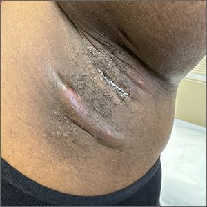
Penile rash
Biopsy revealed a lichenoid infiltrate in the upper dermis with thinning of the epidermis and a predominance of plasma cells. This finding, along with a red-orange, glossy lesion on the glans penis in an uncircumcised man is a classic presentation of Zoon balanitis.
Zoon balanitis is a chronic, idiopathic disorder that affects uncircumcised men who are middle-aged and older.1 Although the exact pathogenesis is unknown, it is hypothesized to be the result of chronic irritation due to poor hygiene/urine retention with prepuce dysfunction. The classic clinical presentation is an asymptomatic, well-defined, orange-to-red glazed patch with symmetric small red “cayenne pepper” spots contained within the glans penis and/or prepuce. Often there are symmetric “kissing lesions” where a second lesion of similar morphology is apparent on the prepuce where it meets the glans penis. While this disorder is typically asymptomatic, it can be associated with itching and/or burning.
There are several other inflammatory disorders in the differential for Zoon balanitis (erosive lichen planus, lichen sclerosus, psoriasis), but the most important consideration is penile intraepithelial carcinoma, specifically erythroplasia of Queyrat. Erythroplasia of Queyrat can be difficult to distinguish clinically and often requires a biopsy. Zoon balanitis will show a plasma cell predominant lichenoid infiltrate, whereas erythroplasia of Queyrat will show squamous cell carcinoma in situ.
It is important to note that Zoon balanitis is a clinicopathologic diagnosis and that zoonoid inflammation on biopsy is not pathognomonic, as this can also be seen in other inflammatory and neoplastic conditions. It is, therefore, advisable to follow up with patients with Zoon balanitis to ensure that the lesion is resolving and/or not getting worse.
Improved hygiene measures are the mainstay of treatment; circumcision is also effective.2 Both can help keep the glans clean and dry. In those with symptomatic disease, low-strength topical steroids including 1% hydrocortisone ointment or cream twice daily or topical calcineurin inhibitors (pimecrolimus 1% or tacrolimus 0.1%) twice daily can be used for symptom management.
Because this patient’s disease was asymptomatic, treatment was deferred. He was counseled to draw back the foreskin when urinating and to do the same while showering so that he could wash, then dry, the glans before returning the foreskin to its normal position. The patient is being monitored clinically.
Photo courtesy of Drew Mitchell, MD. Text courtesy of Drew Mitchell, MD, Department of Dermatology, and Daniel Stulberg, MD, FAAFP, Department of Family and Community Medicine, Western Michigan University Homer Stryker, MD School of Medicine, Kalamazoo.
1. Mallon E, Hawkins D, Dinneen M, et al. Circumcision and genital dermatoses. Arch Dermatol. 2000;136:350-354. doi: 10.1001/archderm.136.3.350
2. Kumar B, Sharma R, Rajagopalan M, et al. Plasma cell balanitis: clinical and histopathological features—response to circumcision. Genitourin Med. 1995;71:32-34. doi: 10.1136/sti.71.1.32
Biopsy revealed a lichenoid infiltrate in the upper dermis with thinning of the epidermis and a predominance of plasma cells. This finding, along with a red-orange, glossy lesion on the glans penis in an uncircumcised man is a classic presentation of Zoon balanitis.
Zoon balanitis is a chronic, idiopathic disorder that affects uncircumcised men who are middle-aged and older.1 Although the exact pathogenesis is unknown, it is hypothesized to be the result of chronic irritation due to poor hygiene/urine retention with prepuce dysfunction. The classic clinical presentation is an asymptomatic, well-defined, orange-to-red glazed patch with symmetric small red “cayenne pepper” spots contained within the glans penis and/or prepuce. Often there are symmetric “kissing lesions” where a second lesion of similar morphology is apparent on the prepuce where it meets the glans penis. While this disorder is typically asymptomatic, it can be associated with itching and/or burning.
There are several other inflammatory disorders in the differential for Zoon balanitis (erosive lichen planus, lichen sclerosus, psoriasis), but the most important consideration is penile intraepithelial carcinoma, specifically erythroplasia of Queyrat. Erythroplasia of Queyrat can be difficult to distinguish clinically and often requires a biopsy. Zoon balanitis will show a plasma cell predominant lichenoid infiltrate, whereas erythroplasia of Queyrat will show squamous cell carcinoma in situ.
It is important to note that Zoon balanitis is a clinicopathologic diagnosis and that zoonoid inflammation on biopsy is not pathognomonic, as this can also be seen in other inflammatory and neoplastic conditions. It is, therefore, advisable to follow up with patients with Zoon balanitis to ensure that the lesion is resolving and/or not getting worse.
Improved hygiene measures are the mainstay of treatment; circumcision is also effective.2 Both can help keep the glans clean and dry. In those with symptomatic disease, low-strength topical steroids including 1% hydrocortisone ointment or cream twice daily or topical calcineurin inhibitors (pimecrolimus 1% or tacrolimus 0.1%) twice daily can be used for symptom management.
Because this patient’s disease was asymptomatic, treatment was deferred. He was counseled to draw back the foreskin when urinating and to do the same while showering so that he could wash, then dry, the glans before returning the foreskin to its normal position. The patient is being monitored clinically.
Photo courtesy of Drew Mitchell, MD. Text courtesy of Drew Mitchell, MD, Department of Dermatology, and Daniel Stulberg, MD, FAAFP, Department of Family and Community Medicine, Western Michigan University Homer Stryker, MD School of Medicine, Kalamazoo.
Biopsy revealed a lichenoid infiltrate in the upper dermis with thinning of the epidermis and a predominance of plasma cells. This finding, along with a red-orange, glossy lesion on the glans penis in an uncircumcised man is a classic presentation of Zoon balanitis.
Zoon balanitis is a chronic, idiopathic disorder that affects uncircumcised men who are middle-aged and older.1 Although the exact pathogenesis is unknown, it is hypothesized to be the result of chronic irritation due to poor hygiene/urine retention with prepuce dysfunction. The classic clinical presentation is an asymptomatic, well-defined, orange-to-red glazed patch with symmetric small red “cayenne pepper” spots contained within the glans penis and/or prepuce. Often there are symmetric “kissing lesions” where a second lesion of similar morphology is apparent on the prepuce where it meets the glans penis. While this disorder is typically asymptomatic, it can be associated with itching and/or burning.
There are several other inflammatory disorders in the differential for Zoon balanitis (erosive lichen planus, lichen sclerosus, psoriasis), but the most important consideration is penile intraepithelial carcinoma, specifically erythroplasia of Queyrat. Erythroplasia of Queyrat can be difficult to distinguish clinically and often requires a biopsy. Zoon balanitis will show a plasma cell predominant lichenoid infiltrate, whereas erythroplasia of Queyrat will show squamous cell carcinoma in situ.
It is important to note that Zoon balanitis is a clinicopathologic diagnosis and that zoonoid inflammation on biopsy is not pathognomonic, as this can also be seen in other inflammatory and neoplastic conditions. It is, therefore, advisable to follow up with patients with Zoon balanitis to ensure that the lesion is resolving and/or not getting worse.
Improved hygiene measures are the mainstay of treatment; circumcision is also effective.2 Both can help keep the glans clean and dry. In those with symptomatic disease, low-strength topical steroids including 1% hydrocortisone ointment or cream twice daily or topical calcineurin inhibitors (pimecrolimus 1% or tacrolimus 0.1%) twice daily can be used for symptom management.
Because this patient’s disease was asymptomatic, treatment was deferred. He was counseled to draw back the foreskin when urinating and to do the same while showering so that he could wash, then dry, the glans before returning the foreskin to its normal position. The patient is being monitored clinically.
Photo courtesy of Drew Mitchell, MD. Text courtesy of Drew Mitchell, MD, Department of Dermatology, and Daniel Stulberg, MD, FAAFP, Department of Family and Community Medicine, Western Michigan University Homer Stryker, MD School of Medicine, Kalamazoo.
1. Mallon E, Hawkins D, Dinneen M, et al. Circumcision and genital dermatoses. Arch Dermatol. 2000;136:350-354. doi: 10.1001/archderm.136.3.350
2. Kumar B, Sharma R, Rajagopalan M, et al. Plasma cell balanitis: clinical and histopathological features—response to circumcision. Genitourin Med. 1995;71:32-34. doi: 10.1136/sti.71.1.32
1. Mallon E, Hawkins D, Dinneen M, et al. Circumcision and genital dermatoses. Arch Dermatol. 2000;136:350-354. doi: 10.1001/archderm.136.3.350
2. Kumar B, Sharma R, Rajagopalan M, et al. Plasma cell balanitis: clinical and histopathological features—response to circumcision. Genitourin Med. 1995;71:32-34. doi: 10.1136/sti.71.1.32
Semaglutide doesn’t improve fibrosis in NASH-related cirrhosis
according to a phase 2 trial.
However, the glucagonlike peptide–1 (GLP-1) receptor agonist led to improvements in liver enzymes, liver steatosis, weight, triglycerides, and very low-density lipoprotein (VLDL) cholesterol. Similar proportions of patients in each group reported adverse events, such as nausea, diarrhea, and vomiting.
“Previous studies in patients with NASH and stage 2 or 3 fibrosis have shown that semaglutide can improve NASH resolution over 72 weeks. However, there are limited data on whether any therapy is effective in patients with NASH cirrhosis,” lead author Rohit Loomba, MD, founding director of the NAFLD Research Center at the University of California, San Diego, said in an interview.
“Although semaglutide did not succeed in improving histological fibrosis, it had success in improving other clinically important parameters, such as cardiometabolic risk factors, liver enzymes, liver fat, and noninvasive biomarkers of fibrosis,” he said.
The study was published online in The Lancet Gastroenterology & Hepatology.
Analyzing safety and efficacy
Dr. Loomba and colleagues conducted a double-blind, placebo-controlled phase 2 trial that enrolled 71 patients at 38 centers in the United States and Europe between June 2019 and April 2021. Adults with biopsy-confirmed NASH-related cirrhosis and a body mass index (BMI) of at least 27 kg/m2 were randomly assigned 2:1 to receive either once-weekly subcutaneous semaglutide at 2.4 mg or a visually matching placebo.
Patients were randomly allocated through an interactive web system, which stratified participants on the basis of the presence or absence of type 2 diabetes. Patients, investigators, and outcomes analysts were masked to the treatment assignment.
The primary endpoint was the proportion of patients with an improvement in liver fibrosis of one stage or more without a worsening of NASH after 48 weeks, which was measured through biopsy in the intention-to-treat population. Safety was also assessed in all patients who received at least one dose of semaglutide.
Among the 71 patients, 47 were randomly assigned to the semaglutide group and 24 to the placebo group. About 90% completed treatment, and 63 had evaluable paired biopsies for primary endpoint assessment.
Between the groups, 49 participants (69%) were women and 22 were men. The average age was 59.5 years, and the average BMI was 34.9. About 75% of patients had diabetes at baseline, with an average hemoglobin A1c of 7.1%.
After 48 weeks, researchers found no statistically significant difference between the groups in the proportion of patients with an improvement in liver fibrosis of one stage or more without worsening of NASH. In the semaglutide group, five patients (11%) had an improvement, compared with seven patients (29%) in the placebo group (odds ratio, 0.28; 95% confidence interval, 0.06-1.24, P = .087).
There also wasn’t a significant difference between groups in the proportion of patients who achieved NASH resolution. In the semaglutide group, 16 patients (34%) had resolution, compared with 5 patients (21%) in the placebo group (OR, 1.97; 95% CI, 0.56-7.91; P = .29).
In addition, a lower proportion of patients achieved both NASH resolution and improvement in liver fibrosis with semaglutide versus placebo, although the difference wasn’t significant. In the semaglutide group, three patients (6%) achieved both, compared with three patients (13%) in the placebo group (OR, 0.48; 95% CI, 0.06-3.91; P = .4). A lower proportion of patients had an improvement in liver fibrosis stage with semaglutide versus placebo.
Some improvements seen
However, the semaglutide group had significantly greater improvements in liver steatosis (but not stiffness), liver fat volume, procollagen 3 peptide, and liver enzymes such as ALT, AST, and gamma-glutamyltransferase.
Body weight decreased by 8.83% in the semaglutide group, compared with 0.09% in the placebo group, which was a significant difference. BMI, waist circumference, triglycerides, and VLDL cholesterol were also significantly lower in the semaglutide group, but total cholesterol and blood pressure measurements weren’t significantly different. Among those with type 2 diabetes, A1c also decreased in the semaglutide group but did not in the placebo group.
Similar proportions of patients in each group reported adverse events. In the semaglutide group, 42 patients (89%) had an adverse event, compared with 19 patients (79%) in the placebo group. In addition, six patients (13%) in the semaglutide group and two patients (8%) in the placebo group reported serious adverse events.
The most common adverse events in the semaglutide and placebo groups were nausea (45% and 17%), diarrhea (19% and 8%), and vomiting (17% and none), which mainly occurred during treatment initiation or dose escalation. No patients withdrew from the trial because of adverse events, although five had a dose reduction. Hepatic and renal function remained stable after semaglutide treatment, and there were no decompensating events or deaths.
“GLP-1 analogue exposure – among patients with compensated cirrhosis who suffer from morbid obesity and type 2 diabetes – for the treatment of diabetes appears to be well-tolerated and may be safe,” Dr. Loomba said. “Further studies are needed in this study population.”
Considering next steps
Dr. Loomba and colleagues are continuing research around risk factors linked to advanced fibrosis, such as type 2 diabetes, a family history of cirrhosis, and the presence of key genetic risk alleles. Gut dysbiosis also appears to increase the risk for advanced fatty liver disease, he said.
Future clinical trials could focus on therapeutic options for patients with advanced fibrosis, particularly those with cirrhosis who face increased risks for liver-related complications and mortality.
“As these patients are oftentimes excluded from initial randomized controlled trials, we have significantly less information on how to address obesity, type 2 diabetes, and NASH in these patients,” Fernando Bril, MD, a physician-scientist focused on NASH-related research at the University of Alabama at Birmingham, said in an interview.
Dr. Bril, who wasn’t involved with this study, wrote an accompanying editorial in The Lancet Gastroenterology & Hepatology.
Patients with NASH-related cirrhosis may have progressed to a point of the disease where fibrosis regression may be more difficult to achieve, he said.
“This emphasizes that early diagnosis of patients with NASH is crucial,” he said.
“Therefore, primary care providers, endocrinologists, and diabetologists need to have a low threshold to suspect liver disease in patients with overweight, obesity, and/or type 2 diabetes. Only this will allow for early initiation of therapy, which may delay the progression of liver disease.”
In further research, investigators may want to consider the lack of NASH resolution, a result that could be caused by this study being underpowered, Dr. Bril noted. The trend in resolution in this study appeared similar to improvements seen in NASH patients without cirrhosis in other studies, he said. The weight reduction and improved diabetes control in this group also shows promise.
“While a purist may be adamant that this was a negative study for histological outcomes, it is essential to take note of the positive results in many secondary outcomes,” he said. “Improving cardiometabolic risk in these patients is essential because many still die of cardiovascular disease and not liver-related complications.”
At the same time, it’s important to note that NASH can’t be oversimplified as “a matter of weight,” Dr. Bril said. Significant weight loss in the study didn’t result in histologic improvement, which means other strategies are needed to treat the disease.
“Negative results from this study emphasize that monotherapy may not be enough to improve NASH and liver fibrosis,” he said. “In a similar way we treat type 2 diabetes and hypertension with combination therapy, we need to consider a similar approach for patients with NASH.”
The study was sponsored by Novo Nordisk, which manufactures semaglutide. The authors declared grant funding, speaker fees, and consultant roles with numerous pharmaceutical companies. Dr. Bril had no relevant disclosures.
A version of this article first appeared on Medscape.com.
according to a phase 2 trial.
However, the glucagonlike peptide–1 (GLP-1) receptor agonist led to improvements in liver enzymes, liver steatosis, weight, triglycerides, and very low-density lipoprotein (VLDL) cholesterol. Similar proportions of patients in each group reported adverse events, such as nausea, diarrhea, and vomiting.
“Previous studies in patients with NASH and stage 2 or 3 fibrosis have shown that semaglutide can improve NASH resolution over 72 weeks. However, there are limited data on whether any therapy is effective in patients with NASH cirrhosis,” lead author Rohit Loomba, MD, founding director of the NAFLD Research Center at the University of California, San Diego, said in an interview.
“Although semaglutide did not succeed in improving histological fibrosis, it had success in improving other clinically important parameters, such as cardiometabolic risk factors, liver enzymes, liver fat, and noninvasive biomarkers of fibrosis,” he said.
The study was published online in The Lancet Gastroenterology & Hepatology.
Analyzing safety and efficacy
Dr. Loomba and colleagues conducted a double-blind, placebo-controlled phase 2 trial that enrolled 71 patients at 38 centers in the United States and Europe between June 2019 and April 2021. Adults with biopsy-confirmed NASH-related cirrhosis and a body mass index (BMI) of at least 27 kg/m2 were randomly assigned 2:1 to receive either once-weekly subcutaneous semaglutide at 2.4 mg or a visually matching placebo.
Patients were randomly allocated through an interactive web system, which stratified participants on the basis of the presence or absence of type 2 diabetes. Patients, investigators, and outcomes analysts were masked to the treatment assignment.
The primary endpoint was the proportion of patients with an improvement in liver fibrosis of one stage or more without a worsening of NASH after 48 weeks, which was measured through biopsy in the intention-to-treat population. Safety was also assessed in all patients who received at least one dose of semaglutide.
Among the 71 patients, 47 were randomly assigned to the semaglutide group and 24 to the placebo group. About 90% completed treatment, and 63 had evaluable paired biopsies for primary endpoint assessment.
Between the groups, 49 participants (69%) were women and 22 were men. The average age was 59.5 years, and the average BMI was 34.9. About 75% of patients had diabetes at baseline, with an average hemoglobin A1c of 7.1%.
After 48 weeks, researchers found no statistically significant difference between the groups in the proportion of patients with an improvement in liver fibrosis of one stage or more without worsening of NASH. In the semaglutide group, five patients (11%) had an improvement, compared with seven patients (29%) in the placebo group (odds ratio, 0.28; 95% confidence interval, 0.06-1.24, P = .087).
There also wasn’t a significant difference between groups in the proportion of patients who achieved NASH resolution. In the semaglutide group, 16 patients (34%) had resolution, compared with 5 patients (21%) in the placebo group (OR, 1.97; 95% CI, 0.56-7.91; P = .29).
In addition, a lower proportion of patients achieved both NASH resolution and improvement in liver fibrosis with semaglutide versus placebo, although the difference wasn’t significant. In the semaglutide group, three patients (6%) achieved both, compared with three patients (13%) in the placebo group (OR, 0.48; 95% CI, 0.06-3.91; P = .4). A lower proportion of patients had an improvement in liver fibrosis stage with semaglutide versus placebo.
Some improvements seen
However, the semaglutide group had significantly greater improvements in liver steatosis (but not stiffness), liver fat volume, procollagen 3 peptide, and liver enzymes such as ALT, AST, and gamma-glutamyltransferase.
Body weight decreased by 8.83% in the semaglutide group, compared with 0.09% in the placebo group, which was a significant difference. BMI, waist circumference, triglycerides, and VLDL cholesterol were also significantly lower in the semaglutide group, but total cholesterol and blood pressure measurements weren’t significantly different. Among those with type 2 diabetes, A1c also decreased in the semaglutide group but did not in the placebo group.
Similar proportions of patients in each group reported adverse events. In the semaglutide group, 42 patients (89%) had an adverse event, compared with 19 patients (79%) in the placebo group. In addition, six patients (13%) in the semaglutide group and two patients (8%) in the placebo group reported serious adverse events.
The most common adverse events in the semaglutide and placebo groups were nausea (45% and 17%), diarrhea (19% and 8%), and vomiting (17% and none), which mainly occurred during treatment initiation or dose escalation. No patients withdrew from the trial because of adverse events, although five had a dose reduction. Hepatic and renal function remained stable after semaglutide treatment, and there were no decompensating events or deaths.
“GLP-1 analogue exposure – among patients with compensated cirrhosis who suffer from morbid obesity and type 2 diabetes – for the treatment of diabetes appears to be well-tolerated and may be safe,” Dr. Loomba said. “Further studies are needed in this study population.”
Considering next steps
Dr. Loomba and colleagues are continuing research around risk factors linked to advanced fibrosis, such as type 2 diabetes, a family history of cirrhosis, and the presence of key genetic risk alleles. Gut dysbiosis also appears to increase the risk for advanced fatty liver disease, he said.
Future clinical trials could focus on therapeutic options for patients with advanced fibrosis, particularly those with cirrhosis who face increased risks for liver-related complications and mortality.
“As these patients are oftentimes excluded from initial randomized controlled trials, we have significantly less information on how to address obesity, type 2 diabetes, and NASH in these patients,” Fernando Bril, MD, a physician-scientist focused on NASH-related research at the University of Alabama at Birmingham, said in an interview.
Dr. Bril, who wasn’t involved with this study, wrote an accompanying editorial in The Lancet Gastroenterology & Hepatology.
Patients with NASH-related cirrhosis may have progressed to a point of the disease where fibrosis regression may be more difficult to achieve, he said.
“This emphasizes that early diagnosis of patients with NASH is crucial,” he said.
“Therefore, primary care providers, endocrinologists, and diabetologists need to have a low threshold to suspect liver disease in patients with overweight, obesity, and/or type 2 diabetes. Only this will allow for early initiation of therapy, which may delay the progression of liver disease.”
In further research, investigators may want to consider the lack of NASH resolution, a result that could be caused by this study being underpowered, Dr. Bril noted. The trend in resolution in this study appeared similar to improvements seen in NASH patients without cirrhosis in other studies, he said. The weight reduction and improved diabetes control in this group also shows promise.
“While a purist may be adamant that this was a negative study for histological outcomes, it is essential to take note of the positive results in many secondary outcomes,” he said. “Improving cardiometabolic risk in these patients is essential because many still die of cardiovascular disease and not liver-related complications.”
At the same time, it’s important to note that NASH can’t be oversimplified as “a matter of weight,” Dr. Bril said. Significant weight loss in the study didn’t result in histologic improvement, which means other strategies are needed to treat the disease.
“Negative results from this study emphasize that monotherapy may not be enough to improve NASH and liver fibrosis,” he said. “In a similar way we treat type 2 diabetes and hypertension with combination therapy, we need to consider a similar approach for patients with NASH.”
The study was sponsored by Novo Nordisk, which manufactures semaglutide. The authors declared grant funding, speaker fees, and consultant roles with numerous pharmaceutical companies. Dr. Bril had no relevant disclosures.
A version of this article first appeared on Medscape.com.
according to a phase 2 trial.
However, the glucagonlike peptide–1 (GLP-1) receptor agonist led to improvements in liver enzymes, liver steatosis, weight, triglycerides, and very low-density lipoprotein (VLDL) cholesterol. Similar proportions of patients in each group reported adverse events, such as nausea, diarrhea, and vomiting.
“Previous studies in patients with NASH and stage 2 or 3 fibrosis have shown that semaglutide can improve NASH resolution over 72 weeks. However, there are limited data on whether any therapy is effective in patients with NASH cirrhosis,” lead author Rohit Loomba, MD, founding director of the NAFLD Research Center at the University of California, San Diego, said in an interview.
“Although semaglutide did not succeed in improving histological fibrosis, it had success in improving other clinically important parameters, such as cardiometabolic risk factors, liver enzymes, liver fat, and noninvasive biomarkers of fibrosis,” he said.
The study was published online in The Lancet Gastroenterology & Hepatology.
Analyzing safety and efficacy
Dr. Loomba and colleagues conducted a double-blind, placebo-controlled phase 2 trial that enrolled 71 patients at 38 centers in the United States and Europe between June 2019 and April 2021. Adults with biopsy-confirmed NASH-related cirrhosis and a body mass index (BMI) of at least 27 kg/m2 were randomly assigned 2:1 to receive either once-weekly subcutaneous semaglutide at 2.4 mg or a visually matching placebo.
Patients were randomly allocated through an interactive web system, which stratified participants on the basis of the presence or absence of type 2 diabetes. Patients, investigators, and outcomes analysts were masked to the treatment assignment.
The primary endpoint was the proportion of patients with an improvement in liver fibrosis of one stage or more without a worsening of NASH after 48 weeks, which was measured through biopsy in the intention-to-treat population. Safety was also assessed in all patients who received at least one dose of semaglutide.
Among the 71 patients, 47 were randomly assigned to the semaglutide group and 24 to the placebo group. About 90% completed treatment, and 63 had evaluable paired biopsies for primary endpoint assessment.
Between the groups, 49 participants (69%) were women and 22 were men. The average age was 59.5 years, and the average BMI was 34.9. About 75% of patients had diabetes at baseline, with an average hemoglobin A1c of 7.1%.
After 48 weeks, researchers found no statistically significant difference between the groups in the proportion of patients with an improvement in liver fibrosis of one stage or more without worsening of NASH. In the semaglutide group, five patients (11%) had an improvement, compared with seven patients (29%) in the placebo group (odds ratio, 0.28; 95% confidence interval, 0.06-1.24, P = .087).
There also wasn’t a significant difference between groups in the proportion of patients who achieved NASH resolution. In the semaglutide group, 16 patients (34%) had resolution, compared with 5 patients (21%) in the placebo group (OR, 1.97; 95% CI, 0.56-7.91; P = .29).
In addition, a lower proportion of patients achieved both NASH resolution and improvement in liver fibrosis with semaglutide versus placebo, although the difference wasn’t significant. In the semaglutide group, three patients (6%) achieved both, compared with three patients (13%) in the placebo group (OR, 0.48; 95% CI, 0.06-3.91; P = .4). A lower proportion of patients had an improvement in liver fibrosis stage with semaglutide versus placebo.
Some improvements seen
However, the semaglutide group had significantly greater improvements in liver steatosis (but not stiffness), liver fat volume, procollagen 3 peptide, and liver enzymes such as ALT, AST, and gamma-glutamyltransferase.
Body weight decreased by 8.83% in the semaglutide group, compared with 0.09% in the placebo group, which was a significant difference. BMI, waist circumference, triglycerides, and VLDL cholesterol were also significantly lower in the semaglutide group, but total cholesterol and blood pressure measurements weren’t significantly different. Among those with type 2 diabetes, A1c also decreased in the semaglutide group but did not in the placebo group.
Similar proportions of patients in each group reported adverse events. In the semaglutide group, 42 patients (89%) had an adverse event, compared with 19 patients (79%) in the placebo group. In addition, six patients (13%) in the semaglutide group and two patients (8%) in the placebo group reported serious adverse events.
The most common adverse events in the semaglutide and placebo groups were nausea (45% and 17%), diarrhea (19% and 8%), and vomiting (17% and none), which mainly occurred during treatment initiation or dose escalation. No patients withdrew from the trial because of adverse events, although five had a dose reduction. Hepatic and renal function remained stable after semaglutide treatment, and there were no decompensating events or deaths.
“GLP-1 analogue exposure – among patients with compensated cirrhosis who suffer from morbid obesity and type 2 diabetes – for the treatment of diabetes appears to be well-tolerated and may be safe,” Dr. Loomba said. “Further studies are needed in this study population.”
Considering next steps
Dr. Loomba and colleagues are continuing research around risk factors linked to advanced fibrosis, such as type 2 diabetes, a family history of cirrhosis, and the presence of key genetic risk alleles. Gut dysbiosis also appears to increase the risk for advanced fatty liver disease, he said.
Future clinical trials could focus on therapeutic options for patients with advanced fibrosis, particularly those with cirrhosis who face increased risks for liver-related complications and mortality.
“As these patients are oftentimes excluded from initial randomized controlled trials, we have significantly less information on how to address obesity, type 2 diabetes, and NASH in these patients,” Fernando Bril, MD, a physician-scientist focused on NASH-related research at the University of Alabama at Birmingham, said in an interview.
Dr. Bril, who wasn’t involved with this study, wrote an accompanying editorial in The Lancet Gastroenterology & Hepatology.
Patients with NASH-related cirrhosis may have progressed to a point of the disease where fibrosis regression may be more difficult to achieve, he said.
“This emphasizes that early diagnosis of patients with NASH is crucial,” he said.
“Therefore, primary care providers, endocrinologists, and diabetologists need to have a low threshold to suspect liver disease in patients with overweight, obesity, and/or type 2 diabetes. Only this will allow for early initiation of therapy, which may delay the progression of liver disease.”
In further research, investigators may want to consider the lack of NASH resolution, a result that could be caused by this study being underpowered, Dr. Bril noted. The trend in resolution in this study appeared similar to improvements seen in NASH patients without cirrhosis in other studies, he said. The weight reduction and improved diabetes control in this group also shows promise.
“While a purist may be adamant that this was a negative study for histological outcomes, it is essential to take note of the positive results in many secondary outcomes,” he said. “Improving cardiometabolic risk in these patients is essential because many still die of cardiovascular disease and not liver-related complications.”
At the same time, it’s important to note that NASH can’t be oversimplified as “a matter of weight,” Dr. Bril said. Significant weight loss in the study didn’t result in histologic improvement, which means other strategies are needed to treat the disease.
“Negative results from this study emphasize that monotherapy may not be enough to improve NASH and liver fibrosis,” he said. “In a similar way we treat type 2 diabetes and hypertension with combination therapy, we need to consider a similar approach for patients with NASH.”
The study was sponsored by Novo Nordisk, which manufactures semaglutide. The authors declared grant funding, speaker fees, and consultant roles with numerous pharmaceutical companies. Dr. Bril had no relevant disclosures.
A version of this article first appeared on Medscape.com.
FROM THE LANCET GASTROENTEROLOGY & HEPATOLOGY
SBRT: Alternative to surgery in early stage lung cancer?
, according to population-based data from a German cancer registry.
“From a public health perspective, SBRT is a good therapeutic option in terms of survival, especially for elderly and inoperable patients,” noted the study authors, led by Jörg Andreas Müller, MD, department of radiation oncology, University Hospital of Halle, Germany.
The analysis was published online in the journal Strahlentherapie Und Onkologie.
Surgery remains the standard of care for early stage NSCLC. However, many patients are not eligible for surgery because of the tumor’s location, age, frailty, or comorbidities.
Before the introduction of SBRT, conventional radiation therapy was the only reasonable option for inoperable patients, with study data showing only a small survival improvement in treated vs. untreated patients.
High-precision, image-guided SBRT offers better tumor control with limited toxicity. And while many radiation oncology centers in Germany adopted SBRT as an alternative treatment for surgery after 2000, few population-based studies evaluating SBRT’s impact on overall survival exist.
Using the German clinical cancer registry of Berlin-Brandenburg, Dr. Müller and colleagues assessed SBRT as an alternative to surgery in 558 patients with stage I and II NSCLC, diagnosed between 2000 and 2015.
More patients received surgery than SBRT (74% vs. 26%). Those who received SBRT were younger than those in the surgery group and had better Karnofsky performance status.
Among patients in the SBRT group, median survival was 19 months overall and 27 months in patients over age 75. In the surgery group, median survival was 22 months overall and 24 months in those over 75.
In a univariate survival model of a propensity-matched sample of 292 patients – half of whom received SBRT – survival rates were similar among those who underwent SBRT versus surgery (hazard ratio [HR], 1.2; P = .2).
Survival was also similar in the two treatment groups in a T1 subanalysis (HR, 1.12; P = .7) as well as in patients over age 75 (HR, 0.86; P = .5).
Better performance status scores were associated with improved survival, and higher histological grades and TNM stages were linked to higher mortality risk. The availability of histological data did not have a significant impact on survival outcomes.
Overall, the findings suggest that SBRT and surgery offer comparable survival outcomes in early stage NSCLC and “the availability of histological data might not be decisive for treatment planning,” Dr. Müller and colleagues said.
Drew Moghanaki, MD, chief of the thoracic oncology service at UCLA Health Jonsson Comprehensive Cancer Center, Los Angeles, highlighted the findings on Twitter.
A thoracic surgeon from Germany responded with several concerns about the study, including the use of statistics with univariate modeling and undiagnosed lymph node (N) status.
Dr. Moghanaki replied that these “concerns summarize how we USED to think. It increasingly seems they aren’t as important as our teachers once thought they were. As we move into the future we need to reassess the data that supported these recommendations as they seem more academic than patient centered.”
The study authors reported no specific funding, and no relevant financial relationships.
A version of this article first appeared on Medscape.com.
, according to population-based data from a German cancer registry.
“From a public health perspective, SBRT is a good therapeutic option in terms of survival, especially for elderly and inoperable patients,” noted the study authors, led by Jörg Andreas Müller, MD, department of radiation oncology, University Hospital of Halle, Germany.
The analysis was published online in the journal Strahlentherapie Und Onkologie.
Surgery remains the standard of care for early stage NSCLC. However, many patients are not eligible for surgery because of the tumor’s location, age, frailty, or comorbidities.
Before the introduction of SBRT, conventional radiation therapy was the only reasonable option for inoperable patients, with study data showing only a small survival improvement in treated vs. untreated patients.
High-precision, image-guided SBRT offers better tumor control with limited toxicity. And while many radiation oncology centers in Germany adopted SBRT as an alternative treatment for surgery after 2000, few population-based studies evaluating SBRT’s impact on overall survival exist.
Using the German clinical cancer registry of Berlin-Brandenburg, Dr. Müller and colleagues assessed SBRT as an alternative to surgery in 558 patients with stage I and II NSCLC, diagnosed between 2000 and 2015.
More patients received surgery than SBRT (74% vs. 26%). Those who received SBRT were younger than those in the surgery group and had better Karnofsky performance status.
Among patients in the SBRT group, median survival was 19 months overall and 27 months in patients over age 75. In the surgery group, median survival was 22 months overall and 24 months in those over 75.
In a univariate survival model of a propensity-matched sample of 292 patients – half of whom received SBRT – survival rates were similar among those who underwent SBRT versus surgery (hazard ratio [HR], 1.2; P = .2).
Survival was also similar in the two treatment groups in a T1 subanalysis (HR, 1.12; P = .7) as well as in patients over age 75 (HR, 0.86; P = .5).
Better performance status scores were associated with improved survival, and higher histological grades and TNM stages were linked to higher mortality risk. The availability of histological data did not have a significant impact on survival outcomes.
Overall, the findings suggest that SBRT and surgery offer comparable survival outcomes in early stage NSCLC and “the availability of histological data might not be decisive for treatment planning,” Dr. Müller and colleagues said.
Drew Moghanaki, MD, chief of the thoracic oncology service at UCLA Health Jonsson Comprehensive Cancer Center, Los Angeles, highlighted the findings on Twitter.
A thoracic surgeon from Germany responded with several concerns about the study, including the use of statistics with univariate modeling and undiagnosed lymph node (N) status.
Dr. Moghanaki replied that these “concerns summarize how we USED to think. It increasingly seems they aren’t as important as our teachers once thought they were. As we move into the future we need to reassess the data that supported these recommendations as they seem more academic than patient centered.”
The study authors reported no specific funding, and no relevant financial relationships.
A version of this article first appeared on Medscape.com.
, according to population-based data from a German cancer registry.
“From a public health perspective, SBRT is a good therapeutic option in terms of survival, especially for elderly and inoperable patients,” noted the study authors, led by Jörg Andreas Müller, MD, department of radiation oncology, University Hospital of Halle, Germany.
The analysis was published online in the journal Strahlentherapie Und Onkologie.
Surgery remains the standard of care for early stage NSCLC. However, many patients are not eligible for surgery because of the tumor’s location, age, frailty, or comorbidities.
Before the introduction of SBRT, conventional radiation therapy was the only reasonable option for inoperable patients, with study data showing only a small survival improvement in treated vs. untreated patients.
High-precision, image-guided SBRT offers better tumor control with limited toxicity. And while many radiation oncology centers in Germany adopted SBRT as an alternative treatment for surgery after 2000, few population-based studies evaluating SBRT’s impact on overall survival exist.
Using the German clinical cancer registry of Berlin-Brandenburg, Dr. Müller and colleagues assessed SBRT as an alternative to surgery in 558 patients with stage I and II NSCLC, diagnosed between 2000 and 2015.
More patients received surgery than SBRT (74% vs. 26%). Those who received SBRT were younger than those in the surgery group and had better Karnofsky performance status.
Among patients in the SBRT group, median survival was 19 months overall and 27 months in patients over age 75. In the surgery group, median survival was 22 months overall and 24 months in those over 75.
In a univariate survival model of a propensity-matched sample of 292 patients – half of whom received SBRT – survival rates were similar among those who underwent SBRT versus surgery (hazard ratio [HR], 1.2; P = .2).
Survival was also similar in the two treatment groups in a T1 subanalysis (HR, 1.12; P = .7) as well as in patients over age 75 (HR, 0.86; P = .5).
Better performance status scores were associated with improved survival, and higher histological grades and TNM stages were linked to higher mortality risk. The availability of histological data did not have a significant impact on survival outcomes.
Overall, the findings suggest that SBRT and surgery offer comparable survival outcomes in early stage NSCLC and “the availability of histological data might not be decisive for treatment planning,” Dr. Müller and colleagues said.
Drew Moghanaki, MD, chief of the thoracic oncology service at UCLA Health Jonsson Comprehensive Cancer Center, Los Angeles, highlighted the findings on Twitter.
A thoracic surgeon from Germany responded with several concerns about the study, including the use of statistics with univariate modeling and undiagnosed lymph node (N) status.
Dr. Moghanaki replied that these “concerns summarize how we USED to think. It increasingly seems they aren’t as important as our teachers once thought they were. As we move into the future we need to reassess the data that supported these recommendations as they seem more academic than patient centered.”
The study authors reported no specific funding, and no relevant financial relationships.
A version of this article first appeared on Medscape.com.
FROM STRAHLENTHERAPIE UND ONKOLOGIE
Mindfulness-based CBT an ‘important’ option for moderate depression
The findings suggest that “offering practitioner-supported MBCT-SH as an intervention for mild to moderate depression would improve outcomes and save money compared with practitioner-supported CBT-SH,” noted the investigators, led by Clara Strauss, PhD, DClinPsy, with the University of Sussex School of Psychology in England.
Practitioner-supported CBT-SH is recommended in U.K. national treatment guidelines for mild to moderate depression. However, some patients’ conditions don’t respond, and dropout rates are high.
The Low-Intensity Guided Help Through Mindfulness (LIGHTMind) trial tested practitioner-supported MBCT-SH as an alternative.
The findings have “important implications” for the more than 100,000 people currently offered CBT-SH for depression in the Improving Access to Psychological Therapies (IAPT) program each year and in publicly funded services elsewhere, the researchers noted.
If translated into routine practice, “this would see many more people recovering from depression while costing health services less money,” they added.
The study was published online in JAMA Psychiatry .
Practice changing?
The trial included 410 adults (mean age, 32 years; 62% women) with mild to moderate depression who were recruited from 10 publicly funded psychological therapy services in England as part of the IAPT program.
Participants were given one of two established self-help workbooks – The Mindful Way Workbook: An 8-Week Program to Free Yourself from Depression and Emotional Distress, written by the pioneers of MBCT, or Overcoming Depression and Low Mood, 3rd Edition: A Five Areas Approach, which is a CBT-SH program widely used in IAPT.
Use of the self-help books was supported by six structured phone or in-person sessions with a trained psychological well-being practitioner.
The primary outcome was depression symptom severity at 16 weeks, which was determined on the basis of Patient Health Questionnaire 9 (PHQ-9) score.
At 16 weeks following randomization, MBCT-SH led to significantly greater reductions in depression symptom severity compared with CBT-SH (mean PHQ-9 score, 7.2 vs. 8.6; between-group difference, 1.5 points; P = .009; d = −0.36).
MBCT-SH also had superior effects on anxiety symptom severity at 16 weeks.
At the 42-week follow-up, between-group effects on depression and anxiety symptom severity remained in the hypothesized direction but were nonsignificant.
This could be due in part by the greater postintervention psychological therapy accessed by participants in the CBT-SH group, the investigators noted.
Practitioner-supported MBCT-SH was more cost-effective than supported CBT-SH.
On average, the CBT-SH intervention cost health services £526 ($631) more per participant than the MBCT-SH intervention over the 42-week follow-up. The probability of MBCT-SH being cost-effective compared with CBT-SH exceeded 95%, the researchers noted.
Useful model for the United States
Commenting for this news organization, Zindel Segal, PhD, professor of psychology, University of Toronto, Scarborough, cautioned against making too much of the differences between the groups, because CBT-SH “trended positive and had a pretty healthy effect size, it just never reached significance.
“I wouldn’t say mindfulness drastically outperformed cognitive therapy. But cognitive therapy is a robust treatment in its own right, and so doing a little bit better is significant,” Dr. Segal said.
He also noted that, appropriately, the trial enrolled adults who were experiencing moderate depression and were not acutely ill. “That’s one of the rationales for self-help compared to providing patients with a more resource-intensive group treatment.
“If you look at the needs of people with moderate depression, what you find is that for cognitive therapy to work, negative thoughts and feelings need to be pervasive in order to make use of the techniques,” Dr. Segal explained.
“With mindfulness, you don’t need any to have constant negative thoughts or feelings. Anything that arises in your experience serves as grist for mill in terms of concentration and focus,” Dr. Segal said.
He also noted that mindfulness-based intervention is “more optimized” for people who are experiencing some measure of recovery or remission.
“It’s well suited for that, as it trends towards the wellness spectrum. But for people who might have greater levels of acuity or severity, cognitive-behavioral therapy might be indicated,” said Dr. Segal.
He also said the U.K. study findings are relevant to U.S. patients with depression.
“While it’s not disseminated in the same way through any kind of national program, the self-help books that are used are widely available, and the support that people were offered, either in person, telephone, or email, could be easily delivered. This would be a very useful model,” Dr. Segal said.
The LIGHTMind trial was funded by the National Institute for Health and Care Research and the Brighton and Sussex Clinical Trials Unit. Dr. Strauss has received grants from Headspace, is research lead for Sussex Mindfulness Centre, and has been chief investigator on National Institute for Health and Care Research. Dr. Segal is one of the authors of the MBCT-SH workbooks used in the study.
A version of this article first appeared on Medscape.com.
The findings suggest that “offering practitioner-supported MBCT-SH as an intervention for mild to moderate depression would improve outcomes and save money compared with practitioner-supported CBT-SH,” noted the investigators, led by Clara Strauss, PhD, DClinPsy, with the University of Sussex School of Psychology in England.
Practitioner-supported CBT-SH is recommended in U.K. national treatment guidelines for mild to moderate depression. However, some patients’ conditions don’t respond, and dropout rates are high.
The Low-Intensity Guided Help Through Mindfulness (LIGHTMind) trial tested practitioner-supported MBCT-SH as an alternative.
The findings have “important implications” for the more than 100,000 people currently offered CBT-SH for depression in the Improving Access to Psychological Therapies (IAPT) program each year and in publicly funded services elsewhere, the researchers noted.
If translated into routine practice, “this would see many more people recovering from depression while costing health services less money,” they added.
The study was published online in JAMA Psychiatry .
Practice changing?
The trial included 410 adults (mean age, 32 years; 62% women) with mild to moderate depression who were recruited from 10 publicly funded psychological therapy services in England as part of the IAPT program.
Participants were given one of two established self-help workbooks – The Mindful Way Workbook: An 8-Week Program to Free Yourself from Depression and Emotional Distress, written by the pioneers of MBCT, or Overcoming Depression and Low Mood, 3rd Edition: A Five Areas Approach, which is a CBT-SH program widely used in IAPT.
Use of the self-help books was supported by six structured phone or in-person sessions with a trained psychological well-being practitioner.
The primary outcome was depression symptom severity at 16 weeks, which was determined on the basis of Patient Health Questionnaire 9 (PHQ-9) score.
At 16 weeks following randomization, MBCT-SH led to significantly greater reductions in depression symptom severity compared with CBT-SH (mean PHQ-9 score, 7.2 vs. 8.6; between-group difference, 1.5 points; P = .009; d = −0.36).
MBCT-SH also had superior effects on anxiety symptom severity at 16 weeks.
At the 42-week follow-up, between-group effects on depression and anxiety symptom severity remained in the hypothesized direction but were nonsignificant.
This could be due in part by the greater postintervention psychological therapy accessed by participants in the CBT-SH group, the investigators noted.
Practitioner-supported MBCT-SH was more cost-effective than supported CBT-SH.
On average, the CBT-SH intervention cost health services £526 ($631) more per participant than the MBCT-SH intervention over the 42-week follow-up. The probability of MBCT-SH being cost-effective compared with CBT-SH exceeded 95%, the researchers noted.
Useful model for the United States
Commenting for this news organization, Zindel Segal, PhD, professor of psychology, University of Toronto, Scarborough, cautioned against making too much of the differences between the groups, because CBT-SH “trended positive and had a pretty healthy effect size, it just never reached significance.
“I wouldn’t say mindfulness drastically outperformed cognitive therapy. But cognitive therapy is a robust treatment in its own right, and so doing a little bit better is significant,” Dr. Segal said.
He also noted that, appropriately, the trial enrolled adults who were experiencing moderate depression and were not acutely ill. “That’s one of the rationales for self-help compared to providing patients with a more resource-intensive group treatment.
“If you look at the needs of people with moderate depression, what you find is that for cognitive therapy to work, negative thoughts and feelings need to be pervasive in order to make use of the techniques,” Dr. Segal explained.
“With mindfulness, you don’t need any to have constant negative thoughts or feelings. Anything that arises in your experience serves as grist for mill in terms of concentration and focus,” Dr. Segal said.
He also noted that mindfulness-based intervention is “more optimized” for people who are experiencing some measure of recovery or remission.
“It’s well suited for that, as it trends towards the wellness spectrum. But for people who might have greater levels of acuity or severity, cognitive-behavioral therapy might be indicated,” said Dr. Segal.
He also said the U.K. study findings are relevant to U.S. patients with depression.
“While it’s not disseminated in the same way through any kind of national program, the self-help books that are used are widely available, and the support that people were offered, either in person, telephone, or email, could be easily delivered. This would be a very useful model,” Dr. Segal said.
The LIGHTMind trial was funded by the National Institute for Health and Care Research and the Brighton and Sussex Clinical Trials Unit. Dr. Strauss has received grants from Headspace, is research lead for Sussex Mindfulness Centre, and has been chief investigator on National Institute for Health and Care Research. Dr. Segal is one of the authors of the MBCT-SH workbooks used in the study.
A version of this article first appeared on Medscape.com.
The findings suggest that “offering practitioner-supported MBCT-SH as an intervention for mild to moderate depression would improve outcomes and save money compared with practitioner-supported CBT-SH,” noted the investigators, led by Clara Strauss, PhD, DClinPsy, with the University of Sussex School of Psychology in England.
Practitioner-supported CBT-SH is recommended in U.K. national treatment guidelines for mild to moderate depression. However, some patients’ conditions don’t respond, and dropout rates are high.
The Low-Intensity Guided Help Through Mindfulness (LIGHTMind) trial tested practitioner-supported MBCT-SH as an alternative.
The findings have “important implications” for the more than 100,000 people currently offered CBT-SH for depression in the Improving Access to Psychological Therapies (IAPT) program each year and in publicly funded services elsewhere, the researchers noted.
If translated into routine practice, “this would see many more people recovering from depression while costing health services less money,” they added.
The study was published online in JAMA Psychiatry .
Practice changing?
The trial included 410 adults (mean age, 32 years; 62% women) with mild to moderate depression who were recruited from 10 publicly funded psychological therapy services in England as part of the IAPT program.
Participants were given one of two established self-help workbooks – The Mindful Way Workbook: An 8-Week Program to Free Yourself from Depression and Emotional Distress, written by the pioneers of MBCT, or Overcoming Depression and Low Mood, 3rd Edition: A Five Areas Approach, which is a CBT-SH program widely used in IAPT.
Use of the self-help books was supported by six structured phone or in-person sessions with a trained psychological well-being practitioner.
The primary outcome was depression symptom severity at 16 weeks, which was determined on the basis of Patient Health Questionnaire 9 (PHQ-9) score.
At 16 weeks following randomization, MBCT-SH led to significantly greater reductions in depression symptom severity compared with CBT-SH (mean PHQ-9 score, 7.2 vs. 8.6; between-group difference, 1.5 points; P = .009; d = −0.36).
MBCT-SH also had superior effects on anxiety symptom severity at 16 weeks.
At the 42-week follow-up, between-group effects on depression and anxiety symptom severity remained in the hypothesized direction but were nonsignificant.
This could be due in part by the greater postintervention psychological therapy accessed by participants in the CBT-SH group, the investigators noted.
Practitioner-supported MBCT-SH was more cost-effective than supported CBT-SH.
On average, the CBT-SH intervention cost health services £526 ($631) more per participant than the MBCT-SH intervention over the 42-week follow-up. The probability of MBCT-SH being cost-effective compared with CBT-SH exceeded 95%, the researchers noted.
Useful model for the United States
Commenting for this news organization, Zindel Segal, PhD, professor of psychology, University of Toronto, Scarborough, cautioned against making too much of the differences between the groups, because CBT-SH “trended positive and had a pretty healthy effect size, it just never reached significance.
“I wouldn’t say mindfulness drastically outperformed cognitive therapy. But cognitive therapy is a robust treatment in its own right, and so doing a little bit better is significant,” Dr. Segal said.
He also noted that, appropriately, the trial enrolled adults who were experiencing moderate depression and were not acutely ill. “That’s one of the rationales for self-help compared to providing patients with a more resource-intensive group treatment.
“If you look at the needs of people with moderate depression, what you find is that for cognitive therapy to work, negative thoughts and feelings need to be pervasive in order to make use of the techniques,” Dr. Segal explained.
“With mindfulness, you don’t need any to have constant negative thoughts or feelings. Anything that arises in your experience serves as grist for mill in terms of concentration and focus,” Dr. Segal said.
He also noted that mindfulness-based intervention is “more optimized” for people who are experiencing some measure of recovery or remission.
“It’s well suited for that, as it trends towards the wellness spectrum. But for people who might have greater levels of acuity or severity, cognitive-behavioral therapy might be indicated,” said Dr. Segal.
He also said the U.K. study findings are relevant to U.S. patients with depression.
“While it’s not disseminated in the same way through any kind of national program, the self-help books that are used are widely available, and the support that people were offered, either in person, telephone, or email, could be easily delivered. This would be a very useful model,” Dr. Segal said.
The LIGHTMind trial was funded by the National Institute for Health and Care Research and the Brighton and Sussex Clinical Trials Unit. Dr. Strauss has received grants from Headspace, is research lead for Sussex Mindfulness Centre, and has been chief investigator on National Institute for Health and Care Research. Dr. Segal is one of the authors of the MBCT-SH workbooks used in the study.
A version of this article first appeared on Medscape.com.
FROM JAMA PSYCHIATRY
Doctor’s checklist for treating long COVID patients
Lisa McCorkell had a mild bout of COVID-19 in March 2020. Young and healthy, she assumed that she would bounce back quickly. But when her fatigue, shortness of breath, and brain fog persisted, she realized that she most likely had long COVID.
“Back then, we as patients basically coined the term,” she said. While her first primary care provider was sympathetic, they were unsure how to treat her. After her insurance changed, she ended up with a second primary care provider who didn’t take her symptoms seriously. “They dismissed my complaints and told me they were all in my head. I didn’t seek care for a while after that.”
Ms. McCorkell’s symptoms improved after her first COVID vaccine in the spring of 2021. She also finally found a new primary care doctor she could trust. But as one of the founders of the Patient-Led Research Collaborative, a group of researchers who study long COVID, she said many doctors still don’t know the hallmark symptoms of the condition or how to treat it.
“There’s still a lack of education on what long COVID is, and the symptoms associated with it,” she said. “Many of the symptoms that occur in long COVID are symptoms of other chronic conditions, such as chronic fatigue syndrome, that are often dismissed. And even if providers believe patients and send them for a workup, many of the routine blood and imaging tests come back normal.”
The term “long COVID” emerged in May 2020. And though the condition was recognized within a few months of the start of the pandemic, doctors weren’t sure how to screen or treat it.
While knowledge has developed since then, primary care doctors are still in a tough spot. They’re often the first providers that patients turn to when they have symptoms of long COVID.
“There’s no clear algorithm to pick up long COVID – there are no definite blood tests or biomarkers, or specific things to look for on a physical exam,” said Lawrence Purpura, MD, an infectious disease specialist and director of the long COVID clinic at Columbia University Medical Center, New York. “It’s a complicated disease that can impact every organ system of the body.”
Even so, emerging research has identified a checklist of sorts that doctors should consider when a patient seeks care for what appears to be long COVID. Among them: the key systems and organs impacted by the disease, the most common symptoms, useful therapeutic options for symptom management that have been found to help people with long COVID, and the best healthy lifestyle choices that doctors can recommend to help their patients
Here’s a closer look at each of these aspects, based on research and interviews with experts, patients, and doctors.
Key systems, organs impacted
About 10% of people who are infected with COVID-19 go on to have long COVID, according to a recent study that Ms. McCorkell helped coauthor. But more than 3 years into the pandemic, much about the condition is still a mystery.
COVID is a unique virus because it can spread far and wide in a patient’s body. A December 2022 study, published in Nature, autopsied 44 people who died of COVID and found that the virus could spread throughout the body and persist, in one case as long as 230 days after symptoms started.
“We know that there are dozens of symptoms across multiple organ systems,” said Ms. McCorkell. “That makes it harder for a primary care physician to connect the dots and associate it with COVID.”
A paper published in Nature Medicine proposed one way to help guide diagnosis. It divided symptoms into four groups:
- Cardiac and renal issues such as heart palpitations, chest pain, and kidney damage
- Sleep and anxiety problems like insomnia, waking up in the middle of the night, and anxiety
- In the musculoskeletal and nervous systems: musculoskeletal pain, osteoarthritis, and problems with mental skills
- In the digestive and respiratory systems: trouble breathing, asthma, stomach pain, nausea, and vomiting
There were also specific patterns in these groups. People in the first group were more likely to be older, male, have other conditions and to have been infected during the first wave of the COVID pandemic. People in the second group were over 60% female, and were more likely to have had previous allergies or asthma. The third group was also about 60% female, and many of them already had autoimmune conditions such as rheumatoid arthritis. Members of the fourth group – also 60% female – were the least likely of all the groups to have another condition.
This research is helpful, because it gives doctors a better sense of what conditions might make a patient more likely to get long COVID, as well as specific symptoms to look out for, said Steven Flanagan, MD, a physical medicine and rehabilitation specialist at New York University Langone Medical Center who also specializes in treating patients with long COVID.
But the “challenge there, though, for health care providers is that not everyone will fall neatly into one of these categories,” he stressed.
Checklist of symptoms
Although long COVID can be confusing, doctors say there are several symptoms that appear consistently that primary care providers should look out for, that could flag long COVID.
Postexertional malaise (PEM). This is different from simply feeling tired. “This term is often conflated with fatigue, but it’s very different,” said David Putrino, PhD, director of rehabilitation innovation at the Mount Sinai Health System in New York, who says that he sees it in about 90% of patients who come to his long COVID clinic.
PEM is the worsening of symptoms after physical or mental exertion. This usually occurs a day or 2 after the activity, but it can last for days, and sometimes weeks.
“It’s very different from fatigue, which is just a generalized tiredness, and exercise intolerance, where someone complains of not being able to do their usual workout on the treadmill,” he noted. “People with PEM are able to push through and do what they need to do, and then are hit with symptoms anywhere from 12 to 72 hours later.”
Dysautonomia. This is an umbrella term used to describe a dysfunction of the autonomic nervous system, which regulates bodily functions that you can’t control, like your blood pressure, heart rate, and breathing. This can cause symptoms such as heart palpitations, along with orthostatic intolerance, which means you can’t stand up for long without feeling faint or dizzy.
“In my practice, about 80% of patients meet criteria for dysautonomia,” said Dr. Putrino. Other research has found that it’s present in about two-thirds of long COVID patients.
One relatively easy way primary care providers can diagnose dysautonomia is to do the tilt table test. This helps check for postural orthostatic tachycardia syndrome (POTS), one of the most common forms of dysautonomia. During this exam, the patient lies flat on a table. As the head of the table is raised to an almost upright position, their heart rate and blood pressure are measured. Signs of POTS include an abnormal heart rate when you’re upright, as well as a worsening of symptoms.
Exercise intolerance. A review published in the journal JAMA Network Open analyzed 38 studies on long COVID and exercise and found that patients with the condition had a much harder time doing physical activity. Exercise capacity was reduced to levels that would be expected about a decade later in life, according to study authors.
“This is especially important because it can’t be explained just by deconditioning,” said Dr. Purpura. “Sometimes these patients are encouraged to ramp up exercise as a way to help with symptoms, but in these cases, encouraging them to push through can cause postexertional malaise, which sets patients back and delays recovery.”
While long COVID can cause dozens of symptoms, a paper Ms. McCorkell coauthored zeroed in on some of the most common ones: chest pain, heart palpitations, coughing, shortness of breath, belly pain, nausea, problems with mental skills, fatigue, disordered sleep, memory loss, ringing in the ears (tinnitus), erectile dysfunction, irregular menstruation, and worsened premenstrual syndrome.
While most primary care providers are familiar with some of these long COVID symptoms, they may not be aware of others.
“COVID itself seems to cause hormonal changes that can lead to erection and menstrual cycle problems,” explained Dr. Putrino. “But these may not be picked up in a visit if the patient is complaining of other signs of long COVID.”
It’s not just what symptoms are, but when they began to occur, he added. “Usually, these symptoms either start with the initial COVID infection, or begin sometime within 3 months after the acute COVID infection. That’s why it’s important for people with COVID to take notice of anything unusual that crops up within a month or 2 after getting sick.”
Can you prevent long COVID?
You can’t, but one of the best ways to reduce your risk is to get vaccinated. Getting at least one dose of a COVID vaccine before you test positive for COVID lowers your risk of long COVID by about 35% according to a study published in Antimicrobial Stewardship & Healthcare Epidemiology. Unvaccinated people who recovered from COVID, and then got a vaccine, lowered their own long COVID risk by 27%.
In addition, a study published in JAMA Internal Medicine found that women who were infected with COVID were less likely to go on to get long COVID and/or have less debilitating symptoms if they had a healthy lifestyle, which included the following: a healthy weight (a body mass index between 18.5 and 24.7 kg/m2), never-smoker, moderate alcohol consumption, a high-quality diet, 7-9 hours of sleep a night, and at least 150 minutes per week of physical activity
But Ms. McCorkell noted that she herself had a healthy preinfection lifestyle but got long COVID anyway, suggesting these approaches don’t work for everyone.
“I think one reason my symptoms weren’t addressed by primary care physicians for so long is because they looked at me and saw that I was young and healthy, so they dismissed my reports as being all in my head,” she explained. “But we know now anyone can get long COVID, regardless of age, health status, or disease severity. That’s why it’s so important that primary care physicians be able to recognize symptoms.”
A version of this article first appeared on WebMD.com.
Lisa McCorkell had a mild bout of COVID-19 in March 2020. Young and healthy, she assumed that she would bounce back quickly. But when her fatigue, shortness of breath, and brain fog persisted, she realized that she most likely had long COVID.
“Back then, we as patients basically coined the term,” she said. While her first primary care provider was sympathetic, they were unsure how to treat her. After her insurance changed, she ended up with a second primary care provider who didn’t take her symptoms seriously. “They dismissed my complaints and told me they were all in my head. I didn’t seek care for a while after that.”
Ms. McCorkell’s symptoms improved after her first COVID vaccine in the spring of 2021. She also finally found a new primary care doctor she could trust. But as one of the founders of the Patient-Led Research Collaborative, a group of researchers who study long COVID, she said many doctors still don’t know the hallmark symptoms of the condition or how to treat it.
“There’s still a lack of education on what long COVID is, and the symptoms associated with it,” she said. “Many of the symptoms that occur in long COVID are symptoms of other chronic conditions, such as chronic fatigue syndrome, that are often dismissed. And even if providers believe patients and send them for a workup, many of the routine blood and imaging tests come back normal.”
The term “long COVID” emerged in May 2020. And though the condition was recognized within a few months of the start of the pandemic, doctors weren’t sure how to screen or treat it.
While knowledge has developed since then, primary care doctors are still in a tough spot. They’re often the first providers that patients turn to when they have symptoms of long COVID.
“There’s no clear algorithm to pick up long COVID – there are no definite blood tests or biomarkers, or specific things to look for on a physical exam,” said Lawrence Purpura, MD, an infectious disease specialist and director of the long COVID clinic at Columbia University Medical Center, New York. “It’s a complicated disease that can impact every organ system of the body.”
Even so, emerging research has identified a checklist of sorts that doctors should consider when a patient seeks care for what appears to be long COVID. Among them: the key systems and organs impacted by the disease, the most common symptoms, useful therapeutic options for symptom management that have been found to help people with long COVID, and the best healthy lifestyle choices that doctors can recommend to help their patients
Here’s a closer look at each of these aspects, based on research and interviews with experts, patients, and doctors.
Key systems, organs impacted
About 10% of people who are infected with COVID-19 go on to have long COVID, according to a recent study that Ms. McCorkell helped coauthor. But more than 3 years into the pandemic, much about the condition is still a mystery.
COVID is a unique virus because it can spread far and wide in a patient’s body. A December 2022 study, published in Nature, autopsied 44 people who died of COVID and found that the virus could spread throughout the body and persist, in one case as long as 230 days after symptoms started.
“We know that there are dozens of symptoms across multiple organ systems,” said Ms. McCorkell. “That makes it harder for a primary care physician to connect the dots and associate it with COVID.”
A paper published in Nature Medicine proposed one way to help guide diagnosis. It divided symptoms into four groups:
- Cardiac and renal issues such as heart palpitations, chest pain, and kidney damage
- Sleep and anxiety problems like insomnia, waking up in the middle of the night, and anxiety
- In the musculoskeletal and nervous systems: musculoskeletal pain, osteoarthritis, and problems with mental skills
- In the digestive and respiratory systems: trouble breathing, asthma, stomach pain, nausea, and vomiting
There were also specific patterns in these groups. People in the first group were more likely to be older, male, have other conditions and to have been infected during the first wave of the COVID pandemic. People in the second group were over 60% female, and were more likely to have had previous allergies or asthma. The third group was also about 60% female, and many of them already had autoimmune conditions such as rheumatoid arthritis. Members of the fourth group – also 60% female – were the least likely of all the groups to have another condition.
This research is helpful, because it gives doctors a better sense of what conditions might make a patient more likely to get long COVID, as well as specific symptoms to look out for, said Steven Flanagan, MD, a physical medicine and rehabilitation specialist at New York University Langone Medical Center who also specializes in treating patients with long COVID.
But the “challenge there, though, for health care providers is that not everyone will fall neatly into one of these categories,” he stressed.
Checklist of symptoms
Although long COVID can be confusing, doctors say there are several symptoms that appear consistently that primary care providers should look out for, that could flag long COVID.
Postexertional malaise (PEM). This is different from simply feeling tired. “This term is often conflated with fatigue, but it’s very different,” said David Putrino, PhD, director of rehabilitation innovation at the Mount Sinai Health System in New York, who says that he sees it in about 90% of patients who come to his long COVID clinic.
PEM is the worsening of symptoms after physical or mental exertion. This usually occurs a day or 2 after the activity, but it can last for days, and sometimes weeks.
“It’s very different from fatigue, which is just a generalized tiredness, and exercise intolerance, where someone complains of not being able to do their usual workout on the treadmill,” he noted. “People with PEM are able to push through and do what they need to do, and then are hit with symptoms anywhere from 12 to 72 hours later.”
Dysautonomia. This is an umbrella term used to describe a dysfunction of the autonomic nervous system, which regulates bodily functions that you can’t control, like your blood pressure, heart rate, and breathing. This can cause symptoms such as heart palpitations, along with orthostatic intolerance, which means you can’t stand up for long without feeling faint or dizzy.
“In my practice, about 80% of patients meet criteria for dysautonomia,” said Dr. Putrino. Other research has found that it’s present in about two-thirds of long COVID patients.
One relatively easy way primary care providers can diagnose dysautonomia is to do the tilt table test. This helps check for postural orthostatic tachycardia syndrome (POTS), one of the most common forms of dysautonomia. During this exam, the patient lies flat on a table. As the head of the table is raised to an almost upright position, their heart rate and blood pressure are measured. Signs of POTS include an abnormal heart rate when you’re upright, as well as a worsening of symptoms.
Exercise intolerance. A review published in the journal JAMA Network Open analyzed 38 studies on long COVID and exercise and found that patients with the condition had a much harder time doing physical activity. Exercise capacity was reduced to levels that would be expected about a decade later in life, according to study authors.
“This is especially important because it can’t be explained just by deconditioning,” said Dr. Purpura. “Sometimes these patients are encouraged to ramp up exercise as a way to help with symptoms, but in these cases, encouraging them to push through can cause postexertional malaise, which sets patients back and delays recovery.”
While long COVID can cause dozens of symptoms, a paper Ms. McCorkell coauthored zeroed in on some of the most common ones: chest pain, heart palpitations, coughing, shortness of breath, belly pain, nausea, problems with mental skills, fatigue, disordered sleep, memory loss, ringing in the ears (tinnitus), erectile dysfunction, irregular menstruation, and worsened premenstrual syndrome.
While most primary care providers are familiar with some of these long COVID symptoms, they may not be aware of others.
“COVID itself seems to cause hormonal changes that can lead to erection and menstrual cycle problems,” explained Dr. Putrino. “But these may not be picked up in a visit if the patient is complaining of other signs of long COVID.”
It’s not just what symptoms are, but when they began to occur, he added. “Usually, these symptoms either start with the initial COVID infection, or begin sometime within 3 months after the acute COVID infection. That’s why it’s important for people with COVID to take notice of anything unusual that crops up within a month or 2 after getting sick.”
Can you prevent long COVID?
You can’t, but one of the best ways to reduce your risk is to get vaccinated. Getting at least one dose of a COVID vaccine before you test positive for COVID lowers your risk of long COVID by about 35% according to a study published in Antimicrobial Stewardship & Healthcare Epidemiology. Unvaccinated people who recovered from COVID, and then got a vaccine, lowered their own long COVID risk by 27%.
In addition, a study published in JAMA Internal Medicine found that women who were infected with COVID were less likely to go on to get long COVID and/or have less debilitating symptoms if they had a healthy lifestyle, which included the following: a healthy weight (a body mass index between 18.5 and 24.7 kg/m2), never-smoker, moderate alcohol consumption, a high-quality diet, 7-9 hours of sleep a night, and at least 150 minutes per week of physical activity
But Ms. McCorkell noted that she herself had a healthy preinfection lifestyle but got long COVID anyway, suggesting these approaches don’t work for everyone.
“I think one reason my symptoms weren’t addressed by primary care physicians for so long is because they looked at me and saw that I was young and healthy, so they dismissed my reports as being all in my head,” she explained. “But we know now anyone can get long COVID, regardless of age, health status, or disease severity. That’s why it’s so important that primary care physicians be able to recognize symptoms.”
A version of this article first appeared on WebMD.com.
Lisa McCorkell had a mild bout of COVID-19 in March 2020. Young and healthy, she assumed that she would bounce back quickly. But when her fatigue, shortness of breath, and brain fog persisted, she realized that she most likely had long COVID.
“Back then, we as patients basically coined the term,” she said. While her first primary care provider was sympathetic, they were unsure how to treat her. After her insurance changed, she ended up with a second primary care provider who didn’t take her symptoms seriously. “They dismissed my complaints and told me they were all in my head. I didn’t seek care for a while after that.”
Ms. McCorkell’s symptoms improved after her first COVID vaccine in the spring of 2021. She also finally found a new primary care doctor she could trust. But as one of the founders of the Patient-Led Research Collaborative, a group of researchers who study long COVID, she said many doctors still don’t know the hallmark symptoms of the condition or how to treat it.
“There’s still a lack of education on what long COVID is, and the symptoms associated with it,” she said. “Many of the symptoms that occur in long COVID are symptoms of other chronic conditions, such as chronic fatigue syndrome, that are often dismissed. And even if providers believe patients and send them for a workup, many of the routine blood and imaging tests come back normal.”
The term “long COVID” emerged in May 2020. And though the condition was recognized within a few months of the start of the pandemic, doctors weren’t sure how to screen or treat it.
While knowledge has developed since then, primary care doctors are still in a tough spot. They’re often the first providers that patients turn to when they have symptoms of long COVID.
“There’s no clear algorithm to pick up long COVID – there are no definite blood tests or biomarkers, or specific things to look for on a physical exam,” said Lawrence Purpura, MD, an infectious disease specialist and director of the long COVID clinic at Columbia University Medical Center, New York. “It’s a complicated disease that can impact every organ system of the body.”
Even so, emerging research has identified a checklist of sorts that doctors should consider when a patient seeks care for what appears to be long COVID. Among them: the key systems and organs impacted by the disease, the most common symptoms, useful therapeutic options for symptom management that have been found to help people with long COVID, and the best healthy lifestyle choices that doctors can recommend to help their patients
Here’s a closer look at each of these aspects, based on research and interviews with experts, patients, and doctors.
Key systems, organs impacted
About 10% of people who are infected with COVID-19 go on to have long COVID, according to a recent study that Ms. McCorkell helped coauthor. But more than 3 years into the pandemic, much about the condition is still a mystery.
COVID is a unique virus because it can spread far and wide in a patient’s body. A December 2022 study, published in Nature, autopsied 44 people who died of COVID and found that the virus could spread throughout the body and persist, in one case as long as 230 days after symptoms started.
“We know that there are dozens of symptoms across multiple organ systems,” said Ms. McCorkell. “That makes it harder for a primary care physician to connect the dots and associate it with COVID.”
A paper published in Nature Medicine proposed one way to help guide diagnosis. It divided symptoms into four groups:
- Cardiac and renal issues such as heart palpitations, chest pain, and kidney damage
- Sleep and anxiety problems like insomnia, waking up in the middle of the night, and anxiety
- In the musculoskeletal and nervous systems: musculoskeletal pain, osteoarthritis, and problems with mental skills
- In the digestive and respiratory systems: trouble breathing, asthma, stomach pain, nausea, and vomiting
There were also specific patterns in these groups. People in the first group were more likely to be older, male, have other conditions and to have been infected during the first wave of the COVID pandemic. People in the second group were over 60% female, and were more likely to have had previous allergies or asthma. The third group was also about 60% female, and many of them already had autoimmune conditions such as rheumatoid arthritis. Members of the fourth group – also 60% female – were the least likely of all the groups to have another condition.
This research is helpful, because it gives doctors a better sense of what conditions might make a patient more likely to get long COVID, as well as specific symptoms to look out for, said Steven Flanagan, MD, a physical medicine and rehabilitation specialist at New York University Langone Medical Center who also specializes in treating patients with long COVID.
But the “challenge there, though, for health care providers is that not everyone will fall neatly into one of these categories,” he stressed.
Checklist of symptoms
Although long COVID can be confusing, doctors say there are several symptoms that appear consistently that primary care providers should look out for, that could flag long COVID.
Postexertional malaise (PEM). This is different from simply feeling tired. “This term is often conflated with fatigue, but it’s very different,” said David Putrino, PhD, director of rehabilitation innovation at the Mount Sinai Health System in New York, who says that he sees it in about 90% of patients who come to his long COVID clinic.
PEM is the worsening of symptoms after physical or mental exertion. This usually occurs a day or 2 after the activity, but it can last for days, and sometimes weeks.
“It’s very different from fatigue, which is just a generalized tiredness, and exercise intolerance, where someone complains of not being able to do their usual workout on the treadmill,” he noted. “People with PEM are able to push through and do what they need to do, and then are hit with symptoms anywhere from 12 to 72 hours later.”
Dysautonomia. This is an umbrella term used to describe a dysfunction of the autonomic nervous system, which regulates bodily functions that you can’t control, like your blood pressure, heart rate, and breathing. This can cause symptoms such as heart palpitations, along with orthostatic intolerance, which means you can’t stand up for long without feeling faint or dizzy.
“In my practice, about 80% of patients meet criteria for dysautonomia,” said Dr. Putrino. Other research has found that it’s present in about two-thirds of long COVID patients.
One relatively easy way primary care providers can diagnose dysautonomia is to do the tilt table test. This helps check for postural orthostatic tachycardia syndrome (POTS), one of the most common forms of dysautonomia. During this exam, the patient lies flat on a table. As the head of the table is raised to an almost upright position, their heart rate and blood pressure are measured. Signs of POTS include an abnormal heart rate when you’re upright, as well as a worsening of symptoms.
Exercise intolerance. A review published in the journal JAMA Network Open analyzed 38 studies on long COVID and exercise and found that patients with the condition had a much harder time doing physical activity. Exercise capacity was reduced to levels that would be expected about a decade later in life, according to study authors.
“This is especially important because it can’t be explained just by deconditioning,” said Dr. Purpura. “Sometimes these patients are encouraged to ramp up exercise as a way to help with symptoms, but in these cases, encouraging them to push through can cause postexertional malaise, which sets patients back and delays recovery.”
While long COVID can cause dozens of symptoms, a paper Ms. McCorkell coauthored zeroed in on some of the most common ones: chest pain, heart palpitations, coughing, shortness of breath, belly pain, nausea, problems with mental skills, fatigue, disordered sleep, memory loss, ringing in the ears (tinnitus), erectile dysfunction, irregular menstruation, and worsened premenstrual syndrome.
While most primary care providers are familiar with some of these long COVID symptoms, they may not be aware of others.
“COVID itself seems to cause hormonal changes that can lead to erection and menstrual cycle problems,” explained Dr. Putrino. “But these may not be picked up in a visit if the patient is complaining of other signs of long COVID.”
It’s not just what symptoms are, but when they began to occur, he added. “Usually, these symptoms either start with the initial COVID infection, or begin sometime within 3 months after the acute COVID infection. That’s why it’s important for people with COVID to take notice of anything unusual that crops up within a month or 2 after getting sick.”
Can you prevent long COVID?
You can’t, but one of the best ways to reduce your risk is to get vaccinated. Getting at least one dose of a COVID vaccine before you test positive for COVID lowers your risk of long COVID by about 35% according to a study published in Antimicrobial Stewardship & Healthcare Epidemiology. Unvaccinated people who recovered from COVID, and then got a vaccine, lowered their own long COVID risk by 27%.
In addition, a study published in JAMA Internal Medicine found that women who were infected with COVID were less likely to go on to get long COVID and/or have less debilitating symptoms if they had a healthy lifestyle, which included the following: a healthy weight (a body mass index between 18.5 and 24.7 kg/m2), never-smoker, moderate alcohol consumption, a high-quality diet, 7-9 hours of sleep a night, and at least 150 minutes per week of physical activity
But Ms. McCorkell noted that she herself had a healthy preinfection lifestyle but got long COVID anyway, suggesting these approaches don’t work for everyone.
“I think one reason my symptoms weren’t addressed by primary care physicians for so long is because they looked at me and saw that I was young and healthy, so they dismissed my reports as being all in my head,” she explained. “But we know now anyone can get long COVID, regardless of age, health status, or disease severity. That’s why it’s so important that primary care physicians be able to recognize symptoms.”
A version of this article first appeared on WebMD.com.