User login
Age alone shouldn’t preclude use of chemo in older adults with head and neck cancer
SCOTTSDALE, ARIZ. – Oncologists should consider not only age, but also comorbidities and disease extent when deciding whether to offer concurrent chemoradiation to older adults with locally advanced head and neck cancer, suggests a cohort study using data from the National Cancer Data Base.
The study of 4,042 patients aged 71 years or older found that adding chemotherapy to radiation therapy (RT) reduced the risk of death by at least one-fourth overall, according to results reported in a poster session and related press briefing at the Multidisciplinary Head and Neck Cancer Symposium.
In further analysis, benefit was limited to those who were aged 81 years or younger with low comorbidity and more advanced disease.

“Does age matter? The answer to that question is yes and no,” commented senior author Dr. Sana Karam of the University of Colorado at Denver, Aurora. “The physician needs to use his or her clinical judgment.”
“Don’t just look at the age of the patient,” she advised. “In this day and age where patients are healthier and living longer, give them the benefit of curative intent with the addition of chemo. Assess the patient clinically. If they are not healthy; their comorbidity score, KPS, ECOG, whatever you are using in your practice, is poor; or if they have earlier-stage disease, early T, no bulky nodes, then it’s okay to just give RT alone. But the addition of chemotherapy can improve survival dramatically” for other patients.
The new findings are likely to temper those of the pivotal MACH-NC (Meta-Analysis of Chemotherapy in Head and Neck Cancers). That analysis found little to no survival benefit from adding concurrent chemotherapy to radiation therapy in patients aged 71 years or older, but only 4% of the included patients fell into this age-group.
“So it was very underpowered, but yet, it has set our clinical practice guidelines,” Dr. Karam noted at the meeting cosponsored by the American Society for Radiation Oncology and the American Society of Clinical Oncology. “And we know from many of our clinical trials this is a patient population that’s generally heavily underrepresented on clinical trials.”
Press briefing moderator Dr. Christine Gourin of Johns Hopkins University, Baltimore, commented, “Your data are very important because we all know the MACH-NC meta-analysis is used by our colleagues in Europe to support not using chemotherapy in elderly patients,” she added. “And in fact your data suggest it’s really not age, but comorbidity” that should be considered.
Dr. Gourin and colleagues performed a similar analysis, but instead used the Surveillance, Epidemiology, and End Results (SEER) Medicare database. Their results suggested that the impact of adding concurrent chemotherapy to radiation depended on the site: Patients with oropharynx cancer benefited, but those with larynx cancer actually fared worse because of late toxicity.
“Did you find any difference when you drilled down between larynx and hypopharynx and oropharynx?” she asked.
“We found that the overall survival benefit was regardless of the subsite,” Dr. Karam replied, noting that the patient populations in the two cohorts differed somewhat. “Unfortunately, we don’t have clear-cut variables for toxicity, but what we did look at is time to completion of RT, and we found that patients who did get concurrent chemoradiation had a longer time to completion of RT, suggesting perhaps more treatment breaks maybe. … But still, despite the treatment breaks, even after controlling for that, we still found an overall survival advantage, regardless of the subsite.”
At her institution, patients are already being treated with a tailored approach, Dr. Gourin commented. “We have young patients who are so sick that they are not great candidates for chemotherapy, and then we have old patients who are healthier than I am who are great candidates for chemotherapy. So I would say that we have been doing what Dr. Karam suggests, which is looking at age not as a number, but rather comorbidity and the overall functional status of the patient.”
“That’s why I really liked your study: It’s great to see that in writing, because I know that colleagues in other countries that I talk to about health care reform and how to cut costs, they actually use the MACH-NC to define who gets treated and who doesn’t,” she added.
For their analysis, Dr. Karam and colleagues identified patients in the database given a diagnosis of locally advanced cancer of the oropharynx, larynx, or hypopharynx between 1998 and 2011 who were treated with radiation therapy.
Overall, 53% received chemotherapy concurrently, defined as starting it in the 14 days before or 14 days after initiation of the radiation therapy, according to Dr. Karam.
The specific agents given could not be ascertained, she acknowledged. “Unfortunately, the NCDB does not give us data in regard to the type of chemotherapy, and they only started collecting cetuximab data in 2013.”
With a median follow-up of 19 months, the unadjusted 5-year overall survival rate was 15.2% with radiation therapy alone and 30.3% with concurrent chemoradiation (hazard ratio, 0.59; P less than .001). Benefit fell only slightly after multivariate adjustment (HR, 0.63; P less than .001).
Findings were similar in a propensity-matched analysis, which showed an 18.1% survival with radiation therapy alone versus 26.4% with concurrent chemoradiation (HR, 0.73; P less than .001).
On recursive partitioning analysis, chemoradiation was associated with better survival among patients 81 years of age or younger who had low comorbidity based on Charlson-Deyo score and either T1-2,N2-3 disease or T3-4,N0-3 disease.
There was no survival benefit in patients older than age 81. And among patients aged 71-80, there was no benefit for those having less advanced disease (stage T1-2,N1) and low comorbidity, or having more advanced disease (T3-4,N1+ disease) and high comorbidity.
SCOTTSDALE, ARIZ. – Oncologists should consider not only age, but also comorbidities and disease extent when deciding whether to offer concurrent chemoradiation to older adults with locally advanced head and neck cancer, suggests a cohort study using data from the National Cancer Data Base.
The study of 4,042 patients aged 71 years or older found that adding chemotherapy to radiation therapy (RT) reduced the risk of death by at least one-fourth overall, according to results reported in a poster session and related press briefing at the Multidisciplinary Head and Neck Cancer Symposium.
In further analysis, benefit was limited to those who were aged 81 years or younger with low comorbidity and more advanced disease.
“Does age matter? The answer to that question is yes and no,” commented senior author Dr. Sana Karam of the University of Colorado at Denver, Aurora. “The physician needs to use his or her clinical judgment.”
“Don’t just look at the age of the patient,” she advised. “In this day and age where patients are healthier and living longer, give them the benefit of curative intent with the addition of chemo. Assess the patient clinically. If they are not healthy; their comorbidity score, KPS, ECOG, whatever you are using in your practice, is poor; or if they have earlier-stage disease, early T, no bulky nodes, then it’s okay to just give RT alone. But the addition of chemotherapy can improve survival dramatically” for other patients.
The new findings are likely to temper those of the pivotal MACH-NC (Meta-Analysis of Chemotherapy in Head and Neck Cancers). That analysis found little to no survival benefit from adding concurrent chemotherapy to radiation therapy in patients aged 71 years or older, but only 4% of the included patients fell into this age-group.
“So it was very underpowered, but yet, it has set our clinical practice guidelines,” Dr. Karam noted at the meeting cosponsored by the American Society for Radiation Oncology and the American Society of Clinical Oncology. “And we know from many of our clinical trials this is a patient population that’s generally heavily underrepresented on clinical trials.”
Press briefing moderator Dr. Christine Gourin of Johns Hopkins University, Baltimore, commented, “Your data are very important because we all know the MACH-NC meta-analysis is used by our colleagues in Europe to support not using chemotherapy in elderly patients,” she added. “And in fact your data suggest it’s really not age, but comorbidity” that should be considered.
Dr. Gourin and colleagues performed a similar analysis, but instead used the Surveillance, Epidemiology, and End Results (SEER) Medicare database. Their results suggested that the impact of adding concurrent chemotherapy to radiation depended on the site: Patients with oropharynx cancer benefited, but those with larynx cancer actually fared worse because of late toxicity.
“Did you find any difference when you drilled down between larynx and hypopharynx and oropharynx?” she asked.
“We found that the overall survival benefit was regardless of the subsite,” Dr. Karam replied, noting that the patient populations in the two cohorts differed somewhat. “Unfortunately, we don’t have clear-cut variables for toxicity, but what we did look at is time to completion of RT, and we found that patients who did get concurrent chemoradiation had a longer time to completion of RT, suggesting perhaps more treatment breaks maybe. … But still, despite the treatment breaks, even after controlling for that, we still found an overall survival advantage, regardless of the subsite.”
At her institution, patients are already being treated with a tailored approach, Dr. Gourin commented. “We have young patients who are so sick that they are not great candidates for chemotherapy, and then we have old patients who are healthier than I am who are great candidates for chemotherapy. So I would say that we have been doing what Dr. Karam suggests, which is looking at age not as a number, but rather comorbidity and the overall functional status of the patient.”
“That’s why I really liked your study: It’s great to see that in writing, because I know that colleagues in other countries that I talk to about health care reform and how to cut costs, they actually use the MACH-NC to define who gets treated and who doesn’t,” she added.
For their analysis, Dr. Karam and colleagues identified patients in the database given a diagnosis of locally advanced cancer of the oropharynx, larynx, or hypopharynx between 1998 and 2011 who were treated with radiation therapy.
Overall, 53% received chemotherapy concurrently, defined as starting it in the 14 days before or 14 days after initiation of the radiation therapy, according to Dr. Karam.
The specific agents given could not be ascertained, she acknowledged. “Unfortunately, the NCDB does not give us data in regard to the type of chemotherapy, and they only started collecting cetuximab data in 2013.”
With a median follow-up of 19 months, the unadjusted 5-year overall survival rate was 15.2% with radiation therapy alone and 30.3% with concurrent chemoradiation (hazard ratio, 0.59; P less than .001). Benefit fell only slightly after multivariate adjustment (HR, 0.63; P less than .001).
Findings were similar in a propensity-matched analysis, which showed an 18.1% survival with radiation therapy alone versus 26.4% with concurrent chemoradiation (HR, 0.73; P less than .001).
On recursive partitioning analysis, chemoradiation was associated with better survival among patients 81 years of age or younger who had low comorbidity based on Charlson-Deyo score and either T1-2,N2-3 disease or T3-4,N0-3 disease.
There was no survival benefit in patients older than age 81. And among patients aged 71-80, there was no benefit for those having less advanced disease (stage T1-2,N1) and low comorbidity, or having more advanced disease (T3-4,N1+ disease) and high comorbidity.
AT THE HEAD AND NECK CANCER SYMPOSIUM
Key clinical point: Age alone is not a reason to withhold concurrent chemoradiation for head and neck cancer.
Major finding: Addition of concurrent chemotherapy improved overall survival for patients aged 81 years or younger who had low comorbidity and more advanced disease.
Data source: A retrospective cohort study of 4,042 patients aged 71 years or older with locally advanced head and neck cancer given radiation therapy (National Cancer Data Base).
Disclosures: Dr. Karam disclosed that she had no relevant conflicts of interest.

Antibiotic-resistant infections remain a persistent threat
One in every seven infections in acute care hospitals related to catheters and surgeries was caused by antibiotic-resistant bacteria. In long-term acute care hospitals, that number increased to one in four.
Those are key findings from a study published March 3 in the Centers for Disease Control and Prevention’s Morbidity and Mortality Weekly Report that is the first to combine national data on antibiotic-resistant (AR) bacteria threats with progress on health care–associated infections (HAIs).
“Antibiotic resistance threatens to return us to a time when a simple infection could kill,” CDC Director Thomas Frieden said during a March 3 telebriefing. “The more people who get infected with resistant bacteria, the more people who suffer complications, the more who, tragically, may die from preventable infections. On any given day about one in 25 hospitalized patients has at least one health care–associated infection that they didn’t come in with. No one should get sick when they’re trying to get well.”
For the study, researchers led by Dr. Clifford McDonald of the CDC’s Division of Healthcare Quality Promotion, collected data on specific infections that were reported to the National Healthcare Safety Network in 2014 by approximately 4,000 short-term acute care hospitals, 501 long-term acute care hospitals, and 1,135 inpatient rehabilitation facilities in all 50 states (MMWR. 2016 Mar 3. doi: 10.15585/mmwr.mm6509e1er). Next, they determined the proportions of AR pathogens and HAIs caused by any of six resistant bacteria highlighted by the CDC in 2013 as urgent or serious threats: CRE (carbapenem-resistant Enterobacteriaceae), MRSA (methicillin-resistant Staphylococcus aureus), ESBL-producing Enterobacteriaceae (extended-spectrum beta-lactamases), VRE (vancomycin-resistant enterococci), multidrug-resistant pseudomonas, and multidrug-resistant Acinetobacter.
The researchers found that, compared with historical data from 5-8 years earlier, central line–associated bloodstream infections decreased by 50% and surgical site infections (SSIs) by 17% in 2014.

“There is encouraging news here,” Dr. Frieden said. “Doctors, nurses, hospitals, health care systems and other partners have made progress preventing some health care–associated infections.” However, the study found that one in six remaining central line-associated bloodstream infections were caused by urgent or serious antibiotic-resistant bacteria, while one in seven remaining surgical site infections were caused by urgent or serious antibiotic-resistant bacteria.
While catheter-associated urinary tract infections appear unchanged from baseline, there have been recent decreases, according to the study. In addition, C. difficile infections in hospitals decreased 8% between 2011 and 2014.
Dr. McDonald and his associates determined that in 2014, one in seven infections in acute care hospitals related to catheters and surgeries was caused by one of the six antibiotic-resistance threat bacteria, “which is deeply concerning,” Dr. Frieden said. That number increased to one in four infections in long-term acute care hospitals, a proportion that he characterized as “chilling.”
The CDC recommends three strategies that doctors, nurses, and other health care providers should take with every patient, to prevent HAIs and stop the spread of antibiotic resistance:
• Prevent the spread of bacteria between patients. Dr. Peter Pronovost, who participated in the telebriefing, said that he and his associates at Johns Hopkins University in Baltimore “do this by practicing good hand hygiene techniques by wearing sterile equipment when inserting lines.”
• Prevent surgery-related infections and/or placement of a catheter. “Check catheters frequently and remove them when you no longer need them,” advised Dr. Pronovost, director of the Armstrong Institute for Patient Safety and Quality at Johns Hopkins. “Ask if you actually need them before you even place them.”
• Improve antibiotic use through stewardship. This means using “the right antibiotics for the right duration,” Dr. Pronovost said. “Antibiotics could be lifesaving and are necessary for critically ill patients, especially those with septic shock. But these antibiotics need to be adjusted based on lab results and new information about the organisms that are causing these infections. Forty-eight hours after antibiotics are initiated, take a ‘time out.’ Perform a brief but focused assessment to determine if antibiotic therapy is still needed, or if it should be refined. A common mistake we make is to continue vancomycin when there is no presence of MRSA. We often tell our staff at Johns Hopkins, ‘if it doesn’t grow, let it go.’ ”
Dr. Frieden concluded his remarks by noting that physicians and other clinicians on the front lines “need support of their facility leadership,” to prevent HAIs. “Health care facilities, CEOs, and administrators are a major part of the solution. It’s important that they make a priority of infection prevention, sepsis prevention, and antibiotic stewardship. Know your facility’s data and target prevention efforts to ensure improvements in patient safety.”
One in every seven infections in acute care hospitals related to catheters and surgeries was caused by antibiotic-resistant bacteria. In long-term acute care hospitals, that number increased to one in four.
Those are key findings from a study published March 3 in the Centers for Disease Control and Prevention’s Morbidity and Mortality Weekly Report that is the first to combine national data on antibiotic-resistant (AR) bacteria threats with progress on health care–associated infections (HAIs).
“Antibiotic resistance threatens to return us to a time when a simple infection could kill,” CDC Director Thomas Frieden said during a March 3 telebriefing. “The more people who get infected with resistant bacteria, the more people who suffer complications, the more who, tragically, may die from preventable infections. On any given day about one in 25 hospitalized patients has at least one health care–associated infection that they didn’t come in with. No one should get sick when they’re trying to get well.”
For the study, researchers led by Dr. Clifford McDonald of the CDC’s Division of Healthcare Quality Promotion, collected data on specific infections that were reported to the National Healthcare Safety Network in 2014 by approximately 4,000 short-term acute care hospitals, 501 long-term acute care hospitals, and 1,135 inpatient rehabilitation facilities in all 50 states (MMWR. 2016 Mar 3. doi: 10.15585/mmwr.mm6509e1er). Next, they determined the proportions of AR pathogens and HAIs caused by any of six resistant bacteria highlighted by the CDC in 2013 as urgent or serious threats: CRE (carbapenem-resistant Enterobacteriaceae), MRSA (methicillin-resistant Staphylococcus aureus), ESBL-producing Enterobacteriaceae (extended-spectrum beta-lactamases), VRE (vancomycin-resistant enterococci), multidrug-resistant pseudomonas, and multidrug-resistant Acinetobacter.
The researchers found that, compared with historical data from 5-8 years earlier, central line–associated bloodstream infections decreased by 50% and surgical site infections (SSIs) by 17% in 2014.
“There is encouraging news here,” Dr. Frieden said. “Doctors, nurses, hospitals, health care systems and other partners have made progress preventing some health care–associated infections.” However, the study found that one in six remaining central line-associated bloodstream infections were caused by urgent or serious antibiotic-resistant bacteria, while one in seven remaining surgical site infections were caused by urgent or serious antibiotic-resistant bacteria.
While catheter-associated urinary tract infections appear unchanged from baseline, there have been recent decreases, according to the study. In addition, C. difficile infections in hospitals decreased 8% between 2011 and 2014.
Dr. McDonald and his associates determined that in 2014, one in seven infections in acute care hospitals related to catheters and surgeries was caused by one of the six antibiotic-resistance threat bacteria, “which is deeply concerning,” Dr. Frieden said. That number increased to one in four infections in long-term acute care hospitals, a proportion that he characterized as “chilling.”
The CDC recommends three strategies that doctors, nurses, and other health care providers should take with every patient, to prevent HAIs and stop the spread of antibiotic resistance:
• Prevent the spread of bacteria between patients. Dr. Peter Pronovost, who participated in the telebriefing, said that he and his associates at Johns Hopkins University in Baltimore “do this by practicing good hand hygiene techniques by wearing sterile equipment when inserting lines.”
• Prevent surgery-related infections and/or placement of a catheter. “Check catheters frequently and remove them when you no longer need them,” advised Dr. Pronovost, director of the Armstrong Institute for Patient Safety and Quality at Johns Hopkins. “Ask if you actually need them before you even place them.”
• Improve antibiotic use through stewardship. This means using “the right antibiotics for the right duration,” Dr. Pronovost said. “Antibiotics could be lifesaving and are necessary for critically ill patients, especially those with septic shock. But these antibiotics need to be adjusted based on lab results and new information about the organisms that are causing these infections. Forty-eight hours after antibiotics are initiated, take a ‘time out.’ Perform a brief but focused assessment to determine if antibiotic therapy is still needed, or if it should be refined. A common mistake we make is to continue vancomycin when there is no presence of MRSA. We often tell our staff at Johns Hopkins, ‘if it doesn’t grow, let it go.’ ”
Dr. Frieden concluded his remarks by noting that physicians and other clinicians on the front lines “need support of their facility leadership,” to prevent HAIs. “Health care facilities, CEOs, and administrators are a major part of the solution. It’s important that they make a priority of infection prevention, sepsis prevention, and antibiotic stewardship. Know your facility’s data and target prevention efforts to ensure improvements in patient safety.”
FROM MMWR
Haseeb Rahman, MD
The video associated with this article is no longer available on this site. Please view all of our videos on the MDedge YouTube channel
The video associated with this article is no longer available on this site. Please view all of our videos on the MDedge YouTube channel
The video associated with this article is no longer available on this site. Please view all of our videos on the MDedge YouTube channel

Painful area above cesarean scar
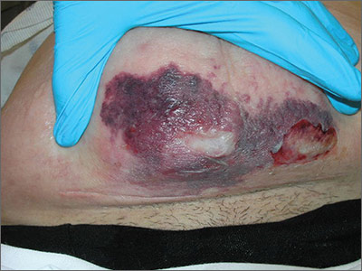
The ED physician suspected necrotizing fasciitis (NF) and immediately called for a surgical consult. This patient had type II NF, which can be caused by common skin organisms, such as Streptococcus pyogenes and Staphylococcus aureus.
Surgical debridement is the primary therapeutic modality. If the first debridement occurs within 24 hours of the onset of symptoms, there is a significantly improved chance of survival. Extensive, definitive debridement should be the goal with the first surgery. This may require amputation of an extremity to control the disease. Surgical debridement is repeated until all infected devitalized tissue is removed.
Antibiotics are the main adjunctive therapy to surgery. Broad-spectrum empiric antibiotics should be started immediately when NF is suspected and should include coverage of Gram-positive, Gram-negative, and anaerobic organisms. Antimicrobial therapy must be directed at the known or suspected pathogens and used in appropriate doses until repeated operative procedures are no longer needed. Empiric vancomycin should be considered while awaiting culture results to cover for the increasing incidence of methicillin-resistant Staphylococcus aureus (MRSA). Aggressive fluid resuscitation is often necessary because of massive capillary leak syndrome.
In this case, the patient was started on empiric broad-spectrum intravenous antibiotics to cover Gram-positive, Gram-negative, and anaerobic organisms. The surgical team then rapidly admitted the patient to the operating room (OR) for debridement. From the OR, she was transferred to the surgical intensive care unit where she received ongoing supportive and postoperative care. The intraoperative tissue cultures grew out MRSA.
The patient in this case was fortunate. She survived because of the speed with which she received a proper diagnosis and treatment.
Photo courtesy of Michael Babcock, MD. Text for Photo Rounds Friday courtesy of Richard P. Usatine, MD. This case was adapted from: Usatine R, Franklin J. Necrotizing fasciitis. In: Usatine R, Smith M, Mayeaux EJ, et al, eds. Color Atlas of Family Medicine. 2nd ed. New York, NY: McGraw-Hill; 2013:702-706.
To learn more about the Color Atlas of Family Medicine, see: www.amazon.com/Color-Family-Medicine-Richard-Usatine/dp/0071769641/
You can now get the second edition of the Color Atlas of Family Medicine as an app by clicking on this link: usatinemedia.com

Swollen, painful leg

The ED physician suspected necrotizing fasciitis (NF), a rapidly progressive infection of the deep fascia with necrosis of the subcutaneous tissues. It usually occurs after surgery or trauma. Patients have erythema and pain disproportionate to the physical findings.
This patient had type I NF, a polymicrobial infection with aerobic and anaerobic bacteria. It is frequently caused by enteric Gram-negative pathogens including Enterobacteriaceae organisms and Bacteroides. It can also occur with Gram-positive organisms such as non–group A streptococci and Peptostreptococcus.
Risk factors for type I NF include diabetes, severe peripheral vascular disease, obesity, alcoholism, cirrhosis, intravenous drug use, decubitus ulcers, poor nutritional status, surgery, and penetrating trauma. Early recognition based on signs and symptoms is potentially lifesaving. Although lab tests and imaging studies can confirm one’s clinical impression, rapid treatment with antibiotics and surgery are crucial to improving survival.
The patient in this case was taken to the operating room for debridement of her NF. Broad-spectrum antibiotics were started, but the infection continued to quickly advance. The patient died the following day. Her wound culture later grew Escherichia coli, Proteus vulgaris, Corynebacterium, Enterococcus, Staphylococcus sp., and Peptostreptococcus.
Adapted from: Dufel S, Martino M. Simple cellulitis or a more serious infection? J Fam Pract. 2006;55:396-400.
To learn more about the Color Atlas of Family Medicine, see: www.amazon.com/Color-Family-Medicine-Richard-Usatine/dp/0071769641/
You can now get the second edition of the Color Atlas of Family Medicine as an app by clicking on this link: usatinemedia.com

Swollen neck

The physician recognized that his patient had a neck abscess that was likely related to a dental abscess (odontogenic abscess). The patient’s alcoholism most likely weakened his ability to fight the infection, causing it to become potentially life-threatening. Risk factors for abscess formation include intravenous drug abuse, alcoholism, homelessness, dental disease, contact sports, and incarceration.
The physician transferred the patient to the local emergency department for hospitalization under the care of the ear, nose, and throat (ENT) service. The ENT physicians drained the abscess in the operating room without any complications. They cultured the abscess and started the patient on appropriate antibiotics, including penicillin G for dental aerobes and anaerobes.
The hospital team observed the patient for signs of alcohol withdrawal, but there were no complications because the patient hadn’t been drinking for the 5 days prior to hospitalization. Social Services was consulted and the patient was discharged to a respite bed in a local shelter that also had an alcohol and drug rehabilitation program. Arrangements were made for dental work in a charity dental clinic.
Photos and text for Photo Rounds Friday courtesy of Richard P. Usatine, MD. This case was adapted from: Usatine R. Abscess. In: Usatine R, Smith M, Mayeaux EJ, et al, eds. Color Atlas of Family Medicine. 2nd ed. New York, NY: McGraw-Hill;2013:698-701.
To learn more about the Color Atlas of Family Medicine, see: www.amazon.com/Color-Family-Medicine-Richard-Usatine/dp/0071769641/
You can now get the second edition of the Color Atlas of Family Medicine as an app by clicking on this link: usatinemedia.com

ECT may improve course of bipolar disorder
Electroconvulsive therapy increased illness-free intervals and reduced manic and depressive episodes in bipolar patients, reported Dr. Gian Paolo Minnai of the psychiatry unit at San Martino Hospital in Oristano, Italy, and coauthors.
In a retrospective study of 41 patients with bipolar disorder, the investigators analyzed the number of episodes and admissions 5 years before and after ECT treatment. The duration of free intervals before and after ECT was also studied.

The results showed “significantly longer” free intervals after treatment (13.2 ± 9.0 months before ECT to 25.1 ± 19.1 months after treatment [t = 3.8; P less than .0001]), Dr. Minnai and colleagues reported. In addition, the analysis found “significant reductions” in the number of manic and depressive episodes (5.9 ± 3.0 before ECT to 1.0 ± 1.7 after treatment [t = 9.3; P less than .0001]) and admissions (2.2 ± 1.3 before ECT to 0.2 ± 0.5 after treatment [t = 9.4; P less than .0001]).
The study suggests that “it is plausible that ECT, along with suspending antidepressant treatment, might carry intrinsic stabilizing effect on the course of [bipolar disorder],” the authors concluded.
No financial conflicts of interest were disclosed.
Read the full article in the Journal of Affective Disorders (May 2016;195:180-4).
Electroconvulsive therapy increased illness-free intervals and reduced manic and depressive episodes in bipolar patients, reported Dr. Gian Paolo Minnai of the psychiatry unit at San Martino Hospital in Oristano, Italy, and coauthors.
In a retrospective study of 41 patients with bipolar disorder, the investigators analyzed the number of episodes and admissions 5 years before and after ECT treatment. The duration of free intervals before and after ECT was also studied.
The results showed “significantly longer” free intervals after treatment (13.2 ± 9.0 months before ECT to 25.1 ± 19.1 months after treatment [t = 3.8; P less than .0001]), Dr. Minnai and colleagues reported. In addition, the analysis found “significant reductions” in the number of manic and depressive episodes (5.9 ± 3.0 before ECT to 1.0 ± 1.7 after treatment [t = 9.3; P less than .0001]) and admissions (2.2 ± 1.3 before ECT to 0.2 ± 0.5 after treatment [t = 9.4; P less than .0001]).
The study suggests that “it is plausible that ECT, along with suspending antidepressant treatment, might carry intrinsic stabilizing effect on the course of [bipolar disorder],” the authors concluded.
No financial conflicts of interest were disclosed.
Read the full article in the Journal of Affective Disorders (May 2016;195:180-4).

Practice pathway addresses problem behaviors for children with ASD
A new practice pathway provides primary care providers with evidence-based steps in assessing and addressing irritability and problem behavior in patients at least 3 years old who have been diagnosed with an autism spectrum disorder (ASD).
“As the medical professionals whom children with ASD will probably encounter first and most frequently, pediatric primary care providers need a breadth of knowledge to enable them to identify contributing factors to irritability and problem behavior, decide when and how to initiate treatment, and judge when to refer to specialists,” reported Dr. Kelly McGuire of Columbia University Medical Center and New York State Psychiatric Institute, and her associates (Pediatrics. 2016 Feb;137 Suppl 2:S136-48). “Therefore, this practice pathway can be viewed more as a comprehensive rather than an exhaustive recommendation for [primary care physicians], who must weigh their expertise and resources when addressing the range of issues that a child with ASD and irritability and problem behavior may present.”

The Irritability Workgroup comprised eight child psychiatrists, a developmental pediatrician, and a behavioral psychologist who met regularly to define irritability and problem behavior, and then to review the evidence base on assessing and treating factors that contribute to irritability and problem behavior. They grouped these factors into five domains: “co-occurring medical conditions, lack of functional communication, psychosocial stressors, maladaptive reinforcement patterns, and co-occurring psychiatric conditions.”
Then the group developed a consensus in determining each step in the practice pathway and when it should occur, along with opportunities for a primary care provider to refer the patient to a specialist or to collaborate with school and community providers. The 10 steps in the practice pathway are:
1. Assess the patient for irritability and problem behavior based on the presence of tantrums, meltdowns or rages; property destruction; aggression toward others; and self-injury.
2. Assess the safety of the patient and others in the home, enlisting home-based crisis services or considering hospitalization if safety is threatened.
3. Review the patient’s medical, developmental, communication, environmental, and psychiatric history before and after the problem behavior. Include information on the caretaker’s characteristics, any loss of skills by patient, and any interference occurring with functioning or relationships.
4. Prioritize the behaviors to address based on safety, severity, and impact on daily life.
5. Consider all contributing factors to the problem behavior, including medical problems, difficulties functionally communicating, psychosocial stressors, maladaptive reinforcement patterns (reducing inadvertent triggers), and comorbid psychiatric disorders.
6. Consider possible medication treatments, such as N-acetylcysteine, clonidine, risperidone or aripiprazole, depending on circumstances.
7. Develop an individualized treatment and safety plan.
8. Implement and monitor the treatment plan, setting clear and measurable treatment goals.
9. Follow up at 3 months to assess whether symptoms are continuing and a reassessment is needed.
10. Reevaluate every 3 months.
The article provides a useful and extensive checklist with a variety of specific things to consider when implementing the 10 steps.
“No other provider in the patient’s life combines the medical expertise and first-hand knowledge of the individual patient’s health and development” than the primary care provider, the authors wrote. “The practice pathway is most likely to be efficient and effective in generating a treatment plan if it is systematically followed and the specific combination of individual contributing factors is identified for each patient.”
The research was conducted through the Autism Speaks Autism Treatment Network and funded by the U.S. Department of Health and Human Services with the Autism Intervention Research Network on Physical Health and by Marilyn and James Simons Family Giving. Dr. Jeremy Veenstra-VanderWeele has received research funding from Seaside Therapeutics, Roche, Novartis, Forest, Sunovion, and SynapDx and has consulted with Roche, Novartis and SynapDx.
A new practice pathway provides primary care providers with evidence-based steps in assessing and addressing irritability and problem behavior in patients at least 3 years old who have been diagnosed with an autism spectrum disorder (ASD).
“As the medical professionals whom children with ASD will probably encounter first and most frequently, pediatric primary care providers need a breadth of knowledge to enable them to identify contributing factors to irritability and problem behavior, decide when and how to initiate treatment, and judge when to refer to specialists,” reported Dr. Kelly McGuire of Columbia University Medical Center and New York State Psychiatric Institute, and her associates (Pediatrics. 2016 Feb;137 Suppl 2:S136-48). “Therefore, this practice pathway can be viewed more as a comprehensive rather than an exhaustive recommendation for [primary care physicians], who must weigh their expertise and resources when addressing the range of issues that a child with ASD and irritability and problem behavior may present.”
The Irritability Workgroup comprised eight child psychiatrists, a developmental pediatrician, and a behavioral psychologist who met regularly to define irritability and problem behavior, and then to review the evidence base on assessing and treating factors that contribute to irritability and problem behavior. They grouped these factors into five domains: “co-occurring medical conditions, lack of functional communication, psychosocial stressors, maladaptive reinforcement patterns, and co-occurring psychiatric conditions.”
Then the group developed a consensus in determining each step in the practice pathway and when it should occur, along with opportunities for a primary care provider to refer the patient to a specialist or to collaborate with school and community providers. The 10 steps in the practice pathway are:
1. Assess the patient for irritability and problem behavior based on the presence of tantrums, meltdowns or rages; property destruction; aggression toward others; and self-injury.
2. Assess the safety of the patient and others in the home, enlisting home-based crisis services or considering hospitalization if safety is threatened.
3. Review the patient’s medical, developmental, communication, environmental, and psychiatric history before and after the problem behavior. Include information on the caretaker’s characteristics, any loss of skills by patient, and any interference occurring with functioning or relationships.
4. Prioritize the behaviors to address based on safety, severity, and impact on daily life.
5. Consider all contributing factors to the problem behavior, including medical problems, difficulties functionally communicating, psychosocial stressors, maladaptive reinforcement patterns (reducing inadvertent triggers), and comorbid psychiatric disorders.
6. Consider possible medication treatments, such as N-acetylcysteine, clonidine, risperidone or aripiprazole, depending on circumstances.
7. Develop an individualized treatment and safety plan.
8. Implement and monitor the treatment plan, setting clear and measurable treatment goals.
9. Follow up at 3 months to assess whether symptoms are continuing and a reassessment is needed.
10. Reevaluate every 3 months.
The article provides a useful and extensive checklist with a variety of specific things to consider when implementing the 10 steps.
“No other provider in the patient’s life combines the medical expertise and first-hand knowledge of the individual patient’s health and development” than the primary care provider, the authors wrote. “The practice pathway is most likely to be efficient and effective in generating a treatment plan if it is systematically followed and the specific combination of individual contributing factors is identified for each patient.”
The research was conducted through the Autism Speaks Autism Treatment Network and funded by the U.S. Department of Health and Human Services with the Autism Intervention Research Network on Physical Health and by Marilyn and James Simons Family Giving. Dr. Jeremy Veenstra-VanderWeele has received research funding from Seaside Therapeutics, Roche, Novartis, Forest, Sunovion, and SynapDx and has consulted with Roche, Novartis and SynapDx.
FROM PEDIATRICS
Key clinical point: A 10-step practice pathway helps primary care providers address irritability and problem behavior in children with ASD.
Major finding: Ten steps in the pathway include assessment of problems, identification of contributing factors, and formulation of a plan to address the underlying factors and the behavior.
Data source: The pathway is based on a consensus by a 10-member multidisciplinary work group charged with identifying available evidence on effective ways to assess and address irritability and problem behavior in children with ASD.
Disclosures: The research was conducted through the Autism Speaks Autism Treatment Network and funded by the U.S. Department of Health and Human Services with the Autism Intervention Research Network on Physical Health and by Marilyn and James Simons Family Giving. Dr. Jeremy Veenstra-VanderWeele has received research funding from Seaside Therapeutics, Roche, Novartis, Forest, Sunovion and SynapDx and has consulted with Roche, Novartis and SynapDx.

Safety of bioresorbable stents does not match that of metal stents
Bioresorbable vascular scaffold stents are improving rapidly but they are still associated with a higher risk of complications compared with drug-eluting metal stents, according to a meta-analysis of published studies presented at Cardiovascular Research Technologies 2016.
“Bioresorbable stents are clearly an attractive strategy, but our data suggest that physicians and patients should remain aware of the risks,” reported Dr. Alok Saurav of Creighton University Medical Center, Omaha, Neb.

The first bioresorbable vascular scaffold (BVS) device, Synergy, was approved this past October, but this stent, despite bioresorbable struts, still has body parts that are not fully bioresorbable. However, several fully bioresorbable devices have reached late stages of testing and may receive regulatory approval this year.
In the meta-analysis, eight studies – five randomized trials, two studies with propensity matching, and an observational study –the primary goal was to compare BVS to drug eluting metal (DEM) stents for definite stent thrombosis. Secondary outcomes included subacute stent thrombosis within 30 days and within 1 year and cardiac death, all-cause death, MI, and ischemia-driven target vessel revascularization (TVR).
Despite the fact that the mean age and gender distribution was the same when the 2,760 patients receiving BVS stents were compared to the 2,212 receiving DEM stents, and both received comparable antiplatelet regimens after the stent was placed, there was an 80% greater relative risk for definite stent thrombosis in the BVS group. Although this difference fell short of statistical significance (P = .06), Dr. Saurav called it a “strong trend.”
Several of the adverse events that were analyzed as secondary outcomes in this study were less frequent with the BVS, such as cardiac death (relative risk, 0.83) and all-cause death (RR, 0.74), but the statistics did not suggest a trend, so Dr. Saurav characterized these outcomes as similar. MI was an exception. This was more frequent in those received a BVS stent (RR, 1.35; P = .049), and this reached significance.
Most of the studies included in this analysis were conducted with the everolimus-eluting Absorb BVS device, which many are predicting will be the first fully bioresorbable stent to receive regulatory approval.
It is notable that another meta-analysis including some of the same studies and published just weeks prior to the CRT meeting drew the same conclusion about the increased risk of stent thrombosis with BVS relative to DEM stents (Lancet 2016;387:537-44). This meta-analysis was restricted to six trials with 3,738 randomized patients. Unlike the meta-analysis presented at CRT, this study compared the two types of stents for both definite and probable stent thrombosis. For BVS relative to DEM stents, the relative risk for this outcome was 1.99 (P = .05).
“We think our restriction to definite stent thrombosis provides a stricter endpoint, but it’s notable that the results were relatively consistent,” Dr. Saurav reported.
Acknowledging that the increased risk of stent thrombosis appears to be modest for BVS relative to DEM stents, Dr. Saurav emphasized that these data should not discourage further development of bioresorbable stents, which are conceptually attractive.
“We cannot take these bioresorbable devices off the table,” he said. “But we do need more data to evaluate their risks relative to the conventional devices that are now available.”
The meeting was sponsored by the Cardiovascular Research Institute at Washington Hospital Center. Dr. Saurav reported no conflicts of interest.
Bioresorbable vascular scaffold stents are improving rapidly but they are still associated with a higher risk of complications compared with drug-eluting metal stents, according to a meta-analysis of published studies presented at Cardiovascular Research Technologies 2016.
“Bioresorbable stents are clearly an attractive strategy, but our data suggest that physicians and patients should remain aware of the risks,” reported Dr. Alok Saurav of Creighton University Medical Center, Omaha, Neb.
The first bioresorbable vascular scaffold (BVS) device, Synergy, was approved this past October, but this stent, despite bioresorbable struts, still has body parts that are not fully bioresorbable. However, several fully bioresorbable devices have reached late stages of testing and may receive regulatory approval this year.
In the meta-analysis, eight studies – five randomized trials, two studies with propensity matching, and an observational study –the primary goal was to compare BVS to drug eluting metal (DEM) stents for definite stent thrombosis. Secondary outcomes included subacute stent thrombosis within 30 days and within 1 year and cardiac death, all-cause death, MI, and ischemia-driven target vessel revascularization (TVR).
Despite the fact that the mean age and gender distribution was the same when the 2,760 patients receiving BVS stents were compared to the 2,212 receiving DEM stents, and both received comparable antiplatelet regimens after the stent was placed, there was an 80% greater relative risk for definite stent thrombosis in the BVS group. Although this difference fell short of statistical significance (P = .06), Dr. Saurav called it a “strong trend.”
Several of the adverse events that were analyzed as secondary outcomes in this study were less frequent with the BVS, such as cardiac death (relative risk, 0.83) and all-cause death (RR, 0.74), but the statistics did not suggest a trend, so Dr. Saurav characterized these outcomes as similar. MI was an exception. This was more frequent in those received a BVS stent (RR, 1.35; P = .049), and this reached significance.
Most of the studies included in this analysis were conducted with the everolimus-eluting Absorb BVS device, which many are predicting will be the first fully bioresorbable stent to receive regulatory approval.
It is notable that another meta-analysis including some of the same studies and published just weeks prior to the CRT meeting drew the same conclusion about the increased risk of stent thrombosis with BVS relative to DEM stents (Lancet 2016;387:537-44). This meta-analysis was restricted to six trials with 3,738 randomized patients. Unlike the meta-analysis presented at CRT, this study compared the two types of stents for both definite and probable stent thrombosis. For BVS relative to DEM stents, the relative risk for this outcome was 1.99 (P = .05).
“We think our restriction to definite stent thrombosis provides a stricter endpoint, but it’s notable that the results were relatively consistent,” Dr. Saurav reported.
Acknowledging that the increased risk of stent thrombosis appears to be modest for BVS relative to DEM stents, Dr. Saurav emphasized that these data should not discourage further development of bioresorbable stents, which are conceptually attractive.
“We cannot take these bioresorbable devices off the table,” he said. “But we do need more data to evaluate their risks relative to the conventional devices that are now available.”
The meeting was sponsored by the Cardiovascular Research Institute at Washington Hospital Center. Dr. Saurav reported no conflicts of interest.
AT CARDIOVASCULAR RESEARCH TECHNOLOGIES 2016
Key clinical point: Trial data suggest the risk of thrombosis and other adverse events remains higher with bioresorbable stents than with conventional drug-eluting metal stents.
Major finding: In a meta-analysis, the 80% increased risk of definite stent thrombosis for bioresorbable relative to metal stents fell just short of significance (P = .06) but the 35% increased risk of subsequent MI was significant (P = .049).
Data source: Meta-analysis of eight studies.
Disclosures: Dr. Saurav reported no conflicts of interest.

Limited posttreatment imaging suffices in HPV-positive oropharyngeal cancer
SCOTTSDALE, ARIZ. – Most patients who are treated for human papillomavirus (HPV)–positive oropharyngeal cancer can safely skip routine imaging after a negative 3-month posttreatment scan, suggest results of a retrospective cohort study reported at the Multidisciplinary Head and Neck Cancer Symposium.
Investigators led by Dr. Jessica M. Frakes of the H. Lee Moffitt Cancer Center in Tampa, Fla., studied 246 patients treated nonsurgically between 2006 and 2014 for nonmetastatic HPV-positive disease.

With a median follow-up of 36 months, all local recurrences and the large majority of regional and distant recurrences were detected from symptoms, physical exam, and the 3-month posttreatment PET-CT imaging, according to data reported in a session and related press briefing.
“Routine imaging is not recommended after posttreatment imaging shows a complete response to treatment, unless the patient presents with symptoms or something else that would warrant imaging,” Dr. Frakes commented. “Follow-up should include history and physical examination with direct visualization.”
Currently, the National Comprehensive Cancer Network advises a one-size-fits-all approach to follow-up that does not consider tumor HPV status, she noted. But reducing surveillance imaging for HPV-positive patients would likely have considerable benefit in terms of less stress and anxiety (provided patients are educated about the safety of clinical follow-up) and lower financial burden for both the patient and the health care system as a whole.
Results additionally showed that the majority of recurrences occurred within the first 6 months, a pattern that was consistent whether or not patients had risk factors for recurrence. The 3-year rate of freedom from local failure exceeded 97%, and only 2% of patients had grade 3 or worse toxicity at their last follow-up.
“Our outcomes are excellent with low rates of permanent toxicity, and we think that this partly is due to the fact that they are treated by specialized multidisciplinary team,” Dr. Frakes commented at the meeting.
Press briefing moderator Dr. Christine Gourin of Johns Hopkins University in Baltimore, commented, “This study is one that’s dear to my heart because I think that we probably do too much posttreatment surveillance, and they are exactly right that the NCCN is fairly vague about when to perform imaging.”
“I can tell you that we have stopped routinely imaging patients after 3 months if the PET is negative, and it’s true that we do pick up recurrences more clinically than we do radiologically,” she added. “And of course the false positives are causing much morbidity.”
Introducing the study, Dr. Frakes commented, “Several retrospective and prospective trials have shown increased survivals and decreased toxicity in patients with HPV-associated oropharynx cancer. As the number of patients and survivors grows, so does the need to determine general time to recurrence and the most effective modes of recurrence detection, thereby guiding our standards for optimal follow-up care.”
All patients studied received definitive radiation therapy, and 85% of them also received concurrent chemotherapy.
The patients had a 3-month posttreatment PET-CT scan, plus physical exams every 3 months in the first year post treatment, every 4 months in the second year, every 6 months in the third through fifth years, and annually thereafter.
Results showed that the 3-year rate of local control was 97.8%, and 100% of the local failures were detected by physical exam, including direct visualization or flexible laryngoscopy.
The rate of regional control was 95.3%, and 89% of cases of regional failure were detected through symptoms or the 3-month posttreatment imaging. Risk factors for regional recurrence included involvement of five or more lymph nodes in the neck and involvement of level 4 (low neck) lymph nodes (P less than .05 for each).
The rate of freedom from distant metastases was 91%, and 71% of cases of distant metastases were detected through symptoms or the 3-month posttreatment imaging. Risk factors for distant metastases included tumor in the lymph nodes measuring greater than 6 cm, involvement of bilateral lymph nodes, involvement of five or more lymph nodes in the neck, and involvement of level 4 lymph nodes (P less than .05 for each).
Overall, 9% of patients experienced grade 3 or worse late toxicity (occurring at 3 months or thereafter), consisting of feeding/gastrostomy tube (G-tube) placement, necrosis or ulcer, and tracheostomy. However, these toxicities had resolved as of the last follow-up in most cases, with a final rate of toxicity of only 2%.
The center follows an aggressive approach to preventing and managing toxicity, noted Dr. Frakes.
“Even when the patients have their G-tube in place, we really do encourage p.o. [oral] intake as much as possible with pain medication. And I think that really does make a big difference for our patients,” she said. “They do meet with a speech pathologist and our nutritionist weekly when they are on treatment.” Patients are also allowed to have the G-tube removed by last follow-up, she added.
SCOTTSDALE, ARIZ. – Most patients who are treated for human papillomavirus (HPV)–positive oropharyngeal cancer can safely skip routine imaging after a negative 3-month posttreatment scan, suggest results of a retrospective cohort study reported at the Multidisciplinary Head and Neck Cancer Symposium.
Investigators led by Dr. Jessica M. Frakes of the H. Lee Moffitt Cancer Center in Tampa, Fla., studied 246 patients treated nonsurgically between 2006 and 2014 for nonmetastatic HPV-positive disease.
With a median follow-up of 36 months, all local recurrences and the large majority of regional and distant recurrences were detected from symptoms, physical exam, and the 3-month posttreatment PET-CT imaging, according to data reported in a session and related press briefing.
“Routine imaging is not recommended after posttreatment imaging shows a complete response to treatment, unless the patient presents with symptoms or something else that would warrant imaging,” Dr. Frakes commented. “Follow-up should include history and physical examination with direct visualization.”
Currently, the National Comprehensive Cancer Network advises a one-size-fits-all approach to follow-up that does not consider tumor HPV status, she noted. But reducing surveillance imaging for HPV-positive patients would likely have considerable benefit in terms of less stress and anxiety (provided patients are educated about the safety of clinical follow-up) and lower financial burden for both the patient and the health care system as a whole.
Results additionally showed that the majority of recurrences occurred within the first 6 months, a pattern that was consistent whether or not patients had risk factors for recurrence. The 3-year rate of freedom from local failure exceeded 97%, and only 2% of patients had grade 3 or worse toxicity at their last follow-up.
“Our outcomes are excellent with low rates of permanent toxicity, and we think that this partly is due to the fact that they are treated by specialized multidisciplinary team,” Dr. Frakes commented at the meeting.
Press briefing moderator Dr. Christine Gourin of Johns Hopkins University in Baltimore, commented, “This study is one that’s dear to my heart because I think that we probably do too much posttreatment surveillance, and they are exactly right that the NCCN is fairly vague about when to perform imaging.”
“I can tell you that we have stopped routinely imaging patients after 3 months if the PET is negative, and it’s true that we do pick up recurrences more clinically than we do radiologically,” she added. “And of course the false positives are causing much morbidity.”
Introducing the study, Dr. Frakes commented, “Several retrospective and prospective trials have shown increased survivals and decreased toxicity in patients with HPV-associated oropharynx cancer. As the number of patients and survivors grows, so does the need to determine general time to recurrence and the most effective modes of recurrence detection, thereby guiding our standards for optimal follow-up care.”
All patients studied received definitive radiation therapy, and 85% of them also received concurrent chemotherapy.
The patients had a 3-month posttreatment PET-CT scan, plus physical exams every 3 months in the first year post treatment, every 4 months in the second year, every 6 months in the third through fifth years, and annually thereafter.
Results showed that the 3-year rate of local control was 97.8%, and 100% of the local failures were detected by physical exam, including direct visualization or flexible laryngoscopy.
The rate of regional control was 95.3%, and 89% of cases of regional failure were detected through symptoms or the 3-month posttreatment imaging. Risk factors for regional recurrence included involvement of five or more lymph nodes in the neck and involvement of level 4 (low neck) lymph nodes (P less than .05 for each).
The rate of freedom from distant metastases was 91%, and 71% of cases of distant metastases were detected through symptoms or the 3-month posttreatment imaging. Risk factors for distant metastases included tumor in the lymph nodes measuring greater than 6 cm, involvement of bilateral lymph nodes, involvement of five or more lymph nodes in the neck, and involvement of level 4 lymph nodes (P less than .05 for each).
Overall, 9% of patients experienced grade 3 or worse late toxicity (occurring at 3 months or thereafter), consisting of feeding/gastrostomy tube (G-tube) placement, necrosis or ulcer, and tracheostomy. However, these toxicities had resolved as of the last follow-up in most cases, with a final rate of toxicity of only 2%.
The center follows an aggressive approach to preventing and managing toxicity, noted Dr. Frakes.
“Even when the patients have their G-tube in place, we really do encourage p.o. [oral] intake as much as possible with pain medication. And I think that really does make a big difference for our patients,” she said. “They do meet with a speech pathologist and our nutritionist weekly when they are on treatment.” Patients are also allowed to have the G-tube removed by last follow-up, she added.
AT THE HEAD AND NECK CANCER SYMPOSIUM
Key clinical point: Most patients with HPV-positive oropharyngeal cancer do not need routine imaging after a negative 3-month posttreatment scan.
Major finding: Overall, 100%, 89%, and 71% of local, regional, and distant recurrences, respectively, were detected from symptoms, physical exam, and 3-month posttreatment imaging.
Data source: A retrospective cohort study of 246 patients treated for HPV-positive oropharyngeal cancer.
Disclosures: Dr. Frakes disclosed that she had no relevant conflicts of interest.