User login
FDG-PET/CT leads pack for small-cell lung cancer staging
For pretreatment staging of small-cell lung cancer (SCLC) the use of positron-emission tomography combined with CT was more sensitive compared with several other tests, according to a new report on a review of studies.
Overall, positron emission tomography using [F]-fluorodeoxyglucose as a radiotracer combined with CT (FDG-PET/CT) had greater sensitivity to detect osseous metastases than did bone scintigraphy or CT alone, according to Dr. Jonathan R. Treadwell, Ph.D., of ECRI Institute–Penn Medicine’s Evidence-based Practice Center in Plymouth Meeting, Pa., and colleagues. In addition, the researchers concluded that adding FDG-PET/CT to the protocol for patients who have undergone standard staging increased the sensitivity for detecting additional metastases. Data on endobronchial ultrasound were insufficient to draw any conclusions.
The findings generally line up with recent guidelines from the American College of Radiology (ACR) and American College of Chest Physicians (ACCP). In 2014, the ACR gave the highest rating of “usually appropriate” (with regard to staging SCLC) to FDG-PET/CT from skull base to mid-thigh, while bone scintigraphy was rated as “may be appropriate” and not necessary if PET/CT had been done, the researchers wrote. The 2013 ACCP guideline “suggested” FDG PET instead of bone scintigraphy for patients with limited disease, they added.
The researchers reviewed data from seven studies to assess the accuracy and effectiveness of several imaging modalities for the pretreatment staging of SCLC. The report was generated for the Agency for Healthcare Research and Quality (AHRQ) as part of its Comparative Effectiveness Review series, and is not an official AHRQ position, the researchers noted.
Combining FDG-PET with CT scanning has demonstrated even greater effectiveness at identifying malignant tumors and metabolically active metastases than has PET alone, because the CT allows for more localized anatomic detail, the researchers explained. “False negative scans usually result from non–metabolically active sites of tumor or from suboptimal quality studies,” they said, while false positives using FDG-PET are usually attributed to inflammation or metabolically active infection.
The meta-analysis included data on endobronchial ultrasound, which involves ultrasound to view structures inside and adjacent to the airway; bone scintigraphy, a less expensive planar molecular imaging technique; and CT alone.
Comparative evidence on pretreatment staging for SCLC is limited, according to the researchers. The data did not allow them to determine how FDG-PET/CT compared to other imaging in terms of specificity, and any type of imaging can yield false positives, they said. However, higher sensitivity alone can benefit patients in terms of improving patient selection for optimal therapy, sparing patients chemotherapy if not needed, and improving the prediction value of ongoing research, they noted.
“Although high-quality evidence may not be voluminous, I think most physicians would agree with the conclusion that a bone scan is not mandatory in the work-up of possible SCLC, if a PET/CT has been done,” Dr. W. Michael Alberts of the Moffitt Cancer Center in Tampa, Fla., said in an interview.
Cost might play a role in why the guidelines are being issued at this time, he noted, because “the initial work-up of the patient with suspected SCLC may prove to be quite expensive, and the elimination of a superfluous test may be a fiscal winner.” However, more research is needed in this area, particularly in the areas of including the order of pretreatment testing and the incorporation of new procedures and imaging modalities, he added. “Perhaps more intellectually challenging, however, might be the question of why SCLC is becoming less common, or why has improvement in treatment been so slow compared to NSCLC,” he added.
The researchers had no financial conflicts to disclose.
More connective tissue disease–associated PAH seen in older patients
Patient age contributes to significant differences in the characteristics and etiology of pulmonary arterial hypertension seen in randomized, controlled trials, including more frequent connective tissue disease–associated disease in older patients, according to a post-hoc analysis of trials.
Additionally, older age was associated with worse baseline functional status, worse outcomes in the 6-minute walk distance, and an overall reduced response to treatment, while hemodynamic severity was higher in younger patients.
“Although registry data have shown that idiopathic PAH [pulmonary arterial hypertension] is increasingly recognized in older populations, our analysis shows that idiopathic etiology was less frequent in the older group. In contrast, CTD [connective tissue disease]–associated PAH accounted for a higher proportion of PAH etiology in the oldest age group,” wrote Jonathan A. Rose of Case Western Reserve University, Cleveland, and his colleagues (Chest. 2016;149[5]:1234-44. doi: 10.1016/j.chest.2015.11.008).
The researchers analyzed seven multicenter, randomized, double-blind, placebo-controlled treatment trials for PAH conducted by United Therapeutics and one open-label extension study that involved following patients being treated with subcutaneous treprostinil for 4 additional years. Data from the open-label extension trial and one of the trials were combined and reported as one trial. All trials excluded patients with a history of left-sided heart disease, pulmonary artery wedge pressure (PAWP) greater than 15 mmHg, or pulmonary vascular resistance (PVR) less than 3 Woods units. The researchers categorized the 2,627 patients included in the trials in the following three age groups: 50 years or younger, 51-64 years, and 65 years or older.
Between 53% and 74% of patients in all trials across all age groups had idiopathic PAH, but older patients comprised a significantly smaller proportion of the patients with idiopathic PAH in three of the trials (P = .004) and a significantly higher percentage of the patients with CTD-associated PAH in all of the trials (P less than .001). Across the trials, CTD-associated PAH occurred in 15%-21% of patients 50 years or younger, 25%-40% of those aged 51-64 years, and 27%-49% of those aged 65 years or older.
From baseline to the end in three of the studies, a smaller change in the 6-minute walk distance was seen in older patients and a higher proportion of older patients had an overall decrease in total 6-minute walk distance. Older age remained associated with this outcome measure when only data from treatment-naive patients or the subgroup of patients with CTD-associated PAH were included.
The association between age and lower baseline functional status was observed in four of the trials; A lower proportion of patients in the oldest age group were classified as being of the World Health Organization functional classes I and II, with 9%-32% of patients aged 65 years or older, 10%-33% in those aged 51-64 years, and 16%-43% of those aged 50 years or younger.
Hemodynamic severity was among the areas in which older patients performed better than younger patients, with the oldest age group having lower baseline mean pulmonary artery pressure (mPAP) and PVR in the two trials that measured hemodynamics.
“The difference in the clinical function and hemodynamics in older patients may reflect different mechanisms of disease. Older patients may have diminished cardiopulmonary reserve, and similar degrees of hemodynamic severity manifest with a larger functional impairment. Alternatively, additional comorbidities in older patients may contribute to a larger extent, and clinical function may not be as directly related to hemodynamic severity,” according to the researchers.
Mortality was generally small in these studies and not significantly different among age groups, except for in the open-label extension study of Subcutaneous Infusion of Treprostinil in Patients with PAH (SC-TRE) and the Study of Intravenous Remodulin in Patients in India with PAH (TRUST) trials, which were combined and reported on as one trial (P = .0004).
Nausea was the only adverse effect found to be associated with age in more than one trial, and the relationship between it and age in two trials were inconsistent. While a higher incidence of nausea was found in the oldest age group in the Oral Treprostinil as Monotherapy for the Treatment of Pulmonary Arterial Hypertension (FREEDOM-M) trial (P = .03), a lower incidence of nausea was found in the oldest age group in the Treprostinil Sodium Inhalation Used in the Management of Pulmonary Arterial Hypertension (TRIUMPH) trial (P = .02).
The analysis was supported by a grant from the National Institutes of Health. Two of the authors, Jody M. Cleveland and Youlan Rao, Ph.D., reported being employees of United Therapeutics, while Dr. Omar A. Minai, another author of the report, serves on the scientific advisory board of United Therapeutics. The other authors declared no conflicts.
FROM CHEST
FDA issues warning about counterfeit BiCNU
A counterfeit version of BiCNU has been detected in foreign countries, the Food and Drug Administration reports.
BiCNU is approved to treat different types of brain cancer, multiple myeloma, and lymphoma (Hodgkin’s and non-Hodgkin’s), manufactured by Emcure Pharmaceuticals, and distributed by Heritage Pharmaceuticals.
There has been no counterfeit BiCNU detected in the United States, but the FDA encourages health care professionals to diligently inspect BiCNU vials before administering the drug to patients.
“While the [National Drug Code] on the outer package of the authentic and counterfeit version might match, the best way to distinguish a counterfeit is to look at the BiCNU vial inside the packaging,” the FDA reported in a written statement, which includes a list of the counterfeit lots.
To report sales solicitation of suspect drugs call the FDA’s office of criminal investigations at 800-551-3989 or e-mail DrugSupplyChainIntegrity@fda.hhs.gov. To report adverse events related to suspect medications, submit a report online at www.fda.gov/medwatch/report.htm.
On Twitter @jess_craig94
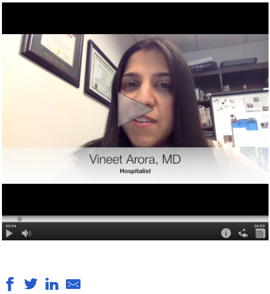
WATCH: Mentoring in Hospital Medicine
Drs. Vineet Arora and Hyung "Harry" Cho offer insight on how mentorship—giving, and receiving—is an essential part of all stages of an hospitalist career, in academic or community-based HM.
The video associated with this article is no longer available on this site. Please view all of our videos on the MDedge YouTube channel
Attend SVS PAC Reception in Washington, D.C.
When meeting near Washington, D.C., meeting with members of Congress is a natural choice.

The SVS Political Action Committee Reception will be held from 5:30 to 7 p.m. Wednesday, June 8, on Capitol Hill, at 2261 Rayburn House Office Building. Round-trip transportation will be provided from the Gaylord National’s Maryland Ballroom bus loop entrance on Level 2. The shuttle bus will depart promptly at 4:45 p.m.
The reception is for Capitol Club contributors, those SVS members who have contributed $1,000 or more since Jan. 1, 2015, including those who contribute during the 2016 Vascular Annual Meeting, up until June 8. Attendees can meet members of Congress and thank them for their support of vascular surgery issues.
PAC contributions allow SVS members and staff to gain important access to members of Congress by attending meetings and discussing issues that have a major impact on vascular surgery.
See vsweb.org/PAC to donate and secure a spot at the reception – and to invest in the future.
When meeting near Washington, D.C., meeting with members of Congress is a natural choice.
The SVS Political Action Committee Reception will be held from 5:30 to 7 p.m. Wednesday, June 8, on Capitol Hill, at 2261 Rayburn House Office Building. Round-trip transportation will be provided from the Gaylord National’s Maryland Ballroom bus loop entrance on Level 2. The shuttle bus will depart promptly at 4:45 p.m.
The reception is for Capitol Club contributors, those SVS members who have contributed $1,000 or more since Jan. 1, 2015, including those who contribute during the 2016 Vascular Annual Meeting, up until June 8. Attendees can meet members of Congress and thank them for their support of vascular surgery issues.
PAC contributions allow SVS members and staff to gain important access to members of Congress by attending meetings and discussing issues that have a major impact on vascular surgery.
See vsweb.org/PAC to donate and secure a spot at the reception – and to invest in the future.
Earn CME, Maintenance of Certification Credits
Beyond learning what’s new in the world of vascular surgery, paid physicians also can earn both Continuing Medical Education credits and Maintenance of Certification Part 2 Lifelong Learning and Self-Assessment credits.
The Society for Vascular Surgery is accredited by the Accreditation Council for Continuing Medical Education to provide continuing medical education for physicians. SVS has designated the 2016 Vascular Annual Meeting for a maximum of 29.25 AMA PRA Category 1 Credits™.
Physicians should claim only the credits commensurate with the extent of their participation in the activity.
Of the 29.25 credits, 13.25 AMA PRA Category 1 Credits™ meet the requirements for American Board of Surgery MOC Part 2 self-assessment.
To claim credits, physicians must attend that session and then complete the appropriate self-assessment exam with a passing score of 75%. Access to the exams will be available via a link in the 2016 VAM Mobile App and a link on the SVS website. New this year is the ability to have access to the exams immediately after a session.
Attendees may access the exams and claim CME and self-assessment credits through Dec. 31, 2016.
Separate certificates are issued for postgraduate courses (a total of nine credits are available and people may register for postgraduate courses only and not participate in VAM), the Registered Physician Vascular Interpretation exam course (3.25 CME and 3.25 MOC credits) and for VAM itself (up to 29.25 and 13.25 CME and MOC credits, respectively, depending on session participation). A grand total of 25.5 MOC credits is available to someone who takes three postgraduate courses, participates in two breakfast and all plenary sessions and also completes the RPVI review course.
Industry-Supported Satellite Symposia Offered at VAM
Vascular Annual Meeting attendees are invited to the satellite symposia offered by industry, including three breakfast sessions on Thursday, June 9. These sessions are not part of the ACCME-accredited portion of the Vascular Annual Meeting.
Thursday, June 9, 6:30 - 8 a.m.
B1: Complex SFA Disease: Maximizing Outcomes While Minimizing Costs
Presenting: Dr. Brian G. DeRubertis, Dr. Peter L. Faries
Sponsored by Abbott
B2: EndoAnchors in Practice: Treatment Algorithms from the Experts
Sponsored by Medtronic
Presenting: EndoAnchors: What Are They and How They Bring Value to Your Practice – Dr. Bart Muhs
How I Utilize ANCHOR Data to Make Clinically Relevant Treatment Decisions – Dr. Jeffrey Jim
How EndoAnchors Can be Used Safely and Effectively in the Treatment of Abdominal Aortic Aneurysms – Dr. Frank R. Arko
How EndoAnchors Can be Used Safely and Effectively in the Treatment of Thoracic Aortic Aneurysms – Dr. Jean M. Panneton
B3: New Techniques in Advanced Imaging and 3D Navigation for Complex Endovascular Procedures
Sponsored by Philips
Presenting: Dr. Marc L. Schermerhorn and Dr. Wayne K. Nelson
Thursday, June 9, 7:30 - 9 p.m.
“The Battle Continues ... The Battle of the Bulge: Debating the Future of Aortic Aneurysm Repair”
Sponsored by Endologix, Inc.
Moderator: Dr. Christopher J. Kwolek; Presenter: TBA
Friday, June 10, 6:30 - 8:30 p.m.
“Internal Iliac Artery Preservation Expert Panel”
Sponsored by Gore
Presenting: Moderator: Dr. Darren B. Schneider
Pros and Cons of Current Treatment Options – Dr. Jason T. Lee
GORE® EXCLUDER® Iliac Branch Device (IBE) and Procedural Overview – Dr. Gustavo Oderich
IBE Clinical Trial Outcomes and the Benefits of Internal Iliac Artery Preservation – Dr. Darren B. Schneider
Case Report: First Patient Treated and Two-Year Follow-up – Dr. Brian G. Peterson
Case Report: First U.S. Bilateral Case and One-Year Follow-up – Dr. Sharif Ellozy
Rapid-Fire Case Presentations
As of 5/6/16
Postgraduate Courses: Six Sessions Offered
The 2016 Vascular Annual Meeting is offering six postgraduate courses on Wednesday, with two held concurrently in three time slots. SVS members get in free with their registration, a $300 value. Registration is required.
Topics for both the postgraduate courses and the hands-on workshops (also held Wednesday; see story in the May Vascular Specialist) are selected based on member feedback and requests, said Dr. Kellie Brown, chair of the Postgraduate Education Committee that is responsible for the sessions. “We try to cover topics that are current, timely, and necessary for the majority of our membership,” she said.
That includes a broader focus this year on both dialysis access and thoracic outlet, for example. “We’ve always done something on access, but a lot of members asked for more,” she said. “Not only is dialysis access a common issue, particularly for those in community practice, but it is also an area of innovation.”
Another session covers management of the diabetic foot. “Vascular surgeons deal with this every single day,” Dr. Brown said. “It’s a really common problem – so let’s talk about the best way to manage it during a session at VAM.”
This session on diabetic foot management also introduces a new VAM partner, the American Podiatric Medical Association, which worked with SVS and the Society for Vascular Medicine on a comprehensive new guideline for managing the diabetic foot. “Having joint sessions – and we have several this year – adds an extra layer to VAM sessions,” Dr. Brown said. “We get to offer examination of other aspects of a particular topic, which adds not only more information but also a certain level of excitement.”
Last year, “Standing on the Shoulders of Giants” drew the most attendees. For 2016, “Giants” is back, with different experts and a continued focus on open surgery. “People are so comfortable with the endovascular side, but open surgery is becoming less common. This offers an opportunity to learn the techniques from the masters,” she said.
As for next year, Dr. Brown advised anyone who has a topic idea to submit it with his or her VAM evaluation.
Session 1: 7 – 10 a.m.
P1: Controversies and Management of Lower Extremity Arterial Occlusive Disease
Moderators: Dr. Christopher J. Abularrage and Dr. Niten Singh
P2: Complete Management of Venous Disease: From Superficial to Deep
Moderators: Dr. Ruth L. Bush and Dr. Mark Meissner
Session 2: 10:15 a.m. – 1:15 p.m.
P3: SVS/APMA Joint Session: Management of the Diabetic Foot
Moderators: Dr. James R. Christina (APMA) and Dr. Matthew J. Eagleton (SVS)
P4: Hemodialysis: Challenges, Controversies and New Techniques
Moderators: Dr. Melissa L. Kirkwood and Dr. Jill Zink
Session 3: 2 – 5 p.m.
P5: Endovascular Strategies for Complex Aortic Scenarios
Moderators: Dr. Ali Azizzadeh and Dr. Edward Woo
P6: Standing on the Shoulders of Giants: Open Operative Techniques by the Masters
Moderators: Dr. Hasan Dosluoglu and Dr. Michael Rohrer
Vascular Live
The Vascular Live stage is located in Exhibit Halls A-C, on Level 1. On the Vascular Live stage, exhibitors will present new ideas, showcase breakthrough technologies and discuss the latest trends in vascular surgery.
Thursday, June 9
12:15 – 1:15 p.m.
Sponsored by Bard Peripheral Vascular, Inc.
“Latest Outcomes from Lutonix® Global SFA Registry and Head to Head Comparison between IN.PACT and LUTONIX® DCB”
Speakers: Dr. Yvonne Bausback, Dr. Chad Laurich, and Dr. Renu Virmani
3:30 – 4 p.m.
Sponsored by Abbott
“Treatment Strategies for Complex Femoropopliteal Disease”
Speaker: Dr. Niten Singh
5:30 – 6 p.m.
Sponsored by Philips
“Physician Perspective on Outpatient Based Labs (OBLs)”
6 – 6:30 p.m.
Sponsored by GE Healthcare
“Integrated Workflow for Endograft Sizing, 3D Fusion, and Assessment, Using New EVAR ASSIST”
Speaker: Dr. Stephan Haulon
Friday, June 10
9:30 – 10 a.m.
Sponsored by Silk Road Medical
“TransCarotid Artery Revascularization (TCAR): The Way Forward in Treating Carotid Artery Disease and Stroke Prevention”
Moderator: Dr. Sumaira Macdonald
Speakers: Dr. Vikram S. Kashyap, Dr. Peter A. Schneider, and Dr. Raghu L. Motaganahalli
12:30 – 1 p.m.
Sponsored by Gore
“Five-year Patency Rates for GORE® PROPATEN® Vascular Grafts vs. ePTFE Grafts Without Heparin. See the Results”
Speaker: Dr. Russell H. Samson
1 – 1:30 p.m.
Sponsored by Gore
“Understanding How Device Design Can Impact Clinical Outcomes in Type B Dissection”
Speaker: Dr. Alan B. Lumsden
“Durable TEVAR – Results from 100,000 Implants”
Speaker: Dr. Mark A. Farber
3 – 3:30 p.m.
Sponsored by Hansen Medical
“Flexible Robotics with Electromagnetic Tracking and the Impacts on Safety and Efficiency”
Speaker: Dr. Alan B. Lumsden
Vascular Live presentations are not eligible for CME credit.
Homans Lecture: A Look Ahead in Vascular Surgery
Vascular surgery has a rich, varied and enormously interesting history, filled with innovators and creators whose inventions changed the course of patient care. This history has been well-documented innumerable times.
Dr. Frank J. Veith is a part of that storied history. However, during the John Homans Distinguished Lecture Thursday morning, he will look ahead, and not back.
During “A Look at the Future of Vascular Surgery,” he plans to discuss the implications of future changes in the field, not to mention the world of medical care, and “what vascular surgery has to do to survive,” he said.
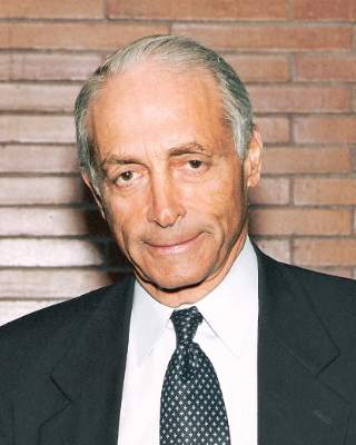
In mid-April he was working on the VEITHsymposium he created more than 40 years ago and so had not yet finalized his thoughts on his lecture.
But at that time he believed he would revisit his long-ago Presidential Address, “Charles Darwin and Vascular Surgery,” delivered in June 1996 and which also discussed the future of vascular surgery.
He noted then that it was a “time of enormous upheaval in American medicine, and holding this office has also forced me to focus on the future of Vascular Surgery and how well it will survive these turbulent times.” As Darwin studied extinct and contemporary species, his thoughts on survival could be correlated to that of the subspecialty itself, Dr. Veith said.
The endovascular revolution had begun and he was convinced – and stated – that vascular surgeons would need to learn endovascular skills. “People thought I was crazy when I said that endovascular was going to replace most of what we did,” he said. “I turned out to be very right.”
Some of his other pronouncements that day turned out to be incorrect, he said. But because change is once again part of the field of medicine, he wants to address issues he sees and possible solutions to those issues.
“I want it to be interesting and provocative,” he said. “I don’t want the listeners to fall asleep.”
Dr. Veith is professor of surgery at New York University Medical Center and at the Cleveland Clinic Lerner College of Medicine of Case Western Reserve University. He holds the William J. von Liebig chair in Vascular Surgery at the Cleveland Clinic Foundation and has lectured throughout the world.
He is a pioneer in the area of limb-salvage surgery and endovascular grafting for traumatic, aneurysmal, and occlusive arterial disease. Each year he hosts the VEITHsymposium on what is new and important in the treatment of vascular disease.
Vascular surgery has a rich, varied and enormously interesting history, filled with innovators and creators whose inventions changed the course of patient care. This history has been well-documented innumerable times.
Dr. Frank J. Veith is a part of that storied history. However, during the John Homans Distinguished Lecture Thursday morning, he will look ahead, and not back.
During “A Look at the Future of Vascular Surgery,” he plans to discuss the implications of future changes in the field, not to mention the world of medical care, and “what vascular surgery has to do to survive,” he said.
In mid-April he was working on the VEITHsymposium he created more than 40 years ago and so had not yet finalized his thoughts on his lecture.
But at that time he believed he would revisit his long-ago Presidential Address, “Charles Darwin and Vascular Surgery,” delivered in June 1996 and which also discussed the future of vascular surgery.
He noted then that it was a “time of enormous upheaval in American medicine, and holding this office has also forced me to focus on the future of Vascular Surgery and how well it will survive these turbulent times.” As Darwin studied extinct and contemporary species, his thoughts on survival could be correlated to that of the subspecialty itself, Dr. Veith said.
The endovascular revolution had begun and he was convinced – and stated – that vascular surgeons would need to learn endovascular skills. “People thought I was crazy when I said that endovascular was going to replace most of what we did,” he said. “I turned out to be very right.”
Some of his other pronouncements that day turned out to be incorrect, he said. But because change is once again part of the field of medicine, he wants to address issues he sees and possible solutions to those issues.
“I want it to be interesting and provocative,” he said. “I don’t want the listeners to fall asleep.”
Dr. Veith is professor of surgery at New York University Medical Center and at the Cleveland Clinic Lerner College of Medicine of Case Western Reserve University. He holds the William J. von Liebig chair in Vascular Surgery at the Cleveland Clinic Foundation and has lectured throughout the world.
He is a pioneer in the area of limb-salvage surgery and endovascular grafting for traumatic, aneurysmal, and occlusive arterial disease. Each year he hosts the VEITHsymposium on what is new and important in the treatment of vascular disease.
