User login
Getting to know our incoming CHEST President
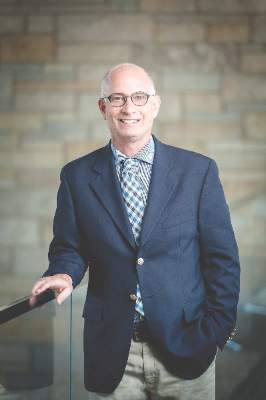
Gerard Silvestri, MD, MS, FCCP, will be inaugurated as the new President of CHEST next month in Los Angeles during CHEST 2016. He is the Hillenbrand Professor of Thoracic Oncology and Vice Chair of Medicine for Faculty Development at the Medical University of South Carolina, Charleston. Dr. Silvestri completed his fellowship training in pulmonary and critical care at Dartmouth, Hanover, N.H. He has an advanced degree in the evaluative clinical sciences, also from Dartmouth. He is a lung cancer and interventional pulmonologist with an interest in health services research, lung cancer screening, nodule evaluation and management, and staging of lung cancer.
After becoming a Fellow of the American College of Chest Physicians in 1998, Dr. Silvestri became active with the NetWorks, serving on the Steering Committees of the Thoracic Oncology and the Interventional Chest/Diagnostic Procedures NetWorks, eventually chairing the Thoracic Oncology NetWork. Dr. Silvestri has also served on the Nominating Committee, the CHEST Scientific Program Committee, the CHEST Foundation Development Committee, as Treasurer and Trustee on the foundation’s Board of Trustees, and as a Regent-at-Large for the American College of Chest Physicians for 3 years. At CHEST 2012, Dr. Silvestri was awarded the Pasquale Ciaglia Memorial Lecture in Interventional Medicine, and at CHEST 2014, he received the Edward C. Rosenow III, MD, Master FCCP/Master Teacher Honor Lecture award. Dr. Silvestri has authored more than 200 scientific articles, book chapters, and editorials, and he currently serves on the editorial board of the journal CHEST.

We asked Dr. Silvestri for some thoughts on his upcoming CHEST presidency.
1. What would you like to accomplish as President of CHEST?
As boring as this may sound, the role of the President is to oversee and carry out the strategic plan set forth by a very capable Board of Regents. It is an ambitious undertaking and among other things, it includes increasing the output of clinical practice guidelines to better serve pulmonologists and their patients, and educating as many physicians as possible through our national meeting, board review courses, our journal CHEST, our SEEK Library app and publication, and the CHEST headquarters, which has a state-of-the art education and simulation center. Our strategic vision aims to provide education to our global colleagues as well as evidenced by our commitment to regional meetings on different continents and our efforts at collaborating with our Chinese colleagues to establish the first pulmonary and critical care fellowships in that populous nation.
In support of these efforts, there are a few other projects we will get off the ground. Because education is our core mission, CHEST has a goal of helping to increase our faculty development offerings culminating in a master educator certification for those who are interested and qualify. We also will be piloting an app for practice guidelines, which will help with the implementation and dissemination of our valuable clinical practice guidelines.
2. What do you consider to be the greatest strength of CHEST, and how will you build upon this during your presidency?
The greatest strength of the College is the amazing staff and physician volunteers who give tirelessly to support the mission of the College, and ultimately, the membership as a whole. We already have begun to take measures to ensure that our most precious resources, our people, are supported in every way possible to better do their work. In the next year, it is my commitment that we continue to provide the resources and recognition so that our faculty and staff can deliver the best educational content to our membership.
Our CHEST Foundation continues to champion lung health by supporting clinical research grants, community service grants, and patient education. CHEST members, their patients, and many others have benefited from the various clinical research and humanitarian projects that the Foundation has supported. This year, we celebrate the 20th anniversary of the CHEST Foundation, and I am sure that the innovative initiatives of our charitable foundation will continue to move forward, making a difference for people throughout the world.
3. What are some challenges facing CHEST, and how will you address these challenges?
In a day in which physicians have limited resources, decisions about which medical society, if any, they should belong to have become increasingly real. Our members are using electronic media to find the tools they need to care for patients and may be less likely to follow the traditional medical association path. The challenge facing CHEST is to provide value, and it is the job of CHEST leadership to be certain that all of our members find that value in this organization. To do that, we must find or expand in creative ways a means to deliver our content in ways that resonate with our membership.
4. And finally, what is your charge to the members and new Fellows of CHEST?
The simple and overused answer would be to get involved. Without question, I believe that, and my start with the American College of Chest Physicians began as a member of the Thoracic Oncology NetWork, but I want to be a bit more specific. I challenge our members to find a niche within the College that they have a passion for, and in turn, they should challenge us to do better for our members and patients within that chosen area of expertise. There are so many ways to get involved, whether it be our NetWorks, the e-communities, practice guidelines, or helping to teach in our simulation center. CHEST is an extremely welcoming organization, and your passion will find a home here and will be nurtured and supported by other like members and the CHEST staff.
Gerard Silvestri, MD, MS, FCCP, will be inaugurated as the new President of CHEST next month in Los Angeles during CHEST 2016. He is the Hillenbrand Professor of Thoracic Oncology and Vice Chair of Medicine for Faculty Development at the Medical University of South Carolina, Charleston. Dr. Silvestri completed his fellowship training in pulmonary and critical care at Dartmouth, Hanover, N.H. He has an advanced degree in the evaluative clinical sciences, also from Dartmouth. He is a lung cancer and interventional pulmonologist with an interest in health services research, lung cancer screening, nodule evaluation and management, and staging of lung cancer.
After becoming a Fellow of the American College of Chest Physicians in 1998, Dr. Silvestri became active with the NetWorks, serving on the Steering Committees of the Thoracic Oncology and the Interventional Chest/Diagnostic Procedures NetWorks, eventually chairing the Thoracic Oncology NetWork. Dr. Silvestri has also served on the Nominating Committee, the CHEST Scientific Program Committee, the CHEST Foundation Development Committee, as Treasurer and Trustee on the foundation’s Board of Trustees, and as a Regent-at-Large for the American College of Chest Physicians for 3 years. At CHEST 2012, Dr. Silvestri was awarded the Pasquale Ciaglia Memorial Lecture in Interventional Medicine, and at CHEST 2014, he received the Edward C. Rosenow III, MD, Master FCCP/Master Teacher Honor Lecture award. Dr. Silvestri has authored more than 200 scientific articles, book chapters, and editorials, and he currently serves on the editorial board of the journal CHEST.
We asked Dr. Silvestri for some thoughts on his upcoming CHEST presidency.
1. What would you like to accomplish as President of CHEST?
As boring as this may sound, the role of the President is to oversee and carry out the strategic plan set forth by a very capable Board of Regents. It is an ambitious undertaking and among other things, it includes increasing the output of clinical practice guidelines to better serve pulmonologists and their patients, and educating as many physicians as possible through our national meeting, board review courses, our journal CHEST, our SEEK Library app and publication, and the CHEST headquarters, which has a state-of-the art education and simulation center. Our strategic vision aims to provide education to our global colleagues as well as evidenced by our commitment to regional meetings on different continents and our efforts at collaborating with our Chinese colleagues to establish the first pulmonary and critical care fellowships in that populous nation.
In support of these efforts, there are a few other projects we will get off the ground. Because education is our core mission, CHEST has a goal of helping to increase our faculty development offerings culminating in a master educator certification for those who are interested and qualify. We also will be piloting an app for practice guidelines, which will help with the implementation and dissemination of our valuable clinical practice guidelines.
2. What do you consider to be the greatest strength of CHEST, and how will you build upon this during your presidency?
The greatest strength of the College is the amazing staff and physician volunteers who give tirelessly to support the mission of the College, and ultimately, the membership as a whole. We already have begun to take measures to ensure that our most precious resources, our people, are supported in every way possible to better do their work. In the next year, it is my commitment that we continue to provide the resources and recognition so that our faculty and staff can deliver the best educational content to our membership.
Our CHEST Foundation continues to champion lung health by supporting clinical research grants, community service grants, and patient education. CHEST members, their patients, and many others have benefited from the various clinical research and humanitarian projects that the Foundation has supported. This year, we celebrate the 20th anniversary of the CHEST Foundation, and I am sure that the innovative initiatives of our charitable foundation will continue to move forward, making a difference for people throughout the world.
3. What are some challenges facing CHEST, and how will you address these challenges?
In a day in which physicians have limited resources, decisions about which medical society, if any, they should belong to have become increasingly real. Our members are using electronic media to find the tools they need to care for patients and may be less likely to follow the traditional medical association path. The challenge facing CHEST is to provide value, and it is the job of CHEST leadership to be certain that all of our members find that value in this organization. To do that, we must find or expand in creative ways a means to deliver our content in ways that resonate with our membership.
4. And finally, what is your charge to the members and new Fellows of CHEST?
The simple and overused answer would be to get involved. Without question, I believe that, and my start with the American College of Chest Physicians began as a member of the Thoracic Oncology NetWork, but I want to be a bit more specific. I challenge our members to find a niche within the College that they have a passion for, and in turn, they should challenge us to do better for our members and patients within that chosen area of expertise. There are so many ways to get involved, whether it be our NetWorks, the e-communities, practice guidelines, or helping to teach in our simulation center. CHEST is an extremely welcoming organization, and your passion will find a home here and will be nurtured and supported by other like members and the CHEST staff.
In Memoriam
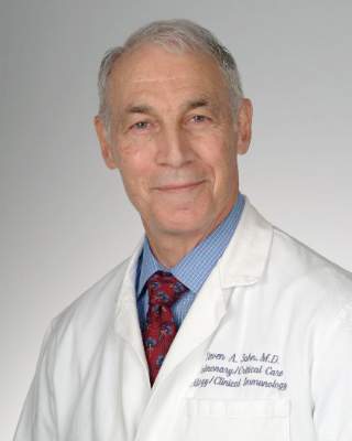
Steven A. Sahn, MD, FCCP, died on August 16, 2016, after an illustrious academic career. Born in Brooklyn, N.Y., he attended Duke University as an undergraduate and subsequently graduated from the University of Louisville School of Medicine. He completed a pulmonary–critical care fellowship at the University of Colorado, where he served the first 12 years of his academic career. As an investigator, Steve’s early pioneering work in weaning from mechanical ventilation and pleural physiology set the stage for almost all subsequent research in these fields. He was recruited in 1983 to the Medical University of South Carolina as Director of the Division of Pulmonary and Critical Care Medicine. During the next 30 years, he built the Division from three physicians to an internationally prominent team of clinicians and investigators. His passion for teaching blended his mentoring style with a love for sports and positive coaching.

He was a master clinician with remarkable diagnostic skills who attracted patients from around the world who valued his exceptional warmth and compassion. Steve’s extensive contributions to the literature resulted in numerous awards, which included CHEST’s Alfred Soffer Award for Editorial Excellence, ATS Trudeau Medal, CHEST Distinguished Lecturer for Pleural Disease, induction into the Colorado Trudeau Society Pulmonary Hall of Fame, and Distinguished University Professor of Medicine at MUSC. He contributed to the American College of Chest Physicians throughout his career serving on editorial boards of CHEST and PCCSU (Editor in Chief), on numerous committees, as co-editor of the CHEST “Pearls” section, and on the Council of Governors representing South Carolina. We extend our heartfelt condolences to his wife, Claire, and the entire Sahn family.
Steven A. Sahn, MD, FCCP, died on August 16, 2016, after an illustrious academic career. Born in Brooklyn, N.Y., he attended Duke University as an undergraduate and subsequently graduated from the University of Louisville School of Medicine. He completed a pulmonary–critical care fellowship at the University of Colorado, where he served the first 12 years of his academic career. As an investigator, Steve’s early pioneering work in weaning from mechanical ventilation and pleural physiology set the stage for almost all subsequent research in these fields. He was recruited in 1983 to the Medical University of South Carolina as Director of the Division of Pulmonary and Critical Care Medicine. During the next 30 years, he built the Division from three physicians to an internationally prominent team of clinicians and investigators. His passion for teaching blended his mentoring style with a love for sports and positive coaching.
He was a master clinician with remarkable diagnostic skills who attracted patients from around the world who valued his exceptional warmth and compassion. Steve’s extensive contributions to the literature resulted in numerous awards, which included CHEST’s Alfred Soffer Award for Editorial Excellence, ATS Trudeau Medal, CHEST Distinguished Lecturer for Pleural Disease, induction into the Colorado Trudeau Society Pulmonary Hall of Fame, and Distinguished University Professor of Medicine at MUSC. He contributed to the American College of Chest Physicians throughout his career serving on editorial boards of CHEST and PCCSU (Editor in Chief), on numerous committees, as co-editor of the CHEST “Pearls” section, and on the Council of Governors representing South Carolina. We extend our heartfelt condolences to his wife, Claire, and the entire Sahn family.
Sleep apnea and myocardial preconditioning: A paradigm shift?
The phenomenon of preconditioning reflects complex adaptive responses by living organisms to stimuli such as ischemia, hypoxia, hypothermia, or starvation. Acute ischemic preconditioning, initially described by Murry in 1986 (Circulation. 1986;74[5]:1124), occurs when multiple brief episodes of ischemia followed by reperfusion elicit a protective effect on the heart from a subsequent prolonged period of ischemia, such as a heart attack. This protective effect from ischemic preconditioning can be in the form of a smaller heart attack, lower chance of cardiac arrhythmias, less myocardial cell death, and lower risk of heart muscle failure. The cardioprotective effect of ischemic preconditioning is dependent on the duration and strength of the preconditioning stimulus. If the preconditioning stimulus is too strong or prolonged, detrimental effects on the heart may be observed.

Like ischemic preconditioning, hypoxic preconditioning represents a complex adaptive response that organisms have developed to offset damage inflicted by oxygen deprivation. The concept of hypoxic preconditioning is familiar to humans; for years, athletes have been using hypoxic training (high altitude and other newer technologies) to boost their performance in sporting events. Additionally, there is evidence dating back to before the breakthrough findings of Murry and colleagues who confirm the cardioprotective effects of hypoxia. In 1973, Meerson and colleagues (Am J Cardiol. 1973;31[1]:30) reported that mice exposed to high-altitude hypoxia have reduced mortality and smaller areas of necrotic myocardium after coronary artery occlusion.
Both of the ischemic and hypoxic preconditioning animal experiments mentioned above involve acute exposure to the preconditioning stimuli, resulting in a cardioprotective response for a limited time period. In order to afford a sustained period of cardioprotection, recurrent hypoxic exposure may be necessary. Indeed, recent studies have concentrated on just that; repeated exposure to intermittent hypoxia over a few weeks (Manukhina et al. Exp Biol Med. 2013;238[12]:1413) results in robust cardioprotection after coronary artery occlusion and reperfusion.
Despite the convincing cardioprotective discoveries from ischemic and hypoxic preconditioning, translation into clinical practice as a therapeutic modality is absent. This is partly because human beings are more complex than animals. They have comorbidities and are affected by aging, both of which may alter the milieu for preconditioning stimuli. Furthermore, the therapeutic range for any given preconditioning stimulus is unknown.
Sleep apnea (SA) is exceedingly prevalent in the United States. In SA, an individual stops breathing either completely (apnea) or partially (hypopnea) during sleep resulting in intermittent hypoxia, with arousal from sleep and resumption of breathing leading to reoxygenation. Hence, SA is characterized by intermittent hypoxia followed by reoxygenation. So, one can speculate that SA could exert cardioprotective effects as seen in hypoxic preconditioning and ischemic preconditioning. It is important to note, however, that SA is associated with hypercapnic intermittent hypoxia, whereas most of the investigations on ischemic preconditioning and intermittent hypoxia are with eucapnia or hypocapnia.
The potentially cardioprotective role of SA is supported by a growing body of complementary research that indicates that coronary collateral flow is higher in patients with SA vs. control subjects (Steiner. Chest. 2010;137[3]:516) and that there is an increased mobilization, proliferation, and angiogenic capacity of endothelial progenitor cells from patients with myocardial infarction and SA as compared with cells from controls subjects without SA (Berger. Am J Respir Crit Care Med. 2013;187[1]:90). Some epidemiologic data support a weaker relationship between SA and coronary ischemic events compared with other cardiovascular events (Gottlieb. Circulation. 2010;122[4]:352). Hypoxic preconditioning may explain the relatively decreased pro-thrombotic influence of SA in the coronary vascular bed. Nevertheless, more research is needed to determine if SA is cardioprotective. If, however, cardioprotection from SA is confirmed, it may contribute to a paradigm shift in how SA is considered in relationship to coronary heart disease. Furthermore, future investigations would need to focus on what dose and duration of SA is needed for cardioprotection to occur. Prospective studies may also provide an opportunity for investigating interindividual variability in the susceptibility of the myocardium to the hypoxic preconditioning stimulus from SA.
This article highlights how complex the relationship between hypoxia and myocardial response is. This is further supported by results from a recent clinical trial, AVOID (Air Versus Oxygen In ST-elevation MyocarDial Infarction) (Stub. Circulation. 2015;131[24]:2143). Results from the AVOID trial report that routine oxygen use in normoxic patients hospitalized with a heart attack was not beneficial and, in fact, was harmful. Patients who received oxygen had more myocardial injury than those who did not. Therefore, even though for decades we thought that oxygen therapy helps hospitalized heart attack patients, results from the AVOID trial have initiated a paradigm shift. It remains to be determined whether such a paradigm shift will follow for sleep apnea.
Dr. Shah is with the department of epidemiology and population health, Albert Einstein College of Medicine, N.Y.
The phenomenon of preconditioning reflects complex adaptive responses by living organisms to stimuli such as ischemia, hypoxia, hypothermia, or starvation. Acute ischemic preconditioning, initially described by Murry in 1986 (Circulation. 1986;74[5]:1124), occurs when multiple brief episodes of ischemia followed by reperfusion elicit a protective effect on the heart from a subsequent prolonged period of ischemia, such as a heart attack. This protective effect from ischemic preconditioning can be in the form of a smaller heart attack, lower chance of cardiac arrhythmias, less myocardial cell death, and lower risk of heart muscle failure. The cardioprotective effect of ischemic preconditioning is dependent on the duration and strength of the preconditioning stimulus. If the preconditioning stimulus is too strong or prolonged, detrimental effects on the heart may be observed.
Like ischemic preconditioning, hypoxic preconditioning represents a complex adaptive response that organisms have developed to offset damage inflicted by oxygen deprivation. The concept of hypoxic preconditioning is familiar to humans; for years, athletes have been using hypoxic training (high altitude and other newer technologies) to boost their performance in sporting events. Additionally, there is evidence dating back to before the breakthrough findings of Murry and colleagues who confirm the cardioprotective effects of hypoxia. In 1973, Meerson and colleagues (Am J Cardiol. 1973;31[1]:30) reported that mice exposed to high-altitude hypoxia have reduced mortality and smaller areas of necrotic myocardium after coronary artery occlusion.
Both of the ischemic and hypoxic preconditioning animal experiments mentioned above involve acute exposure to the preconditioning stimuli, resulting in a cardioprotective response for a limited time period. In order to afford a sustained period of cardioprotection, recurrent hypoxic exposure may be necessary. Indeed, recent studies have concentrated on just that; repeated exposure to intermittent hypoxia over a few weeks (Manukhina et al. Exp Biol Med. 2013;238[12]:1413) results in robust cardioprotection after coronary artery occlusion and reperfusion.
Despite the convincing cardioprotective discoveries from ischemic and hypoxic preconditioning, translation into clinical practice as a therapeutic modality is absent. This is partly because human beings are more complex than animals. They have comorbidities and are affected by aging, both of which may alter the milieu for preconditioning stimuli. Furthermore, the therapeutic range for any given preconditioning stimulus is unknown.
Sleep apnea (SA) is exceedingly prevalent in the United States. In SA, an individual stops breathing either completely (apnea) or partially (hypopnea) during sleep resulting in intermittent hypoxia, with arousal from sleep and resumption of breathing leading to reoxygenation. Hence, SA is characterized by intermittent hypoxia followed by reoxygenation. So, one can speculate that SA could exert cardioprotective effects as seen in hypoxic preconditioning and ischemic preconditioning. It is important to note, however, that SA is associated with hypercapnic intermittent hypoxia, whereas most of the investigations on ischemic preconditioning and intermittent hypoxia are with eucapnia or hypocapnia.
The potentially cardioprotective role of SA is supported by a growing body of complementary research that indicates that coronary collateral flow is higher in patients with SA vs. control subjects (Steiner. Chest. 2010;137[3]:516) and that there is an increased mobilization, proliferation, and angiogenic capacity of endothelial progenitor cells from patients with myocardial infarction and SA as compared with cells from controls subjects without SA (Berger. Am J Respir Crit Care Med. 2013;187[1]:90). Some epidemiologic data support a weaker relationship between SA and coronary ischemic events compared with other cardiovascular events (Gottlieb. Circulation. 2010;122[4]:352). Hypoxic preconditioning may explain the relatively decreased pro-thrombotic influence of SA in the coronary vascular bed. Nevertheless, more research is needed to determine if SA is cardioprotective. If, however, cardioprotection from SA is confirmed, it may contribute to a paradigm shift in how SA is considered in relationship to coronary heart disease. Furthermore, future investigations would need to focus on what dose and duration of SA is needed for cardioprotection to occur. Prospective studies may also provide an opportunity for investigating interindividual variability in the susceptibility of the myocardium to the hypoxic preconditioning stimulus from SA.
This article highlights how complex the relationship between hypoxia and myocardial response is. This is further supported by results from a recent clinical trial, AVOID (Air Versus Oxygen In ST-elevation MyocarDial Infarction) (Stub. Circulation. 2015;131[24]:2143). Results from the AVOID trial report that routine oxygen use in normoxic patients hospitalized with a heart attack was not beneficial and, in fact, was harmful. Patients who received oxygen had more myocardial injury than those who did not. Therefore, even though for decades we thought that oxygen therapy helps hospitalized heart attack patients, results from the AVOID trial have initiated a paradigm shift. It remains to be determined whether such a paradigm shift will follow for sleep apnea.
Dr. Shah is with the department of epidemiology and population health, Albert Einstein College of Medicine, N.Y.
Networks
Occupational and Environmental Health
Incident sarcoidosis
The history of sarcoidosis dates back to 1869, when Dr. Jonathan Hutchinson described symmetrical purple skin plaques on the legs and hands of a coal-wharf worker (James and Sharma. Curr Opin Pulm Med. 2002;8[5]:416). However, despite its distant beginning, much remains unknown. It has been hypothesized that environmental factors play a pivotal role in disease onset and course, as is evidenced by the notable exposure in the first historical case.
Research has shown environmental factors, such as wood smoke, tree pollen, insecticides, and mold; as well as occupational exposures, such as flight deck work on aircraft carriers, metalworking, construction, and firefighting, carry increased risk of sarcoidosis (Newman et al. Am J Respir Crit Care Med. 2004;170[12]:1324; Newman and Newman. Curr Opin Allergy Clin Immunol. 2012;12[2]:145). A significantly high annual incidence of sarcoidosis was first demonstrated in FDNY firefighters between 1985 and 1998; 12.9/100,000, as compared with 2.5 to 7.6/100,000 for U.S. white men (Prezant et al. Chest. 1999;116[5]:1183).

Following the attack of the World Trade Center (WTC) on September 11, 2001, a further increase in sarcoidosis incidence was found in FDNY firefighters exposed to WTC “dust” during the collapse and rescue/recovery effort (Izbicki et al. Chest. 2007;131[5]:1414). As of 2015, a total of 75 FDNY firefighters have been identified as having new post-9/11 sarcoidosis.
Since the WTC-exposed FDNY firefighters with new-onset sarcoidosis since September 11, 2001 can be considered to have had a WTC “trigger,” we have a unique opportunity to define the clinical patterns and outcomes of incident sarcoidosis following a distinct exposure. Members of the Occupational and Environmental NetWork Steering Committee are currently investigating this aim and others in a National Institute of Occupational Safety and Health (NIOSH)-granted cohort study. We hypothesize that the patterns of organ involvement, and time course of disease progression or resolution, may significantly differ in this group as compared with the general population. Preliminary results of our study of WTC-exposed FDNY firefighters will be presented at CHEST 2016 in Los Angeles.
Kerry Hena, MD
Physician-in-Training Member
Palliative and End-of-Life Care
Integrated palliative care for mechanical circulatory support
Patients with advanced heart failure (AHF) have well-documented needs for comprehensive supportive care services in the critical care setting. Notable symptom burden, high morbidity and mortality, prognostic uncertainty, and need for care coordination across hospital settings pave the way for palliative care (PC) teams to work symbiotically with advanced heart failure specialists and intensivists. Furthermore, the expanded availability of mechanical circulatory support (MCS) technology extends these clinical and ethical challenges to balancing longevity, quality of life, and resource utilization, most prominently in the ICU.
To date, collaborations between PC, AHF specialists, and critical care have tended to be reactive, not proactive – palliative consultation usually occurs after a medical or surgical crisis (for example, the massive stroke, MCS thrombus, sepsis, and multiorgan failure) or after a prolonged ICU stay without clear improvement in patient function or prognosis. This reactive consult may be misperceived by patient and family as “giving up.”
At our institution, we have worked to develop a model of seamless integration of interdisciplinary palliative care consultation upstream in advanced heart failure patient care that aims to preempt many dilemmas in the ICU around complex medical decision making and end-of-life care. Through development of therapeutic supportive care relationships, preparedness planning, and discussions of goals of care early in treatment pathways involving critical care resources – including MCS evaluation and cardiac transplantation – this model purports to strengthen appropriate critical care delivery for patients with advanced heart failure. This model has evolved to where PC consultation becomes a structured part of the preoperative evaluation of all candidates for left-ventricular assist device as destination therapy (LVAD-DT). The result is a collaborative approach where patients and families see PC as part of the continuum of whole-person AHF care, rather than a negative alternative.
MCS implantation is on the rise. While MCS technology continues to evolve, its recipients remain seriously ill. Normalizing and integrating PC consultation as part of high quality AHF and critical care sends an important message to patients and families: regardless of clinical outcome, relief from suffering matters throughout the trajectory of the illness experience.
Hunter Groninger, MD
Steering Committee Member
Respiratory Care NetWork
Professional relationships in RC
At the 2015 meeting of the American Association for Respiratory Care (AARC) in Tampa, there were more than 20 presentations given by FCCPs! Also, a majority of CHEST’s Respiratory Care NetWork’s steering committee was in attendance at the meeting. To other members of CHEST, that might seem rather unusual. However, many CHEST members have connections with the field of respiratory care. In addition, CHEST as an organization has a professional relationship with the respiratory care field. CHEST has more than 10 official liaisons to respiratory care professional organizations.
Those organizations include: The Commission for Accreditation for Respiratory Care, which credentials all RC educational programs; The National Board for Respiratory Care, which provides the credentialing examinations for all RC practitioners in the United States; The National Association for Medical Direction for Respiratory Care (NAMDRC); the Board of Medical Advisors to the AARC; and the Respiratory Compromise Institute.
The Respiratory Care NetWork has the responsibility of identifying and nominating CHEST members for these liaison positions. These volunteer positions do involve work, yet past and present liaisons have enthusiastically fulfilled their respective roles. As one recently noted, “This work has been some of the most important endeavors of my professional career.”
We are always seeking volunteers for these positions, which vary in time commitment and type of work involved. Please contact the Respiratory Care NetWork (mkosinski@chestnet.org) for further information. These organizations accomplish the type of things that made us all want to get into medicine. Be a part of those important efforts!
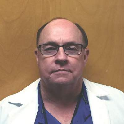
Thomas Fuhrman, MD, FCCP
Steering Committee Member
Sleep Medicine
Listening to patient voices: Sleep Apnea Patient-Centered Outcomes Network (MyApnea.org)
The US Department of Transportation’s (DOT) Federal Motor Carrier Safety Administration (FMCSA) and Federal Railroad Administration (FRA) recently called for input for obstructive sleep apnea screening and treatment for transportation workers. The DOT (https://www.transportation.gov) encouraged input from the public regarding this important transportation safety issue. This concept of engaging the public (which includes patients) with sleep disorders is gaining momentum as patients are increasingly partnering with researchers, clinicians, and policy makers to improve the delivery of care and research efforts in sleep medicine.
A remarkable example of such an effort is the Sleep Apnea Patient-Centered Outcomes Network (SAPCON; MyApnea.Org) (Redline et al. JCSM. 2016;12[7]:1053). This patient-powered research network was initiated in 2013 to improve the diagnosis and treatment of sleep apnea through the active engagement of patients, families, researchers, and healthcare providers in a virtual community that facilitates patient-centered research. The need for such an initiative reflects the paucity of patient-centric evidence from large populations to inform insurers, public policy makers, medical schools, and clinicians on the best ways to screen, diagnose, and treat patients with sleep apnea.
As of August 2016, over 8,000 individuals across the globe have joined SAPCON. There are approximately 500 unique visitors to the site per day, with over 2,500 posts on over 250 topics, including blogs on a variety of emerging research and public health topics. Among these topics are driving and general transportation safety concerns. Further engagement of patients and key stakeholders through forums and patient-centered networks can promote the “patient voice” in public policy, while linking patient needs for better information with responsive research and policy development.
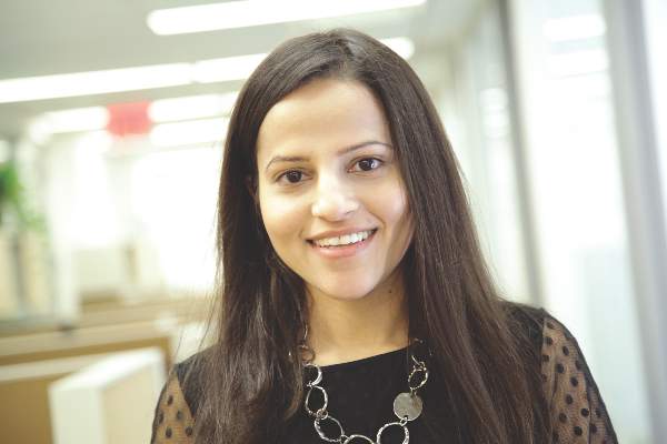
Neomi Shah, MD, MPH
Steering Committee Member
Occupational and Environmental Health
Incident sarcoidosis
The history of sarcoidosis dates back to 1869, when Dr. Jonathan Hutchinson described symmetrical purple skin plaques on the legs and hands of a coal-wharf worker (James and Sharma. Curr Opin Pulm Med. 2002;8[5]:416). However, despite its distant beginning, much remains unknown. It has been hypothesized that environmental factors play a pivotal role in disease onset and course, as is evidenced by the notable exposure in the first historical case.
Research has shown environmental factors, such as wood smoke, tree pollen, insecticides, and mold; as well as occupational exposures, such as flight deck work on aircraft carriers, metalworking, construction, and firefighting, carry increased risk of sarcoidosis (Newman et al. Am J Respir Crit Care Med. 2004;170[12]:1324; Newman and Newman. Curr Opin Allergy Clin Immunol. 2012;12[2]:145). A significantly high annual incidence of sarcoidosis was first demonstrated in FDNY firefighters between 1985 and 1998; 12.9/100,000, as compared with 2.5 to 7.6/100,000 for U.S. white men (Prezant et al. Chest. 1999;116[5]:1183).
Following the attack of the World Trade Center (WTC) on September 11, 2001, a further increase in sarcoidosis incidence was found in FDNY firefighters exposed to WTC “dust” during the collapse and rescue/recovery effort (Izbicki et al. Chest. 2007;131[5]:1414). As of 2015, a total of 75 FDNY firefighters have been identified as having new post-9/11 sarcoidosis.
Since the WTC-exposed FDNY firefighters with new-onset sarcoidosis since September 11, 2001 can be considered to have had a WTC “trigger,” we have a unique opportunity to define the clinical patterns and outcomes of incident sarcoidosis following a distinct exposure. Members of the Occupational and Environmental NetWork Steering Committee are currently investigating this aim and others in a National Institute of Occupational Safety and Health (NIOSH)-granted cohort study. We hypothesize that the patterns of organ involvement, and time course of disease progression or resolution, may significantly differ in this group as compared with the general population. Preliminary results of our study of WTC-exposed FDNY firefighters will be presented at CHEST 2016 in Los Angeles.
Kerry Hena, MD
Physician-in-Training Member
Palliative and End-of-Life Care
Integrated palliative care for mechanical circulatory support
Patients with advanced heart failure (AHF) have well-documented needs for comprehensive supportive care services in the critical care setting. Notable symptom burden, high morbidity and mortality, prognostic uncertainty, and need for care coordination across hospital settings pave the way for palliative care (PC) teams to work symbiotically with advanced heart failure specialists and intensivists. Furthermore, the expanded availability of mechanical circulatory support (MCS) technology extends these clinical and ethical challenges to balancing longevity, quality of life, and resource utilization, most prominently in the ICU.
To date, collaborations between PC, AHF specialists, and critical care have tended to be reactive, not proactive – palliative consultation usually occurs after a medical or surgical crisis (for example, the massive stroke, MCS thrombus, sepsis, and multiorgan failure) or after a prolonged ICU stay without clear improvement in patient function or prognosis. This reactive consult may be misperceived by patient and family as “giving up.”
At our institution, we have worked to develop a model of seamless integration of interdisciplinary palliative care consultation upstream in advanced heart failure patient care that aims to preempt many dilemmas in the ICU around complex medical decision making and end-of-life care. Through development of therapeutic supportive care relationships, preparedness planning, and discussions of goals of care early in treatment pathways involving critical care resources – including MCS evaluation and cardiac transplantation – this model purports to strengthen appropriate critical care delivery for patients with advanced heart failure. This model has evolved to where PC consultation becomes a structured part of the preoperative evaluation of all candidates for left-ventricular assist device as destination therapy (LVAD-DT). The result is a collaborative approach where patients and families see PC as part of the continuum of whole-person AHF care, rather than a negative alternative.
MCS implantation is on the rise. While MCS technology continues to evolve, its recipients remain seriously ill. Normalizing and integrating PC consultation as part of high quality AHF and critical care sends an important message to patients and families: regardless of clinical outcome, relief from suffering matters throughout the trajectory of the illness experience.
Hunter Groninger, MD
Steering Committee Member
Respiratory Care NetWork
Professional relationships in RC
At the 2015 meeting of the American Association for Respiratory Care (AARC) in Tampa, there were more than 20 presentations given by FCCPs! Also, a majority of CHEST’s Respiratory Care NetWork’s steering committee was in attendance at the meeting. To other members of CHEST, that might seem rather unusual. However, many CHEST members have connections with the field of respiratory care. In addition, CHEST as an organization has a professional relationship with the respiratory care field. CHEST has more than 10 official liaisons to respiratory care professional organizations.
Those organizations include: The Commission for Accreditation for Respiratory Care, which credentials all RC educational programs; The National Board for Respiratory Care, which provides the credentialing examinations for all RC practitioners in the United States; The National Association for Medical Direction for Respiratory Care (NAMDRC); the Board of Medical Advisors to the AARC; and the Respiratory Compromise Institute.
The Respiratory Care NetWork has the responsibility of identifying and nominating CHEST members for these liaison positions. These volunteer positions do involve work, yet past and present liaisons have enthusiastically fulfilled their respective roles. As one recently noted, “This work has been some of the most important endeavors of my professional career.”
We are always seeking volunteers for these positions, which vary in time commitment and type of work involved. Please contact the Respiratory Care NetWork (mkosinski@chestnet.org) for further information. These organizations accomplish the type of things that made us all want to get into medicine. Be a part of those important efforts!
Thomas Fuhrman, MD, FCCP
Steering Committee Member
Sleep Medicine
Listening to patient voices: Sleep Apnea Patient-Centered Outcomes Network (MyApnea.org)
The US Department of Transportation’s (DOT) Federal Motor Carrier Safety Administration (FMCSA) and Federal Railroad Administration (FRA) recently called for input for obstructive sleep apnea screening and treatment for transportation workers. The DOT (https://www.transportation.gov) encouraged input from the public regarding this important transportation safety issue. This concept of engaging the public (which includes patients) with sleep disorders is gaining momentum as patients are increasingly partnering with researchers, clinicians, and policy makers to improve the delivery of care and research efforts in sleep medicine.
A remarkable example of such an effort is the Sleep Apnea Patient-Centered Outcomes Network (SAPCON; MyApnea.Org) (Redline et al. JCSM. 2016;12[7]:1053). This patient-powered research network was initiated in 2013 to improve the diagnosis and treatment of sleep apnea through the active engagement of patients, families, researchers, and healthcare providers in a virtual community that facilitates patient-centered research. The need for such an initiative reflects the paucity of patient-centric evidence from large populations to inform insurers, public policy makers, medical schools, and clinicians on the best ways to screen, diagnose, and treat patients with sleep apnea.
As of August 2016, over 8,000 individuals across the globe have joined SAPCON. There are approximately 500 unique visitors to the site per day, with over 2,500 posts on over 250 topics, including blogs on a variety of emerging research and public health topics. Among these topics are driving and general transportation safety concerns. Further engagement of patients and key stakeholders through forums and patient-centered networks can promote the “patient voice” in public policy, while linking patient needs for better information with responsive research and policy development.
Neomi Shah, MD, MPH
Steering Committee Member
Los Angeles Inspires With Arts, Culture
Los Angeles has a flare for the dramatic, and we’re not just talking about Hollywood’s fast-paced, larger-than-life movie industry. When you visit Los Angeles, October 22 – 26, for CHEST 2016, be sure to check out the assortment of arts and culture venues located nearby your home base at CHEST 2016.
Los Angeles has more museums and theaters than any other U.S. city, and we’ll highlight a few local favorites below. For more information on L.A.’s thriving arts and culture scene, check out discoverlosangeles.com.

• The Dorothy Chandler Pavilion – (7-minute drive) – This hall is part of the Los Angeles music center. On October 22 – 23, watch three major U.S. ballet companies share the stage in Celebrate Forsythe. Or, take in The Source, a music-theater production about Chelsea (formerly Bradley) Manning and WikiLeaks.
• The Ahmanson Theatre – (7-minute drive) – This theater is also part of the Los Angeles music center. Be captivated by a 2016 Tony Award–winning play, A View from the Bridge.
• Walt Disney Concert Hall – (6-minute drive) – Home to the Los Angeles Philharmonic Orchestra and the Los Angeles Master Chorale, it is also part of the Los Angeles music center. Listen to the beautiful sounds of Mahler’s Ninth or Hilary Hahn on violin.
• MOCA Grand – (5-minute drive) – The Museum of Contemporary Art has three locations in Los Angeles. The main branch, located on Grand Avenue, is the closest to the convention center. Check out the museum’s main galleries at this location.
• The Getty Center – (30-minute drive) – See spectacular art and architecture at the top of Los Angeles.
Note: all estimated times assume you are starting at the Los Angeles Convention Center.
Los Angeles’ arts and culture scene is sure to inspire you, and CHEST 2016 will move you with the latest clinical information in chest medicine. Join us at CHEST 2016, and you won’t miss a beat with cutting-edge sessions and simulation training designed to update you on the latest patient care strategies. You will be part of an international community of innovative problem solvers. Learn more and register today at chestmeeting.chestnet.org.
Los Angeles has a flare for the dramatic, and we’re not just talking about Hollywood’s fast-paced, larger-than-life movie industry. When you visit Los Angeles, October 22 – 26, for CHEST 2016, be sure to check out the assortment of arts and culture venues located nearby your home base at CHEST 2016.
Los Angeles has more museums and theaters than any other U.S. city, and we’ll highlight a few local favorites below. For more information on L.A.’s thriving arts and culture scene, check out discoverlosangeles.com.
• The Dorothy Chandler Pavilion – (7-minute drive) – This hall is part of the Los Angeles music center. On October 22 – 23, watch three major U.S. ballet companies share the stage in Celebrate Forsythe. Or, take in The Source, a music-theater production about Chelsea (formerly Bradley) Manning and WikiLeaks.
• The Ahmanson Theatre – (7-minute drive) – This theater is also part of the Los Angeles music center. Be captivated by a 2016 Tony Award–winning play, A View from the Bridge.
• Walt Disney Concert Hall – (6-minute drive) – Home to the Los Angeles Philharmonic Orchestra and the Los Angeles Master Chorale, it is also part of the Los Angeles music center. Listen to the beautiful sounds of Mahler’s Ninth or Hilary Hahn on violin.
• MOCA Grand – (5-minute drive) – The Museum of Contemporary Art has three locations in Los Angeles. The main branch, located on Grand Avenue, is the closest to the convention center. Check out the museum’s main galleries at this location.
• The Getty Center – (30-minute drive) – See spectacular art and architecture at the top of Los Angeles.
Note: all estimated times assume you are starting at the Los Angeles Convention Center.
Los Angeles’ arts and culture scene is sure to inspire you, and CHEST 2016 will move you with the latest clinical information in chest medicine. Join us at CHEST 2016, and you won’t miss a beat with cutting-edge sessions and simulation training designed to update you on the latest patient care strategies. You will be part of an international community of innovative problem solvers. Learn more and register today at chestmeeting.chestnet.org.

This Month in CHEST: Editor’s Picks
A Novel PF4-Dependent Platelet Activation Assay Identifies Patients Likely to Have
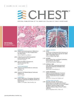
Heparin-Induced Thrombocytopenia/Thrombosis.By Dr. A Padmanabhan et al.
Safety and Tolerability of Alveolar Type II Cell Transplantation in Idiopathic
Pulmonary Fibrosis.By Dr. A. Serrano-Mollar et al.
Hypertension Is Associated With Undiagnosed OSA During Rapid Eye Movement Sleep.By Dr. S. L. Appleton et al.
A Novel PF4-Dependent Platelet Activation Assay Identifies Patients Likely to Have
Heparin-Induced Thrombocytopenia/Thrombosis.By Dr. A Padmanabhan et al.
Safety and Tolerability of Alveolar Type II Cell Transplantation in Idiopathic
Pulmonary Fibrosis.By Dr. A. Serrano-Mollar et al.
Hypertension Is Associated With Undiagnosed OSA During Rapid Eye Movement Sleep.By Dr. S. L. Appleton et al.

From the CEO
As a 22-year member of the senior staff of the American College of Chest Physicians, I am absolutely thrilled to have the opportunity to serve as its interim EVP/CEO for the current 2016-2017 fiscal year. Over the course of the years, I’ve been fortunate to oversee a number of CHEST’s business units and divisions, including Publications, Marketing, Communications, Membership, International Development, and Information Technology (IT). This background has provided a stable foundation for a smooth transition and ensured that the college continues to move to achieve its strategic plan and operational goals. That plan and those goals ensure that we will fulfill CHEST’s mission and vision: “To champion the prevention, diagnosis, and treatment of chest diseases through education, communication, and research” and to be “the global leader in advancing best patient outcomes through innovative chest medicine education, clinical research, and team-based care,” respectively.

To this end, we are executing well as an organization. Our state-of-the-art Innovation, Simulation, and Training Center at the CHEST Global Headquarters in Glenview, Illinois, continues to provide outstanding hands-on educational events and opportunities, and our Education Calendar has something for just about everyone. Our annual Board Review courses continue to provide excellent content. The CHEST 2016 annual meeting in LA this October will showcase all that CHEST has to offer. And the list goes on.
One of CHEST’s strengths is its spirit of innovation. Whether it’s revamping the highly successful SEEK app into an easily accessible online library, adding more simulation and procedure-based training to our educational offerings, or providing our live courses as captured online “on-demand” programs, we are committed to finding ways to package and deliver meaningful education to our members and community. Our for-profit subsidiary, CHEST Enterprises, is providing professional education to industry through the PREP disease-state immersion program, and developing a data analytics product line that will provide insights into physician behavior. Our charitable foundation, the CHEST Foundation, gives nearly $500,000 in research and community service grants each year, to champion lung health. It has also expanded the number of available patient education resources in partnership with the ALA. And in the past year, we have fully implemented CHEST’s new innovative membership model to welcome more nonphysician health care providers and give them opportunities to engage, learn, and participate. All of these things are incredibly exciting to me, and I’m grateful to be part of them.
But what I’m most excited and grateful for are the people who impact our organization. We have a diverse and passionate membership of physician and nonphysician health care providers who want to provide the best care possible and positively impact outcomes. Our dedicated faculty and volunteers generously give their time to the organization’s work groups and programs so that they can give back to others in the field. Our hard-working leaders take responsibility and ownership of our programs and content. And, our outstanding staff operationalizes the strategic plan and goals of the organization hand-in-hand with those leaders, volunteers, faculty, and members. Together, it all results in the excellent programs you have come to expect from CHEST.
Thank you for participating and supporting this robust, dynamic organization. I am excited for the future for CHEST, and I look forward to seeing you at CHEST 2016 in Los Angeles! If you have thoughts or ideas about how we can enhance our work to be a global leader in chest medicine, connect with me anytime. I invite you to follow and connect with me on Twitter (@RocketSurgery99), or look for me at upcoming CHEST events.
As a 22-year member of the senior staff of the American College of Chest Physicians, I am absolutely thrilled to have the opportunity to serve as its interim EVP/CEO for the current 2016-2017 fiscal year. Over the course of the years, I’ve been fortunate to oversee a number of CHEST’s business units and divisions, including Publications, Marketing, Communications, Membership, International Development, and Information Technology (IT). This background has provided a stable foundation for a smooth transition and ensured that the college continues to move to achieve its strategic plan and operational goals. That plan and those goals ensure that we will fulfill CHEST’s mission and vision: “To champion the prevention, diagnosis, and treatment of chest diseases through education, communication, and research” and to be “the global leader in advancing best patient outcomes through innovative chest medicine education, clinical research, and team-based care,” respectively.
To this end, we are executing well as an organization. Our state-of-the-art Innovation, Simulation, and Training Center at the CHEST Global Headquarters in Glenview, Illinois, continues to provide outstanding hands-on educational events and opportunities, and our Education Calendar has something for just about everyone. Our annual Board Review courses continue to provide excellent content. The CHEST 2016 annual meeting in LA this October will showcase all that CHEST has to offer. And the list goes on.
One of CHEST’s strengths is its spirit of innovation. Whether it’s revamping the highly successful SEEK app into an easily accessible online library, adding more simulation and procedure-based training to our educational offerings, or providing our live courses as captured online “on-demand” programs, we are committed to finding ways to package and deliver meaningful education to our members and community. Our for-profit subsidiary, CHEST Enterprises, is providing professional education to industry through the PREP disease-state immersion program, and developing a data analytics product line that will provide insights into physician behavior. Our charitable foundation, the CHEST Foundation, gives nearly $500,000 in research and community service grants each year, to champion lung health. It has also expanded the number of available patient education resources in partnership with the ALA. And in the past year, we have fully implemented CHEST’s new innovative membership model to welcome more nonphysician health care providers and give them opportunities to engage, learn, and participate. All of these things are incredibly exciting to me, and I’m grateful to be part of them.
But what I’m most excited and grateful for are the people who impact our organization. We have a diverse and passionate membership of physician and nonphysician health care providers who want to provide the best care possible and positively impact outcomes. Our dedicated faculty and volunteers generously give their time to the organization’s work groups and programs so that they can give back to others in the field. Our hard-working leaders take responsibility and ownership of our programs and content. And, our outstanding staff operationalizes the strategic plan and goals of the organization hand-in-hand with those leaders, volunteers, faculty, and members. Together, it all results in the excellent programs you have come to expect from CHEST.
Thank you for participating and supporting this robust, dynamic organization. I am excited for the future for CHEST, and I look forward to seeing you at CHEST 2016 in Los Angeles! If you have thoughts or ideas about how we can enhance our work to be a global leader in chest medicine, connect with me anytime. I invite you to follow and connect with me on Twitter (@RocketSurgery99), or look for me at upcoming CHEST events.
Connect with the CHEST Foundation at CHEST 2016
At this year’s annual meeting, the CHEST Foundation will have several new and exciting events, including three networking happy hours, October 23-25, for women in lung health, international members, and nonphysician providers, and two educational sessions developed by CHEST Foundation leaders and volunteers, Chris Carroll, MD, FCCP, and Muhammad Adrish, MD, FCCP. The new sessions will provide a focus on essential tactics for conducting effective research and a panel discussion featuring past CHEST Foundation grant winners, on how to create a successful community service program. In addition to these events, we will be hosting another “Young Professionals Reception” Monday evening. Be sure to stop by the Donor Lounge to network and mingle with leadership, meet the foundation staff, grab a coffee, and learn how you can engage with the CHEST Foundation. If you arrive early on Saturday, October 22, don’t miss out on our afternoon “Champions for Lung Health Event,” where CHEST Foundation leadership will be giving back to the Los Angeles community by volunteering their time at a COPD screening.

We are also proud to introduce our 2016 CHEST Foundation grantees at this year’s Opening Session on Monday, October 24. This year, we will be awarding nearly a half-million dollars in funding to the next generation of lung health champions. Our grants and programs have made a difference in the lives of our members and their patients through the impactful clinical research and impressive humanitarian projects our grantees have created. Since 1996, we’ve provided over $10 million in funding for clinical research and community service, with a reach that spans from Texas to Tanzania. The foundation is an important go-to resource for young investigators seeking research funding, and the projects we support lead to breakthroughs in the treatment of chest diseases and patient care.
We hope to see you at one of our open invitation activities to learn more about how the CHEST Foundation can support you in your efforts to champion lung health.
At this year’s annual meeting, the CHEST Foundation will have several new and exciting events, including three networking happy hours, October 23-25, for women in lung health, international members, and nonphysician providers, and two educational sessions developed by CHEST Foundation leaders and volunteers, Chris Carroll, MD, FCCP, and Muhammad Adrish, MD, FCCP. The new sessions will provide a focus on essential tactics for conducting effective research and a panel discussion featuring past CHEST Foundation grant winners, on how to create a successful community service program. In addition to these events, we will be hosting another “Young Professionals Reception” Monday evening. Be sure to stop by the Donor Lounge to network and mingle with leadership, meet the foundation staff, grab a coffee, and learn how you can engage with the CHEST Foundation. If you arrive early on Saturday, October 22, don’t miss out on our afternoon “Champions for Lung Health Event,” where CHEST Foundation leadership will be giving back to the Los Angeles community by volunteering their time at a COPD screening.
We are also proud to introduce our 2016 CHEST Foundation grantees at this year’s Opening Session on Monday, October 24. This year, we will be awarding nearly a half-million dollars in funding to the next generation of lung health champions. Our grants and programs have made a difference in the lives of our members and their patients through the impactful clinical research and impressive humanitarian projects our grantees have created. Since 1996, we’ve provided over $10 million in funding for clinical research and community service, with a reach that spans from Texas to Tanzania. The foundation is an important go-to resource for young investigators seeking research funding, and the projects we support lead to breakthroughs in the treatment of chest diseases and patient care.
We hope to see you at one of our open invitation activities to learn more about how the CHEST Foundation can support you in your efforts to champion lung health.

Early days of IVF marked by competition, innovation
In 1978, when England’s Louise Brown became the world’s first baby born through in vitro fertilization, physicians at academic centers all over the United States scrambled to figure out how they, too, could provide IVF to the thousands of infertile couples for whom nothing else had worked.
Interest in IVF was strong even before British physiologist Robert Edwards and gynecologist Patrick Steptoe announced their success. “We knew that IVF was being developed, that it had been accomplished in animals, and ultimately we knew it was going to succeed in humans,” said reproductive endocrinologist Zev Rosenwaks, MD, of the Weill Cornell Center for Reproductive Medicine in New York.
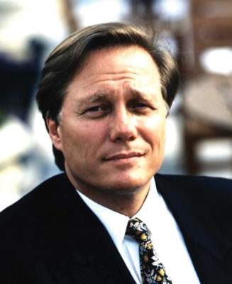
In the late 1970s, “we were able to help only about two-thirds of couples with infertility, either with tubal surgery, insemination – often with donor sperm – or ovulation induction. A full third could not be helped. We predicted that IVF would allow us to treat virtually everyone,” Dr. Rosenwaks said.
But even after the first IVF birth, information on the revolutionary procedure remained frustratingly scarce.
“Edwards and Steptoe would talk to nobody,” said Richard Marrs, MD, a reproductive endocrinologist and infertility specialist in Los Angeles.
And federal research support for “test-tube babies,” as IVF was known in the media then, was nil thanks to a ban on government-funded human embryo research that persists to this day.
The U.S. physicians who took part in the rush to achieve an IVF birth – most of them young fellows at the time – recall a period of improvisation, collaboration, shoestring budgets, and surprise findings.
“People who just started 10 or even 20 years ago don’t realize what it took for us to learn how to go about doing IVF,” said Dr. Rosenwaks, who in the first years of IVF worked closely with Dr. Howard Jones and Dr. Georgeanna Jones, the first team in the U.S. to announce an IVF baby.
Labs in closets
In the late 1970s, Dr. Marrs, then a fellow at the University of Southern California, was focused on surgical methods to treat infertility – and demand was sky-high. Intrauterine devices used in the 1970s left many women with severe scarring and inflammation of the fallopian tubes.
“I was very surgically oriented,” Dr. Marrs said. “I thought I could fix any disaster in the pelvis that was put in front of me, especially with microsurgery.”
After the news of IVF success in England, Dr. Marrs threw himself into a side project at a nearby cancer center, working on single-cell cultures. “I thought if I could grow tumor cells, I could one day grow embryos,” he said.
A year later, Dr. Marrs set up the first IVF lab at USC – in a storage closet. “I sterilized the place and that was our first IVF lab, literally a closet with an incubator and a microscope.” Its budget was accordingly thin, as the director at the time felt certain that IVF was a dead end. To fund his work, Dr. Marrs asked IVF candidate patients for research donations in lieu of payment.
But before Dr. Marrs attempted to perform his first IVF, two centers in Australia announced their own IVF babies. “I decided I really needed to go see someone who had had a baby,” he said. He used his vacation time to fly to Melbourne, shuttling between two competing clinics that were “four blocks apart and wouldn’t even talk to each other,” he recalled.
Over 6 weeks, “I learned how to stimulate, how to time ovulation. I watched the PhDs in the lab – how they handled the eggs and the sperm, what the conditions were, the incubator settings,” he said.
The first IVF babies in the United States were born only months apart: The first, in December 1981, was at the Jones Institute for Reproductive Medicine in Norfolk, Va., where Dr. Rosenwaks served as the first director.
The second baby born was at USC. After that, “we had 4,000 women on a waiting list, all under age 35,” Dr. Marrs said. The Jones Institute reportedly had 5,000.
As demand soared and more IVF babies arrived, the cloak of secrecy surrounding the procedure started to lift. British, Australian, and U.S. clinicians started getting together regularly. “We would pick a spot in the world, present our data: what we’d done, how many cycles, what we used for stimulation, when we took the eggs out,” Dr. Marrs said. “I don’t know how many hundreds of thousands of miles I flew in the first years of IVF, because it was the only way I could get information. We would literally stay up all night talking.”
Answering safety questions
Alan H. DeCherney, MD, currently an infertility researcher at the National Institutes of Health, started Yale University’s IVF program at around the same time Dr. Marrs and the Joneses were starting theirs. Yale already had a large infertility practice, and only academic centers had the laboratory resources and skilled staff needed to attempt IVF in those years.
In 1983, when Yale announced the birth of its first IVF baby – the fifth in the United States – Dr. DeCherney was starting to think about measuring outcomes, as there was concern over the potential for congenital anomalies related to IVF. “This was such a change in the way conception occurred, people were afraid that all kinds of crazy things would happen,” he said.
One concern was about ovarian stimulation with fertility drugs or gonadotropins. The earliest efforts – including by Dr. Steptoe and Dr. Edwards – used no drugs, instead trying to pinpoint the moment of natural egg release by measuring a woman’s hormone levels constantly, but these proved disappointing. Use of clomiphene citrate and human menopausal gonadotropin allowed for more control over timing, and for multiple mature eggs to be harvested at once.
But there were still many unanswered questions related to these agents’ safety and dosing, both for women and for babies.
When the NIH refused to fund a study of IVF outcomes, Dr. DeCherney and Dr. Marrs collaborated on a registry funded by a gonadotropin maker. “The drug company didn’t want to be associated with some terrible abnormal outcomes,” Dr. DeCherney recalled, though by then, “there were 10, maybe even 20 babies around the world, and they seemed to be fine,” he said.
The first registry results affirmed no changes in the rate of congenital abnormalities. (Larger, more recent studies have shown a small but significant elevation in birth defect risk associated with IVF.) A few years later, ovarian stimulation was adjusted to correspond with ovarian reserve, reducing the risk of ovarian hyperstimulation syndrome.
But even by the late 1980s, success rates for IVF per attempted cycle were still low overall, leading many critics, even within the profession, to accuse practitioners of misleading couples. Charles E. Miller, MD, an infertility specialist in Chicago, recalled an early investigation by a major newspaper “that looked at all the IVF clinics in Chicago and found the chances of having a baby was under 3%.”

It was true, Dr. Miller acknowledged – “the rates were dismal. But remember that IVF at the time was still considered a procedure of last resort.” Complex diagnostic testing to determine the cause of infertility, surgery, and fertility drugs all came first.
Some important innovations would soon change that and turn IVF into a mainstay of infertility treatment that could help women not only with damaged tubes but also with ovarian failure, low ovarian reserve, or dense pelvic adhesions. Even some types of male factor infertility would find an answer in IVF, by way of intracytoplasmic sperm transfer.
Eggs without surgery
Laparoscopic egg retrieval was the norm in the first decade of IVF. “We went through the belly button, allowing us to directly visualize the ovary and see whether ovulation had already occurred or we had to retrieve it by introducing a needle into the follicle,” Dr. Rosenwaks recalled.
“Some of us were doing 6 or even 10 laparoscopies a day, and it was physically quite challenging,” he said. “There were no video screens in those days. You had to bend over the scope.” And it was worse still for patients, who had to endure multiple surgeries.
Though egg and embryo cryopreservation were already being worked on, it would be years before these techniques were optimized, giving women more chances from a single retrieval of oocytes.
Finding a less invasive means of retrieving eggs was crucial.
Maria Bustillo, MD, an infertility specialist in Miami, recalled being criticized by peers when she and her then-colleagues at the Genetics & IVF Institute in Fairfax, Va., began retrieving eggs via a needle placed in the vagina, using abdominal ultrasound as a guide.
While the technique was far less invasive than laparoscopy, “we were doing it semi-blindly, and were told it was dangerous,” Dr. Bustillo said.
But these freehand ultrasound retrievals paved the way for what would become a revolutionary advance – the vaginal ultrasound probe, which by the end of the 1980s made nonsurgical extraction of eggs the norm.
Dr. Marrs recalled receiving a prototype of a vaginal ultrasound probe, in the mid-1980s, and finding patients unwilling to use it, except one who relented only because she had an empty bladder. Abdominal ultrasonography required a full bladder to work.
“It was as though somebody had removed the cloud cover,” he said. “I couldn’t believe it. I could see everything: her ovaries, tiny follicles, the uterus.”
Later probes were fitted with a needle and aspirator to retrieve eggs. Multiple IVF cycles no longer meant multiple surgeries, and the less-invasive procedure helped in recruiting egg donors, allowing women with ovarian disease or low ovarian reserves, including older women, to receive IVF.
“It didn’t make sense for a volunteer to go through a surgery, especially back in the early ’80s when the results were not all that great,” Dr. Bustillo said.
Improving ‘home brews’
The culture media in which embryos were grown was another strong factor limiting the success rates of early IVF. James Toner, MD, PhD, an IVF specialist in Atlanta, called the early media “home brews.”
“Everyone made them themselves,” said Dr. Toner, who spent 15 years at the Jones Institute. “You had to do a hamster or mouse embryo test on every batch to make sure embryos would grow.” And often they did not.
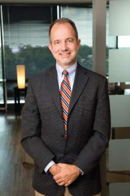
Poor success rates resulted in the emergence of alternative procedures: GIFT (gamete intrafallopian transfer) and ZIFT (zygote intrafallopian transfer). Both aimed to get embryos back into the patient as soon as possible, with the thought that the natural environment offered a better chance for success.
But advances in culture media allowed more time for embryos to be observed. With longer development, “you could do a better job selecting the ones that had a chance, and de-selecting those with no chance,” Dr. Toner said.
This also meant fewer embryos could be transferred back into patients, lowering the likelihood of multiples. Ultimately, for young women, single-embryo transfer would become the norm. “The problem of multiple pregnancy that we used to have no longer exists for IVF,” Dr. Toner said.
Allowing embryos to reach the blastocyst stage – day 5 or 6 – opened other, previously unthinkable possibilities: placing embryos directly into the uterus, without surgery, and pre-implementation genetic screening for abnormalities.
“As the cell number went up, the idea that you could do a genetic test with minimal impact on the embryo eventually became true,” Dr. Toner said.
A genetic revolution?
While many important IVF innovations were achieved in countries with staunch government support, one of the remarkable things about IVF’s evolution in the United States is that so many occurred with virtually none.
By the mid-1990s, most of the early practitioners had moved from academic settings into private practice, though they continued to publish. “After a while it didn’t help to be in academics. It just sort of slowed you down. Because you weren’t going to get any [government] money anyway, you might as well be in a place that’s a little more nimble,” Dr. Toner said.
At the same time, he said, IVF remains a costly, usually unreimbursed procedure – limiting patients’ willingness to take part in randomized trials. “IVF research is built more on cohort studies.”
Most of the current research focus in IVF is on possibilities for genetic screening. Dr. Miller said that rapid DNA sequencing is allowing specialists to “look at more, pick up more abnormalities. That will continue to improve so that we will be able to see virtually everything.”
But he cautioned there is still much to be done in IVF apart from the genetics – he’s concerned, he said, that the field has moved too far from its surgical origins, and is working with the academic societies to encourage more surgical training.
“We don’t do the same work we did before on fallopian tubes, which is good,” Dr. Miller said, noting that there have been many advances, particularly minimally invasive surgeries in the uterus or ovaries, that have occurred parallel to IVF and can improve success rates. “I think we have a better understanding of what kind of patients require surgical treatments and what kind of surgeries can help enhance fertility, and also what not to do.”
Dr. Bustillo said that “cytogenetics is wonderful, but not everything. You have embryos that are genetically normal and still don’t implant. There’s a lot of work to be done on the interaction between the mother and the embryo.”
Dr. Marrs said that even safety questions related to stimulation have yet to be fully answered. “I’ve always been a big believer that lower is better, but we need to know whether stimulation creates genetic abnormalities and whether less stimulation produces fewer – and we need more data to prove it,” he said. Dr. Marrs is an investigator on a national randomized trial comparing outcomes from IVF with standard-dose and ultra-low dose stimulation.
Access, income, and age
The IVF pioneers agree broadly that access to IVF is nowhere near what it should be in the United States, where only 15 states mandate any insurance coverage for infertility.
“Our limited access to care is a crime,” Dr. Toner said. “People who, through no fault of their own, find themselves infertile are asked to write a check for $15,000 to get pregnant. That’s not fair.”
Dr. DeCherney called access “an ethical issue, because who gets IVF? People with higher incomes. And if IVF allows you to select better embryos – whatever that means – it gives that group another advantage.”
Dr. Toner warned that the push toward genetic testing of embryos, especially in the absence of known hereditary disease, could create new problems for the profession – not unlike in the early days of IVF, when the Jones Institute and other clinics were picketed over the specter of “test tube babies.”
“It’s one thing to say this embryo does not have the right number of chromosomes and couldn’t possibly be a child, so let’s not use it, but what about looking for traits? Sex selection? We have this privileged position in which the government does not really interfere in what we do, but to retain this status we need to stay within the bounds that our society accepts,” Dr. Toner said.
In recent years, IVF uptake has been high among women of advanced reproductive age, which poses its own set of challenges. Outcomes in older women using their own eggs become progressively poorer with age, though donor eggs drastically improve their chances, and egg freezing offers the possibility of preserving quality eggs for later pregnancies.
“We could make this situation better by promoting social freezing, doing more work for women early in their lives to get out their own eggs and store them,” Dr. Miller said. “But again, you still face the issue of access.”
Regardless of what technologies are available or become available in assisted reproduction, doctors and women alike need to be better educated on their options and chances early, with a clearer understanding of what happens as they age, Dr. Bustillo said.
“This is not to pressure them, but just so they understand that when they get to be 42 and are just thinking about reproducing, it’s not a major surprise when I tell them this could be a problem,” she said.
Throughout 2016, Ob.Gyn. News is celebrating its 50th anniversary with exclusive articles looking at the evolution of the specialty, including the history of contraception, changes in gynecologic surgery, and the transformation of the well-woman visit.
In 1978, when England’s Louise Brown became the world’s first baby born through in vitro fertilization, physicians at academic centers all over the United States scrambled to figure out how they, too, could provide IVF to the thousands of infertile couples for whom nothing else had worked.
Interest in IVF was strong even before British physiologist Robert Edwards and gynecologist Patrick Steptoe announced their success. “We knew that IVF was being developed, that it had been accomplished in animals, and ultimately we knew it was going to succeed in humans,” said reproductive endocrinologist Zev Rosenwaks, MD, of the Weill Cornell Center for Reproductive Medicine in New York.
In the late 1970s, “we were able to help only about two-thirds of couples with infertility, either with tubal surgery, insemination – often with donor sperm – or ovulation induction. A full third could not be helped. We predicted that IVF would allow us to treat virtually everyone,” Dr. Rosenwaks said.
But even after the first IVF birth, information on the revolutionary procedure remained frustratingly scarce.
“Edwards and Steptoe would talk to nobody,” said Richard Marrs, MD, a reproductive endocrinologist and infertility specialist in Los Angeles.
And federal research support for “test-tube babies,” as IVF was known in the media then, was nil thanks to a ban on government-funded human embryo research that persists to this day.
The U.S. physicians who took part in the rush to achieve an IVF birth – most of them young fellows at the time – recall a period of improvisation, collaboration, shoestring budgets, and surprise findings.
“People who just started 10 or even 20 years ago don’t realize what it took for us to learn how to go about doing IVF,” said Dr. Rosenwaks, who in the first years of IVF worked closely with Dr. Howard Jones and Dr. Georgeanna Jones, the first team in the U.S. to announce an IVF baby.
Labs in closets
In the late 1970s, Dr. Marrs, then a fellow at the University of Southern California, was focused on surgical methods to treat infertility – and demand was sky-high. Intrauterine devices used in the 1970s left many women with severe scarring and inflammation of the fallopian tubes.
“I was very surgically oriented,” Dr. Marrs said. “I thought I could fix any disaster in the pelvis that was put in front of me, especially with microsurgery.”
After the news of IVF success in England, Dr. Marrs threw himself into a side project at a nearby cancer center, working on single-cell cultures. “I thought if I could grow tumor cells, I could one day grow embryos,” he said.
A year later, Dr. Marrs set up the first IVF lab at USC – in a storage closet. “I sterilized the place and that was our first IVF lab, literally a closet with an incubator and a microscope.” Its budget was accordingly thin, as the director at the time felt certain that IVF was a dead end. To fund his work, Dr. Marrs asked IVF candidate patients for research donations in lieu of payment.
But before Dr. Marrs attempted to perform his first IVF, two centers in Australia announced their own IVF babies. “I decided I really needed to go see someone who had had a baby,” he said. He used his vacation time to fly to Melbourne, shuttling between two competing clinics that were “four blocks apart and wouldn’t even talk to each other,” he recalled.
Over 6 weeks, “I learned how to stimulate, how to time ovulation. I watched the PhDs in the lab – how they handled the eggs and the sperm, what the conditions were, the incubator settings,” he said.
The first IVF babies in the United States were born only months apart: The first, in December 1981, was at the Jones Institute for Reproductive Medicine in Norfolk, Va., where Dr. Rosenwaks served as the first director.
The second baby born was at USC. After that, “we had 4,000 women on a waiting list, all under age 35,” Dr. Marrs said. The Jones Institute reportedly had 5,000.
As demand soared and more IVF babies arrived, the cloak of secrecy surrounding the procedure started to lift. British, Australian, and U.S. clinicians started getting together regularly. “We would pick a spot in the world, present our data: what we’d done, how many cycles, what we used for stimulation, when we took the eggs out,” Dr. Marrs said. “I don’t know how many hundreds of thousands of miles I flew in the first years of IVF, because it was the only way I could get information. We would literally stay up all night talking.”
Answering safety questions
Alan H. DeCherney, MD, currently an infertility researcher at the National Institutes of Health, started Yale University’s IVF program at around the same time Dr. Marrs and the Joneses were starting theirs. Yale already had a large infertility practice, and only academic centers had the laboratory resources and skilled staff needed to attempt IVF in those years.
In 1983, when Yale announced the birth of its first IVF baby – the fifth in the United States – Dr. DeCherney was starting to think about measuring outcomes, as there was concern over the potential for congenital anomalies related to IVF. “This was such a change in the way conception occurred, people were afraid that all kinds of crazy things would happen,” he said.
One concern was about ovarian stimulation with fertility drugs or gonadotropins. The earliest efforts – including by Dr. Steptoe and Dr. Edwards – used no drugs, instead trying to pinpoint the moment of natural egg release by measuring a woman’s hormone levels constantly, but these proved disappointing. Use of clomiphene citrate and human menopausal gonadotropin allowed for more control over timing, and for multiple mature eggs to be harvested at once.
But there were still many unanswered questions related to these agents’ safety and dosing, both for women and for babies.
When the NIH refused to fund a study of IVF outcomes, Dr. DeCherney and Dr. Marrs collaborated on a registry funded by a gonadotropin maker. “The drug company didn’t want to be associated with some terrible abnormal outcomes,” Dr. DeCherney recalled, though by then, “there were 10, maybe even 20 babies around the world, and they seemed to be fine,” he said.
The first registry results affirmed no changes in the rate of congenital abnormalities. (Larger, more recent studies have shown a small but significant elevation in birth defect risk associated with IVF.) A few years later, ovarian stimulation was adjusted to correspond with ovarian reserve, reducing the risk of ovarian hyperstimulation syndrome.
But even by the late 1980s, success rates for IVF per attempted cycle were still low overall, leading many critics, even within the profession, to accuse practitioners of misleading couples. Charles E. Miller, MD, an infertility specialist in Chicago, recalled an early investigation by a major newspaper “that looked at all the IVF clinics in Chicago and found the chances of having a baby was under 3%.”
It was true, Dr. Miller acknowledged – “the rates were dismal. But remember that IVF at the time was still considered a procedure of last resort.” Complex diagnostic testing to determine the cause of infertility, surgery, and fertility drugs all came first.
Some important innovations would soon change that and turn IVF into a mainstay of infertility treatment that could help women not only with damaged tubes but also with ovarian failure, low ovarian reserve, or dense pelvic adhesions. Even some types of male factor infertility would find an answer in IVF, by way of intracytoplasmic sperm transfer.
Eggs without surgery
Laparoscopic egg retrieval was the norm in the first decade of IVF. “We went through the belly button, allowing us to directly visualize the ovary and see whether ovulation had already occurred or we had to retrieve it by introducing a needle into the follicle,” Dr. Rosenwaks recalled.
“Some of us were doing 6 or even 10 laparoscopies a day, and it was physically quite challenging,” he said. “There were no video screens in those days. You had to bend over the scope.” And it was worse still for patients, who had to endure multiple surgeries.
Though egg and embryo cryopreservation were already being worked on, it would be years before these techniques were optimized, giving women more chances from a single retrieval of oocytes.
Finding a less invasive means of retrieving eggs was crucial.
Maria Bustillo, MD, an infertility specialist in Miami, recalled being criticized by peers when she and her then-colleagues at the Genetics & IVF Institute in Fairfax, Va., began retrieving eggs via a needle placed in the vagina, using abdominal ultrasound as a guide.
While the technique was far less invasive than laparoscopy, “we were doing it semi-blindly, and were told it was dangerous,” Dr. Bustillo said.
But these freehand ultrasound retrievals paved the way for what would become a revolutionary advance – the vaginal ultrasound probe, which by the end of the 1980s made nonsurgical extraction of eggs the norm.
Dr. Marrs recalled receiving a prototype of a vaginal ultrasound probe, in the mid-1980s, and finding patients unwilling to use it, except one who relented only because she had an empty bladder. Abdominal ultrasonography required a full bladder to work.
“It was as though somebody had removed the cloud cover,” he said. “I couldn’t believe it. I could see everything: her ovaries, tiny follicles, the uterus.”
Later probes were fitted with a needle and aspirator to retrieve eggs. Multiple IVF cycles no longer meant multiple surgeries, and the less-invasive procedure helped in recruiting egg donors, allowing women with ovarian disease or low ovarian reserves, including older women, to receive IVF.
“It didn’t make sense for a volunteer to go through a surgery, especially back in the early ’80s when the results were not all that great,” Dr. Bustillo said.
Improving ‘home brews’
The culture media in which embryos were grown was another strong factor limiting the success rates of early IVF. James Toner, MD, PhD, an IVF specialist in Atlanta, called the early media “home brews.”
“Everyone made them themselves,” said Dr. Toner, who spent 15 years at the Jones Institute. “You had to do a hamster or mouse embryo test on every batch to make sure embryos would grow.” And often they did not.
Poor success rates resulted in the emergence of alternative procedures: GIFT (gamete intrafallopian transfer) and ZIFT (zygote intrafallopian transfer). Both aimed to get embryos back into the patient as soon as possible, with the thought that the natural environment offered a better chance for success.
But advances in culture media allowed more time for embryos to be observed. With longer development, “you could do a better job selecting the ones that had a chance, and de-selecting those with no chance,” Dr. Toner said.
This also meant fewer embryos could be transferred back into patients, lowering the likelihood of multiples. Ultimately, for young women, single-embryo transfer would become the norm. “The problem of multiple pregnancy that we used to have no longer exists for IVF,” Dr. Toner said.
Allowing embryos to reach the blastocyst stage – day 5 or 6 – opened other, previously unthinkable possibilities: placing embryos directly into the uterus, without surgery, and pre-implementation genetic screening for abnormalities.
“As the cell number went up, the idea that you could do a genetic test with minimal impact on the embryo eventually became true,” Dr. Toner said.
A genetic revolution?
While many important IVF innovations were achieved in countries with staunch government support, one of the remarkable things about IVF’s evolution in the United States is that so many occurred with virtually none.
By the mid-1990s, most of the early practitioners had moved from academic settings into private practice, though they continued to publish. “After a while it didn’t help to be in academics. It just sort of slowed you down. Because you weren’t going to get any [government] money anyway, you might as well be in a place that’s a little more nimble,” Dr. Toner said.
At the same time, he said, IVF remains a costly, usually unreimbursed procedure – limiting patients’ willingness to take part in randomized trials. “IVF research is built more on cohort studies.”
Most of the current research focus in IVF is on possibilities for genetic screening. Dr. Miller said that rapid DNA sequencing is allowing specialists to “look at more, pick up more abnormalities. That will continue to improve so that we will be able to see virtually everything.”
But he cautioned there is still much to be done in IVF apart from the genetics – he’s concerned, he said, that the field has moved too far from its surgical origins, and is working with the academic societies to encourage more surgical training.
“We don’t do the same work we did before on fallopian tubes, which is good,” Dr. Miller said, noting that there have been many advances, particularly minimally invasive surgeries in the uterus or ovaries, that have occurred parallel to IVF and can improve success rates. “I think we have a better understanding of what kind of patients require surgical treatments and what kind of surgeries can help enhance fertility, and also what not to do.”
Dr. Bustillo said that “cytogenetics is wonderful, but not everything. You have embryos that are genetically normal and still don’t implant. There’s a lot of work to be done on the interaction between the mother and the embryo.”
Dr. Marrs said that even safety questions related to stimulation have yet to be fully answered. “I’ve always been a big believer that lower is better, but we need to know whether stimulation creates genetic abnormalities and whether less stimulation produces fewer – and we need more data to prove it,” he said. Dr. Marrs is an investigator on a national randomized trial comparing outcomes from IVF with standard-dose and ultra-low dose stimulation.
Access, income, and age
The IVF pioneers agree broadly that access to IVF is nowhere near what it should be in the United States, where only 15 states mandate any insurance coverage for infertility.
“Our limited access to care is a crime,” Dr. Toner said. “People who, through no fault of their own, find themselves infertile are asked to write a check for $15,000 to get pregnant. That’s not fair.”
Dr. DeCherney called access “an ethical issue, because who gets IVF? People with higher incomes. And if IVF allows you to select better embryos – whatever that means – it gives that group another advantage.”
Dr. Toner warned that the push toward genetic testing of embryos, especially in the absence of known hereditary disease, could create new problems for the profession – not unlike in the early days of IVF, when the Jones Institute and other clinics were picketed over the specter of “test tube babies.”
“It’s one thing to say this embryo does not have the right number of chromosomes and couldn’t possibly be a child, so let’s not use it, but what about looking for traits? Sex selection? We have this privileged position in which the government does not really interfere in what we do, but to retain this status we need to stay within the bounds that our society accepts,” Dr. Toner said.
In recent years, IVF uptake has been high among women of advanced reproductive age, which poses its own set of challenges. Outcomes in older women using their own eggs become progressively poorer with age, though donor eggs drastically improve their chances, and egg freezing offers the possibility of preserving quality eggs for later pregnancies.
“We could make this situation better by promoting social freezing, doing more work for women early in their lives to get out their own eggs and store them,” Dr. Miller said. “But again, you still face the issue of access.”
Regardless of what technologies are available or become available in assisted reproduction, doctors and women alike need to be better educated on their options and chances early, with a clearer understanding of what happens as they age, Dr. Bustillo said.
“This is not to pressure them, but just so they understand that when they get to be 42 and are just thinking about reproducing, it’s not a major surprise when I tell them this could be a problem,” she said.
Throughout 2016, Ob.Gyn. News is celebrating its 50th anniversary with exclusive articles looking at the evolution of the specialty, including the history of contraception, changes in gynecologic surgery, and the transformation of the well-woman visit.

Expert Recommendations for Optimizing Outcomes in the Management Of Biofilm to Promote Healing of Chronic Wounds in the Veterans Health Administration System

A supplement to Federal Practitioner. This supplement is sponsored by Organogenesis, Inc.
Chronic non-healing wounds are a major problem for patients and wound care practitioners in the VA system. It is self-evident among experienced wound care practitioners that wound infection can delay (or completely prevent) wound healing. Biofilm is a common form of wound contamination that is now recognized to be a major factor in delaying the healing of wounds.
This supplement discusses the awareness of the biology of biofilm, its prevalence, its clinical significance, and optimal treatment approaches that need to be improved. The supplement also reviews the current status of evidence-based management of biofilm with a focus on the optimal use of PuraPly™ Antimicrobial, a native purified collagen matrix containing the antimicrobial polyhexamethylene biguanide (PHMB).
| Meeting Participants |
Gregory Shultz, PhD |
Stephen C. Davis |
Jonathan N. Brantley, DPM |
Mark Couture, DPM |
Sean L. Kersh, DPM |
Jake G. Ruff, DPM |
Patrick J. Sanchez, DPM |
Janette Thompson, DPM |
Catherine M. Wittgen, MD |
| CLICK HERE TO READ FULL SUPPLEMENT |