User login
VA Confronts Fallout of SAIL Facility Ratings
VA Secretary Robert McDonald minced few words in his reaction to the USA Today story that published VA’s 5-star ratings for VAMCs based on its Strategic Analytics for Improvement and Learning (SAIL) data. “What concerns all of us at VA is that USA Today has a consistent narrative of negativity in their news of VA,” McDonald charged. Specifically, use of the word secret in the headline was an “egregious hyperbole,” he insisted.
Still, within days, a number of VA facilities were using the story as an opportunity to tout their success. VA Central Western Massachusetts Healthcare System in Worcester, for example, used the opportunity to tell local media that it was “in the top 10% of the 152 VA medical centers in the nation for quality of care,” and 1 of 3 5-star facilities in Massachusetts.
Other facilities, however, were forced to explain the relative nature of the 5-star scale. Keith Sullivan, director of the Chillicothe VAMC in Ohio, which received a 3-star rating, responded to reporters by noting that, “Although SAIL is a tool that helps look at areas in need of improvements, we have many other ways to ensure quality care is in place,” he said.
Many VA officials point out that the comparison to other VA facilities may be unfair, especially in underserved locations. The VA Health Care Center at Harlingen in Texas recently improved from 1 to 2 stars. It received this score, in part, because of its difficulty filling open vacancies. “Compared to the nurse retention rates in our local communities we are doing very well, [but] not so much as compared to other VA health care systems in the nation,” a Harlingen customer service manager told a local newspaper.
To allay veterans’ fears, the VA Southern Oregon Rehabilitation Center and Clinics (VA SORCC) sent out a news release to explain the ratings and reassure the public that progress was being made even as it struggles with a shortage of providers. Insisting it had made “absolute performance improvements in wait times for both mental health and specialty care appointments” while acknowledging that the rating also shows “continued frustrations with difficulty in navigating the system and with coordination of care." These frustrations impact Veterans’ experience at the facility, leading to lower scores.” The release pledged that “VA SORCC is currently a 1-star facility but fully expects to move to a 2-star facility in the next quarter.”
The worry for many at the VA is that the veterans might avoid 1- and 2-star facilities. “My concern is that veterans are going to see that their hospital is a ‘one’ in our star system, assume that’s bad quality and veterans that need care are not going to get care,” VA Under Secretary of Health David J. Shulkin, MD, told USA Today. “And they’re going to stay away from hospitals and that’s going to hurt people.”
VA Secretary Robert McDonald minced few words in his reaction to the USA Today story that published VA’s 5-star ratings for VAMCs based on its Strategic Analytics for Improvement and Learning (SAIL) data. “What concerns all of us at VA is that USA Today has a consistent narrative of negativity in their news of VA,” McDonald charged. Specifically, use of the word secret in the headline was an “egregious hyperbole,” he insisted.
Still, within days, a number of VA facilities were using the story as an opportunity to tout their success. VA Central Western Massachusetts Healthcare System in Worcester, for example, used the opportunity to tell local media that it was “in the top 10% of the 152 VA medical centers in the nation for quality of care,” and 1 of 3 5-star facilities in Massachusetts.
Other facilities, however, were forced to explain the relative nature of the 5-star scale. Keith Sullivan, director of the Chillicothe VAMC in Ohio, which received a 3-star rating, responded to reporters by noting that, “Although SAIL is a tool that helps look at areas in need of improvements, we have many other ways to ensure quality care is in place,” he said.
Many VA officials point out that the comparison to other VA facilities may be unfair, especially in underserved locations. The VA Health Care Center at Harlingen in Texas recently improved from 1 to 2 stars. It received this score, in part, because of its difficulty filling open vacancies. “Compared to the nurse retention rates in our local communities we are doing very well, [but] not so much as compared to other VA health care systems in the nation,” a Harlingen customer service manager told a local newspaper.
To allay veterans’ fears, the VA Southern Oregon Rehabilitation Center and Clinics (VA SORCC) sent out a news release to explain the ratings and reassure the public that progress was being made even as it struggles with a shortage of providers. Insisting it had made “absolute performance improvements in wait times for both mental health and specialty care appointments” while acknowledging that the rating also shows “continued frustrations with difficulty in navigating the system and with coordination of care." These frustrations impact Veterans’ experience at the facility, leading to lower scores.” The release pledged that “VA SORCC is currently a 1-star facility but fully expects to move to a 2-star facility in the next quarter.”
The worry for many at the VA is that the veterans might avoid 1- and 2-star facilities. “My concern is that veterans are going to see that their hospital is a ‘one’ in our star system, assume that’s bad quality and veterans that need care are not going to get care,” VA Under Secretary of Health David J. Shulkin, MD, told USA Today. “And they’re going to stay away from hospitals and that’s going to hurt people.”
VA Secretary Robert McDonald minced few words in his reaction to the USA Today story that published VA’s 5-star ratings for VAMCs based on its Strategic Analytics for Improvement and Learning (SAIL) data. “What concerns all of us at VA is that USA Today has a consistent narrative of negativity in their news of VA,” McDonald charged. Specifically, use of the word secret in the headline was an “egregious hyperbole,” he insisted.
Still, within days, a number of VA facilities were using the story as an opportunity to tout their success. VA Central Western Massachusetts Healthcare System in Worcester, for example, used the opportunity to tell local media that it was “in the top 10% of the 152 VA medical centers in the nation for quality of care,” and 1 of 3 5-star facilities in Massachusetts.
Other facilities, however, were forced to explain the relative nature of the 5-star scale. Keith Sullivan, director of the Chillicothe VAMC in Ohio, which received a 3-star rating, responded to reporters by noting that, “Although SAIL is a tool that helps look at areas in need of improvements, we have many other ways to ensure quality care is in place,” he said.
Many VA officials point out that the comparison to other VA facilities may be unfair, especially in underserved locations. The VA Health Care Center at Harlingen in Texas recently improved from 1 to 2 stars. It received this score, in part, because of its difficulty filling open vacancies. “Compared to the nurse retention rates in our local communities we are doing very well, [but] not so much as compared to other VA health care systems in the nation,” a Harlingen customer service manager told a local newspaper.
To allay veterans’ fears, the VA Southern Oregon Rehabilitation Center and Clinics (VA SORCC) sent out a news release to explain the ratings and reassure the public that progress was being made even as it struggles with a shortage of providers. Insisting it had made “absolute performance improvements in wait times for both mental health and specialty care appointments” while acknowledging that the rating also shows “continued frustrations with difficulty in navigating the system and with coordination of care." These frustrations impact Veterans’ experience at the facility, leading to lower scores.” The release pledged that “VA SORCC is currently a 1-star facility but fully expects to move to a 2-star facility in the next quarter.”
The worry for many at the VA is that the veterans might avoid 1- and 2-star facilities. “My concern is that veterans are going to see that their hospital is a ‘one’ in our star system, assume that’s bad quality and veterans that need care are not going to get care,” VA Under Secretary of Health David J. Shulkin, MD, told USA Today. “And they’re going to stay away from hospitals and that’s going to hurt people.”
‘Unprecedented’ MRD negativity with daratumumab in MM

© Todd Buchanan 2016
SAN DIEGO—Daratumumab added to standard of care regimens drives deep clinical responses beyond complete response (CR), a magnitude that is “unprecedented” in the relapsed/refractory multiple myeloma (MM) setting, according to a speaker at the 2016 ASH Annual Meeting.
Investigators added daratumumab to lenalidomide/dexamethasone in the POLLUX trial and to bortezomib/dexamethasone in the CASTOR trial.
In both phase 3 trials, the addition of daratumumab resulted in significant improvements in progression-free survival (PFS), overall response rate, and minimal residual disease (MRD) negativity when compared to control groups.
“The magnitude of daratumumab-induced MRD negativity in the relapsed setting is unprecedented and, for me, was not expected,” said Hervé Avet-Loiseau, MD, of Centre Hospitalier Universitaire Rangueil, Unité de Genomique du Myelome in Toulouse, France.
Dr Avet-Loiseau presented the MRD findings from CASTOR and POLLUX at the ASH Annual Meeting as abstract 246.*
He noted that, based on these studies, daratumumab received US Food and Drug Administration approvals for use in combination with standard of care regimens for MM patients who had received 1 or more prior lines of treatment.
Daratumumab had been previously approved as monotherapy for relapsed or refractory MM.
Study designs and findings from the POLLUX and CASTOR trials have been described earlier in Hematology Times.
Dr Avet-Loiseau provided updated PFS figures for the 2 studies.
At 18 months’ follow-up in the POLLUX study, the PFS rate for patients treated with daratumumab/lenalidomide/dexamethasone was 76%, compared to 49% in the lenalidomide/dexamethasone arm (P<0.0001).
At 12 months’ follow-up in the CASTOR study, the PFS with daratumumab was 60%, compared to 22% for bortezomib/dexamethasone (P<0.0001).
MRD criteria
In both studies, MRD assessments were conducted at suspected complete response (CR). Assessments were also conducted at 3 months and 6 months after CR in the POLLUX study and at 6 months and 12 months after the first study dose in the CASTOR study.
For the assessment of MRD, investigators used bone marrow aspirate samples and the ClonoSEQTM NGS-based assay.
Investigators evaluated MRD at 3 sensitivity thresholds: 10-4, 10-5, and 10-6.
And they used a stringent, unbiased evaluation, Dr Avet-Loiseau said. Any patient in the intent-to-treat population who was not assessed to be MRD negative was scored as MRD positive.
And the minimum cell input equivalent to the sensitivity threshold was required to determine MRD negativity.
MRD results
In the POLLUX study, 24.8% of patients achieved MRD negativity at the 10-5 cutoff, and 11.9% achieved MRD negativity at the 10-6 cutoff with the daratumumab combination.
This compared to 5.7% and 2.5% MRD negativity at the 10-5 and 10-6 cutoffs, respectively, without daratumumab (P<0.0001).
In the CASTOR study, the daratumumab-treated patients achieved 10.4% and 4.4% MRD negativity at the 10-5 and 10-6 cutoffs, respectively.
This compared to 2.4% and 0.8% MRD negativity in the control arm at the 10-5 and 10-6 cutoffs (P<0.005 and P<0.05), respectively.
“So, definitely, the addition of daratumumab improved the MRD negativity rate in both studies,” Dr Avet-Loiseau said.
“If you just look at the patients who did achieve CR in the POLLUX study, almost 50% of the patients [treated with daratumumab] achieved CR, and half of them were MRD negative at the cutoff of 10-5.”
In the CASTOR study, 25% of the patients treated with daratumumab achieved a CR. The MRD negativity rate was one-third in these patients.
“So again, we have consistently higher MRD negative rates in patients who achieve CR when they were treated in the daratumumab arms,” Dr Avet-Loiseau said.
“What is interesting, I think, is that the achievement of molecular CR was very rapid. [A]t 3 months, some patients did already achieve MRD negativity, and so we continued to see an improvement. [W]e still continue to see some achievement of MRD negativity.”
Investigators continue to follow the patients annually.
The investigators also analyzed MRD at 10-5 by cytogenetic risk and did not observe any MRD negativity in the control arm in either the POLLUX or CASTOR study.
“In contrast, we did observe some significant MRD negativity in the experimental arm with daratumumab—18% (POLLUX) and 14% (CASTOR) in high-risk patients,” Dr Avet-Loiseau said. “The most important prognostic factor is to achieve MRD negativity.”
However, even for patients who did not achieve MRD negativity, the PFS was much better in the experimental arms than in the control arms, he added.
This study, presented as a “Best of ASH” abstract, was funded by Janssen Research & Development, LLC. 
*Information in the abstract differs from that presented at the meeting.
© Todd Buchanan 2016
SAN DIEGO—Daratumumab added to standard of care regimens drives deep clinical responses beyond complete response (CR), a magnitude that is “unprecedented” in the relapsed/refractory multiple myeloma (MM) setting, according to a speaker at the 2016 ASH Annual Meeting.
Investigators added daratumumab to lenalidomide/dexamethasone in the POLLUX trial and to bortezomib/dexamethasone in the CASTOR trial.
In both phase 3 trials, the addition of daratumumab resulted in significant improvements in progression-free survival (PFS), overall response rate, and minimal residual disease (MRD) negativity when compared to control groups.
“The magnitude of daratumumab-induced MRD negativity in the relapsed setting is unprecedented and, for me, was not expected,” said Hervé Avet-Loiseau, MD, of Centre Hospitalier Universitaire Rangueil, Unité de Genomique du Myelome in Toulouse, France.
Dr Avet-Loiseau presented the MRD findings from CASTOR and POLLUX at the ASH Annual Meeting as abstract 246.*
He noted that, based on these studies, daratumumab received US Food and Drug Administration approvals for use in combination with standard of care regimens for MM patients who had received 1 or more prior lines of treatment.
Daratumumab had been previously approved as monotherapy for relapsed or refractory MM.
Study designs and findings from the POLLUX and CASTOR trials have been described earlier in Hematology Times.
Dr Avet-Loiseau provided updated PFS figures for the 2 studies.
At 18 months’ follow-up in the POLLUX study, the PFS rate for patients treated with daratumumab/lenalidomide/dexamethasone was 76%, compared to 49% in the lenalidomide/dexamethasone arm (P<0.0001).
At 12 months’ follow-up in the CASTOR study, the PFS with daratumumab was 60%, compared to 22% for bortezomib/dexamethasone (P<0.0001).
MRD criteria
In both studies, MRD assessments were conducted at suspected complete response (CR). Assessments were also conducted at 3 months and 6 months after CR in the POLLUX study and at 6 months and 12 months after the first study dose in the CASTOR study.
For the assessment of MRD, investigators used bone marrow aspirate samples and the ClonoSEQTM NGS-based assay.
Investigators evaluated MRD at 3 sensitivity thresholds: 10-4, 10-5, and 10-6.
And they used a stringent, unbiased evaluation, Dr Avet-Loiseau said. Any patient in the intent-to-treat population who was not assessed to be MRD negative was scored as MRD positive.
And the minimum cell input equivalent to the sensitivity threshold was required to determine MRD negativity.
MRD results
In the POLLUX study, 24.8% of patients achieved MRD negativity at the 10-5 cutoff, and 11.9% achieved MRD negativity at the 10-6 cutoff with the daratumumab combination.
This compared to 5.7% and 2.5% MRD negativity at the 10-5 and 10-6 cutoffs, respectively, without daratumumab (P<0.0001).
In the CASTOR study, the daratumumab-treated patients achieved 10.4% and 4.4% MRD negativity at the 10-5 and 10-6 cutoffs, respectively.
This compared to 2.4% and 0.8% MRD negativity in the control arm at the 10-5 and 10-6 cutoffs (P<0.005 and P<0.05), respectively.
“So, definitely, the addition of daratumumab improved the MRD negativity rate in both studies,” Dr Avet-Loiseau said.
“If you just look at the patients who did achieve CR in the POLLUX study, almost 50% of the patients [treated with daratumumab] achieved CR, and half of them were MRD negative at the cutoff of 10-5.”
In the CASTOR study, 25% of the patients treated with daratumumab achieved a CR. The MRD negativity rate was one-third in these patients.
“So again, we have consistently higher MRD negative rates in patients who achieve CR when they were treated in the daratumumab arms,” Dr Avet-Loiseau said.
“What is interesting, I think, is that the achievement of molecular CR was very rapid. [A]t 3 months, some patients did already achieve MRD negativity, and so we continued to see an improvement. [W]e still continue to see some achievement of MRD negativity.”
Investigators continue to follow the patients annually.
The investigators also analyzed MRD at 10-5 by cytogenetic risk and did not observe any MRD negativity in the control arm in either the POLLUX or CASTOR study.
“In contrast, we did observe some significant MRD negativity in the experimental arm with daratumumab—18% (POLLUX) and 14% (CASTOR) in high-risk patients,” Dr Avet-Loiseau said. “The most important prognostic factor is to achieve MRD negativity.”
However, even for patients who did not achieve MRD negativity, the PFS was much better in the experimental arms than in the control arms, he added.
This study, presented as a “Best of ASH” abstract, was funded by Janssen Research & Development, LLC.
*Information in the abstract differs from that presented at the meeting.
© Todd Buchanan 2016
SAN DIEGO—Daratumumab added to standard of care regimens drives deep clinical responses beyond complete response (CR), a magnitude that is “unprecedented” in the relapsed/refractory multiple myeloma (MM) setting, according to a speaker at the 2016 ASH Annual Meeting.
Investigators added daratumumab to lenalidomide/dexamethasone in the POLLUX trial and to bortezomib/dexamethasone in the CASTOR trial.
In both phase 3 trials, the addition of daratumumab resulted in significant improvements in progression-free survival (PFS), overall response rate, and minimal residual disease (MRD) negativity when compared to control groups.
“The magnitude of daratumumab-induced MRD negativity in the relapsed setting is unprecedented and, for me, was not expected,” said Hervé Avet-Loiseau, MD, of Centre Hospitalier Universitaire Rangueil, Unité de Genomique du Myelome in Toulouse, France.
Dr Avet-Loiseau presented the MRD findings from CASTOR and POLLUX at the ASH Annual Meeting as abstract 246.*
He noted that, based on these studies, daratumumab received US Food and Drug Administration approvals for use in combination with standard of care regimens for MM patients who had received 1 or more prior lines of treatment.
Daratumumab had been previously approved as monotherapy for relapsed or refractory MM.
Study designs and findings from the POLLUX and CASTOR trials have been described earlier in Hematology Times.
Dr Avet-Loiseau provided updated PFS figures for the 2 studies.
At 18 months’ follow-up in the POLLUX study, the PFS rate for patients treated with daratumumab/lenalidomide/dexamethasone was 76%, compared to 49% in the lenalidomide/dexamethasone arm (P<0.0001).
At 12 months’ follow-up in the CASTOR study, the PFS with daratumumab was 60%, compared to 22% for bortezomib/dexamethasone (P<0.0001).
MRD criteria
In both studies, MRD assessments were conducted at suspected complete response (CR). Assessments were also conducted at 3 months and 6 months after CR in the POLLUX study and at 6 months and 12 months after the first study dose in the CASTOR study.
For the assessment of MRD, investigators used bone marrow aspirate samples and the ClonoSEQTM NGS-based assay.
Investigators evaluated MRD at 3 sensitivity thresholds: 10-4, 10-5, and 10-6.
And they used a stringent, unbiased evaluation, Dr Avet-Loiseau said. Any patient in the intent-to-treat population who was not assessed to be MRD negative was scored as MRD positive.
And the minimum cell input equivalent to the sensitivity threshold was required to determine MRD negativity.
MRD results
In the POLLUX study, 24.8% of patients achieved MRD negativity at the 10-5 cutoff, and 11.9% achieved MRD negativity at the 10-6 cutoff with the daratumumab combination.
This compared to 5.7% and 2.5% MRD negativity at the 10-5 and 10-6 cutoffs, respectively, without daratumumab (P<0.0001).
In the CASTOR study, the daratumumab-treated patients achieved 10.4% and 4.4% MRD negativity at the 10-5 and 10-6 cutoffs, respectively.
This compared to 2.4% and 0.8% MRD negativity in the control arm at the 10-5 and 10-6 cutoffs (P<0.005 and P<0.05), respectively.
“So, definitely, the addition of daratumumab improved the MRD negativity rate in both studies,” Dr Avet-Loiseau said.
“If you just look at the patients who did achieve CR in the POLLUX study, almost 50% of the patients [treated with daratumumab] achieved CR, and half of them were MRD negative at the cutoff of 10-5.”
In the CASTOR study, 25% of the patients treated with daratumumab achieved a CR. The MRD negativity rate was one-third in these patients.
“So again, we have consistently higher MRD negative rates in patients who achieve CR when they were treated in the daratumumab arms,” Dr Avet-Loiseau said.
“What is interesting, I think, is that the achievement of molecular CR was very rapid. [A]t 3 months, some patients did already achieve MRD negativity, and so we continued to see an improvement. [W]e still continue to see some achievement of MRD negativity.”
Investigators continue to follow the patients annually.
The investigators also analyzed MRD at 10-5 by cytogenetic risk and did not observe any MRD negativity in the control arm in either the POLLUX or CASTOR study.
“In contrast, we did observe some significant MRD negativity in the experimental arm with daratumumab—18% (POLLUX) and 14% (CASTOR) in high-risk patients,” Dr Avet-Loiseau said. “The most important prognostic factor is to achieve MRD negativity.”
However, even for patients who did not achieve MRD negativity, the PFS was much better in the experimental arms than in the control arms, he added.
This study, presented as a “Best of ASH” abstract, was funded by Janssen Research & Development, LLC.
*Information in the abstract differs from that presented at the meeting.
How old is too old to be on a kids’ protocol for ALL?

Photo by Bill Branson
SAN DIEGO—In recent years, pediatric or pediatric-inspired protocols have become the preferred treatment approach for younger adults with acute lymphoblastic leukemia (ALL).
These protocols include higher doses of steroids, vincristine, methotrexate, and L-asparaginase.
However, the upper age limit for this strategy has not been defined.
With the GRAALL-2005 study, investigators set out to determine how old is too old to be treated on pediatric protocols.
Their results suggest 55 is likely the upper age limit for patients with Ph-negative ALL.
The investigators also evaluated a hyper-fractionated (hyper-C) versus standard dose (standard-C) of cyclophosphamide during induction and late intensification.
They found that hyper-C did not provide an event-free survival (EFS) benefit in the overall study population, but patients age 55 and older did appear to benefit from hyper-C.
Françoise Huguet, MD, of the Institut Universitaire du Cancer de Toulouse in Toulouse, France, presented these findings at the 2016 ASH Annual Meeting (abstract 762).
GRAALL investigators had previously evaluated a pediatric-inspired protocol for adult patients in the GRAALL-2003 study, which validated the approach.
Study design
Patients with newly diagnosed, Ph-negative ALL were eligible to enroll if they were 18 to 59 years of age.
Treatment comprised a steroid pre-phase, a 5-drug induction, two 3-block dose-dense consolidation phases, a late intensification, a third consolidation phase, CNS irradiation, and a 2-year maintenance phase.
Patients could proceed to allogeneic transplant in first complete remission (CR) if eligible.
During induction and late intensification, patients received cyclophosphamide at 750 mg/m2 on day 1 and were then randomized to hyper-C (300 mg/m2/every 12 hours on days 15 to 17) or standard-C (750 mg/m2 on day 15).
The primary endpoint was EFS.
Patient population
Investigators randomized 787 evaluable patients—398 in the standard-C arm and 389 in the hyper-C arm.
Their median age was 36 years, 67% of patients had B-cell precursor ALL, and 33% had T-ALL.
Most had high-risk ALL, 72% of them receiving standard-C and 66% receiving hyper-C.
About a third of the patients in each arm proceeded to allogeneic stem cell transplant in first CR.
Results
The CR rate after induction therapy was 90.2% in the standard-C arm and 93.6% in the hyper-C arm, for an overall CR rate of 92%.
Most patients—87.5% in the standard-C arm and 91.8% in the hyper-C arm—achieved a response in 1 course of therapy.
Sixty percent of patients tested in the standard-C arm and 66% of those tested in the hyper-C arm were minimal residual disease negative at less than 10-4.
There were 26 (6.5%) deaths in the standard-C arm and 18 (4.6%) in the hyper-C arm.
The 5-year EFS rate was 52% overall, and hyper-C treatment had no impact on EFS (hazard ratio=0.89 [range, 0.7-1.1]; P=0.26).
Impact of age
Investigators conducted a post-hoc subgroup analysis of 5 age groups—18-24 years (n=200), 25-34 (n=172), 35-44 (n=171), 45-54 (n=151), and 55+ (n=93).
Overall, the CR rate tended to decrease with age. The rates were 98.5% (18-24), 95.3% (25-34), 87.7% (35-44), 89.4% (45-54), and 79.6% (55+).
Induction death rates increased from 0.5% in the youngest group to 18.3% in the oldest, but the rate of cumulative incidences of failure at 5 years was similar among all the age groups.
The cumulative incidence of treatment-related mortality, without censoring for transplant, ranged from 7.6% in the youngest group to 39.7% in the oldest.
And the 5-year EFS for the youngest patients was 60%, while, for the oldest, it was 26%.
“Above 50 years, the increase in age became highly significant,” Dr Huguet emphasized. “There were fewer CRs and lower survival.”
Treatment compliance
In terms of treatment compliance and median dose received in the induction course, patients aged 55-59 received significantly less L-asparaginase than those aged 18-54 (P<0.001).
During all 3 consolidation phases, patients aged 55-59 received significantly lower median doses of all medications—cytarabine, methotrexate, cyclophosphamide—than patients aged 18-54.
And in late intensification, patients aged 55-59 received significantly lower median doses of vincristine, prednisone, daunorubicin, and hyper-C than all other patients. The median doses of L-asparaginase and standard-C received were lower in the older patients but not significantly so.
EFS by age and randomization
The 5-year EFS for patients aged 18-54 was 57% with hyper-C, compared with 55% in the standard-C arm (P=0.66).
However, for older patients, there was a significant advantage for those receiving hyper-C. The 5-year EFS was 38% with hyper-C, compared to 12% with standard-C (P=0.007).
Dr Huguet explained that inferior compliance in patients 55 and older “might explain why a benefit associated with early hyper-C reinforcement became apparent in these older patients only.”
Dr Huguet concluded that the results “suggest that 55 years is likely to be the upper age limit to tolerate a pediatric-like therapy for younger adults with Ph-negative ALL.”
She added that patients over 54 might benefit from alternative front-line strategies.
Accordingly, investigators are planning to use new agents, such as blinatumomab or inotuzumab ozogamicin, in the next European Working Group on Adult ALL studies.
Photo by Bill Branson
SAN DIEGO—In recent years, pediatric or pediatric-inspired protocols have become the preferred treatment approach for younger adults with acute lymphoblastic leukemia (ALL).
These protocols include higher doses of steroids, vincristine, methotrexate, and L-asparaginase.
However, the upper age limit for this strategy has not been defined.
With the GRAALL-2005 study, investigators set out to determine how old is too old to be treated on pediatric protocols.
Their results suggest 55 is likely the upper age limit for patients with Ph-negative ALL.
The investigators also evaluated a hyper-fractionated (hyper-C) versus standard dose (standard-C) of cyclophosphamide during induction and late intensification.
They found that hyper-C did not provide an event-free survival (EFS) benefit in the overall study population, but patients age 55 and older did appear to benefit from hyper-C.
Françoise Huguet, MD, of the Institut Universitaire du Cancer de Toulouse in Toulouse, France, presented these findings at the 2016 ASH Annual Meeting (abstract 762).
GRAALL investigators had previously evaluated a pediatric-inspired protocol for adult patients in the GRAALL-2003 study, which validated the approach.
Study design
Patients with newly diagnosed, Ph-negative ALL were eligible to enroll if they were 18 to 59 years of age.
Treatment comprised a steroid pre-phase, a 5-drug induction, two 3-block dose-dense consolidation phases, a late intensification, a third consolidation phase, CNS irradiation, and a 2-year maintenance phase.
Patients could proceed to allogeneic transplant in first complete remission (CR) if eligible.
During induction and late intensification, patients received cyclophosphamide at 750 mg/m2 on day 1 and were then randomized to hyper-C (300 mg/m2/every 12 hours on days 15 to 17) or standard-C (750 mg/m2 on day 15).
The primary endpoint was EFS.
Patient population
Investigators randomized 787 evaluable patients—398 in the standard-C arm and 389 in the hyper-C arm.
Their median age was 36 years, 67% of patients had B-cell precursor ALL, and 33% had T-ALL.
Most had high-risk ALL, 72% of them receiving standard-C and 66% receiving hyper-C.
About a third of the patients in each arm proceeded to allogeneic stem cell transplant in first CR.
Results
The CR rate after induction therapy was 90.2% in the standard-C arm and 93.6% in the hyper-C arm, for an overall CR rate of 92%.
Most patients—87.5% in the standard-C arm and 91.8% in the hyper-C arm—achieved a response in 1 course of therapy.
Sixty percent of patients tested in the standard-C arm and 66% of those tested in the hyper-C arm were minimal residual disease negative at less than 10-4.
There were 26 (6.5%) deaths in the standard-C arm and 18 (4.6%) in the hyper-C arm.
The 5-year EFS rate was 52% overall, and hyper-C treatment had no impact on EFS (hazard ratio=0.89 [range, 0.7-1.1]; P=0.26).
Impact of age
Investigators conducted a post-hoc subgroup analysis of 5 age groups—18-24 years (n=200), 25-34 (n=172), 35-44 (n=171), 45-54 (n=151), and 55+ (n=93).
Overall, the CR rate tended to decrease with age. The rates were 98.5% (18-24), 95.3% (25-34), 87.7% (35-44), 89.4% (45-54), and 79.6% (55+).
Induction death rates increased from 0.5% in the youngest group to 18.3% in the oldest, but the rate of cumulative incidences of failure at 5 years was similar among all the age groups.
The cumulative incidence of treatment-related mortality, without censoring for transplant, ranged from 7.6% in the youngest group to 39.7% in the oldest.
And the 5-year EFS for the youngest patients was 60%, while, for the oldest, it was 26%.
“Above 50 years, the increase in age became highly significant,” Dr Huguet emphasized. “There were fewer CRs and lower survival.”
Treatment compliance
In terms of treatment compliance and median dose received in the induction course, patients aged 55-59 received significantly less L-asparaginase than those aged 18-54 (P<0.001).
During all 3 consolidation phases, patients aged 55-59 received significantly lower median doses of all medications—cytarabine, methotrexate, cyclophosphamide—than patients aged 18-54.
And in late intensification, patients aged 55-59 received significantly lower median doses of vincristine, prednisone, daunorubicin, and hyper-C than all other patients. The median doses of L-asparaginase and standard-C received were lower in the older patients but not significantly so.
EFS by age and randomization
The 5-year EFS for patients aged 18-54 was 57% with hyper-C, compared with 55% in the standard-C arm (P=0.66).
However, for older patients, there was a significant advantage for those receiving hyper-C. The 5-year EFS was 38% with hyper-C, compared to 12% with standard-C (P=0.007).
Dr Huguet explained that inferior compliance in patients 55 and older “might explain why a benefit associated with early hyper-C reinforcement became apparent in these older patients only.”
Dr Huguet concluded that the results “suggest that 55 years is likely to be the upper age limit to tolerate a pediatric-like therapy for younger adults with Ph-negative ALL.”
She added that patients over 54 might benefit from alternative front-line strategies.
Accordingly, investigators are planning to use new agents, such as blinatumomab or inotuzumab ozogamicin, in the next European Working Group on Adult ALL studies.
Photo by Bill Branson
SAN DIEGO—In recent years, pediatric or pediatric-inspired protocols have become the preferred treatment approach for younger adults with acute lymphoblastic leukemia (ALL).
These protocols include higher doses of steroids, vincristine, methotrexate, and L-asparaginase.
However, the upper age limit for this strategy has not been defined.
With the GRAALL-2005 study, investigators set out to determine how old is too old to be treated on pediatric protocols.
Their results suggest 55 is likely the upper age limit for patients with Ph-negative ALL.
The investigators also evaluated a hyper-fractionated (hyper-C) versus standard dose (standard-C) of cyclophosphamide during induction and late intensification.
They found that hyper-C did not provide an event-free survival (EFS) benefit in the overall study population, but patients age 55 and older did appear to benefit from hyper-C.
Françoise Huguet, MD, of the Institut Universitaire du Cancer de Toulouse in Toulouse, France, presented these findings at the 2016 ASH Annual Meeting (abstract 762).
GRAALL investigators had previously evaluated a pediatric-inspired protocol for adult patients in the GRAALL-2003 study, which validated the approach.
Study design
Patients with newly diagnosed, Ph-negative ALL were eligible to enroll if they were 18 to 59 years of age.
Treatment comprised a steroid pre-phase, a 5-drug induction, two 3-block dose-dense consolidation phases, a late intensification, a third consolidation phase, CNS irradiation, and a 2-year maintenance phase.
Patients could proceed to allogeneic transplant in first complete remission (CR) if eligible.
During induction and late intensification, patients received cyclophosphamide at 750 mg/m2 on day 1 and were then randomized to hyper-C (300 mg/m2/every 12 hours on days 15 to 17) or standard-C (750 mg/m2 on day 15).
The primary endpoint was EFS.
Patient population
Investigators randomized 787 evaluable patients—398 in the standard-C arm and 389 in the hyper-C arm.
Their median age was 36 years, 67% of patients had B-cell precursor ALL, and 33% had T-ALL.
Most had high-risk ALL, 72% of them receiving standard-C and 66% receiving hyper-C.
About a third of the patients in each arm proceeded to allogeneic stem cell transplant in first CR.
Results
The CR rate after induction therapy was 90.2% in the standard-C arm and 93.6% in the hyper-C arm, for an overall CR rate of 92%.
Most patients—87.5% in the standard-C arm and 91.8% in the hyper-C arm—achieved a response in 1 course of therapy.
Sixty percent of patients tested in the standard-C arm and 66% of those tested in the hyper-C arm were minimal residual disease negative at less than 10-4.
There were 26 (6.5%) deaths in the standard-C arm and 18 (4.6%) in the hyper-C arm.
The 5-year EFS rate was 52% overall, and hyper-C treatment had no impact on EFS (hazard ratio=0.89 [range, 0.7-1.1]; P=0.26).
Impact of age
Investigators conducted a post-hoc subgroup analysis of 5 age groups—18-24 years (n=200), 25-34 (n=172), 35-44 (n=171), 45-54 (n=151), and 55+ (n=93).
Overall, the CR rate tended to decrease with age. The rates were 98.5% (18-24), 95.3% (25-34), 87.7% (35-44), 89.4% (45-54), and 79.6% (55+).
Induction death rates increased from 0.5% in the youngest group to 18.3% in the oldest, but the rate of cumulative incidences of failure at 5 years was similar among all the age groups.
The cumulative incidence of treatment-related mortality, without censoring for transplant, ranged from 7.6% in the youngest group to 39.7% in the oldest.
And the 5-year EFS for the youngest patients was 60%, while, for the oldest, it was 26%.
“Above 50 years, the increase in age became highly significant,” Dr Huguet emphasized. “There were fewer CRs and lower survival.”
Treatment compliance
In terms of treatment compliance and median dose received in the induction course, patients aged 55-59 received significantly less L-asparaginase than those aged 18-54 (P<0.001).
During all 3 consolidation phases, patients aged 55-59 received significantly lower median doses of all medications—cytarabine, methotrexate, cyclophosphamide—than patients aged 18-54.
And in late intensification, patients aged 55-59 received significantly lower median doses of vincristine, prednisone, daunorubicin, and hyper-C than all other patients. The median doses of L-asparaginase and standard-C received were lower in the older patients but not significantly so.
EFS by age and randomization
The 5-year EFS for patients aged 18-54 was 57% with hyper-C, compared with 55% in the standard-C arm (P=0.66).
However, for older patients, there was a significant advantage for those receiving hyper-C. The 5-year EFS was 38% with hyper-C, compared to 12% with standard-C (P=0.007).
Dr Huguet explained that inferior compliance in patients 55 and older “might explain why a benefit associated with early hyper-C reinforcement became apparent in these older patients only.”
Dr Huguet concluded that the results “suggest that 55 years is likely to be the upper age limit to tolerate a pediatric-like therapy for younger adults with Ph-negative ALL.”
She added that patients over 54 might benefit from alternative front-line strategies.
Accordingly, investigators are planning to use new agents, such as blinatumomab or inotuzumab ozogamicin, in the next European Working Group on Adult ALL studies.
Two mutations may help drive CBF-AML
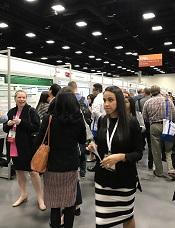
2016 ASH Annual Meeting
SAN DIEGO—Researchers have found evidence to suggest that mutations in the CCND1 and CCND2 genes may contribute to the development of core-binding factor acute myeloid leukemia (CBF-AML).
The team noted that CBF-AML is defined by the presence of either t(8;21)(q22;q22)/RUNX1-RUNX1T1 or inv(16)(p13.1q22)/t(16;16)(p13.1;q22)/CBFB-MYH11.
However, the fusion genes alone are not capable of causing CBF-AML.
“The hematology community has long sought to determine what other factors in addition to the fusion genes occur in this special type of leukemia,” said Ann-Kathrin Eisfeld, MD, of The Ohio State University Comprehensive Cancer Center in Columbus.
“We are now the first to describe that mutations in CCND1—and among the first to describe that mutations in the sister gene CCND2—are unique features of CBF-AML with t(8;21). In addition, we have collected the first evidence that mutations in CCND2 lead to more aggressive growth of leukemia cell lines.”
Dr Eisfeld and her colleagues reported these findings in a paper published in Leukemia and in a poster presented at the 2016 ASH Annual Meeting (abstract 2740).
A previous study of genetic mutations in CBF-AML revealed the presence of at least 1 mutation in 85% of patients studied. This meant the remaining 15% of patients harbored other, undiscovered mutations.
For the current study, Dr Eisfeld and her colleagues searched CBF-AML samples for the missing mutations that, together with the fusion genes, might contribute to the leukemia in this subgroup of cases.
The team analyzed pretreatment bone marrow and peripheral blood samples from 177 adult CBF-AML patients who received similar treatment through a clinical trial conducted at multiple centers across the US.
Using a targeted, next-generation sequencing approach, the researchers looked for mutations in 84 leukemia- and/or cancer-associated genes. They also performed tests on blood or bone marrow cells to look for chromosomal irregularities.
The team discovered 2 significant mutations in the CCND1 and CCND2 genes, representing the first dual evidence of these recurrent mutations in patients with t(8;21)-positive CBF-AML.
CCND1 and CCND2 mutations were found in 15% (n=10) of patients with t(8;21)-positive CBF-AML. Two patients had mutations in CCND1, and 8 had mutations in CCND2.
The researchers also found a single CCND2 mutation in 1 (0.9%) patient with inv(16)-positive CBF-AML.
In comparison, the incidence of CCND1 and CCND2 mutations was 0.77% (n=11) in a cohort of 1426 patients with non-CBF-AML.
“This is extremely valuable information that was previously unknown,” Dr Eisfeld said, “and it might help us develop targeted therapies more likely to help patients with [CBF-AML] in the near future.”
2016 ASH Annual Meeting
SAN DIEGO—Researchers have found evidence to suggest that mutations in the CCND1 and CCND2 genes may contribute to the development of core-binding factor acute myeloid leukemia (CBF-AML).
The team noted that CBF-AML is defined by the presence of either t(8;21)(q22;q22)/RUNX1-RUNX1T1 or inv(16)(p13.1q22)/t(16;16)(p13.1;q22)/CBFB-MYH11.
However, the fusion genes alone are not capable of causing CBF-AML.
“The hematology community has long sought to determine what other factors in addition to the fusion genes occur in this special type of leukemia,” said Ann-Kathrin Eisfeld, MD, of The Ohio State University Comprehensive Cancer Center in Columbus.
“We are now the first to describe that mutations in CCND1—and among the first to describe that mutations in the sister gene CCND2—are unique features of CBF-AML with t(8;21). In addition, we have collected the first evidence that mutations in CCND2 lead to more aggressive growth of leukemia cell lines.”
Dr Eisfeld and her colleagues reported these findings in a paper published in Leukemia and in a poster presented at the 2016 ASH Annual Meeting (abstract 2740).
A previous study of genetic mutations in CBF-AML revealed the presence of at least 1 mutation in 85% of patients studied. This meant the remaining 15% of patients harbored other, undiscovered mutations.
For the current study, Dr Eisfeld and her colleagues searched CBF-AML samples for the missing mutations that, together with the fusion genes, might contribute to the leukemia in this subgroup of cases.
The team analyzed pretreatment bone marrow and peripheral blood samples from 177 adult CBF-AML patients who received similar treatment through a clinical trial conducted at multiple centers across the US.
Using a targeted, next-generation sequencing approach, the researchers looked for mutations in 84 leukemia- and/or cancer-associated genes. They also performed tests on blood or bone marrow cells to look for chromosomal irregularities.
The team discovered 2 significant mutations in the CCND1 and CCND2 genes, representing the first dual evidence of these recurrent mutations in patients with t(8;21)-positive CBF-AML.
CCND1 and CCND2 mutations were found in 15% (n=10) of patients with t(8;21)-positive CBF-AML. Two patients had mutations in CCND1, and 8 had mutations in CCND2.
The researchers also found a single CCND2 mutation in 1 (0.9%) patient with inv(16)-positive CBF-AML.
In comparison, the incidence of CCND1 and CCND2 mutations was 0.77% (n=11) in a cohort of 1426 patients with non-CBF-AML.
“This is extremely valuable information that was previously unknown,” Dr Eisfeld said, “and it might help us develop targeted therapies more likely to help patients with [CBF-AML] in the near future.”
2016 ASH Annual Meeting
SAN DIEGO—Researchers have found evidence to suggest that mutations in the CCND1 and CCND2 genes may contribute to the development of core-binding factor acute myeloid leukemia (CBF-AML).
The team noted that CBF-AML is defined by the presence of either t(8;21)(q22;q22)/RUNX1-RUNX1T1 or inv(16)(p13.1q22)/t(16;16)(p13.1;q22)/CBFB-MYH11.
However, the fusion genes alone are not capable of causing CBF-AML.
“The hematology community has long sought to determine what other factors in addition to the fusion genes occur in this special type of leukemia,” said Ann-Kathrin Eisfeld, MD, of The Ohio State University Comprehensive Cancer Center in Columbus.
“We are now the first to describe that mutations in CCND1—and among the first to describe that mutations in the sister gene CCND2—are unique features of CBF-AML with t(8;21). In addition, we have collected the first evidence that mutations in CCND2 lead to more aggressive growth of leukemia cell lines.”
Dr Eisfeld and her colleagues reported these findings in a paper published in Leukemia and in a poster presented at the 2016 ASH Annual Meeting (abstract 2740).
A previous study of genetic mutations in CBF-AML revealed the presence of at least 1 mutation in 85% of patients studied. This meant the remaining 15% of patients harbored other, undiscovered mutations.
For the current study, Dr Eisfeld and her colleagues searched CBF-AML samples for the missing mutations that, together with the fusion genes, might contribute to the leukemia in this subgroup of cases.
The team analyzed pretreatment bone marrow and peripheral blood samples from 177 adult CBF-AML patients who received similar treatment through a clinical trial conducted at multiple centers across the US.
Using a targeted, next-generation sequencing approach, the researchers looked for mutations in 84 leukemia- and/or cancer-associated genes. They also performed tests on blood or bone marrow cells to look for chromosomal irregularities.
The team discovered 2 significant mutations in the CCND1 and CCND2 genes, representing the first dual evidence of these recurrent mutations in patients with t(8;21)-positive CBF-AML.
CCND1 and CCND2 mutations were found in 15% (n=10) of patients with t(8;21)-positive CBF-AML. Two patients had mutations in CCND1, and 8 had mutations in CCND2.
The researchers also found a single CCND2 mutation in 1 (0.9%) patient with inv(16)-positive CBF-AML.
In comparison, the incidence of CCND1 and CCND2 mutations was 0.77% (n=11) in a cohort of 1426 patients with non-CBF-AML.
“This is extremely valuable information that was previously unknown,” Dr Eisfeld said, “and it might help us develop targeted therapies more likely to help patients with [CBF-AML] in the near future.”
EC authorizes new use for ofatumumab in CLL
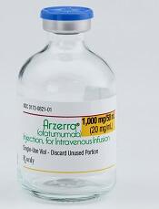
Photo courtesy of GSK
The European Commission (EC) has granted marketing authorization for ofatumumab (Arzerra®) to be used in combination with fludarabine and cyclophosphamide (FC) in the treatment of adults with relapsed chronic lymphocytic leukemia (CLL).
Ofatumumab is a monoclonal antibody designed to target CD20. The drug is marketed under a collaboration agreement between Genmab and Novartis.
The EC previously authorized the use of ofatumumab as a single agent to treat CLL patients who are refractory to fludarabine and alemtuzumab.
The agency also authorized the use of ofatumumab in combination with chlorambucil or bendamustine in CLL patients who have not received prior therapy and are not eligible for fludarabine-based therapy.
The EC’s decision to approve the use of ofatumumab in combination with FC was based on results from the phase 3 COMPLEMENT 2 study, which were published in Leukemia & Lymphoma in October.
The trial enrolled 365 patients with relapsed CLL. The patients were randomized 1:1 to receive up to 6 cycles of ofatumumab in combination with FC or up to 6 cycles of FC alone.
The primary endpoint was progression-free survival, as assessed by an independent review committee.
The median progression-free survival was 28.9 months for patients receiving ofatumumab plus FC, compared to 18.8 months for patients receiving FC only (hazard ratio=0.67, P=0.0032).
The incidence of grade 3 or higher adverse events was 74% in the ofatumumab-plus-FC arm and 69% in the FC-only arm. Neutropenia was the most common of these events, occurring in 49% and 36% of patients, respectively.
Photo courtesy of GSK
The European Commission (EC) has granted marketing authorization for ofatumumab (Arzerra®) to be used in combination with fludarabine and cyclophosphamide (FC) in the treatment of adults with relapsed chronic lymphocytic leukemia (CLL).
Ofatumumab is a monoclonal antibody designed to target CD20. The drug is marketed under a collaboration agreement between Genmab and Novartis.
The EC previously authorized the use of ofatumumab as a single agent to treat CLL patients who are refractory to fludarabine and alemtuzumab.
The agency also authorized the use of ofatumumab in combination with chlorambucil or bendamustine in CLL patients who have not received prior therapy and are not eligible for fludarabine-based therapy.
The EC’s decision to approve the use of ofatumumab in combination with FC was based on results from the phase 3 COMPLEMENT 2 study, which were published in Leukemia & Lymphoma in October.
The trial enrolled 365 patients with relapsed CLL. The patients were randomized 1:1 to receive up to 6 cycles of ofatumumab in combination with FC or up to 6 cycles of FC alone.
The primary endpoint was progression-free survival, as assessed by an independent review committee.
The median progression-free survival was 28.9 months for patients receiving ofatumumab plus FC, compared to 18.8 months for patients receiving FC only (hazard ratio=0.67, P=0.0032).
The incidence of grade 3 or higher adverse events was 74% in the ofatumumab-plus-FC arm and 69% in the FC-only arm. Neutropenia was the most common of these events, occurring in 49% and 36% of patients, respectively.
Photo courtesy of GSK
The European Commission (EC) has granted marketing authorization for ofatumumab (Arzerra®) to be used in combination with fludarabine and cyclophosphamide (FC) in the treatment of adults with relapsed chronic lymphocytic leukemia (CLL).
Ofatumumab is a monoclonal antibody designed to target CD20. The drug is marketed under a collaboration agreement between Genmab and Novartis.
The EC previously authorized the use of ofatumumab as a single agent to treat CLL patients who are refractory to fludarabine and alemtuzumab.
The agency also authorized the use of ofatumumab in combination with chlorambucil or bendamustine in CLL patients who have not received prior therapy and are not eligible for fludarabine-based therapy.
The EC’s decision to approve the use of ofatumumab in combination with FC was based on results from the phase 3 COMPLEMENT 2 study, which were published in Leukemia & Lymphoma in October.
The trial enrolled 365 patients with relapsed CLL. The patients were randomized 1:1 to receive up to 6 cycles of ofatumumab in combination with FC or up to 6 cycles of FC alone.
The primary endpoint was progression-free survival, as assessed by an independent review committee.
The median progression-free survival was 28.9 months for patients receiving ofatumumab plus FC, compared to 18.8 months for patients receiving FC only (hazard ratio=0.67, P=0.0032).
The incidence of grade 3 or higher adverse events was 74% in the ofatumumab-plus-FC arm and 69% in the FC-only arm. Neutropenia was the most common of these events, occurring in 49% and 36% of patients, respectively.
MS & Pregnancy: What's Safe?
Q)What are the considerations and recommendations for pregnancy and breastfeeding in women with multiple sclerosis? When should women discontinue their disease-modifying therapies?
Multiple sclerosis (MS) is an inflammatory, demyelinating, degenerative disease. Three times more common in women than men, it may affect pregnancy planning and childbearing experiences.6 Evidence demonstrates a reduction in annualized relapse rate during pregnancy and the postpartum period with exclusive breastfeeding. Therefore, pregnancy and exclusive breastfeeding provide a favorable immunomodulatory effect in women with MS, which combats the increased relapse risk associated with the postpartum period.7,8
Reproductive education—including conception, pregnancy, and breastfeeding—is critical for patients with MS and their partners during a woman’s childbearing years. However, women with MS do not require special considerations during pregnancy unless they have remarkable disability. As soon as these women and their partners decide upon pregnancy, a plan should be established that includes a discussion about potential risks to the fetus due to drug exposure, as well as risks to the mother. The goal should be to minimize risk for disease activity and optimize the health of the baby.
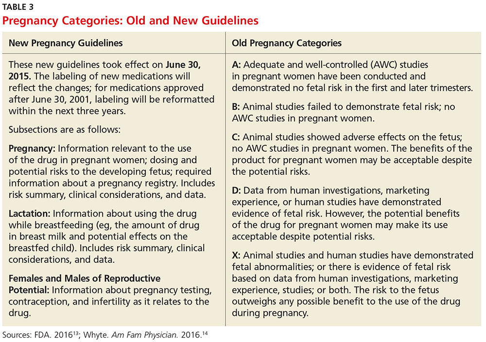
Use of DMTs during conception, pregnancy, and breastfeeding. All disease-modifying therapies (DMTs) are usually discontinued during pregnancy and breastfeeding. Common practice among MS experts is to discontinue DMTs prior to conception, with a few exceptions. There is no consensus about timing of discontinuation and washout period for each DMT. The decision is based on the half-life of each DMT, the opinions of the woman and her partner, and risk tolerance. Note: For the purposes of this discussion, the old pregnancy category designations are used, since they are familiar to clinicians. New guidelines took effect in June 2015; Table 3 outlines the change in format.
Injectables. Glatiramer acetate (GA) is the safest drug in relation to pregnancy and breastfeeding (category B). There is no evidence of congenital malformation or spontaneous abortion. The common recommendation is to discontinue the drug one to two months before conception, although some clinicians allow continuation of the injections throughout pregnancy and into breastfeeding.
Interferon-ßs are category C and therefore pose minimal risk for the fetus. The washout period before conception is two to three months, varying among clinicians. Although there is no evidence of spontaneous abortion or birth defects in humans, animal data show increased risk for abortion.9
Both GA and interferon-ßs are large molecules; there is a very minimal chance that the medication will transmit to the baby via breast milk. Thus, both DMTs are considered safe during lactation.7
Oral MS medications. The three approved MS oral medications are fingolimod, dimethyl fumarate (DMF), and teriflunomide. Fingolimod and DMF are both category C. Women on DMF must discontinue use of the medication one month prior to conception due to its short half-life. There are no reports of birth defects or spontaneous abortion in women taking DMF. Fingolimod needs to be discontinued two months prior to conception. Animal data show evidence for teratogenicity and embryolethality at lower doses of fingolimod than those used in humans.7
Teriflunomide is category X, posing high risk for the health of the fetus. It stays in the blood for approximately eight months after discontinuation of use. Animal data show teratogenicity and embryotoxicity; therefore, teriflunomide is contraindicated in pregnancy. Women on teriflunomide who plan to become pregnant need to undergo an elimination procedure with cholestyramine or charcoal.
Infusions and injections (monoclonal antibodies). The approved monoclonal antibodies include natalizumab, alemtuzumab, and daclizumab. (Currently, use of rituximab in MS is off label, and the FDA is reviewing the efficacy and safety data for ocrelizumab.) The monoclonal antibodies are category C. The recommendation is to discontinue natalizumab one to two months prior to conception and discontinue alemtuzumab four months preconception.10 There is no evidence of spontaneous abortion or birth defects in women on alemtuzumab, but there is potentially increased risk for spontaneous abortion in those on natalizumab.
The spontaneous abortion rate in daclizumab-exposed women is consistent with early pregnancy loss in the general population (12% to 26%). Data on a small number of pregnancies exposed to daclizumab did not suggest an increased risk for adverse fetal or maternal outcomes.11 However, the recommendation is to discontinue daclizumab four months prior to conception. Rituximab should be discontinued 12 months prior to conception, based on the manufacturer recommendation, although it is potentially safe to conceive when the B cell counts return to normal.
Chemotherapy agents. Mitoxantrone is the only FDA-approved chemotherapeutic drug used in MS. However, a few chemotherapy drugs—among them, azathioprine, methotrexate, and cyclophosphamide—are used off label. Chemotherapeutic agents are category D, except methotrexate (category X). Women on category D medications must use a method of birth control for three months after stopping the DMT. Often, clinicians will recommend women initiate GA or interferon during this period, in the hope of minimizing disease activity. Women on mitoxantrone and methotrexate need to use birth control for six months after stopping these immunosuppressive medications, before conceiving.12 These women likely need to switch to a safer pregnancy DMT during the long washout period.
Pregnancy and breastfeeding among women with MS require planning and decision making. The recommendations differ among clinicians and MS experts since there is no definitive evidence about the risks of the DMTs on the mother and/or the fetus. Clinicians should discuss the potential risks with women based on their knowledge and experience, and the data available based on animal research and the pregnancy registries. —ABB-Z
Aliza Bitton Ben-Zacharia, DNP, ANP
President-Elect of IOMSN
Neurology Faculty, Icahn School of Medicine, Mount Sinai Hospital System, New York, New York
6. Tullman MJ. Overview of the epidemiology, diagnosis, and disease progression associated with multiple sclerosis. Am J Manag Care. 2013;19(2):S15-S20.
7. Fabian M. Pregnancy in the setting of multiple sclerosis. Continuum (Minneap Minn). 2016; 22(3):837-850.
8. Vukusic S, Hutchinson M, Hours M, et al. Pregnancy and multiple sclerosis (the PRIMS study): clinical predictors of post-partum relapse. Brain. 2004;127(6):1353-1360.
9. Sandberg-Wollheim M, Alteri E, Moraga MS, Kornmann G. Pregnancy outcomes in multiple sclerosis following subcutaneous interferon beta-1a therapy. Mult Scler. 2011; 17(4):423-430.
10. Coyle PK. Multiple sclerosis in pregnancy. Continuum (Minneap Minn). 2014;20(1):42-59.
11. Gold SM, Voskuhl RR. Pregnancy and multiple sclerosis: from molecular mechanisms to clinical application. Semin Immunopathol. 2016;38:709.
12. Vukusic S, Marignier R. Multiple sclerosis and pregnancy in the ‘treatment era’. Nat Rev Neurol. 2015;11(5):280-289.
13. FDA. Pregnancy and lactation labeling (drugs) final rule. www.fda.gov/Drugs/DevelopmentApprovalProcess/DevelopmentResources/Labeling/ucm093307.htm. Accessed November 2, 2016.
14. Whyte J. FDA implements new labeling for medications used during pregnancy and lactation. Am Fam Physician. 2016;94(1):12-13.
Clinician Reviews in partnership with
MS Consult is edited by Colleen J. Harris, MN, NP, MSCN, Nurse Practitioner/Manager of the Multiple Sclerosis Clinic at Foothills Medical Centre in Calgary, Alberta, Canada, and Bryan Walker, MHS, PA-C, who is in the Department of Neurology, Division of MS and Neuroimmunology, at Duke University Medical Center in Durham, North Carolina. This month’s responses were authored by Megan Weigel, DNP, ARNP-C, MSCN, President of IOMSN, who practices at Baptist Neurology in Jacksonville Beach, Florida, and Aliza Bitton Ben-Zacharia, DNP, ANP, President-Elect of IOMSN, who is Neurology Faculty in the Icahn School of Medicine at Mount Sinai Hospital System in New York, New York.
Clinician Reviews in partnership with
MS Consult is edited by Colleen J. Harris, MN, NP, MSCN, Nurse Practitioner/Manager of the Multiple Sclerosis Clinic at Foothills Medical Centre in Calgary, Alberta, Canada, and Bryan Walker, MHS, PA-C, who is in the Department of Neurology, Division of MS and Neuroimmunology, at Duke University Medical Center in Durham, North Carolina. This month’s responses were authored by Megan Weigel, DNP, ARNP-C, MSCN, President of IOMSN, who practices at Baptist Neurology in Jacksonville Beach, Florida, and Aliza Bitton Ben-Zacharia, DNP, ANP, President-Elect of IOMSN, who is Neurology Faculty in the Icahn School of Medicine at Mount Sinai Hospital System in New York, New York.
Clinician Reviews in partnership with
MS Consult is edited by Colleen J. Harris, MN, NP, MSCN, Nurse Practitioner/Manager of the Multiple Sclerosis Clinic at Foothills Medical Centre in Calgary, Alberta, Canada, and Bryan Walker, MHS, PA-C, who is in the Department of Neurology, Division of MS and Neuroimmunology, at Duke University Medical Center in Durham, North Carolina. This month’s responses were authored by Megan Weigel, DNP, ARNP-C, MSCN, President of IOMSN, who practices at Baptist Neurology in Jacksonville Beach, Florida, and Aliza Bitton Ben-Zacharia, DNP, ANP, President-Elect of IOMSN, who is Neurology Faculty in the Icahn School of Medicine at Mount Sinai Hospital System in New York, New York.
Q)What are the considerations and recommendations for pregnancy and breastfeeding in women with multiple sclerosis? When should women discontinue their disease-modifying therapies?
Multiple sclerosis (MS) is an inflammatory, demyelinating, degenerative disease. Three times more common in women than men, it may affect pregnancy planning and childbearing experiences.6 Evidence demonstrates a reduction in annualized relapse rate during pregnancy and the postpartum period with exclusive breastfeeding. Therefore, pregnancy and exclusive breastfeeding provide a favorable immunomodulatory effect in women with MS, which combats the increased relapse risk associated with the postpartum period.7,8
Reproductive education—including conception, pregnancy, and breastfeeding—is critical for patients with MS and their partners during a woman’s childbearing years. However, women with MS do not require special considerations during pregnancy unless they have remarkable disability. As soon as these women and their partners decide upon pregnancy, a plan should be established that includes a discussion about potential risks to the fetus due to drug exposure, as well as risks to the mother. The goal should be to minimize risk for disease activity and optimize the health of the baby.
Use of DMTs during conception, pregnancy, and breastfeeding. All disease-modifying therapies (DMTs) are usually discontinued during pregnancy and breastfeeding. Common practice among MS experts is to discontinue DMTs prior to conception, with a few exceptions. There is no consensus about timing of discontinuation and washout period for each DMT. The decision is based on the half-life of each DMT, the opinions of the woman and her partner, and risk tolerance. Note: For the purposes of this discussion, the old pregnancy category designations are used, since they are familiar to clinicians. New guidelines took effect in June 2015; Table 3 outlines the change in format.
Injectables. Glatiramer acetate (GA) is the safest drug in relation to pregnancy and breastfeeding (category B). There is no evidence of congenital malformation or spontaneous abortion. The common recommendation is to discontinue the drug one to two months before conception, although some clinicians allow continuation of the injections throughout pregnancy and into breastfeeding.
Interferon-ßs are category C and therefore pose minimal risk for the fetus. The washout period before conception is two to three months, varying among clinicians. Although there is no evidence of spontaneous abortion or birth defects in humans, animal data show increased risk for abortion.9
Both GA and interferon-ßs are large molecules; there is a very minimal chance that the medication will transmit to the baby via breast milk. Thus, both DMTs are considered safe during lactation.7
Oral MS medications. The three approved MS oral medications are fingolimod, dimethyl fumarate (DMF), and teriflunomide. Fingolimod and DMF are both category C. Women on DMF must discontinue use of the medication one month prior to conception due to its short half-life. There are no reports of birth defects or spontaneous abortion in women taking DMF. Fingolimod needs to be discontinued two months prior to conception. Animal data show evidence for teratogenicity and embryolethality at lower doses of fingolimod than those used in humans.7
Teriflunomide is category X, posing high risk for the health of the fetus. It stays in the blood for approximately eight months after discontinuation of use. Animal data show teratogenicity and embryotoxicity; therefore, teriflunomide is contraindicated in pregnancy. Women on teriflunomide who plan to become pregnant need to undergo an elimination procedure with cholestyramine or charcoal.
Infusions and injections (monoclonal antibodies). The approved monoclonal antibodies include natalizumab, alemtuzumab, and daclizumab. (Currently, use of rituximab in MS is off label, and the FDA is reviewing the efficacy and safety data for ocrelizumab.) The monoclonal antibodies are category C. The recommendation is to discontinue natalizumab one to two months prior to conception and discontinue alemtuzumab four months preconception.10 There is no evidence of spontaneous abortion or birth defects in women on alemtuzumab, but there is potentially increased risk for spontaneous abortion in those on natalizumab.
The spontaneous abortion rate in daclizumab-exposed women is consistent with early pregnancy loss in the general population (12% to 26%). Data on a small number of pregnancies exposed to daclizumab did not suggest an increased risk for adverse fetal or maternal outcomes.11 However, the recommendation is to discontinue daclizumab four months prior to conception. Rituximab should be discontinued 12 months prior to conception, based on the manufacturer recommendation, although it is potentially safe to conceive when the B cell counts return to normal.
Chemotherapy agents. Mitoxantrone is the only FDA-approved chemotherapeutic drug used in MS. However, a few chemotherapy drugs—among them, azathioprine, methotrexate, and cyclophosphamide—are used off label. Chemotherapeutic agents are category D, except methotrexate (category X). Women on category D medications must use a method of birth control for three months after stopping the DMT. Often, clinicians will recommend women initiate GA or interferon during this period, in the hope of minimizing disease activity. Women on mitoxantrone and methotrexate need to use birth control for six months after stopping these immunosuppressive medications, before conceiving.12 These women likely need to switch to a safer pregnancy DMT during the long washout period.
Pregnancy and breastfeeding among women with MS require planning and decision making. The recommendations differ among clinicians and MS experts since there is no definitive evidence about the risks of the DMTs on the mother and/or the fetus. Clinicians should discuss the potential risks with women based on their knowledge and experience, and the data available based on animal research and the pregnancy registries. —ABB-Z
Aliza Bitton Ben-Zacharia, DNP, ANP
President-Elect of IOMSN
Neurology Faculty, Icahn School of Medicine, Mount Sinai Hospital System, New York, New York
Q)What are the considerations and recommendations for pregnancy and breastfeeding in women with multiple sclerosis? When should women discontinue their disease-modifying therapies?
Multiple sclerosis (MS) is an inflammatory, demyelinating, degenerative disease. Three times more common in women than men, it may affect pregnancy planning and childbearing experiences.6 Evidence demonstrates a reduction in annualized relapse rate during pregnancy and the postpartum period with exclusive breastfeeding. Therefore, pregnancy and exclusive breastfeeding provide a favorable immunomodulatory effect in women with MS, which combats the increased relapse risk associated with the postpartum period.7,8
Reproductive education—including conception, pregnancy, and breastfeeding—is critical for patients with MS and their partners during a woman’s childbearing years. However, women with MS do not require special considerations during pregnancy unless they have remarkable disability. As soon as these women and their partners decide upon pregnancy, a plan should be established that includes a discussion about potential risks to the fetus due to drug exposure, as well as risks to the mother. The goal should be to minimize risk for disease activity and optimize the health of the baby.
Use of DMTs during conception, pregnancy, and breastfeeding. All disease-modifying therapies (DMTs) are usually discontinued during pregnancy and breastfeeding. Common practice among MS experts is to discontinue DMTs prior to conception, with a few exceptions. There is no consensus about timing of discontinuation and washout period for each DMT. The decision is based on the half-life of each DMT, the opinions of the woman and her partner, and risk tolerance. Note: For the purposes of this discussion, the old pregnancy category designations are used, since they are familiar to clinicians. New guidelines took effect in June 2015; Table 3 outlines the change in format.
Injectables. Glatiramer acetate (GA) is the safest drug in relation to pregnancy and breastfeeding (category B). There is no evidence of congenital malformation or spontaneous abortion. The common recommendation is to discontinue the drug one to two months before conception, although some clinicians allow continuation of the injections throughout pregnancy and into breastfeeding.
Interferon-ßs are category C and therefore pose minimal risk for the fetus. The washout period before conception is two to three months, varying among clinicians. Although there is no evidence of spontaneous abortion or birth defects in humans, animal data show increased risk for abortion.9
Both GA and interferon-ßs are large molecules; there is a very minimal chance that the medication will transmit to the baby via breast milk. Thus, both DMTs are considered safe during lactation.7
Oral MS medications. The three approved MS oral medications are fingolimod, dimethyl fumarate (DMF), and teriflunomide. Fingolimod and DMF are both category C. Women on DMF must discontinue use of the medication one month prior to conception due to its short half-life. There are no reports of birth defects or spontaneous abortion in women taking DMF. Fingolimod needs to be discontinued two months prior to conception. Animal data show evidence for teratogenicity and embryolethality at lower doses of fingolimod than those used in humans.7
Teriflunomide is category X, posing high risk for the health of the fetus. It stays in the blood for approximately eight months after discontinuation of use. Animal data show teratogenicity and embryotoxicity; therefore, teriflunomide is contraindicated in pregnancy. Women on teriflunomide who plan to become pregnant need to undergo an elimination procedure with cholestyramine or charcoal.
Infusions and injections (monoclonal antibodies). The approved monoclonal antibodies include natalizumab, alemtuzumab, and daclizumab. (Currently, use of rituximab in MS is off label, and the FDA is reviewing the efficacy and safety data for ocrelizumab.) The monoclonal antibodies are category C. The recommendation is to discontinue natalizumab one to two months prior to conception and discontinue alemtuzumab four months preconception.10 There is no evidence of spontaneous abortion or birth defects in women on alemtuzumab, but there is potentially increased risk for spontaneous abortion in those on natalizumab.
The spontaneous abortion rate in daclizumab-exposed women is consistent with early pregnancy loss in the general population (12% to 26%). Data on a small number of pregnancies exposed to daclizumab did not suggest an increased risk for adverse fetal or maternal outcomes.11 However, the recommendation is to discontinue daclizumab four months prior to conception. Rituximab should be discontinued 12 months prior to conception, based on the manufacturer recommendation, although it is potentially safe to conceive when the B cell counts return to normal.
Chemotherapy agents. Mitoxantrone is the only FDA-approved chemotherapeutic drug used in MS. However, a few chemotherapy drugs—among them, azathioprine, methotrexate, and cyclophosphamide—are used off label. Chemotherapeutic agents are category D, except methotrexate (category X). Women on category D medications must use a method of birth control for three months after stopping the DMT. Often, clinicians will recommend women initiate GA or interferon during this period, in the hope of minimizing disease activity. Women on mitoxantrone and methotrexate need to use birth control for six months after stopping these immunosuppressive medications, before conceiving.12 These women likely need to switch to a safer pregnancy DMT during the long washout period.
Pregnancy and breastfeeding among women with MS require planning and decision making. The recommendations differ among clinicians and MS experts since there is no definitive evidence about the risks of the DMTs on the mother and/or the fetus. Clinicians should discuss the potential risks with women based on their knowledge and experience, and the data available based on animal research and the pregnancy registries. —ABB-Z
Aliza Bitton Ben-Zacharia, DNP, ANP
President-Elect of IOMSN
Neurology Faculty, Icahn School of Medicine, Mount Sinai Hospital System, New York, New York
6. Tullman MJ. Overview of the epidemiology, diagnosis, and disease progression associated with multiple sclerosis. Am J Manag Care. 2013;19(2):S15-S20.
7. Fabian M. Pregnancy in the setting of multiple sclerosis. Continuum (Minneap Minn). 2016; 22(3):837-850.
8. Vukusic S, Hutchinson M, Hours M, et al. Pregnancy and multiple sclerosis (the PRIMS study): clinical predictors of post-partum relapse. Brain. 2004;127(6):1353-1360.
9. Sandberg-Wollheim M, Alteri E, Moraga MS, Kornmann G. Pregnancy outcomes in multiple sclerosis following subcutaneous interferon beta-1a therapy. Mult Scler. 2011; 17(4):423-430.
10. Coyle PK. Multiple sclerosis in pregnancy. Continuum (Minneap Minn). 2014;20(1):42-59.
11. Gold SM, Voskuhl RR. Pregnancy and multiple sclerosis: from molecular mechanisms to clinical application. Semin Immunopathol. 2016;38:709.
12. Vukusic S, Marignier R. Multiple sclerosis and pregnancy in the ‘treatment era’. Nat Rev Neurol. 2015;11(5):280-289.
13. FDA. Pregnancy and lactation labeling (drugs) final rule. www.fda.gov/Drugs/DevelopmentApprovalProcess/DevelopmentResources/Labeling/ucm093307.htm. Accessed November 2, 2016.
14. Whyte J. FDA implements new labeling for medications used during pregnancy and lactation. Am Fam Physician. 2016;94(1):12-13.
6. Tullman MJ. Overview of the epidemiology, diagnosis, and disease progression associated with multiple sclerosis. Am J Manag Care. 2013;19(2):S15-S20.
7. Fabian M. Pregnancy in the setting of multiple sclerosis. Continuum (Minneap Minn). 2016; 22(3):837-850.
8. Vukusic S, Hutchinson M, Hours M, et al. Pregnancy and multiple sclerosis (the PRIMS study): clinical predictors of post-partum relapse. Brain. 2004;127(6):1353-1360.
9. Sandberg-Wollheim M, Alteri E, Moraga MS, Kornmann G. Pregnancy outcomes in multiple sclerosis following subcutaneous interferon beta-1a therapy. Mult Scler. 2011; 17(4):423-430.
10. Coyle PK. Multiple sclerosis in pregnancy. Continuum (Minneap Minn). 2014;20(1):42-59.
11. Gold SM, Voskuhl RR. Pregnancy and multiple sclerosis: from molecular mechanisms to clinical application. Semin Immunopathol. 2016;38:709.
12. Vukusic S, Marignier R. Multiple sclerosis and pregnancy in the ‘treatment era’. Nat Rev Neurol. 2015;11(5):280-289.
13. FDA. Pregnancy and lactation labeling (drugs) final rule. www.fda.gov/Drugs/DevelopmentApprovalProcess/DevelopmentResources/Labeling/ucm093307.htm. Accessed November 2, 2016.
14. Whyte J. FDA implements new labeling for medications used during pregnancy and lactation. Am Fam Physician. 2016;94(1):12-13.
VIDEO: NAFLD costing U.S. $290 billion annually
BOSTON – A sophisticated mathematical analysis shows that nonalcoholic fatty liver disease is costing the United States over $290 billion annually. A similar economic burden was seen in several Western European countries as well.
The study, which incorporated available data about current cases of nonalcoholic fatty liver disease (NAFLD), looked at both medical and nonmedical costs, as the disease’s economic and social impact extends far beyond medical bills.
“It impacts patients’ quality of life, their work productivity; there’s a significant fatigue associated with that. In addition to this, there is an economic burden of nonalcoholic fatty liver disease,” said the study’s lead author, Zobair Younossi, MD, in a video interview at the annual meeting of the American Association for the Study of Liver Diseases.
The video associated with this article is no longer available on this site. Please view all of our videos on the MDedge YouTube channel
Dr. Younossi, executive director of the Center for Liver Diseases at Inova Fairfax Hospital, Falls Church, Va., and his coinvestigators constructed a Markov chain to model costs associated with NAFLD. This mathematical technique allows the probability of the occurrence of an event to influence the model’s prediction of the probability of later events, a useful technique when trying to model real-world, dynamic conditions.
They used the Markov chain technique to model the transition of patients along the path of NAFLD progression. States included in the model were nonalcoholic steatohepatitis (NASH), NASH with fibrosis, compensated and decompensated cirrhosis, hepatocellular carcinoma, transplant, posttransplant, and death. The probabilities of progressing through these states were built from a meta-analysis and systematic literature review conducted by the authors, which they then adjusted by incorporating real-world data.
According to the model, annual direct medical costs are projected at $103 billion, or $1,613 for each of the 64 million NAFLD patients in the United States. The four European Union countries included in the model were Germany, France, Italy, and the United Kingdom. In aggregate, these countries are projected to have about 52 million people with NAFLD, for an annual cost of 35 billion euros. The estimated annual direct cost per patient varies by country, ranging from 354 euros to 1,163 euros.
When societal costs are incorporated, the numbers leap higher: to $292.19 billion in the United States, and over 200 billion euros in the four European countries studied.
In recent work, Dr. Younossi and his colleagues have reached an estimate that about a quarter of the world’s population has NAFLD. He said he was surprised to learn that the highest prevalences were in some areas of South America and the Middle East, with rapid increases in Asia as well.
However, the etiology of NAFLD helps explain these increases. “It’s really a phenotype. A number of different pathways lead to this disease; the most common is the one that is associated with obesity, type 2 diabetes, and insulin resistance,” said Dr. Younossi.
The subset of NAFLD patients who have NASH also risks progression to cirrhosis and hepatocellular carcinoma. According to Dr. Younossi’s examination of the UNOS transplant database, NASH is the second leading cause of liver transplantation in the United States, and one of the top three causes of death from liver disease.
These sobering data make the need for medical treatment for NASH an urgent priority, said Dr. Younossi. Currently, lifestyle modification such as weight loss is the only known treatment for NAFLD and NASH, “which is not easy to do,” he noted.
Several candidate drugs are in the pipeline currently. Also, said Dr. Younossi, NASH can be diagnosed only by liver biopsy currently, a risky and expensive procedure. The search is on for accurate and reliable imaging and serum biomarkers for NASH, so physicians can understand who’s most at risk for progression to more serious liver disease.
“The analysis quantifies the enormity of the clinical and economic burden of NAFLD, which will likely increase as incidence of NAFLD continues to rise with the increasing obesity and diabetes rates,” wrote Dr. Younossi and his coauthors.
Dr. Younossi reported having financial relationships with several pharmaceutical companies.
koakes@frontlinemedcom.com
On Twitter @karioakes
BOSTON – A sophisticated mathematical analysis shows that nonalcoholic fatty liver disease is costing the United States over $290 billion annually. A similar economic burden was seen in several Western European countries as well.
The study, which incorporated available data about current cases of nonalcoholic fatty liver disease (NAFLD), looked at both medical and nonmedical costs, as the disease’s economic and social impact extends far beyond medical bills.
“It impacts patients’ quality of life, their work productivity; there’s a significant fatigue associated with that. In addition to this, there is an economic burden of nonalcoholic fatty liver disease,” said the study’s lead author, Zobair Younossi, MD, in a video interview at the annual meeting of the American Association for the Study of Liver Diseases.
The video associated with this article is no longer available on this site. Please view all of our videos on the MDedge YouTube channel
Dr. Younossi, executive director of the Center for Liver Diseases at Inova Fairfax Hospital, Falls Church, Va., and his coinvestigators constructed a Markov chain to model costs associated with NAFLD. This mathematical technique allows the probability of the occurrence of an event to influence the model’s prediction of the probability of later events, a useful technique when trying to model real-world, dynamic conditions.
They used the Markov chain technique to model the transition of patients along the path of NAFLD progression. States included in the model were nonalcoholic steatohepatitis (NASH), NASH with fibrosis, compensated and decompensated cirrhosis, hepatocellular carcinoma, transplant, posttransplant, and death. The probabilities of progressing through these states were built from a meta-analysis and systematic literature review conducted by the authors, which they then adjusted by incorporating real-world data.
According to the model, annual direct medical costs are projected at $103 billion, or $1,613 for each of the 64 million NAFLD patients in the United States. The four European Union countries included in the model were Germany, France, Italy, and the United Kingdom. In aggregate, these countries are projected to have about 52 million people with NAFLD, for an annual cost of 35 billion euros. The estimated annual direct cost per patient varies by country, ranging from 354 euros to 1,163 euros.
When societal costs are incorporated, the numbers leap higher: to $292.19 billion in the United States, and over 200 billion euros in the four European countries studied.
In recent work, Dr. Younossi and his colleagues have reached an estimate that about a quarter of the world’s population has NAFLD. He said he was surprised to learn that the highest prevalences were in some areas of South America and the Middle East, with rapid increases in Asia as well.
However, the etiology of NAFLD helps explain these increases. “It’s really a phenotype. A number of different pathways lead to this disease; the most common is the one that is associated with obesity, type 2 diabetes, and insulin resistance,” said Dr. Younossi.
The subset of NAFLD patients who have NASH also risks progression to cirrhosis and hepatocellular carcinoma. According to Dr. Younossi’s examination of the UNOS transplant database, NASH is the second leading cause of liver transplantation in the United States, and one of the top three causes of death from liver disease.
These sobering data make the need for medical treatment for NASH an urgent priority, said Dr. Younossi. Currently, lifestyle modification such as weight loss is the only known treatment for NAFLD and NASH, “which is not easy to do,” he noted.
Several candidate drugs are in the pipeline currently. Also, said Dr. Younossi, NASH can be diagnosed only by liver biopsy currently, a risky and expensive procedure. The search is on for accurate and reliable imaging and serum biomarkers for NASH, so physicians can understand who’s most at risk for progression to more serious liver disease.
“The analysis quantifies the enormity of the clinical and economic burden of NAFLD, which will likely increase as incidence of NAFLD continues to rise with the increasing obesity and diabetes rates,” wrote Dr. Younossi and his coauthors.
Dr. Younossi reported having financial relationships with several pharmaceutical companies.
koakes@frontlinemedcom.com
On Twitter @karioakes
BOSTON – A sophisticated mathematical analysis shows that nonalcoholic fatty liver disease is costing the United States over $290 billion annually. A similar economic burden was seen in several Western European countries as well.
The study, which incorporated available data about current cases of nonalcoholic fatty liver disease (NAFLD), looked at both medical and nonmedical costs, as the disease’s economic and social impact extends far beyond medical bills.
“It impacts patients’ quality of life, their work productivity; there’s a significant fatigue associated with that. In addition to this, there is an economic burden of nonalcoholic fatty liver disease,” said the study’s lead author, Zobair Younossi, MD, in a video interview at the annual meeting of the American Association for the Study of Liver Diseases.
The video associated with this article is no longer available on this site. Please view all of our videos on the MDedge YouTube channel
Dr. Younossi, executive director of the Center for Liver Diseases at Inova Fairfax Hospital, Falls Church, Va., and his coinvestigators constructed a Markov chain to model costs associated with NAFLD. This mathematical technique allows the probability of the occurrence of an event to influence the model’s prediction of the probability of later events, a useful technique when trying to model real-world, dynamic conditions.
They used the Markov chain technique to model the transition of patients along the path of NAFLD progression. States included in the model were nonalcoholic steatohepatitis (NASH), NASH with fibrosis, compensated and decompensated cirrhosis, hepatocellular carcinoma, transplant, posttransplant, and death. The probabilities of progressing through these states were built from a meta-analysis and systematic literature review conducted by the authors, which they then adjusted by incorporating real-world data.
According to the model, annual direct medical costs are projected at $103 billion, or $1,613 for each of the 64 million NAFLD patients in the United States. The four European Union countries included in the model were Germany, France, Italy, and the United Kingdom. In aggregate, these countries are projected to have about 52 million people with NAFLD, for an annual cost of 35 billion euros. The estimated annual direct cost per patient varies by country, ranging from 354 euros to 1,163 euros.
When societal costs are incorporated, the numbers leap higher: to $292.19 billion in the United States, and over 200 billion euros in the four European countries studied.
In recent work, Dr. Younossi and his colleagues have reached an estimate that about a quarter of the world’s population has NAFLD. He said he was surprised to learn that the highest prevalences were in some areas of South America and the Middle East, with rapid increases in Asia as well.
However, the etiology of NAFLD helps explain these increases. “It’s really a phenotype. A number of different pathways lead to this disease; the most common is the one that is associated with obesity, type 2 diabetes, and insulin resistance,” said Dr. Younossi.
The subset of NAFLD patients who have NASH also risks progression to cirrhosis and hepatocellular carcinoma. According to Dr. Younossi’s examination of the UNOS transplant database, NASH is the second leading cause of liver transplantation in the United States, and one of the top three causes of death from liver disease.
These sobering data make the need for medical treatment for NASH an urgent priority, said Dr. Younossi. Currently, lifestyle modification such as weight loss is the only known treatment for NAFLD and NASH, “which is not easy to do,” he noted.
Several candidate drugs are in the pipeline currently. Also, said Dr. Younossi, NASH can be diagnosed only by liver biopsy currently, a risky and expensive procedure. The search is on for accurate and reliable imaging and serum biomarkers for NASH, so physicians can understand who’s most at risk for progression to more serious liver disease.
“The analysis quantifies the enormity of the clinical and economic burden of NAFLD, which will likely increase as incidence of NAFLD continues to rise with the increasing obesity and diabetes rates,” wrote Dr. Younossi and his coauthors.
Dr. Younossi reported having financial relationships with several pharmaceutical companies.
koakes@frontlinemedcom.com
On Twitter @karioakes
Key clinical point:
Major finding: Annual direct medical costs alone are $1,613 for each of the estimated 64 million U.S. NAFLD patients.
Data source: Markov-chain modeling of NAFLD prevalence and morbidity in the United States and four European countries.
Disclosures: Dr. Younossi reported financial relationships with several pharmaceutical companies.

Four-part ‘safety bundle’ targets gynecologic surgery infections
In a new consensus safety bundle designed to reduce the frequency of infections related to gynecologic surgery, an expert panel is calling for preoperative evaluation of infection risk in every patient and surgical timeouts in every case.
The bundle, from the Council on Patient Safety in Women & Mother’s Health Care, seeks to compile existing guidelines and evidence-based recommendations in a way that can be easily implemented with whatever resources an individual institution has available.
The safety bundle covers four domains:
1. Readiness (every facility). The statement calls for standardized preoperative and postoperative care instructions and clearly defined roles for each surgical team member.
Standards should also be established regarding skin preparation, use of prophylactic antibiotics (terminate them within 24 hours after surgery completion unless medical indications are present), and temperature, such as ambient operating room temperature.
“Although the effect of temperature maintenance on surgical site infection is not definitive,” the consensus statement says, “there is no denying other benefits of normothermia; foremost among these is overall patient satisfaction and comfort.”
2. Recognition and prevention (every patient). Every patient should undergo a preoperative evaluation of infection risk based on blood glucose level, body mass index, immunodeficiency, methicillin-resistant Staphylococcus aureus (MRSA) status, nutritional status, and smoking status.
3. Response (every case). Develop “timeouts” during operations, as mandated by the Joint Commission, to address antibiotic dosage and timing, and reassess risk for infection following the procedure based on the length of surgery and blood loss.
4. Reporting and systems learning (every facility). Develop “huddles” – brief team meetings of less than 15 minutes – for high-risk patients. Surgeons and hospital officials should also create a system to report and analyze data about surgical site infections and share physician-specific infection data with all surgeons as part of ongoing professional practice evaluation.
The statement appeared in Obstetrics & Gynecology (2017;129:50-61) and was published concurrently in Anesthesia & Analgesia, the Journal of Obstetric, Gynecologic & Neonatal Nursing, and the AANA Journal.
The authors reported having no potential conflicts of interest.
In a new consensus safety bundle designed to reduce the frequency of infections related to gynecologic surgery, an expert panel is calling for preoperative evaluation of infection risk in every patient and surgical timeouts in every case.
The bundle, from the Council on Patient Safety in Women & Mother’s Health Care, seeks to compile existing guidelines and evidence-based recommendations in a way that can be easily implemented with whatever resources an individual institution has available.
The safety bundle covers four domains:
1. Readiness (every facility). The statement calls for standardized preoperative and postoperative care instructions and clearly defined roles for each surgical team member.
Standards should also be established regarding skin preparation, use of prophylactic antibiotics (terminate them within 24 hours after surgery completion unless medical indications are present), and temperature, such as ambient operating room temperature.
“Although the effect of temperature maintenance on surgical site infection is not definitive,” the consensus statement says, “there is no denying other benefits of normothermia; foremost among these is overall patient satisfaction and comfort.”
2. Recognition and prevention (every patient). Every patient should undergo a preoperative evaluation of infection risk based on blood glucose level, body mass index, immunodeficiency, methicillin-resistant Staphylococcus aureus (MRSA) status, nutritional status, and smoking status.
3. Response (every case). Develop “timeouts” during operations, as mandated by the Joint Commission, to address antibiotic dosage and timing, and reassess risk for infection following the procedure based on the length of surgery and blood loss.
4. Reporting and systems learning (every facility). Develop “huddles” – brief team meetings of less than 15 minutes – for high-risk patients. Surgeons and hospital officials should also create a system to report and analyze data about surgical site infections and share physician-specific infection data with all surgeons as part of ongoing professional practice evaluation.
The statement appeared in Obstetrics & Gynecology (2017;129:50-61) and was published concurrently in Anesthesia & Analgesia, the Journal of Obstetric, Gynecologic & Neonatal Nursing, and the AANA Journal.
The authors reported having no potential conflicts of interest.
In a new consensus safety bundle designed to reduce the frequency of infections related to gynecologic surgery, an expert panel is calling for preoperative evaluation of infection risk in every patient and surgical timeouts in every case.
The bundle, from the Council on Patient Safety in Women & Mother’s Health Care, seeks to compile existing guidelines and evidence-based recommendations in a way that can be easily implemented with whatever resources an individual institution has available.
The safety bundle covers four domains:
1. Readiness (every facility). The statement calls for standardized preoperative and postoperative care instructions and clearly defined roles for each surgical team member.
Standards should also be established regarding skin preparation, use of prophylactic antibiotics (terminate them within 24 hours after surgery completion unless medical indications are present), and temperature, such as ambient operating room temperature.
“Although the effect of temperature maintenance on surgical site infection is not definitive,” the consensus statement says, “there is no denying other benefits of normothermia; foremost among these is overall patient satisfaction and comfort.”
2. Recognition and prevention (every patient). Every patient should undergo a preoperative evaluation of infection risk based on blood glucose level, body mass index, immunodeficiency, methicillin-resistant Staphylococcus aureus (MRSA) status, nutritional status, and smoking status.
3. Response (every case). Develop “timeouts” during operations, as mandated by the Joint Commission, to address antibiotic dosage and timing, and reassess risk for infection following the procedure based on the length of surgery and blood loss.
4. Reporting and systems learning (every facility). Develop “huddles” – brief team meetings of less than 15 minutes – for high-risk patients. Surgeons and hospital officials should also create a system to report and analyze data about surgical site infections and share physician-specific infection data with all surgeons as part of ongoing professional practice evaluation.
The statement appeared in Obstetrics & Gynecology (2017;129:50-61) and was published concurrently in Anesthesia & Analgesia, the Journal of Obstetric, Gynecologic & Neonatal Nursing, and the AANA Journal.
The authors reported having no potential conflicts of interest.
FROM OBSTETRICS & GYNECOLOGY

Autologous stem cell transplantation beat bortezomib regimen in myeloma
SAN DIEGO – Autologous stem cell transplantation outperformed bortezomib-based intensification in fit patients younger than 66 years of age with newly diagnosed multiple myeloma, based on a prespecified interim analysis of 1,192 patients from a randomized phase III trial.
After a median follow-up of 32 months, median progression-free survival (PFS) had not been reached among patients who received high-dose melphalan plus single or double autologous stem cell transplantation, but was 42.5 months among patients who instead received standard-dose bortezomib-melphalan-prednisone (VMP), Michele Cavo, MD, reported at the annual meeting of the American Society of Hematology. Three-year rates of progression free survival were 65% with ASCT and 57% with VMP (hazard ratio, 0.73; 95% confidence interval, 0.61-0.88; P = .001), he reported.

There was a trend toward better outcomes with double ASCT instead of single ASCT, said Dr. Cavo of Bologna (Italy) University. At 3 years, PFS rates were 74% with double ASCT and 62% with single ASCT (HR, 0.7; P = .05).
The effect was stronger among patients with high-risk cytogenetics, for whom 3-year PFS rates were 65% and 41% (HR, 0.49; P = .046). Those patients had median PFS times of 47 months and 27 months, respectively, Dr. Cavo said. In a multivariable analysis, double ASCT also reduced the chances of death or progression by about 35% compared with single ASCT, even after controlling for high-risk cytogenetics, age, and other risk factors for poor prognosis (HR, 0.65; P = .03).
This is the first trial of its type to prospectively compare single and double ASCT with a novel myeloma regimen, according to Dr. Cavo. The data are not yet mature enough to support firm conclusions, but do highlight the role of ASCT in the bortezomib era and the potential for double ASCT to benefit patients with poor prognostic risk factors, particularly high-risk cytogenetics, he said.
The EMN02/HO95 trial enrolled more than 1,500 patients aged 18-65 years with symptomatic, newly diagnosed multiple myeloma. Patients underwent induction therapy with three to four cycles of bortezomib plus cyclophosphamide and dexamethasone (VCD), and then were randomly assigned to either high-dose melphalan (200 mg/m2) plus single or double ASCT, or to four cycles of bortezomib (1.3 mg/m2), melphalan (9 mg/m2), and prednisone (60 mg/m2; VMP). Patients were then re-randomized to receive lenalidomide maintenance alone or after consolidation with two cycles of bortezomib, lenalidomide, and dexamethasone (VRD).
This prespecified analysis was triggered in early November 2016, when 33% of required events occurred. Future analyses will examine the effects of consolidation as well as safety, toxicity, and quality of life, Dr. Cavo noted.
Celgene and Janssen provided funding for the study. Dr. Cavo disclosed ties to Celgene, Janssen, Takeda, Bristol-Myers Squibb, and Amgen.
SAN DIEGO – Autologous stem cell transplantation outperformed bortezomib-based intensification in fit patients younger than 66 years of age with newly diagnosed multiple myeloma, based on a prespecified interim analysis of 1,192 patients from a randomized phase III trial.
After a median follow-up of 32 months, median progression-free survival (PFS) had not been reached among patients who received high-dose melphalan plus single or double autologous stem cell transplantation, but was 42.5 months among patients who instead received standard-dose bortezomib-melphalan-prednisone (VMP), Michele Cavo, MD, reported at the annual meeting of the American Society of Hematology. Three-year rates of progression free survival were 65% with ASCT and 57% with VMP (hazard ratio, 0.73; 95% confidence interval, 0.61-0.88; P = .001), he reported.
There was a trend toward better outcomes with double ASCT instead of single ASCT, said Dr. Cavo of Bologna (Italy) University. At 3 years, PFS rates were 74% with double ASCT and 62% with single ASCT (HR, 0.7; P = .05).
The effect was stronger among patients with high-risk cytogenetics, for whom 3-year PFS rates were 65% and 41% (HR, 0.49; P = .046). Those patients had median PFS times of 47 months and 27 months, respectively, Dr. Cavo said. In a multivariable analysis, double ASCT also reduced the chances of death or progression by about 35% compared with single ASCT, even after controlling for high-risk cytogenetics, age, and other risk factors for poor prognosis (HR, 0.65; P = .03).
This is the first trial of its type to prospectively compare single and double ASCT with a novel myeloma regimen, according to Dr. Cavo. The data are not yet mature enough to support firm conclusions, but do highlight the role of ASCT in the bortezomib era and the potential for double ASCT to benefit patients with poor prognostic risk factors, particularly high-risk cytogenetics, he said.
The EMN02/HO95 trial enrolled more than 1,500 patients aged 18-65 years with symptomatic, newly diagnosed multiple myeloma. Patients underwent induction therapy with three to four cycles of bortezomib plus cyclophosphamide and dexamethasone (VCD), and then were randomly assigned to either high-dose melphalan (200 mg/m2) plus single or double ASCT, or to four cycles of bortezomib (1.3 mg/m2), melphalan (9 mg/m2), and prednisone (60 mg/m2; VMP). Patients were then re-randomized to receive lenalidomide maintenance alone or after consolidation with two cycles of bortezomib, lenalidomide, and dexamethasone (VRD).
This prespecified analysis was triggered in early November 2016, when 33% of required events occurred. Future analyses will examine the effects of consolidation as well as safety, toxicity, and quality of life, Dr. Cavo noted.
Celgene and Janssen provided funding for the study. Dr. Cavo disclosed ties to Celgene, Janssen, Takeda, Bristol-Myers Squibb, and Amgen.
SAN DIEGO – Autologous stem cell transplantation outperformed bortezomib-based intensification in fit patients younger than 66 years of age with newly diagnosed multiple myeloma, based on a prespecified interim analysis of 1,192 patients from a randomized phase III trial.
After a median follow-up of 32 months, median progression-free survival (PFS) had not been reached among patients who received high-dose melphalan plus single or double autologous stem cell transplantation, but was 42.5 months among patients who instead received standard-dose bortezomib-melphalan-prednisone (VMP), Michele Cavo, MD, reported at the annual meeting of the American Society of Hematology. Three-year rates of progression free survival were 65% with ASCT and 57% with VMP (hazard ratio, 0.73; 95% confidence interval, 0.61-0.88; P = .001), he reported.
There was a trend toward better outcomes with double ASCT instead of single ASCT, said Dr. Cavo of Bologna (Italy) University. At 3 years, PFS rates were 74% with double ASCT and 62% with single ASCT (HR, 0.7; P = .05).
The effect was stronger among patients with high-risk cytogenetics, for whom 3-year PFS rates were 65% and 41% (HR, 0.49; P = .046). Those patients had median PFS times of 47 months and 27 months, respectively, Dr. Cavo said. In a multivariable analysis, double ASCT also reduced the chances of death or progression by about 35% compared with single ASCT, even after controlling for high-risk cytogenetics, age, and other risk factors for poor prognosis (HR, 0.65; P = .03).
This is the first trial of its type to prospectively compare single and double ASCT with a novel myeloma regimen, according to Dr. Cavo. The data are not yet mature enough to support firm conclusions, but do highlight the role of ASCT in the bortezomib era and the potential for double ASCT to benefit patients with poor prognostic risk factors, particularly high-risk cytogenetics, he said.
The EMN02/HO95 trial enrolled more than 1,500 patients aged 18-65 years with symptomatic, newly diagnosed multiple myeloma. Patients underwent induction therapy with three to four cycles of bortezomib plus cyclophosphamide and dexamethasone (VCD), and then were randomly assigned to either high-dose melphalan (200 mg/m2) plus single or double ASCT, or to four cycles of bortezomib (1.3 mg/m2), melphalan (9 mg/m2), and prednisone (60 mg/m2; VMP). Patients were then re-randomized to receive lenalidomide maintenance alone or after consolidation with two cycles of bortezomib, lenalidomide, and dexamethasone (VRD).
This prespecified analysis was triggered in early November 2016, when 33% of required events occurred. Future analyses will examine the effects of consolidation as well as safety, toxicity, and quality of life, Dr. Cavo noted.
Celgene and Janssen provided funding for the study. Dr. Cavo disclosed ties to Celgene, Janssen, Takeda, Bristol-Myers Squibb, and Amgen.
AT ASH 2016
Key clinical point: Autologous stem cell transplantation outperformed bortezomib-based intensification in patients with newly diagnosed multiple myeloma.
Major finding: Progression-free survival at 3 years was 65% with melphalan plus ASCT and 57% with bortezomib, melphalan, and prednisone (HR, 0.73; P = .001).
Data source: An interim analysis of a phase III study of 1,510 patients with newly diagnosed multiple myeloma.
Disclosures: Celgene and Janssen provided funding. Dr. Cavo disclosed ties to Celgene, Janssen, Takeda, Bristol-Myers Squibb, and Amgen.

Initial data on biosimilars in IBD are encouraging, but more needed
ORLANDO – The rapidly evolving field of biologic and biosimilar agents can leave gastroenterologists reeling in terms of what role biosimilars will play in practice and how best to utilize them for a patient with ulcerative colitis or Crohn’s disease. Limited data so far – primarily case series and limited postmarketing findings – suggest that a biosimilar can be as safe and effective as the innovator biologic. However, more rigorous research and more research overall are needed to confirm this comparative efficacy and to address unanswered questions around switching and cost effectiveness.
The uncertainty stems in part from a lack of prospective, randomized comparison studies to guide evidence-based clinical practice, Miguel D. Regueiro, MD, gastroenterologist and IBD clinical medical director at the University of Pittsburgh Medical Center.

As clinicians await more definitive evidence, a number of limited studies have been published in the past year assessing biosimilars in inflammatory bowel disease (IBD). “The clinical response ranges were high, in most studies more than two-thirds [of patients] had a response, but again there are no head-to-head studies,” Dr. Regueiro said at the meeting sponsored by the Crohn’s & Colitis Foundation of America.
“We don’t have the data yet in IBD.”
A review of the literature on anti-tumor necrosis factor antibody biosimilar Inflectra (infliximab-dyyb, Celltrion) supports its bioequivalence to infliximab (Remicade, Janssen) in ankylosing spondylitis (Aliment Pharmacol Ther. 2015;42:1158-69). However, dosing regimens and use of concomitant medications differ in the IBD population, the investigators noted, so the case series and short-duration postmarketing results published in the literature leave unanswered questions for gastroenterologists.
Just because biosimilars appear bioequivalent so far in IBD, it does not mean this new class of agents is free of controversy. When the U.S. Food and Drug Administration approved Inflectra in April 2016 based on evidence in ankylosing spondylitis and rheumatoid arthritis, it extrapolated the approval to all conditions for which infliximab is approved, including ulcerative colitis and Crohn’s disease. However, this “highly controversial approach has been criticized by various rheumatology and gastroenterology professional societies around the world,” according to authors of a critical review on biosimilars in IBD (Inflamm Bowel Dis. 2016;22:2513-26).
Clinicians can safely switch patients from infliximab to Inflectra, according to the noninferiority phase IV NOR-SWITCH study conducted in Norway. Researchers assessed almost 500 patients on stable infliximab treatment for a minimum of 6 months prior to switching. However, this was a multi-indication study that included inflammatory bowel disease patients and many others, Dr. Regueiro said. “When you read the fine print in these studies, especially with Crohn’s disease, you start to see differences,” he added.
Although the evidence is encouraging so far, “we do not have prospective, comparative studies yet.”
During a Q&A panel session, a meeting attendee asked if clinicians will be able to use the same laboratory assay used to check a biologic’s drug and antibody levels for its biosimilar. “Our understanding is you will not be able to use the same assay as you do for infliximab. There are companies developing assays specifically for the biosimilars, and I think they will be available in addition to the ones for the innovator agents.”
ORLANDO – The rapidly evolving field of biologic and biosimilar agents can leave gastroenterologists reeling in terms of what role biosimilars will play in practice and how best to utilize them for a patient with ulcerative colitis or Crohn’s disease. Limited data so far – primarily case series and limited postmarketing findings – suggest that a biosimilar can be as safe and effective as the innovator biologic. However, more rigorous research and more research overall are needed to confirm this comparative efficacy and to address unanswered questions around switching and cost effectiveness.
The uncertainty stems in part from a lack of prospective, randomized comparison studies to guide evidence-based clinical practice, Miguel D. Regueiro, MD, gastroenterologist and IBD clinical medical director at the University of Pittsburgh Medical Center.
As clinicians await more definitive evidence, a number of limited studies have been published in the past year assessing biosimilars in inflammatory bowel disease (IBD). “The clinical response ranges were high, in most studies more than two-thirds [of patients] had a response, but again there are no head-to-head studies,” Dr. Regueiro said at the meeting sponsored by the Crohn’s & Colitis Foundation of America.
“We don’t have the data yet in IBD.”
A review of the literature on anti-tumor necrosis factor antibody biosimilar Inflectra (infliximab-dyyb, Celltrion) supports its bioequivalence to infliximab (Remicade, Janssen) in ankylosing spondylitis (Aliment Pharmacol Ther. 2015;42:1158-69). However, dosing regimens and use of concomitant medications differ in the IBD population, the investigators noted, so the case series and short-duration postmarketing results published in the literature leave unanswered questions for gastroenterologists.
Just because biosimilars appear bioequivalent so far in IBD, it does not mean this new class of agents is free of controversy. When the U.S. Food and Drug Administration approved Inflectra in April 2016 based on evidence in ankylosing spondylitis and rheumatoid arthritis, it extrapolated the approval to all conditions for which infliximab is approved, including ulcerative colitis and Crohn’s disease. However, this “highly controversial approach has been criticized by various rheumatology and gastroenterology professional societies around the world,” according to authors of a critical review on biosimilars in IBD (Inflamm Bowel Dis. 2016;22:2513-26).
Clinicians can safely switch patients from infliximab to Inflectra, according to the noninferiority phase IV NOR-SWITCH study conducted in Norway. Researchers assessed almost 500 patients on stable infliximab treatment for a minimum of 6 months prior to switching. However, this was a multi-indication study that included inflammatory bowel disease patients and many others, Dr. Regueiro said. “When you read the fine print in these studies, especially with Crohn’s disease, you start to see differences,” he added.
Although the evidence is encouraging so far, “we do not have prospective, comparative studies yet.”
During a Q&A panel session, a meeting attendee asked if clinicians will be able to use the same laboratory assay used to check a biologic’s drug and antibody levels for its biosimilar. “Our understanding is you will not be able to use the same assay as you do for infliximab. There are companies developing assays specifically for the biosimilars, and I think they will be available in addition to the ones for the innovator agents.”
ORLANDO – The rapidly evolving field of biologic and biosimilar agents can leave gastroenterologists reeling in terms of what role biosimilars will play in practice and how best to utilize them for a patient with ulcerative colitis or Crohn’s disease. Limited data so far – primarily case series and limited postmarketing findings – suggest that a biosimilar can be as safe and effective as the innovator biologic. However, more rigorous research and more research overall are needed to confirm this comparative efficacy and to address unanswered questions around switching and cost effectiveness.
The uncertainty stems in part from a lack of prospective, randomized comparison studies to guide evidence-based clinical practice, Miguel D. Regueiro, MD, gastroenterologist and IBD clinical medical director at the University of Pittsburgh Medical Center.
As clinicians await more definitive evidence, a number of limited studies have been published in the past year assessing biosimilars in inflammatory bowel disease (IBD). “The clinical response ranges were high, in most studies more than two-thirds [of patients] had a response, but again there are no head-to-head studies,” Dr. Regueiro said at the meeting sponsored by the Crohn’s & Colitis Foundation of America.
“We don’t have the data yet in IBD.”
A review of the literature on anti-tumor necrosis factor antibody biosimilar Inflectra (infliximab-dyyb, Celltrion) supports its bioequivalence to infliximab (Remicade, Janssen) in ankylosing spondylitis (Aliment Pharmacol Ther. 2015;42:1158-69). However, dosing regimens and use of concomitant medications differ in the IBD population, the investigators noted, so the case series and short-duration postmarketing results published in the literature leave unanswered questions for gastroenterologists.
Just because biosimilars appear bioequivalent so far in IBD, it does not mean this new class of agents is free of controversy. When the U.S. Food and Drug Administration approved Inflectra in April 2016 based on evidence in ankylosing spondylitis and rheumatoid arthritis, it extrapolated the approval to all conditions for which infliximab is approved, including ulcerative colitis and Crohn’s disease. However, this “highly controversial approach has been criticized by various rheumatology and gastroenterology professional societies around the world,” according to authors of a critical review on biosimilars in IBD (Inflamm Bowel Dis. 2016;22:2513-26).
Clinicians can safely switch patients from infliximab to Inflectra, according to the noninferiority phase IV NOR-SWITCH study conducted in Norway. Researchers assessed almost 500 patients on stable infliximab treatment for a minimum of 6 months prior to switching. However, this was a multi-indication study that included inflammatory bowel disease patients and many others, Dr. Regueiro said. “When you read the fine print in these studies, especially with Crohn’s disease, you start to see differences,” he added.
Although the evidence is encouraging so far, “we do not have prospective, comparative studies yet.”
During a Q&A panel session, a meeting attendee asked if clinicians will be able to use the same laboratory assay used to check a biologic’s drug and antibody levels for its biosimilar. “Our understanding is you will not be able to use the same assay as you do for infliximab. There are companies developing assays specifically for the biosimilars, and I think they will be available in addition to the ones for the innovator agents.”
AT AIBD 2016
Key clinical point: Initial research on biosimilars in ulcerative colitis and Crohn’s disease suggests similar efficacy and safety to originator biologic agents, which researchers hope to confirm in larger, prospective studies underway.
Major finding: The clinical response ranges were high – in most studies more than two-thirds of patients had a response to a biosimilar agent.
Data source: Literature review, expert opinion.
Disclosures: Dr. Regueiro reported he is a consultant for AbbVie, Janssen, Pfizer, Takeda, and UCB.