User login
Patients Pick Pazopanib Over Sunitinib for Metastatic RCC
CHICAGO – Here’s a novel idea: Ask patients which chemotherapy drug they prefer.
Given the choice between pazopanib (Votrient) and sunitinib (Sutent), patients with metastatic renal cell carcinoma who had undergone 10 weeks of chemotherapy with each drug said that they preferred pazopanib by a more than 3 to 1 margin, reported Dr. Bernard J. Escudier of the Institut Gustave Roussy in Villejuif, France.

In all, 70% of patients in the randomized, double-blind trial – sponsored by GlaxoSmithKline, maker of pazopanib – expressed a preference for pazopanib, compared with 22% for sunitinib (P less than .001). The remaining 8% of patients did not have a preference.
Physician preferences echoed those of their patients, with 60% saying they favored pazopanib, 21% liking sunitnib, and 19% reporting no preference.
"We didn’t ever expect such a big difference between the two drugs," Dr. Escudier said in a briefing at the meeting.
The investigators attributed the lopsided patient preference to less fatigue and better general quality of life associated with pazopanib. Patients also reported less mouth, throat, hand, and foot soreness with pazopanib; less nausea and vomiting; and less diarrhea.
"I think patient preference is relevant, but I don’t think this study actually captures patient preference in its entirety," commented Dr. Robin Wiltshire, global medical affairs lead for sunitinib for Pfizer Oncology.
He pointed out that the length of the study – 10 weeks on each drug, with a 2-week washout in between – is a relatively short amount of time and does not reflect how the drugs are used in the real world.
"Patients on these drugs nowadays are on treatment for a year, 2 years, or even more, and are surviving 2, 3, 4 years, so it’s a very long journey, and I think to base the preference on that very early time scale is quite naive. But more importantly, we deal with patients all the time in patient groups, and we know what motivates them the most is efficacy, and this study tells us nothing about efficacy," Dr. Wiltshire said in an interview.
Dr. Escudier and his colleagues randomly assigned patients to receive one of the two drugs as first-line therapy – either pazopanib 800 mg or sunitinib 50 mg – followed by crossover to the other drug after the washout period. The investigators, patients, pharmacists, and sponsor were blinded to the drugs the patients were receiving.
A total of 114 patients did not have disease progression after the first dosing period, completed at least one dose of each drug, and filled out the study questionnaire. These patients were included in the final analysis.
Preplanned sensitivity analyses significantly favored pazopanib, including a "worst-case" assumption imputing sunitinib for all unavailable patient-preference data.
There were also fewer dose reductions among patients on pazopanib (13% vs. 20%), and fewer interruptions (6% vs. 12%), Dr. Escudier said.
Although the study was not powered to look at efficacy, investigator-assessed Response Evaluation Criteria in Solid Tumors (RECIST) responses were seen in 22% of patients on pazopanib, compared with 24% of those on sunitinib, although this difference was not statistically significant.
The study was funded by GlaxoSmithKline, maker of pazopanib. Dr. Escudier disclosed serving as a consultant or advisor to the company. His coauthors also have served as advisors or consultants or have received honoraria from the company. Some coauthors are employees and own GSK stock. Dr. Wiltshire is an employee of Pfizer, maker of sunitinib.
CHICAGO – Here’s a novel idea: Ask patients which chemotherapy drug they prefer.
Given the choice between pazopanib (Votrient) and sunitinib (Sutent), patients with metastatic renal cell carcinoma who had undergone 10 weeks of chemotherapy with each drug said that they preferred pazopanib by a more than 3 to 1 margin, reported Dr. Bernard J. Escudier of the Institut Gustave Roussy in Villejuif, France.
In all, 70% of patients in the randomized, double-blind trial – sponsored by GlaxoSmithKline, maker of pazopanib – expressed a preference for pazopanib, compared with 22% for sunitinib (P less than .001). The remaining 8% of patients did not have a preference.
Physician preferences echoed those of their patients, with 60% saying they favored pazopanib, 21% liking sunitnib, and 19% reporting no preference.
"We didn’t ever expect such a big difference between the two drugs," Dr. Escudier said in a briefing at the meeting.
The investigators attributed the lopsided patient preference to less fatigue and better general quality of life associated with pazopanib. Patients also reported less mouth, throat, hand, and foot soreness with pazopanib; less nausea and vomiting; and less diarrhea.
"I think patient preference is relevant, but I don’t think this study actually captures patient preference in its entirety," commented Dr. Robin Wiltshire, global medical affairs lead for sunitinib for Pfizer Oncology.
He pointed out that the length of the study – 10 weeks on each drug, with a 2-week washout in between – is a relatively short amount of time and does not reflect how the drugs are used in the real world.
"Patients on these drugs nowadays are on treatment for a year, 2 years, or even more, and are surviving 2, 3, 4 years, so it’s a very long journey, and I think to base the preference on that very early time scale is quite naive. But more importantly, we deal with patients all the time in patient groups, and we know what motivates them the most is efficacy, and this study tells us nothing about efficacy," Dr. Wiltshire said in an interview.
Dr. Escudier and his colleagues randomly assigned patients to receive one of the two drugs as first-line therapy – either pazopanib 800 mg or sunitinib 50 mg – followed by crossover to the other drug after the washout period. The investigators, patients, pharmacists, and sponsor were blinded to the drugs the patients were receiving.
A total of 114 patients did not have disease progression after the first dosing period, completed at least one dose of each drug, and filled out the study questionnaire. These patients were included in the final analysis.
Preplanned sensitivity analyses significantly favored pazopanib, including a "worst-case" assumption imputing sunitinib for all unavailable patient-preference data.
There were also fewer dose reductions among patients on pazopanib (13% vs. 20%), and fewer interruptions (6% vs. 12%), Dr. Escudier said.
Although the study was not powered to look at efficacy, investigator-assessed Response Evaluation Criteria in Solid Tumors (RECIST) responses were seen in 22% of patients on pazopanib, compared with 24% of those on sunitinib, although this difference was not statistically significant.
The study was funded by GlaxoSmithKline, maker of pazopanib. Dr. Escudier disclosed serving as a consultant or advisor to the company. His coauthors also have served as advisors or consultants or have received honoraria from the company. Some coauthors are employees and own GSK stock. Dr. Wiltshire is an employee of Pfizer, maker of sunitinib.
FROM THE ANNUAL MEETING OF THE AMERICAN SOCIETY OF CLINICAL ONCOLOGY
Major Finding: 70% of patients with metastatic renal cell carcinoma expressed a preference for pazopanib, compared with 22% for sunitinib (P less than .001).
Data Source: The randomized, doubled blind trial involved 114 patients.
Disclosures: The study was funded by GlaxoSmithKline, maker of pazopanib. Dr. Escudier disclosed serving as a consultant or advisor to the company. His coauthors also have served as advisors or consultants or have received honoraria from the company. Some coauthors are employees and own GSK stock. Dr. Wiltshire is an employee of Pfizer, maker of sunitinib.
Alcohol, Street Drugs Account for One in Eight Toddler Poisonings
BOSTON – Alcohol and illicit drugs account for about one in eight accidental drug poisonings of infants and toddlers in the United States, according to prospective registry data from 31 U.S. toxicology centers.
A review of confirmed poisoning cases from these centers showed that cardiac drugs accounted for 16% of poisonings of children under 2 years of age, followed by psychotropic drugs (15%), and recreational drugs and alcohol (13%), reported Dr. Yaron Finkelstein, a pediatric emergency medicine physician at the University of Toronto.

"Infant and toddler poisonings pose a unique public health concern. They involve among the most helpless and vulnerable populations in our society, partly because of their inability to protect themselves from environmental hazards, or communicate the circumstances of their injury," Dr. Finkelstein said.
Emergency department visits by children aged 5 years and under for poisoning in the United States rose 30% from 2001 to 2008, suggesting that better prevention methods and better data on the extent of the problem are needed, he said at the annual meeting of the Pediatric Academic Societies.
However, the National Poison Data System (NPDS), run by the American Association of Poison Control Centers, relies largely on voluntary information, and reported poisonings are not verified.
"The NPDS system probably underestimates the true magnitude of the problems, since less than 20% of poisoned children who actually present to the emergency department have contacted the regional poison control center," he said.
To get a clearer picture of accidental poisonings in children under 2 years, Dr. Finkelstein and his colleagues reviewed data from the Toxicology Investigators Consortium (ToxIC) Case Registry of the American College of Medical Toxicology (ACMT), a prospective, nationwide toxicology database with 31 U.S. registry sites.
They identified a total of 6,810 poisoning cases from April 2010 through June 2011, 248 (3.6%) of which involved children under age 2 years. The cases were all confirmed by a certified medical toxicologist at bedside.
Of these children, 51% were boys, and 63% were symptomatic at the time of consultation. Slightly more than half of the consultations (54%) occurred when the child was being admitted to the hospital, 42% occurred in the emergency department, and 4% took place in an outpatient setting.
The top seven exposures according to the ToxIC registry were to cardiac drugs , psychotropic agents, street drugs/alcohol, analgesics (9%), cleaning products (6.5%), scorpion stings (4.5%), and toxic alkaloids (3.9%).
In contrast, NPDS data for the same categories implicate, in descending order from most to least frequent, analgesics, cleaning products, psychotropics, alkaloids, cardiac drugs, street drugs, and scorpion stings, Dr. Finkelstein said.
He noted that because the ToxIC registry is a sentinel system based primarily in academic tertiary care centers, it may not be representative of the experience in community or primary care practice settings, but the ToxIC and NPDS registry data complement each other to provide effective real-time surveillance of poisonings in the United States.
The finding that one in eight children presenting with poisoning had been exposed to alcohol or illicit drugs "highlights again the issues of unsafe environment, child neglect, or maltreatment. Additionally, malicious intent should be considered, especially in first-year-of-life exposures," he concluded.
The study was supported by the Toxicology Investigators Consortium. Dr. Finkelstein reported having no relevant financial disclosures.
BOSTON – Alcohol and illicit drugs account for about one in eight accidental drug poisonings of infants and toddlers in the United States, according to prospective registry data from 31 U.S. toxicology centers.
A review of confirmed poisoning cases from these centers showed that cardiac drugs accounted for 16% of poisonings of children under 2 years of age, followed by psychotropic drugs (15%), and recreational drugs and alcohol (13%), reported Dr. Yaron Finkelstein, a pediatric emergency medicine physician at the University of Toronto.
"Infant and toddler poisonings pose a unique public health concern. They involve among the most helpless and vulnerable populations in our society, partly because of their inability to protect themselves from environmental hazards, or communicate the circumstances of their injury," Dr. Finkelstein said.
Emergency department visits by children aged 5 years and under for poisoning in the United States rose 30% from 2001 to 2008, suggesting that better prevention methods and better data on the extent of the problem are needed, he said at the annual meeting of the Pediatric Academic Societies.
However, the National Poison Data System (NPDS), run by the American Association of Poison Control Centers, relies largely on voluntary information, and reported poisonings are not verified.
"The NPDS system probably underestimates the true magnitude of the problems, since less than 20% of poisoned children who actually present to the emergency department have contacted the regional poison control center," he said.
To get a clearer picture of accidental poisonings in children under 2 years, Dr. Finkelstein and his colleagues reviewed data from the Toxicology Investigators Consortium (ToxIC) Case Registry of the American College of Medical Toxicology (ACMT), a prospective, nationwide toxicology database with 31 U.S. registry sites.
They identified a total of 6,810 poisoning cases from April 2010 through June 2011, 248 (3.6%) of which involved children under age 2 years. The cases were all confirmed by a certified medical toxicologist at bedside.
Of these children, 51% were boys, and 63% were symptomatic at the time of consultation. Slightly more than half of the consultations (54%) occurred when the child was being admitted to the hospital, 42% occurred in the emergency department, and 4% took place in an outpatient setting.
The top seven exposures according to the ToxIC registry were to cardiac drugs , psychotropic agents, street drugs/alcohol, analgesics (9%), cleaning products (6.5%), scorpion stings (4.5%), and toxic alkaloids (3.9%).
In contrast, NPDS data for the same categories implicate, in descending order from most to least frequent, analgesics, cleaning products, psychotropics, alkaloids, cardiac drugs, street drugs, and scorpion stings, Dr. Finkelstein said.
He noted that because the ToxIC registry is a sentinel system based primarily in academic tertiary care centers, it may not be representative of the experience in community or primary care practice settings, but the ToxIC and NPDS registry data complement each other to provide effective real-time surveillance of poisonings in the United States.
The finding that one in eight children presenting with poisoning had been exposed to alcohol or illicit drugs "highlights again the issues of unsafe environment, child neglect, or maltreatment. Additionally, malicious intent should be considered, especially in first-year-of-life exposures," he concluded.
The study was supported by the Toxicology Investigators Consortium. Dr. Finkelstein reported having no relevant financial disclosures.
FROM THE ANNUAL MEETING OF THE PEDIATRIC ACADEMIC SOCIETIES
Major Finding: Cardiac drugs accounted for 16% of poisonings of children under 2 years of age in the United States, followed by psychotropic drugs (15%), and recreational drugs and alcohol (13%).
Data Source: Findings are based on a prospective registry of data from 31 U.S. toxicology centers.
Disclosures: The study was supported by the Toxicology Investigators Consortium. Dr. Finkelstein reported having no relevant financial disclosures.
Longer ICD Detection Window Reduces Inappropriate Shocks
BOSTON – Tweaking implantable cardioverter defibrillator settings to lengthen the detection window is safe and significantly reduces inappropriate antitachycardia pacing and shocks, an investigator said at the annual meeting of the Heart Rhythm Society.
Patients with ICDs programmed with a number of intervals to detect (NID) of 30/40 beats had a 37% reduction in ventricular therapies (antitachycardia pacing and shocks), compared with patients with ICDs programmed with an NID of 18/24 beats, with no significant differences in syncope or deaths between the groups, reported Dr. Maurizio Gasparini of Istituto Clinico Humanitas IRCCS, Milan.
"This strategy is demonstrated to be safe and effective in reducing unnecessary ICD therapy, and increasing consequently the quality of life of these patients," Dr. Gasparini said on behalf of coinvestigators in the randomized ADVANCE III (Avoid Delivering Therapies for Nonsustained Arrhythmias in ICD Patients III) trial.
In a previous trial, Dr. Gasparini and colleagues showed that 66% of ventricular fibrillation (VF) episodes, and 91% of fast ventricular tachycardia (FVT) episodes terminated spontaneously within 30 beats (Eur. Heart. J. 2009;30:2758-67), yet two major ICD manufacturers still have nominal (in-the-box) settings of only 2-3 seconds for a VF detection window, potentially leading to unpleasant and unnecessary shocks, he said.
The ADVANCE III investigators enrolled 1,902 patients from 94 centers with single-chamber, dual-chamber, or cardiac resynchronization therapy-defibrillator (CRT-D) ICDs. In all, 891 of those assigned to NID 18/24 programming with antitachycardia pacing (ATP) during charging and 876 patients assigned to NID 30/40 with ATP had available clinical data for the primary end point: a 20% or greater reduction in ATP and shocks for spontaneous arrhythmia with a cycle length of 320 ms or less.
The patients were predominantly male (84% in each arm) with a mean age of 65. Nearly half of patients in each group had New York Heart Association class III or IV heart failure, and 60% had coronary artery disease. The mean left ventricular ejection fraction in each group was 30%.
The devices were implanted for primary prevention in about 75% of patients in each arm. About 40% had CRT-Ds, 31% had dual-chamber devices, and 29% had single-chamber ICDs.
At a median follow-up of 12 months in an intention-to-treat analysis, 97 patients assigned to NID 30/40 had experienced 346 therapies (ATP or shock deliveries), compared with 149 patients and 557 therapies in those assigned to NID 18/24 (incidence rate ratio [IRR] 0.63, P less than .001), meeting the primary end point.
A Kaplan-Meier analysis also showed that the longer detection window was significantly better at keeping patients therapy free over 12 months.
There were no significant differences in syncopal events, which occurred in 1.5% of patients in the 30/40 group, compared with 0.8% in the 18/24 group, or in deaths, which occurred in 5.1% of patients randomized to the 30/40 strategy, and 5.9% of those assigned to 18/24.
The results suggest that "in many cases the nominal ICD settings are probably too conservative," Dr. Gasparini said.
Session comoderator Dr. Christine M. Albert, director of the center for arrhythmia prevention at Brigham and Women’s Hospital in Boston, challenged the safety findings, noting that the incidence of syncope in both treatment arms was extremely low.
Dr. Gasparini agreed, but noted that in each arm of the study population, about 20% of participants had experienced one or more syncopal episodes prior to device implantation.
"This was a population that theoretically may have a high incidence of syncope; nonetheless, we did not observe very high incidence of it," he said.
The study was supported by Medtronic. Dr. Gasparini reported having no conflicts of interest. Two of the study coauthors are Medtronic employees. Dr. Albert has previously received research support from St. Jude Medical and was a consultant to Novartis.
FROM THE ANNUAL MEETING OF THE HEART RHYTHM SOCIETY
Major Finding: The incidence rate ratio of ICD therapy (antitachycardia pacing or shocks) was 37% lower among patients with implantable cardioverter defibrillators programmed to a longer arrhythmia detection interval (30/40 beats), compared with patients assigned to ICDs programmed to an 18/24-beat detection interval.
Data Source: This was a randomized prospective multicenter trial.
Disclosures: The study was supported by Medtronic. Dr. Gasparini reported having no conflicts of interest. Two of the study coauthors are Medtronic employees. Dr. Albert has previously received research support from St. Jude Medical and was a consultant to Novartis.
Guidance Offered on Children With Wolff-Parkinson-White Syndrome
BOSTON – Although it ranks behind hypertrophic cardiomyopathy as a cause of sudden cardiac death in children and young adults, the Wolff-Parkinson-White electrocardiogram pattern warrants monitoring and, in some cases, intervention, according to authors of a consensus statement announced at the annual meeting of the Heart Rhythm Society.
The Pediatric and Congenital Electrophysiology Society (PACES) and the Heart Rhythm Society (HRS) issued an expert consensus statement on the care of young, asymptomatic patients with the Wolff-Parkinson-White (WPW) electrocardiographic patterns, caused by an accessory cardiac electrical pathway.

The statement is intended as a guideline for clinicians who treat patients aged 8-21 years who have the WPW pattern but are otherwise asymptomatic, said lead author Dr. Mitchell I. Cohen, chief of pediatric cardiology and director of pediatric electrophysiology at Phoenix Children’s Hospital.
An estimated 65% of young patients with WPW are asymptomatic, Dr. Cohen said in a briefing. In those patients, "essentially one of three things can happen: They may remain asymptomatic; they may develop an arrhythmia that can be managed with medication or ablation; or, more concerning, they may have a life-threatening event and die suddenly. The incidence of sudden death is quite rare, but it’s not zero," he said.
The consensus panel, comprising both pediatric and adult electrophysiologists, estimates the prevalence of the WPW to range from 1 to 3 per 1,000. The incidence of sudden death from WPW, including resuscitated sudden cardiac death (SCD), is about 4.5 per 1,000 patient-years, on the basis of a study of asymptomatic adults with the pattern who were followed for a mean of 38 months (J. Am. Coll. Cardiol. 2003;41:239-44).
In contrast, the incidence of sudden death attributable to hypertrophic cardiomyopathy was about 7.4 per 1,000 person-years in one study. (N. Engl. J. Med. 2000;342:1778-85).
Symptoms of WPW may include palpitations, dizziness, syncope, and supraventricular tachycardia. Many young patients are diagnosed only after they undergo electrocardiograms required by many school districts prior to participation in organized sports.
Dr. Cohen says that although the condition can be effectively treated with catheter-based radiofrequency ablation, invasive techniques may not always be necessary or appropriate for younger patients.
Specifically, the statement recommends the following for patients aged 8-21 years who have the WPW ECG pattern:
• Patients should take an exercise stress test if the ambulatory ECG exhibits persistent pre-excitation.
• Invasive risk stratification (transesophageal or intracardiac) should be performed to assess the shortest pre-excited RR interval in atrial fibrillation in patients in whom noninvasive testing fails to demonstrate clear and abrupt loss of pre-excitation.
• Catheter ablation may be considered in young patients with a measurement of the SPERRI (Shortest Pre-Excited RR Interval) of 250 ms or less in atrial fibrillation, as they are at increased risk for SCD.
• Ablation may be safely deferred in lower-risk young patients with a SPERRI longer than 250 ms in atrial fibrillation.
• Catheter ablation may be considered in previously asymptomatic patients who subsequently develop cardiovascular symptoms such as syncope or palpitations.
• Ablation may be considered regardless of the anterograde characteristics of the accessory pathway in asymptomatic patients with a WPW ECG pattern and structural heart disease.
• Asymptomatic patients with a WPW ECG pattern and ventricular dysfunction secondary to dyssynchronous contractions, regardless of anterograde characteristics of the bypass tract, may benefit from ablation.
• It is safe to prescribe medications for attention-deficit/hyperactivity disorder (ADHD) for asymptomatic patients with a WPW ECG in accordance with American Heart Association guidelines, which state that ADHD medications may be used in this setting after cardiac evaluation and with intermittent monitoring and supervision by a pediatric cardiologist.
The consensus statement has been endorsed by the governing bodies of the PACES, the HRS, the American College of Cardiology Foundation, the American Heart Association, the American Academy of Pediatrics, and the Canadian Heart Rhythm Society.
Dr. Cohen reported having no relevant disclosures.
BOSTON – Although it ranks behind hypertrophic cardiomyopathy as a cause of sudden cardiac death in children and young adults, the Wolff-Parkinson-White electrocardiogram pattern warrants monitoring and, in some cases, intervention, according to authors of a consensus statement announced at the annual meeting of the Heart Rhythm Society.
The Pediatric and Congenital Electrophysiology Society (PACES) and the Heart Rhythm Society (HRS) issued an expert consensus statement on the care of young, asymptomatic patients with the Wolff-Parkinson-White (WPW) electrocardiographic patterns, caused by an accessory cardiac electrical pathway.
The statement is intended as a guideline for clinicians who treat patients aged 8-21 years who have the WPW pattern but are otherwise asymptomatic, said lead author Dr. Mitchell I. Cohen, chief of pediatric cardiology and director of pediatric electrophysiology at Phoenix Children’s Hospital.
An estimated 65% of young patients with WPW are asymptomatic, Dr. Cohen said in a briefing. In those patients, "essentially one of three things can happen: They may remain asymptomatic; they may develop an arrhythmia that can be managed with medication or ablation; or, more concerning, they may have a life-threatening event and die suddenly. The incidence of sudden death is quite rare, but it’s not zero," he said.
The consensus panel, comprising both pediatric and adult electrophysiologists, estimates the prevalence of the WPW to range from 1 to 3 per 1,000. The incidence of sudden death from WPW, including resuscitated sudden cardiac death (SCD), is about 4.5 per 1,000 patient-years, on the basis of a study of asymptomatic adults with the pattern who were followed for a mean of 38 months (J. Am. Coll. Cardiol. 2003;41:239-44).
In contrast, the incidence of sudden death attributable to hypertrophic cardiomyopathy was about 7.4 per 1,000 person-years in one study. (N. Engl. J. Med. 2000;342:1778-85).
Symptoms of WPW may include palpitations, dizziness, syncope, and supraventricular tachycardia. Many young patients are diagnosed only after they undergo electrocardiograms required by many school districts prior to participation in organized sports.
Dr. Cohen says that although the condition can be effectively treated with catheter-based radiofrequency ablation, invasive techniques may not always be necessary or appropriate for younger patients.
Specifically, the statement recommends the following for patients aged 8-21 years who have the WPW ECG pattern:
• Patients should take an exercise stress test if the ambulatory ECG exhibits persistent pre-excitation.
• Invasive risk stratification (transesophageal or intracardiac) should be performed to assess the shortest pre-excited RR interval in atrial fibrillation in patients in whom noninvasive testing fails to demonstrate clear and abrupt loss of pre-excitation.
• Catheter ablation may be considered in young patients with a measurement of the SPERRI (Shortest Pre-Excited RR Interval) of 250 ms or less in atrial fibrillation, as they are at increased risk for SCD.
• Ablation may be safely deferred in lower-risk young patients with a SPERRI longer than 250 ms in atrial fibrillation.
• Catheter ablation may be considered in previously asymptomatic patients who subsequently develop cardiovascular symptoms such as syncope or palpitations.
• Ablation may be considered regardless of the anterograde characteristics of the accessory pathway in asymptomatic patients with a WPW ECG pattern and structural heart disease.
• Asymptomatic patients with a WPW ECG pattern and ventricular dysfunction secondary to dyssynchronous contractions, regardless of anterograde characteristics of the bypass tract, may benefit from ablation.
• It is safe to prescribe medications for attention-deficit/hyperactivity disorder (ADHD) for asymptomatic patients with a WPW ECG in accordance with American Heart Association guidelines, which state that ADHD medications may be used in this setting after cardiac evaluation and with intermittent monitoring and supervision by a pediatric cardiologist.
The consensus statement has been endorsed by the governing bodies of the PACES, the HRS, the American College of Cardiology Foundation, the American Heart Association, the American Academy of Pediatrics, and the Canadian Heart Rhythm Society.
Dr. Cohen reported having no relevant disclosures.
FROM THE ANNUAL MEETING OF THE HEART RHYTHM SOCIETY
Ablation Safe, Effective for First-Line Atrial Fib Treatment
BOSTON – Ablation bested antiarrhythmic drugs at reducing the incidence of time to first recurrence of atrial arrhythmias in patients with paroxysmal atrial fibrillation, results of a randomized, multicenter trial showed.
In the study, radiofrequency catheter–based pulmonary vein isolation was associated with a 44% relative risk reduction, compared with antiarrhythmics drugs in the primary end point of time to first recurrence of symptomatic or asymptomatic atrial fibrillation (AF), atrial tachyarrhythmia (AT), or atrial flutter (AFL), reported Dr. Carlos A. Morillo, coprincipal investigator of the RAAFT-2 (Radiofrequency Ablation vs. Antiarrhythmic Drugs as First-Line Treatment of Symptomatic Atrial Fibrillation) study at the annual meeting of the Heart Rhythm Society.
"Radiofrequency catheter pulmonary vein isolation achieved a significant reduction in all primary efficacy outcomes and most secondary outcomes with similar rates of success," said Dr. Morillo, a professor of cardiology at McMaster University in Hamilton, Ont.
"It certainly is very impressive that in these patients who for the first time have atrial fibrillation, they can do better with a strategy of ablation as a first-line therapy, compared to antiarrhythmic drugs. It’s only one trial, however, and it has to be validated, and we have to see how it carries forward, not only at the 1- or 2-year mark but over the long term," Dr. Richard I. Fogel of the St. Vincent Medical Group, Indianapolis, commented in an interview.
Dr. Fogel moderated the late-breaking abstract session at which these data were presented but was not involved in the study.
Previous studies have shown that ablation of atrial fibrillation results in about a 66% relative risk reduction in recurrence, but most studies have focused on patients with atrial fibrillation refractory to one or more antiarrhythmic drugs, Dr. Morillo noted.
The RAAFT-2 investigators enrolled 127 patients with symptomatic, recurrent paroxysmal AF lasting more than 30 seconds who had at least four episodes within the prior 6 months, with at least one of the episodes documented by Holter monitor, 12-lead ECG, event monitor, or rhythm strip.
In the intention-to-treat population, 66 patients (mean age, 56.3 years) were assigned to receive ablation, and 61 (mean age, 54.3 years) were allocated to receive antiarrhythmic drugs for treatment and follow-up, including flecainide (Tambocor), propafenone (Rythmol), dronedarone (Multaq), amiodarone (Cordarone), dofetilide (Tikosyn), and sotalol (Betapace).
About three-fourths of patients in each group were men, and about 87% had paroxysmal AF, with the remaining patients having persistent AF.
Among patients assigned to ablation, the mean number of AF episodes before enrollment was 47.7, and among patients assigned to antiarrhythmic drugs, enrollment was 33.
In the ablation group, 65 of 66 had the ablation performed. During follow-up one patient was lost to follow-up, nine received a second ablation, and seven were crossed over to antiarrhythmic drugs. In the drug group, 60 of 61 were started on drugs. One patient in this group was also lost follow-up, 36 discontinued antiarrhythmic drugs, and 26 were crossed over to ablation.
At 2-year follow-up, 72% of patients in the antiarrhythmic drug group reached the primary efficacy outcome (time to first recurrence of symptomatic or asymptomatic AF/AT/AFL), compared with 55% in the catheter ablation group, for a statistically significant risk reduction of 44%.
Looking at symptomatic AF/AT/AFL only, the proportion of patients with a first recurrence at 2 years was 59% and 47% for the ablation and medical therapy groups, respectively, for a significant 48% relative risk reduction.
In an analysis conducted to determine whether the interventions reduced the frequency of the primary outcome, the authors compared the percentage of transtelephonic monitor (TTM) transmissions indicating any recurrence of the arrhythmias. In all, 6.6% of transmissions in catheter ablation patients showed recurrence, compared with 14.7% of transmission from the antiarrhythmic drug group, yielding a highly significant risk reduction of 66% (P = .0001).
"Of note, when we excluded the transtelephonic monitor, we couldn’t show any [significant] difference in recurrence of the primary outcome – 31% in the antiarrhythmic drug and 24% in the catheter ablation – highlighting the need of very strict monitoring in these patients to be able to define a successful outcome."
Reported patient quality of life in both groups improved over baseline, and was not significantly different between the groups.
For the primary safety end point, there were no deaths in either group at 2 years. Cardiac tamponade was seen in 6.2% of patients in the ablation group, and severe pulmonary stenosis of 70% or greater in 1.5%. There were no cases of atrioesophageal fistula, thromboembolism, vascular complications, or phrenic nerve injury.
For patients on antiarrhythmic drugs, syncope occurred in 3.3%, atrial flutter with 1:1 conduction in 1.6%, and other significant advents leading to discontinuation of drug therapy in 14.3%.
The trial was sponsored by the Population Health Research Institute and McMaster University and Hamilton Health Sciences. It was supported by grant-in-aid from Biosense Webster. Dr. Morillo disclosed receiving consulting fees/honoraria/research grants from and/or being on a speakers bureau for Boehringer Ingelheim, Sanofi, Medtronic, Merck, St. Jude Medical, Boston Scientific, and Biosense Webster. Dr. Fogel disclosed that he has received grants for clinical research and educational activities from St. Jude Medical, Medtronic, and Guidant, and owns stock in Medtronic and Guidant.
FROM THE ANNUAL MEETING OF THE HEART RHYTHM SOCIETY
Major Finding: Radiofrequency catheter–based pulmonary vein isolation was associated with a 44% relative risk reduction, compared with antiarrhythmics drugs, in the primary end point of time to first recurrence of symptomatic or asymptomatic atrial fibrillation, atrial tachyarrhythmia, or atrial flutter over 2 years of follow-up.
Data Source: Data were taken from the RAAFT-2 randomized multicenter trial in 127 patients with symptomatic, recurrent paroxysmal atrial fibrillation.
Disclosures: The trial was sponsored by the Population Health Research Institute and McMaster University and Hamilton Health Sciences. It was supported by grant-in-aid from Biosense Webster. Dr. Morillo disclosed receiving consulting fees/honoraria/research grants from and/or being on a speakers bureau for Boehringer Ingelheim, Sanofi, Medtronic, Merck, St. Jude Medical, Boston Scientific, and Biosense Webster. Dr. Fogel disclosed that he has received grants for clinical research and educational activities from St. Jude Medical, Medtronic, and Guidant, and owns stock in Medtronic and Guidant.
Abused Children Treated in ED at Risk of Return
BOSTON – Children treated in the emergency department for abuse or neglect are at increased risk for further maltreatment, even after medical or social service intervention, a study has shown.
Among nearly 44,000 pediatric emergency department (ED) visits with at least one ICD-9 code for maltreatment, 3% of the children returned one or more times and were again identified as victims of maltreatment, reported Michael C. Monuteaux, Sc.D., of Harvard Medical School, Boston.

Children who were admitted to a patient floor or to an intensive care unit on their initial visit were twice as likely as those who were treated and released to be readmitted on subsequent ED visits. Children under 5 years of age were the most vulnerable, the authors found.
"Even when maltreatment is identified in the ED, children are at risk for further victimization resulting in future ED care," Dr. Monuteaux said at the annual meeting of the Pediatric Academic Societies.
Coinvestigator Dr. Daniel M. Lindberg, an emergency physician at Brigham and Women’s Hospital in Boston, said in an interview that the Child Protective Services workers have "a tremendously difficult" job made even more difficult by increasing caseloads and proposed reductions in funding.
"If that happens, [there will be] fewer investigators or case workers who can do the kind of checking in to make sure that safety plans are being followed or dangerous people are kept away from kids at risk. My hope is that any intervention to support Child Protective Services workers, and decrease caseloads, will help decrease rates of recurrent abuse," he said.
Dr. Monuteaux and Dr. Lindberg took a retrospective look at data from an administrative database on children under 18 treated in the emergency departments of 41 U.S. hospitals in 2005-2010.
They identified 43,824 ED visits by 42,354 children with one or more ICD-9 principal or secondary diagnoses of physical or sexual abuse, or other/unspecified maltreatment, and used medical record numbers to track patients over time.
In all, 1,286 maltreated children (3.0%) returned for another ED visit and received a second diagnosis of maltreatment. The median age of the children was 3 years (range, 1-8 years), 63% were girls, and 60% were white. The majority of the children (90%) had two ED visits, 8% had three visits, and 2% were seen in the ED four or more times.
One-fourth of the returning patients were seen again in the emergency department within 21 days, half within 150 days, and two-thirds within 1 year.
Abuse and neglect was the primary diagnosis in 38%, sexual abuse in 18%, physical abuse in 17%, and other maltreatment or injury in 27%.
Overall, 20% were admitted to the hospital at the initial visit, 3% were admitted to an ICU, and 6% underwent surgery for their injuries.
Of 253 children admitted at the initial visit, 42% were also admitted on their second visit. In comparison, of the 1,033 children not admitted at their first ED visit, 7% were admitted on the second visit. The odds ratio (OR) for being admitted a second time after a first admission was 2.1 (95% confidence interval [CI], 1.6-2.8).
Similarly, of 78 children with an initial ICU stay, 17% went back to the ICU at the second ED visit, compared with 2% of those who were not put in intensive care at their first ED visit (OR, 2.2; 95% CI, 1.4-3.6).
In a multivariate analysis controlled for demographic and clinical factors, the only significant predictor of repeat ED visits was age younger than 5 years (OR, 1.47; 95% CI, 1.22-1.78).
Dr. Monuteaux noted that the study might underestimate the actual number of repeat abuse cases because of its reliance on ICD-9 codes and because some of the children may have had ED visits for abuse or neglect before the start of the study. It is also possible that the code for physical abuse reflects long-term complications from prior abuse and not a new episode. Additionally, the data were drawn from academic pediatric hospitals and may not reflect the experience of community and general hospitals.
"Despite the dedicated work of ED and child protection workers, children diagnosed with maltreatment in the ED are at risk for additional victimization and subsequent emergency care for maltreatment, which leads us to suggest that improvements in the child protection apparatus should be considered," Dr. Monuteaux concluded.
The study was internally funded. The authors reported having no relevant financial relationships.
Michael C. Monuteaux, Sc.D., intensive care unit, Children under 5 years of age, maltreatment identified in the ED, Pediatric Academic Societies, Dr. Daniel M. Lindberg, Child Protective Services,
BOSTON – Children treated in the emergency department for abuse or neglect are at increased risk for further maltreatment, even after medical or social service intervention, a study has shown.
Among nearly 44,000 pediatric emergency department (ED) visits with at least one ICD-9 code for maltreatment, 3% of the children returned one or more times and were again identified as victims of maltreatment, reported Michael C. Monuteaux, Sc.D., of Harvard Medical School, Boston.
Children who were admitted to a patient floor or to an intensive care unit on their initial visit were twice as likely as those who were treated and released to be readmitted on subsequent ED visits. Children under 5 years of age were the most vulnerable, the authors found.
"Even when maltreatment is identified in the ED, children are at risk for further victimization resulting in future ED care," Dr. Monuteaux said at the annual meeting of the Pediatric Academic Societies.
Coinvestigator Dr. Daniel M. Lindberg, an emergency physician at Brigham and Women’s Hospital in Boston, said in an interview that the Child Protective Services workers have "a tremendously difficult" job made even more difficult by increasing caseloads and proposed reductions in funding.
"If that happens, [there will be] fewer investigators or case workers who can do the kind of checking in to make sure that safety plans are being followed or dangerous people are kept away from kids at risk. My hope is that any intervention to support Child Protective Services workers, and decrease caseloads, will help decrease rates of recurrent abuse," he said.
Dr. Monuteaux and Dr. Lindberg took a retrospective look at data from an administrative database on children under 18 treated in the emergency departments of 41 U.S. hospitals in 2005-2010.
They identified 43,824 ED visits by 42,354 children with one or more ICD-9 principal or secondary diagnoses of physical or sexual abuse, or other/unspecified maltreatment, and used medical record numbers to track patients over time.
In all, 1,286 maltreated children (3.0%) returned for another ED visit and received a second diagnosis of maltreatment. The median age of the children was 3 years (range, 1-8 years), 63% were girls, and 60% were white. The majority of the children (90%) had two ED visits, 8% had three visits, and 2% were seen in the ED four or more times.
One-fourth of the returning patients were seen again in the emergency department within 21 days, half within 150 days, and two-thirds within 1 year.
Abuse and neglect was the primary diagnosis in 38%, sexual abuse in 18%, physical abuse in 17%, and other maltreatment or injury in 27%.
Overall, 20% were admitted to the hospital at the initial visit, 3% were admitted to an ICU, and 6% underwent surgery for their injuries.
Of 253 children admitted at the initial visit, 42% were also admitted on their second visit. In comparison, of the 1,033 children not admitted at their first ED visit, 7% were admitted on the second visit. The odds ratio (OR) for being admitted a second time after a first admission was 2.1 (95% confidence interval [CI], 1.6-2.8).
Similarly, of 78 children with an initial ICU stay, 17% went back to the ICU at the second ED visit, compared with 2% of those who were not put in intensive care at their first ED visit (OR, 2.2; 95% CI, 1.4-3.6).
In a multivariate analysis controlled for demographic and clinical factors, the only significant predictor of repeat ED visits was age younger than 5 years (OR, 1.47; 95% CI, 1.22-1.78).
Dr. Monuteaux noted that the study might underestimate the actual number of repeat abuse cases because of its reliance on ICD-9 codes and because some of the children may have had ED visits for abuse or neglect before the start of the study. It is also possible that the code for physical abuse reflects long-term complications from prior abuse and not a new episode. Additionally, the data were drawn from academic pediatric hospitals and may not reflect the experience of community and general hospitals.
"Despite the dedicated work of ED and child protection workers, children diagnosed with maltreatment in the ED are at risk for additional victimization and subsequent emergency care for maltreatment, which leads us to suggest that improvements in the child protection apparatus should be considered," Dr. Monuteaux concluded.
The study was internally funded. The authors reported having no relevant financial relationships.
Michael C. Monuteaux, Sc.D., intensive care unit, Children under 5 years of age, maltreatment identified in the ED, Pediatric Academic Societies, Dr. Daniel M. Lindberg, Child Protective Services,
Michael C. Monuteaux, Sc.D., intensive care unit, Children under 5 years of age, maltreatment identified in the ED, Pediatric Academic Societies, Dr. Daniel M. Lindberg, Child Protective Services,
FROM THE ANNUAL MEETING OF THE PEDIATRIC ACADEMIC SOCIETIES
Major Finding: In all, 3% of abused or neglected children treated in the emergency department will return with a second diagnosis of maltreatment, often within a year of the first visit.
Data Source: The retrospective study drew on data from pediatric divisions of 41 U.S. academic medical centers.
Disclosures: The study was internally funded. The authors reported having no relevant financial relationships.
Subcutaneous ICD Passes Efficacy, Safety Muster
BOSTON – A subcutaneous implantable cardioverter defibrillator that does not require a transvenous lead appears to be safe and effective for the treatment of ventricular tachyarrhythmias, according to a prospective, nonrandomized multicenter study.
In the trial, the device (S-ICD system, Cameron Health) met its primary effectiveness end point of successful ventricular fibrillation – defined as two consecutive successful conversions out of a possible four attempts in the same shock polarity – in all of 304 evaluable patients, Dr. Martin C. Burke, director of the heart rhythm center at the University of Chicago, reported at the annual meeting of the Heart Rhythm Society.
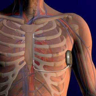
The rates of major complications related to device implantation were 4.4% at 30 days and 7.9% at 180 days.
"This is not a niche device. We actually implanted it in anybody who had an indication for ICD implantation that met the guidelines indications overall," he said.
Unlike ICDs with transvenous leads, however, the subcutaneous device cannot provide pacing, Dr. Burke noted.
Dr. Hugh Calkins, who was not involved in the study, said in an interview that "the data look really terrific. What’s striking is that when this company started, [ICD] lead failures and lead problems weren’t paid much attention, and now this device is coming along just at a time when everyone is being reminded the implanted lead is the weak link of an implantable device of any type."
The device, if approved in the United States, would most benefit younger patients who do not need antitachycardial pacing, such as those with long QT or Brugada syndromes, or with hypertrophic cardiomyopathy, said Dr. Calkins, professor of cardiology at the Johns Hopkins Heart and Vascular Institute in Baltimore and vice president of the Heart Rhythm Society.
The device involves a pulse generator implanted into the left lateral, middle axillary line with a lead tunneled across to a subxiphoid incision, then carried along the left parasternum to the sternal angle (angle of Louis) at the second intercostal space. The lead has two electrodes: one that sits at the angle, and the other down by the xiphoid process, with a coil between the electrodes where the shock vector is continued across to the casing of the generator, or vice versa.
The investigators enrolled 330 patients with any standard indication for an ICD who did not require pacing. The patients came from 33 sites in the United States, New Zealand, the Netherlands, and the United Kingdom.
Common comorbidities in the group included heart failure in 61%, hypertension in 58%, myocardial infarction in 41%, and diabetes in 28%. Nearly one-third of patients (29%) had undergone percutaneous revascularization, 15% had a coronary artery bypass graft, and 13% had previously had a transvenous ICD.
Among the 321 patients in whom the implantation was attempted, the device was indicated for primary prevention in 79% and secondary prevention in 21%, similar to the proportion in the National Cardiovascular Data Registry, Dr. Burke said.
In all but 5% of the implantations, the device was inserted using only anatomical landmarks, with no medical imaging required.
Of the 321 patients, acute induction testing was not performed in 1, and was not evaluable in 16 patients, because they did not complete four required shock episodes, leaving 304 patients for the effectiveness analysis.
The device successfully converted in 100% of the 304 patients. In a sensitivity analysis including 11 additional patients with incomplete testing and one or more failed shocks, the conversion rate was 96.5%.
In a "worst-case" sensitivity analysis, including all nonevaluable patients, the successful conversion rate was 94.7%, above the prespecified lower boundary of a two-sided 95% confidence interval of more than 88%, Dr. Burke said.
There were 109 spontaneous episodes in 16 patients, with 1 patient experiencing a ventricular fibrillation storm of 81 episodes. All the episodes converted either spontaneously after the first shock, or with 80 J of energy.
The device algorithm prevents delivery of therapy for ventricular tachycardia/ventricular fibrillation rhythms that are likely to spontaneously terminate. Therapy was avoided in 63% if patients with VT/VF met criteria to charge the device without any reports of syncope, Dr. Burke said.
The safety analysis showed that the rate of freedom from type I complications (device-related complications requiring invasive interaction) was 99% at 180 days, above the performance goal of 79%. The rate of freedom from all device-, labeling-, and procedure-related complications at 180 days was 92%.
There were 18 suspected or confirmed infections, 14 of which were superficial or incisional infections successfully managed with antibiotics in 13 and sternal wound revision in 1, and 4 cases in which explantation of the device was required.
Inappropriate shocks occurred in 38 patients, 15 with supraventricular tachycardia in the shock-only zone, and 24 patients from oversensing (1 patient had multiple events). No patient had a shock caused by a discrimination error in the conditional shock zone.
Inappropriate shocks were reduced with dual-zone programming, Dr. Burke noted.
The device is currently approved for marketing in Europe (CE Marking) but has not received the Food and Drug Administration’s approval.
The study was funded by Cameron Health, maker of the device. Dr. Burke and his colleagues have received consulting fees, honoraria, and/or research grants from the company. Dr. Calkins reported having no relevant financial disclosures.
BOSTON – A subcutaneous implantable cardioverter defibrillator that does not require a transvenous lead appears to be safe and effective for the treatment of ventricular tachyarrhythmias, according to a prospective, nonrandomized multicenter study.
In the trial, the device (S-ICD system, Cameron Health) met its primary effectiveness end point of successful ventricular fibrillation – defined as two consecutive successful conversions out of a possible four attempts in the same shock polarity – in all of 304 evaluable patients, Dr. Martin C. Burke, director of the heart rhythm center at the University of Chicago, reported at the annual meeting of the Heart Rhythm Society.
The rates of major complications related to device implantation were 4.4% at 30 days and 7.9% at 180 days.
"This is not a niche device. We actually implanted it in anybody who had an indication for ICD implantation that met the guidelines indications overall," he said.
Unlike ICDs with transvenous leads, however, the subcutaneous device cannot provide pacing, Dr. Burke noted.
Dr. Hugh Calkins, who was not involved in the study, said in an interview that "the data look really terrific. What’s striking is that when this company started, [ICD] lead failures and lead problems weren’t paid much attention, and now this device is coming along just at a time when everyone is being reminded the implanted lead is the weak link of an implantable device of any type."
The device, if approved in the United States, would most benefit younger patients who do not need antitachycardial pacing, such as those with long QT or Brugada syndromes, or with hypertrophic cardiomyopathy, said Dr. Calkins, professor of cardiology at the Johns Hopkins Heart and Vascular Institute in Baltimore and vice president of the Heart Rhythm Society.
The device involves a pulse generator implanted into the left lateral, middle axillary line with a lead tunneled across to a subxiphoid incision, then carried along the left parasternum to the sternal angle (angle of Louis) at the second intercostal space. The lead has two electrodes: one that sits at the angle, and the other down by the xiphoid process, with a coil between the electrodes where the shock vector is continued across to the casing of the generator, or vice versa.
The investigators enrolled 330 patients with any standard indication for an ICD who did not require pacing. The patients came from 33 sites in the United States, New Zealand, the Netherlands, and the United Kingdom.
Common comorbidities in the group included heart failure in 61%, hypertension in 58%, myocardial infarction in 41%, and diabetes in 28%. Nearly one-third of patients (29%) had undergone percutaneous revascularization, 15% had a coronary artery bypass graft, and 13% had previously had a transvenous ICD.
Among the 321 patients in whom the implantation was attempted, the device was indicated for primary prevention in 79% and secondary prevention in 21%, similar to the proportion in the National Cardiovascular Data Registry, Dr. Burke said.
In all but 5% of the implantations, the device was inserted using only anatomical landmarks, with no medical imaging required.
Of the 321 patients, acute induction testing was not performed in 1, and was not evaluable in 16 patients, because they did not complete four required shock episodes, leaving 304 patients for the effectiveness analysis.
The device successfully converted in 100% of the 304 patients. In a sensitivity analysis including 11 additional patients with incomplete testing and one or more failed shocks, the conversion rate was 96.5%.
In a "worst-case" sensitivity analysis, including all nonevaluable patients, the successful conversion rate was 94.7%, above the prespecified lower boundary of a two-sided 95% confidence interval of more than 88%, Dr. Burke said.
There were 109 spontaneous episodes in 16 patients, with 1 patient experiencing a ventricular fibrillation storm of 81 episodes. All the episodes converted either spontaneously after the first shock, or with 80 J of energy.
The device algorithm prevents delivery of therapy for ventricular tachycardia/ventricular fibrillation rhythms that are likely to spontaneously terminate. Therapy was avoided in 63% if patients with VT/VF met criteria to charge the device without any reports of syncope, Dr. Burke said.
The safety analysis showed that the rate of freedom from type I complications (device-related complications requiring invasive interaction) was 99% at 180 days, above the performance goal of 79%. The rate of freedom from all device-, labeling-, and procedure-related complications at 180 days was 92%.
There were 18 suspected or confirmed infections, 14 of which were superficial or incisional infections successfully managed with antibiotics in 13 and sternal wound revision in 1, and 4 cases in which explantation of the device was required.
Inappropriate shocks occurred in 38 patients, 15 with supraventricular tachycardia in the shock-only zone, and 24 patients from oversensing (1 patient had multiple events). No patient had a shock caused by a discrimination error in the conditional shock zone.
Inappropriate shocks were reduced with dual-zone programming, Dr. Burke noted.
The device is currently approved for marketing in Europe (CE Marking) but has not received the Food and Drug Administration’s approval.
The study was funded by Cameron Health, maker of the device. Dr. Burke and his colleagues have received consulting fees, honoraria, and/or research grants from the company. Dr. Calkins reported having no relevant financial disclosures.
FROM THE ANNUAL MEETING OF THE HEART RHYTHM SOCIETY
Major Finding: A subcutaneous ICD successfully converted ventricular arrhythmias in 100% of patients.
Data Source: A prospective, nonrandomized multicenter study was conducted.
Disclosures: The study was funded by Cameron Health, maker of the device. Dr. Burke and his coauthors have received consulting fees, honoraria, and/or research grants from the company. Dr. Calkins reported having no relevant financial disclosures.
Independent Study Confirms Higher Riata ICD Lead Failure Rate
BOSTON – An independent investigation by seven U.S. centers has shown that recently recalled Riata implantable cardioverter defibrillator leads are significantly more prone to failure than are leads made by a different manufacturer.
A retrospective study of all patients older than 18 years who received ICDs with either Riata or Riata ST leads (St. Jude Medical) showed a failure rate of 1.93% per patient-year for Riata leads, significantly more than the rate of 0.43% for the comparator lead, Quattro Secure (Medtronic) (P less than .0001). The failure rate for Riata ST leads was 0.50%, a nonsignificant difference, reported Dr. Raed Abdelhadi of the Minneapolis Heart Institute.

The Riata and Riata ST ICD leads are prone to inside-out and outside-in silicone insulation defects, which can cause lead malfunction and externalization (breach of the outer insulation, visible outside the lead body) of the conductor cables, but until this study, there were no independent, multicenter investigations into the rate of lead failure, failure signs, or clinical sequelae, he said.
"Without such data, it is not possible to design evidence-based management strategies or to advise Riata patients of their risks," he said at the annual meeting of the Heart Rhythm Society.
Investigators at the seven centers looked at all-cause failures and electrical malfunctions in 774 patients who received an 8-French Riata lead (mean follow-up, 4.2 years), and 307 patients who received a 7-French Riata ST lead (mean follow-up, 3.3 years). ICD leads with silicone-polyurethane copolymer insulation (RIATA ST Optim and Durata leads) were excluded.
For comparison, they obtained data on the Quattro Secure ICD leads from a three-center study of the longevity and risk factors for failure of Sprint Fidelis leads (Circulation 2011;123:358-63).
The investigators defined lead failure as either "abnormal impedance leading to replacement, electrical noise manifested by nonphysiologic signals, increase in pacing threshold or decline in R-wave amplitude necessitating lead placement, inability to provide effective therapy due to apparent lead abnormality, or externalized conductor, clearly demonstrated on fluoroscopy or x-ray ... even if the lead was functioning normally and electrically intact."
Neither functional abnormalities (for example, exit block and physiologic oversensing in the presence of an electrically intact lead) nor lead displacement (unless caused by a bad fixation mechanism) were considered to be failures.
Of the 1,081 Riata and Riata ST leads implanted, 712 (66%) were active and functioning normally at last follow-up. Of the 302 (28%) removals from service, 266 were owing to death, 10 to transplant, 5 to perforation, 4 to infection, and 17 to other causes.
In all, 10% of leads in the study were examined for externalized conductors, and 27 (25%) were found to have them. Of this group, seven (26%) leads were malfunctioning.
There were 65 Riata failures and 5 Riata ST failures, for a 6.2% incidence of all-cause failures. Of this group, 47 failures were due to electrical malfunctions, including seven from the externalized conductors contributing to the malfunction, as noted before. An additional 20 leads had externalized conductors with normal function, but met the study definition of failure.
In contrast, there were 23 lead failures among 1,668 patients with Quattro Secure leads (1.37%).
In univariate analysis, neither age, gender, primary or secondary indication for ICD implantation, ejection fraction, type of cardiac disease, nor lead placement location were significantly associated with risk for lead failure. The same was true when the Riata ST leads were subtracted from the equation.
Signs of failure included noise resulting in inappropriate therapy, elevated thresholds requiring lead replacement resulting in loss of capture and syncope, abnormal pacing impedance, abnormal high-voltage impedance, decline in R value requiring lead replacement, and failed defibrillation threshold testing with abnormal high-voltage impedance, requiring lead replacement.
Dr. Abdelhadi noted that the study was limited by its retrospective design, the fact that only 10% of leads were examined for externalized conductors, and the relatively short follow-up in the Riata ST group compared with the other two groups. In addition, most lead failures were not confirmed by returned-product analysis.

The study confirms what was already known and what many meeting attendees expected to hear, commented Dr. Hugh Calkins, professor of cardiology at the Johns Hopkins Heart and Vascular Institute, Baltimore, and vice president of the Heart Rhythm Society, in an interview.
"The bottom line is that it’s all about the patients, and how we take care of them, advise them, and whether we explant the leads. The whole international heart rhythm community is focused on this question and what to do so that there aren’t deaths that result from noise, that there aren’t deaths from unnecessary lead extractions," he said. Dr. Calkins was not involved in the study, but he moderated a media briefing where Dr. Abdelhadi presented the data.
Dr. Abdelhadi and Dr. Calkins reported having no current conflicts of interest to disclose.
BOSTON – An independent investigation by seven U.S. centers has shown that recently recalled Riata implantable cardioverter defibrillator leads are significantly more prone to failure than are leads made by a different manufacturer.
A retrospective study of all patients older than 18 years who received ICDs with either Riata or Riata ST leads (St. Jude Medical) showed a failure rate of 1.93% per patient-year for Riata leads, significantly more than the rate of 0.43% for the comparator lead, Quattro Secure (Medtronic) (P less than .0001). The failure rate for Riata ST leads was 0.50%, a nonsignificant difference, reported Dr. Raed Abdelhadi of the Minneapolis Heart Institute.
The Riata and Riata ST ICD leads are prone to inside-out and outside-in silicone insulation defects, which can cause lead malfunction and externalization (breach of the outer insulation, visible outside the lead body) of the conductor cables, but until this study, there were no independent, multicenter investigations into the rate of lead failure, failure signs, or clinical sequelae, he said.
"Without such data, it is not possible to design evidence-based management strategies or to advise Riata patients of their risks," he said at the annual meeting of the Heart Rhythm Society.
Investigators at the seven centers looked at all-cause failures and electrical malfunctions in 774 patients who received an 8-French Riata lead (mean follow-up, 4.2 years), and 307 patients who received a 7-French Riata ST lead (mean follow-up, 3.3 years). ICD leads with silicone-polyurethane copolymer insulation (RIATA ST Optim and Durata leads) were excluded.
For comparison, they obtained data on the Quattro Secure ICD leads from a three-center study of the longevity and risk factors for failure of Sprint Fidelis leads (Circulation 2011;123:358-63).
The investigators defined lead failure as either "abnormal impedance leading to replacement, electrical noise manifested by nonphysiologic signals, increase in pacing threshold or decline in R-wave amplitude necessitating lead placement, inability to provide effective therapy due to apparent lead abnormality, or externalized conductor, clearly demonstrated on fluoroscopy or x-ray ... even if the lead was functioning normally and electrically intact."
Neither functional abnormalities (for example, exit block and physiologic oversensing in the presence of an electrically intact lead) nor lead displacement (unless caused by a bad fixation mechanism) were considered to be failures.
Of the 1,081 Riata and Riata ST leads implanted, 712 (66%) were active and functioning normally at last follow-up. Of the 302 (28%) removals from service, 266 were owing to death, 10 to transplant, 5 to perforation, 4 to infection, and 17 to other causes.
In all, 10% of leads in the study were examined for externalized conductors, and 27 (25%) were found to have them. Of this group, seven (26%) leads were malfunctioning.
There were 65 Riata failures and 5 Riata ST failures, for a 6.2% incidence of all-cause failures. Of this group, 47 failures were due to electrical malfunctions, including seven from the externalized conductors contributing to the malfunction, as noted before. An additional 20 leads had externalized conductors with normal function, but met the study definition of failure.
In contrast, there were 23 lead failures among 1,668 patients with Quattro Secure leads (1.37%).
In univariate analysis, neither age, gender, primary or secondary indication for ICD implantation, ejection fraction, type of cardiac disease, nor lead placement location were significantly associated with risk for lead failure. The same was true when the Riata ST leads were subtracted from the equation.
Signs of failure included noise resulting in inappropriate therapy, elevated thresholds requiring lead replacement resulting in loss of capture and syncope, abnormal pacing impedance, abnormal high-voltage impedance, decline in R value requiring lead replacement, and failed defibrillation threshold testing with abnormal high-voltage impedance, requiring lead replacement.
Dr. Abdelhadi noted that the study was limited by its retrospective design, the fact that only 10% of leads were examined for externalized conductors, and the relatively short follow-up in the Riata ST group compared with the other two groups. In addition, most lead failures were not confirmed by returned-product analysis.
The study confirms what was already known and what many meeting attendees expected to hear, commented Dr. Hugh Calkins, professor of cardiology at the Johns Hopkins Heart and Vascular Institute, Baltimore, and vice president of the Heart Rhythm Society, in an interview.
"The bottom line is that it’s all about the patients, and how we take care of them, advise them, and whether we explant the leads. The whole international heart rhythm community is focused on this question and what to do so that there aren’t deaths that result from noise, that there aren’t deaths from unnecessary lead extractions," he said. Dr. Calkins was not involved in the study, but he moderated a media briefing where Dr. Abdelhadi presented the data.
Dr. Abdelhadi and Dr. Calkins reported having no current conflicts of interest to disclose.
FROM THE ANNUAL MEETING OF THE HEART RHYTHM SOCIETY
Following Concussions, Give Kids' Brains a Rest
BOSTON – Children who present to a primary care practice following a head or facial injury must be carefully evaluated for signs and symptoms of concussion, and those with concussion should be advised to give their brains a break, recommended a pediatric injury specialist at the annual meeting of the Pediatric Academic Societies.
Cognitive rest involves limiting "cognitive activity to a level that does not elicit symptoms." For children, this means forgoing computers, video games, texting/social media, reading for school and/or homework, and avoiding noisy or busy environments.

"The primary focus of pediatric concussion management should be return to cognitive activities and then return to the playing field," Dr. Kristy B. Arbogast said.
A review of records from a large pediatric primary care network showed that 25% of patients with concussion symptoms did not have concussion mentioned in the medical assessment or diagnosis, and nearly half of children in a random sample presented for reevaluation of concussion or persistent concussion symptoms, said Dr. Arbogast, an emergency physician and director of the Pediatric Injury Prevention Program at Children’s Hospital of Philadelphia.
"So-called ‘mild’ traumatic brain injuries are often far from mild. They lead to poor neurological outcomes that can adversely affect a child’s quality of life," she said.
Children with concussion make an estimated 144,000 emergency department visits annually in the United States. As children’s participation in year-round sports has increased, their risk of mild traumatic brain injury (TBI) has also increased, Dr. Arbogast noted.
Concussions among professional athletes receive considerable attention in the popular press, where the stories center on physical recovery and returning to the game. But for school kids, who are at significant risk for learning disabilities, memory problems, and emotional or behavioral changes after a TBI, the focus should be on physical and cognitive rest, she said.
Dr. Arbogast and her colleagues reviewed the concussion management and return-to-school practices of pediatric primary care providers by reviewing records from their hospital’s primary care network.
They looked for data on children aged 5-18 years with any presentation of mild TBI based on ICD-9 codes for skull fracture, concussion, intracranial injury, head injury (unspecified), or facial fracture. The children received initial or continuing care during the study period of July 2010 through June 2011.
The researchers included children who presented with one or more injury characteristics such as amnesia, loss of consciousness, cognitive symptoms (decline in school performance, report of being "in a fog"), emotional symptoms, and physical symptoms or exam findings including eye-tracking problems, headache, nausea/vomiting, poor balance, sleep disturbances, slow reaction times, or visual disturbance. Children with moderate or severe head trauma were excluded from the study.
The investigators randomly selected a sample of 193 patients who met all of the criteria. In all, 51% had a sports-related injury, 8% had previously had a concussion, and 6% had sustained a loss of consciousness.
There was no mention of concussion in the medical assessment or diagnosis of 25% of the sample. Of 94 children who presented for reevaluation of concussion, 33% reported a decline in school performance, 13% said they felt like they were "in a fog," 12% reported vision problems, 12% reported fatigue, and 11% had concentration problems (some reported more than one symptom).
When the authors looked at the written instructions primary care providers gave to the patients, they found that while 54% received return-to-play instructions, only 34% received return-to-school instructions, suggesting that in many cases the practitioners may have failed to recognize the importance of cognitive rest, Dr. Arbogast said. The remaining 12% of patients did not have written instructions documented.
She recommended that clinicians caring for children with suspected concussion ask about all symptoms individually in a systematic fashion, and prescribe a return-to-school protocol with a stepwise approach. The protocol includes a return to the previous step if an action elicits the return of symptoms, sending a standardized letter to notify the child’s school of the plan, and training the child’s parent or guardian to oversee the child’s progression from one step to the next.
The study was funded by the Children’s Hospital of Philadelphia and the University of Pennsylvania, also in Philadelphia. Dr. Arbogast and coinvestigators reported having no conflicts of interest to disclose.
BOSTON – Children who present to a primary care practice following a head or facial injury must be carefully evaluated for signs and symptoms of concussion, and those with concussion should be advised to give their brains a break, recommended a pediatric injury specialist at the annual meeting of the Pediatric Academic Societies.
Cognitive rest involves limiting "cognitive activity to a level that does not elicit symptoms." For children, this means forgoing computers, video games, texting/social media, reading for school and/or homework, and avoiding noisy or busy environments.
"The primary focus of pediatric concussion management should be return to cognitive activities and then return to the playing field," Dr. Kristy B. Arbogast said.
A review of records from a large pediatric primary care network showed that 25% of patients with concussion symptoms did not have concussion mentioned in the medical assessment or diagnosis, and nearly half of children in a random sample presented for reevaluation of concussion or persistent concussion symptoms, said Dr. Arbogast, an emergency physician and director of the Pediatric Injury Prevention Program at Children’s Hospital of Philadelphia.
"So-called ‘mild’ traumatic brain injuries are often far from mild. They lead to poor neurological outcomes that can adversely affect a child’s quality of life," she said.
Children with concussion make an estimated 144,000 emergency department visits annually in the United States. As children’s participation in year-round sports has increased, their risk of mild traumatic brain injury (TBI) has also increased, Dr. Arbogast noted.
Concussions among professional athletes receive considerable attention in the popular press, where the stories center on physical recovery and returning to the game. But for school kids, who are at significant risk for learning disabilities, memory problems, and emotional or behavioral changes after a TBI, the focus should be on physical and cognitive rest, she said.
Dr. Arbogast and her colleagues reviewed the concussion management and return-to-school practices of pediatric primary care providers by reviewing records from their hospital’s primary care network.
They looked for data on children aged 5-18 years with any presentation of mild TBI based on ICD-9 codes for skull fracture, concussion, intracranial injury, head injury (unspecified), or facial fracture. The children received initial or continuing care during the study period of July 2010 through June 2011.
The researchers included children who presented with one or more injury characteristics such as amnesia, loss of consciousness, cognitive symptoms (decline in school performance, report of being "in a fog"), emotional symptoms, and physical symptoms or exam findings including eye-tracking problems, headache, nausea/vomiting, poor balance, sleep disturbances, slow reaction times, or visual disturbance. Children with moderate or severe head trauma were excluded from the study.
The investigators randomly selected a sample of 193 patients who met all of the criteria. In all, 51% had a sports-related injury, 8% had previously had a concussion, and 6% had sustained a loss of consciousness.
There was no mention of concussion in the medical assessment or diagnosis of 25% of the sample. Of 94 children who presented for reevaluation of concussion, 33% reported a decline in school performance, 13% said they felt like they were "in a fog," 12% reported vision problems, 12% reported fatigue, and 11% had concentration problems (some reported more than one symptom).
When the authors looked at the written instructions primary care providers gave to the patients, they found that while 54% received return-to-play instructions, only 34% received return-to-school instructions, suggesting that in many cases the practitioners may have failed to recognize the importance of cognitive rest, Dr. Arbogast said. The remaining 12% of patients did not have written instructions documented.
She recommended that clinicians caring for children with suspected concussion ask about all symptoms individually in a systematic fashion, and prescribe a return-to-school protocol with a stepwise approach. The protocol includes a return to the previous step if an action elicits the return of symptoms, sending a standardized letter to notify the child’s school of the plan, and training the child’s parent or guardian to oversee the child’s progression from one step to the next.
The study was funded by the Children’s Hospital of Philadelphia and the University of Pennsylvania, also in Philadelphia. Dr. Arbogast and coinvestigators reported having no conflicts of interest to disclose.
FROM THE ANNUAL MEETING OF THE PEDIATRIC ACADEMIC SOCIETIES
Major Finding: About 25% of children who presented to a primary care practice with concussion symptoms did not have concussion mentioned in the medical assessment or diagnosis.
Data Source: The data were taken from a random sample of records from a pediatric primary care provider network.
Disclosures: The study was funded by the Children’s Hospital of Philadelphia and the University of Pennsylvania. Dr. Arbogast and coinvestigators reported having no conflicts of interest to disclose.
Athletes With ICDs Have a Sporting Chance for Safe Play
BOSTON – Athletes with implantable cardioverter defibrillators may not need to be sidelined when the action gets fast and furious, say investigators in a multicenter study presented at the annual meeting of the Heart Rhythm Society.
Although international consensus statements caution against anything more vigorous than golf, bowling, or billiards for people with ICDs, an analysis of data from a prospective, multinational registry shows that although 10%* of athletes with ICDs received a shock from their devices during competition or practice, there were no serious adverse events, reported Dr. Rachel Lampert of the division of cardiology at Yale University, New Haven, Conn.

"Shocks were not rare during sports. However, no serious health consequences occurred. Most patients returned to sports having received shocks while playing, implying that the negative impact on quality of life of the shocks was offset for them on the quality of life from sports participation," Dr. Lampert said.
The findings suggest that the decision to return to a sport or quit it after an ICD implantation should be individualized, she added.
Dr. Hugh Calkins, professor of cardiology at the Johns Hopkins Heart and Vascular Institute, Baltimore, and vice president of the Heart Rhythm Society, said in an interview that he is comfortable with allowing many but not all patients with ICDs to return to an active sport.
"The one exception is arrhythmogenic right ventricular dysplasia, where exercise actually causes the disease to progress. That’s very different from the long QT or the Brugada syndrome, where exercise doesn’t have any impact on the underlying disease or disease progression," said Dr. Calkins, who was not involved in the study.
Dr. Lampert and her colleagues recruited 372* athletes aged 10-60 years (median, 33 years) with ICDs who participated in either competitive or dangerous sports (defined as any sport where sudden loss of control could cause injury).
The athletes were self-enrolled and volunteered to participate after learning of the study from patient-advocacy groups, mailing lists, or word of mouth. The investigators interviewed the athletes by telephone and obtained their medical records for capturing sports-related and clinical data, then followed up with phone calls every 6 months.
In all, 67% were male and 94% were white, with a mean time since initial ICD implantation of 27 months. The mean left ventricular ejection fraction was 60%. Nearly two-thirds (62%) were taking beta-blockers. The primary cardiac diagnoses included long QT syndrome, hypertrophic cardiomyopathy, arrhythmogenic right ventricular cardiomyopathy, and coronary artery disease.
Competition was at the elite amateur level, which the authors defined as interscholastic varsity or junior varsity, or regional or national competitions.
During the median 31 months of follow-up, two patients died: a 52 year-old cyclist with coronary artery disease died at his desk at work after receiving multiple shocks; and a 34-year-old volleyball/basketball player with familial cardiomyopathy who died during a hospitalization for heart failure. Additionally, 9 patients were lost to follow-up (all were confirmed still alive), 6 withdrew, and 4 developed worsening cardiac or medical conditions precluding sports, leaving 351* patients for the analysis.
There were no cases of the primary end point: either tachyarrhythmic death or externally resuscitated tachyarrhythmia during or after sports, or of injury due to arrhythmia or shock during sports.

Overall, 77 patients received at least one shock during the study, 37 during sports. Of this group, four stopped sports completely, and seven stopped one or more sports. Five patients stopped participating in at least one sport because of shocks they received at rest or at other times when playing a sport.
Seven patients had eight ventricular arrhythmias requiring multiple shocks to (two to six) to terminate the event, occurring in patients with catecholaminergic polymorphic ventricular tachycardia, idiopathic ventricular fibrillation, or coronary artery disease.
There were 13 definite lead malfunctions, defined as noise on a lead or change in pacing parameters with a visualized lead abnormality, and 14 probable lead malfunctions (change in pacing function only).
Freedom from lead malfunction at 5 years from the time of implant was 93%, and at 10 years was 84%.
Dr. Lampert acknowledged that the study was limited by lack of a control group; patient self-selection, which may have led to underrepresentation of athletes who have received ICDS; and limited follow-up.
Nonetheless, "these data do not support blanket restriction of athletes with ICDs from participating in sports," she concluded.
The investigator-initiated study was supported by Medtronic, St. Jude Medical, and Boston Scientific. Dr. Lampert disclosed receiving honoraria from two of the companies (not specified). Dr. Calkins has previously consulted for and received research support from Medtronic.
*Correction, 5/14/12: An earlier version of this article misstated the number of patients and thus the percentage of patients receiving shocks.
BOSTON – Athletes with implantable cardioverter defibrillators may not need to be sidelined when the action gets fast and furious, say investigators in a multicenter study presented at the annual meeting of the Heart Rhythm Society.
Although international consensus statements caution against anything more vigorous than golf, bowling, or billiards for people with ICDs, an analysis of data from a prospective, multinational registry shows that although 10%* of athletes with ICDs received a shock from their devices during competition or practice, there were no serious adverse events, reported Dr. Rachel Lampert of the division of cardiology at Yale University, New Haven, Conn.
"Shocks were not rare during sports. However, no serious health consequences occurred. Most patients returned to sports having received shocks while playing, implying that the negative impact on quality of life of the shocks was offset for them on the quality of life from sports participation," Dr. Lampert said.
The findings suggest that the decision to return to a sport or quit it after an ICD implantation should be individualized, she added.
Dr. Hugh Calkins, professor of cardiology at the Johns Hopkins Heart and Vascular Institute, Baltimore, and vice president of the Heart Rhythm Society, said in an interview that he is comfortable with allowing many but not all patients with ICDs to return to an active sport.
"The one exception is arrhythmogenic right ventricular dysplasia, where exercise actually causes the disease to progress. That’s very different from the long QT or the Brugada syndrome, where exercise doesn’t have any impact on the underlying disease or disease progression," said Dr. Calkins, who was not involved in the study.
Dr. Lampert and her colleagues recruited 372* athletes aged 10-60 years (median, 33 years) with ICDs who participated in either competitive or dangerous sports (defined as any sport where sudden loss of control could cause injury).
The athletes were self-enrolled and volunteered to participate after learning of the study from patient-advocacy groups, mailing lists, or word of mouth. The investigators interviewed the athletes by telephone and obtained their medical records for capturing sports-related and clinical data, then followed up with phone calls every 6 months.
In all, 67% were male and 94% were white, with a mean time since initial ICD implantation of 27 months. The mean left ventricular ejection fraction was 60%. Nearly two-thirds (62%) were taking beta-blockers. The primary cardiac diagnoses included long QT syndrome, hypertrophic cardiomyopathy, arrhythmogenic right ventricular cardiomyopathy, and coronary artery disease.
Competition was at the elite amateur level, which the authors defined as interscholastic varsity or junior varsity, or regional or national competitions.
During the median 31 months of follow-up, two patients died: a 52 year-old cyclist with coronary artery disease died at his desk at work after receiving multiple shocks; and a 34-year-old volleyball/basketball player with familial cardiomyopathy who died during a hospitalization for heart failure. Additionally, 9 patients were lost to follow-up (all were confirmed still alive), 6 withdrew, and 4 developed worsening cardiac or medical conditions precluding sports, leaving 351* patients for the analysis.
There were no cases of the primary end point: either tachyarrhythmic death or externally resuscitated tachyarrhythmia during or after sports, or of injury due to arrhythmia or shock during sports.
Overall, 77 patients received at least one shock during the study, 37 during sports. Of this group, four stopped sports completely, and seven stopped one or more sports. Five patients stopped participating in at least one sport because of shocks they received at rest or at other times when playing a sport.
Seven patients had eight ventricular arrhythmias requiring multiple shocks to (two to six) to terminate the event, occurring in patients with catecholaminergic polymorphic ventricular tachycardia, idiopathic ventricular fibrillation, or coronary artery disease.
There were 13 definite lead malfunctions, defined as noise on a lead or change in pacing parameters with a visualized lead abnormality, and 14 probable lead malfunctions (change in pacing function only).
Freedom from lead malfunction at 5 years from the time of implant was 93%, and at 10 years was 84%.
Dr. Lampert acknowledged that the study was limited by lack of a control group; patient self-selection, which may have led to underrepresentation of athletes who have received ICDS; and limited follow-up.
Nonetheless, "these data do not support blanket restriction of athletes with ICDs from participating in sports," she concluded.
The investigator-initiated study was supported by Medtronic, St. Jude Medical, and Boston Scientific. Dr. Lampert disclosed receiving honoraria from two of the companies (not specified). Dr. Calkins has previously consulted for and received research support from Medtronic.
*Correction, 5/14/12: An earlier version of this article misstated the number of patients and thus the percentage of patients receiving shocks.
FROM THE ANNUAL MEETING OF THE HEART RHYTHM SOCIETY
Major Finding: While 9% of athletes with ICDs received a shock from their devices during competition or practice, there were no serious adverse events.
Data Source: Data were taken from a prospective registry study.
Disclosures: The investigator-initiated study was supported by Medtronic, St. Jude Medical, and Boston Scientific. Dr. Lampert disclosed receiving honoraria from two of the companies (not specified). Dr. Calkins has previously consulted for and received research support from Medtronic.