User login
Potential cause of R-CHOP failure ruled out
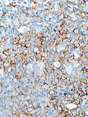
Researchers say they have come one step closer to determining the cause of R-CHOP failure in diffuse large B-cell lymphoma (DLBCL) by ruling out a potential cause.
Randy D. Gascoyne, MD, of the British Columbia Cancer Agency, and colleagues found that CD20 mutations involving the rituximab epitope are not the source of R-CHOP resistance. In fact, the mutations are rare in both de novo and relapsed DLBCL.
The rituximab epitope is located in exon 5 of the MS4A1 gene, so Dr Gascoyne and colleagues sequenced this region in DLBCL samples taken at diagnosis and relapse (1 month after completion of 6 cycles of R-CHOP). The team successfully sequenced 264 diagnostic samples and 15 relapsed samples.
The samples could be considered representative of the DLBCL population in British Columbia because clinical characteristics were similar to those observed in previous studies, according to the researchers. In addition, most of the patients had nodal disease with a minimum of 80% tumor, which was sufficient for detecting mutations.
Only 1 of 264 diagnostic samples showed a CD20 mutation involving the rituximab epitope—a 13 base pair heterozygous deletion at position IVS5(+8) in intron 5. Dr Gascoyne and colleagues were unable to determine if this was a polymorphism or a somatic mutation.
This patient achieved a complete response to R-CHOP and is still in remission more than 2 years after diagnosis. This outcome rules out the possibility of mutation-induced rituximab resistance.
As with the diagnostic samples, only 1 of the relapsed samples showed a CD20 mutation involving the rituximab epitope. This was a heterozygous 4 base pair deletion (TAAT) at nucleotide position 353-356, which predicted for a premature termination at amino acid position 121, well before the critical ANPS binding site.
The researchers were unable to establish whether this mutation was present at diagnosis, but they did determine there were no single nucleotide polymorphisms in exon 5 of the CD20 gene.
The rarity of mutations in the rituximab epitope observed in this study suggests these mutations cannot be responsible for the majority of R-CHOP treatment failures. However, Dr Gascoyne and colleagues said they cannot exclude the possibility that R-CHOP resistance might result from mutations at other sites in MS4A1, as these sites were not evaluated.
These findings appear in the March issue of haematologica. 
Researchers say they have come one step closer to determining the cause of R-CHOP failure in diffuse large B-cell lymphoma (DLBCL) by ruling out a potential cause.
Randy D. Gascoyne, MD, of the British Columbia Cancer Agency, and colleagues found that CD20 mutations involving the rituximab epitope are not the source of R-CHOP resistance. In fact, the mutations are rare in both de novo and relapsed DLBCL.
The rituximab epitope is located in exon 5 of the MS4A1 gene, so Dr Gascoyne and colleagues sequenced this region in DLBCL samples taken at diagnosis and relapse (1 month after completion of 6 cycles of R-CHOP). The team successfully sequenced 264 diagnostic samples and 15 relapsed samples.
The samples could be considered representative of the DLBCL population in British Columbia because clinical characteristics were similar to those observed in previous studies, according to the researchers. In addition, most of the patients had nodal disease with a minimum of 80% tumor, which was sufficient for detecting mutations.
Only 1 of 264 diagnostic samples showed a CD20 mutation involving the rituximab epitope—a 13 base pair heterozygous deletion at position IVS5(+8) in intron 5. Dr Gascoyne and colleagues were unable to determine if this was a polymorphism or a somatic mutation.
This patient achieved a complete response to R-CHOP and is still in remission more than 2 years after diagnosis. This outcome rules out the possibility of mutation-induced rituximab resistance.
As with the diagnostic samples, only 1 of the relapsed samples showed a CD20 mutation involving the rituximab epitope. This was a heterozygous 4 base pair deletion (TAAT) at nucleotide position 353-356, which predicted for a premature termination at amino acid position 121, well before the critical ANPS binding site.
The researchers were unable to establish whether this mutation was present at diagnosis, but they did determine there were no single nucleotide polymorphisms in exon 5 of the CD20 gene.
The rarity of mutations in the rituximab epitope observed in this study suggests these mutations cannot be responsible for the majority of R-CHOP treatment failures. However, Dr Gascoyne and colleagues said they cannot exclude the possibility that R-CHOP resistance might result from mutations at other sites in MS4A1, as these sites were not evaluated.
These findings appear in the March issue of haematologica.
Researchers say they have come one step closer to determining the cause of R-CHOP failure in diffuse large B-cell lymphoma (DLBCL) by ruling out a potential cause.
Randy D. Gascoyne, MD, of the British Columbia Cancer Agency, and colleagues found that CD20 mutations involving the rituximab epitope are not the source of R-CHOP resistance. In fact, the mutations are rare in both de novo and relapsed DLBCL.
The rituximab epitope is located in exon 5 of the MS4A1 gene, so Dr Gascoyne and colleagues sequenced this region in DLBCL samples taken at diagnosis and relapse (1 month after completion of 6 cycles of R-CHOP). The team successfully sequenced 264 diagnostic samples and 15 relapsed samples.
The samples could be considered representative of the DLBCL population in British Columbia because clinical characteristics were similar to those observed in previous studies, according to the researchers. In addition, most of the patients had nodal disease with a minimum of 80% tumor, which was sufficient for detecting mutations.
Only 1 of 264 diagnostic samples showed a CD20 mutation involving the rituximab epitope—a 13 base pair heterozygous deletion at position IVS5(+8) in intron 5. Dr Gascoyne and colleagues were unable to determine if this was a polymorphism or a somatic mutation.
This patient achieved a complete response to R-CHOP and is still in remission more than 2 years after diagnosis. This outcome rules out the possibility of mutation-induced rituximab resistance.
As with the diagnostic samples, only 1 of the relapsed samples showed a CD20 mutation involving the rituximab epitope. This was a heterozygous 4 base pair deletion (TAAT) at nucleotide position 353-356, which predicted for a premature termination at amino acid position 121, well before the critical ANPS binding site.
The researchers were unable to establish whether this mutation was present at diagnosis, but they did determine there were no single nucleotide polymorphisms in exon 5 of the CD20 gene.
The rarity of mutations in the rituximab epitope observed in this study suggests these mutations cannot be responsible for the majority of R-CHOP treatment failures. However, Dr Gascoyne and colleagues said they cannot exclude the possibility that R-CHOP resistance might result from mutations at other sites in MS4A1, as these sites were not evaluated.
These findings appear in the March issue of haematologica.
Study shows imatinib response is durable and improves with time

SAN FRANCISCO—The longest duration study of imatinib treatment for patients with Ph+ CML shows 86% of patients are still alive 7 years after beginning therapy.
The International Randomized Interferon versus STI571 (IRIS) study revealed only 1 early chronic-phase patient progressed to a more advanced phase between years 6 and 7, Stephen O’Brien, MD, PhD, of Newcastle University in the UK, said at the 50th Annual Meeting of the American Society of Hematology.
IRIS is an open-label, phase 3 clinical trial enrolling 1106 newly diagnosed patients with chronic phase Ph+ CML in 177 centers across 16 countries. One group of 553 patients received imatinib 400 mg per day. Another group of 553 patients received a target dose of interferon (IFN) of 5 MIU/m2/day in combination with cytarabine at 20 mg/m2/day for 10 days each month.
Because of tolerability issues, lack of response, or loss of response, 65% of patients in the IFN/cytarabine arm crossed over to the imatinib arm. Only 3% of patients in the imatinib arm crossed over to the IFN/cytarabine arm.
A low rate of progression has been reported every year since this trial began in 2001. Seven percent of patients treated with imatinib progressed to advanced phases of CML after 7 years. Of the 456 patients (82%) who achieved a complete cytogenetic response, 17% lost their response and 3% progressed to advanced phases.
“After 1 year of treatment, there is a small risk of progression,” Dr O’Brien said. “If patients achieve and maintain a complete cytogenetic response after 3 years, they are fairly safe.”
Treatment with imatinib in the IRIS study was well tolerated, he said. No new serious adverse events occurred between the sixth and seventh year of treatment.
The results from the IRIS study also reveal that, by year 6, 85% to 90% of patients still taking imatinib achieved a major molecular response. This key milestone indicates a reduction in the abnormal protein responsible for the uncontrolled production of abnormal white blood cells and may be a sensitive predictor of long-term progression-free survival.
“There was a steady improvement in major molecular responses between 4 and 7 years of treatment,” said Timothy Hughes, MD, of the Institute of Medical and Veterinary Science in Adelaide, Australia. “By 7 years, the vast majority of patients who achieved a complete cytogenetic response also achieved a major molecular response.”
A major molecular response at any time point represents a “safe haven” for patients, Dr Hughes said. Both molecular and cytogenetic evaluations should be used to guide treatment decisions until a complete cytogenetic response is achieved, followed by measurements of molecular assessments.
“In this, the seventh year of the IRIS study, CML patients treated with imatinib continue to demonstrate impressive long-term survival,” Dr O’Brien said. “Imatinib 400 mg daily is confirmed as the standard of care for the initial therapy of chronic-phase CML.”
Imatinib, the first therapy to inhibit the activity of Bcr-Abl, revolutionized the treatment of Ph+ CML, Dr O’Brien said. Prior to imatinib, about 50% of patients with Ph+ CML progressed from the initial phase to more advanced stages after 3 to 5 years. Once patients reached the final blast crisis phase, survival was generally 3 to 6 months. 
SAN FRANCISCO—The longest duration study of imatinib treatment for patients with Ph+ CML shows 86% of patients are still alive 7 years after beginning therapy.
The International Randomized Interferon versus STI571 (IRIS) study revealed only 1 early chronic-phase patient progressed to a more advanced phase between years 6 and 7, Stephen O’Brien, MD, PhD, of Newcastle University in the UK, said at the 50th Annual Meeting of the American Society of Hematology.
IRIS is an open-label, phase 3 clinical trial enrolling 1106 newly diagnosed patients with chronic phase Ph+ CML in 177 centers across 16 countries. One group of 553 patients received imatinib 400 mg per day. Another group of 553 patients received a target dose of interferon (IFN) of 5 MIU/m2/day in combination with cytarabine at 20 mg/m2/day for 10 days each month.
Because of tolerability issues, lack of response, or loss of response, 65% of patients in the IFN/cytarabine arm crossed over to the imatinib arm. Only 3% of patients in the imatinib arm crossed over to the IFN/cytarabine arm.
A low rate of progression has been reported every year since this trial began in 2001. Seven percent of patients treated with imatinib progressed to advanced phases of CML after 7 years. Of the 456 patients (82%) who achieved a complete cytogenetic response, 17% lost their response and 3% progressed to advanced phases.
“After 1 year of treatment, there is a small risk of progression,” Dr O’Brien said. “If patients achieve and maintain a complete cytogenetic response after 3 years, they are fairly safe.”
Treatment with imatinib in the IRIS study was well tolerated, he said. No new serious adverse events occurred between the sixth and seventh year of treatment.
The results from the IRIS study also reveal that, by year 6, 85% to 90% of patients still taking imatinib achieved a major molecular response. This key milestone indicates a reduction in the abnormal protein responsible for the uncontrolled production of abnormal white blood cells and may be a sensitive predictor of long-term progression-free survival.
“There was a steady improvement in major molecular responses between 4 and 7 years of treatment,” said Timothy Hughes, MD, of the Institute of Medical and Veterinary Science in Adelaide, Australia. “By 7 years, the vast majority of patients who achieved a complete cytogenetic response also achieved a major molecular response.”
A major molecular response at any time point represents a “safe haven” for patients, Dr Hughes said. Both molecular and cytogenetic evaluations should be used to guide treatment decisions until a complete cytogenetic response is achieved, followed by measurements of molecular assessments.
“In this, the seventh year of the IRIS study, CML patients treated with imatinib continue to demonstrate impressive long-term survival,” Dr O’Brien said. “Imatinib 400 mg daily is confirmed as the standard of care for the initial therapy of chronic-phase CML.”
Imatinib, the first therapy to inhibit the activity of Bcr-Abl, revolutionized the treatment of Ph+ CML, Dr O’Brien said. Prior to imatinib, about 50% of patients with Ph+ CML progressed from the initial phase to more advanced stages after 3 to 5 years. Once patients reached the final blast crisis phase, survival was generally 3 to 6 months.
SAN FRANCISCO—The longest duration study of imatinib treatment for patients with Ph+ CML shows 86% of patients are still alive 7 years after beginning therapy.
The International Randomized Interferon versus STI571 (IRIS) study revealed only 1 early chronic-phase patient progressed to a more advanced phase between years 6 and 7, Stephen O’Brien, MD, PhD, of Newcastle University in the UK, said at the 50th Annual Meeting of the American Society of Hematology.
IRIS is an open-label, phase 3 clinical trial enrolling 1106 newly diagnosed patients with chronic phase Ph+ CML in 177 centers across 16 countries. One group of 553 patients received imatinib 400 mg per day. Another group of 553 patients received a target dose of interferon (IFN) of 5 MIU/m2/day in combination with cytarabine at 20 mg/m2/day for 10 days each month.
Because of tolerability issues, lack of response, or loss of response, 65% of patients in the IFN/cytarabine arm crossed over to the imatinib arm. Only 3% of patients in the imatinib arm crossed over to the IFN/cytarabine arm.
A low rate of progression has been reported every year since this trial began in 2001. Seven percent of patients treated with imatinib progressed to advanced phases of CML after 7 years. Of the 456 patients (82%) who achieved a complete cytogenetic response, 17% lost their response and 3% progressed to advanced phases.
“After 1 year of treatment, there is a small risk of progression,” Dr O’Brien said. “If patients achieve and maintain a complete cytogenetic response after 3 years, they are fairly safe.”
Treatment with imatinib in the IRIS study was well tolerated, he said. No new serious adverse events occurred between the sixth and seventh year of treatment.
The results from the IRIS study also reveal that, by year 6, 85% to 90% of patients still taking imatinib achieved a major molecular response. This key milestone indicates a reduction in the abnormal protein responsible for the uncontrolled production of abnormal white blood cells and may be a sensitive predictor of long-term progression-free survival.
“There was a steady improvement in major molecular responses between 4 and 7 years of treatment,” said Timothy Hughes, MD, of the Institute of Medical and Veterinary Science in Adelaide, Australia. “By 7 years, the vast majority of patients who achieved a complete cytogenetic response also achieved a major molecular response.”
A major molecular response at any time point represents a “safe haven” for patients, Dr Hughes said. Both molecular and cytogenetic evaluations should be used to guide treatment decisions until a complete cytogenetic response is achieved, followed by measurements of molecular assessments.
“In this, the seventh year of the IRIS study, CML patients treated with imatinib continue to demonstrate impressive long-term survival,” Dr O’Brien said. “Imatinib 400 mg daily is confirmed as the standard of care for the initial therapy of chronic-phase CML.”
Imatinib, the first therapy to inhibit the activity of Bcr-Abl, revolutionized the treatment of Ph+ CML, Dr O’Brien said. Prior to imatinib, about 50% of patients with Ph+ CML progressed from the initial phase to more advanced stages after 3 to 5 years. Once patients reached the final blast crisis phase, survival was generally 3 to 6 months.
Dabigatran safe, effective in elderly surgery patients
San Francisco—Researchers confirmed the safety and efficacy of a lower dose of dabigatran etexilate in elderly hip and total knee replacement surgery patients.
Dabigatran is a new oral thrombin inhibitor recently approved in Europe for the prevention of VTE in patients undergoing this surgery. Ola E. Dahl, MD, of the Thrombosis Research Institute in London, reported the results of a post hoc pooled analysis of 2 pivotal trials comparing dabigatran with enoxaparin at the 50th Annual Meeting of the American Society of Hematology.
Dr Dahl and colleagues analyzed 883 patients older than 75 years who were enrolled in the RE-MODEL and RE-NOVATE trials. Researchers evaluated 220 mg and 150 mg once-daily doses of dabigatran compared to a 40 mg daily dose of enoxaparin.
The primary efficacy endpoint was total number of VTEs and all-cause mortality. Both doses of dabigatran reduced total VTEs compared to enoxaparin, though not significantly.
However, the higher dose of dabigatran produced a significant difference in the secondary endpoint, major VTEs and VTE-related mortality. Four of 216 patients (1.9%) receiving the 220 mg dose had a major VTE, compared with 13 of 218 patients receiving enoxaparin (P=0.045).
The safety endpoint was the difference in major bleeding events, including surgical site bleeding, which accounts for up to 90% of bleeding in these patients. Major bleeding events occurred in 3.7% of the patients receiving dabigatran at 220 mg and 1.4% receiving 150 mg, compared to 2.9% in the enoxaparin group. The study was not powered to show significance in the safety endpoint.
“If you look into the dabigatran regimens versus enoxaparin, you see that we have more efficacious 200 mg dosing with slightly increased bleeding,” Dr Dahl said. “The 150 mg dose has the same efficacy level, but with a little less bleeding. And that is exactly the profile we are looking for in the elderly.”
San Francisco—Researchers confirmed the safety and efficacy of a lower dose of dabigatran etexilate in elderly hip and total knee replacement surgery patients.
Dabigatran is a new oral thrombin inhibitor recently approved in Europe for the prevention of VTE in patients undergoing this surgery. Ola E. Dahl, MD, of the Thrombosis Research Institute in London, reported the results of a post hoc pooled analysis of 2 pivotal trials comparing dabigatran with enoxaparin at the 50th Annual Meeting of the American Society of Hematology.
Dr Dahl and colleagues analyzed 883 patients older than 75 years who were enrolled in the RE-MODEL and RE-NOVATE trials. Researchers evaluated 220 mg and 150 mg once-daily doses of dabigatran compared to a 40 mg daily dose of enoxaparin.
The primary efficacy endpoint was total number of VTEs and all-cause mortality. Both doses of dabigatran reduced total VTEs compared to enoxaparin, though not significantly.
However, the higher dose of dabigatran produced a significant difference in the secondary endpoint, major VTEs and VTE-related mortality. Four of 216 patients (1.9%) receiving the 220 mg dose had a major VTE, compared with 13 of 218 patients receiving enoxaparin (P=0.045).
The safety endpoint was the difference in major bleeding events, including surgical site bleeding, which accounts for up to 90% of bleeding in these patients. Major bleeding events occurred in 3.7% of the patients receiving dabigatran at 220 mg and 1.4% receiving 150 mg, compared to 2.9% in the enoxaparin group. The study was not powered to show significance in the safety endpoint.
“If you look into the dabigatran regimens versus enoxaparin, you see that we have more efficacious 200 mg dosing with slightly increased bleeding,” Dr Dahl said. “The 150 mg dose has the same efficacy level, but with a little less bleeding. And that is exactly the profile we are looking for in the elderly.”
San Francisco—Researchers confirmed the safety and efficacy of a lower dose of dabigatran etexilate in elderly hip and total knee replacement surgery patients.
Dabigatran is a new oral thrombin inhibitor recently approved in Europe for the prevention of VTE in patients undergoing this surgery. Ola E. Dahl, MD, of the Thrombosis Research Institute in London, reported the results of a post hoc pooled analysis of 2 pivotal trials comparing dabigatran with enoxaparin at the 50th Annual Meeting of the American Society of Hematology.
Dr Dahl and colleagues analyzed 883 patients older than 75 years who were enrolled in the RE-MODEL and RE-NOVATE trials. Researchers evaluated 220 mg and 150 mg once-daily doses of dabigatran compared to a 40 mg daily dose of enoxaparin.
The primary efficacy endpoint was total number of VTEs and all-cause mortality. Both doses of dabigatran reduced total VTEs compared to enoxaparin, though not significantly.
However, the higher dose of dabigatran produced a significant difference in the secondary endpoint, major VTEs and VTE-related mortality. Four of 216 patients (1.9%) receiving the 220 mg dose had a major VTE, compared with 13 of 218 patients receiving enoxaparin (P=0.045).
The safety endpoint was the difference in major bleeding events, including surgical site bleeding, which accounts for up to 90% of bleeding in these patients. Major bleeding events occurred in 3.7% of the patients receiving dabigatran at 220 mg and 1.4% receiving 150 mg, compared to 2.9% in the enoxaparin group. The study was not powered to show significance in the safety endpoint.
“If you look into the dabigatran regimens versus enoxaparin, you see that we have more efficacious 200 mg dosing with slightly increased bleeding,” Dr Dahl said. “The 150 mg dose has the same efficacy level, but with a little less bleeding. And that is exactly the profile we are looking for in the elderly.”
Novel agent more effective than standard therapy in NHL
Preliminary results of a phase 3 study indicate that pixantrone is more effective than standard chemotherapy in patients with advanced, relapsed, aggressive non-Hodgkin lymphoma (NHL).
The phase 3 EXTEND PIX301 trial enrolled 140 NHL patients from 130 sites in 17 countries. Patients had received 2 or more prior therapies and were sensitive to anthracycline treatment.
They were randomized to receive either pixantrone or another single-agent drug currently used in this patient population and selected by a physician. The trial assessed patients’ complete remission or unconfirmed complete remission rate, overall survival, and progression-free survival.
Twenty percent of patients who received pixantrone achieved either a confirmed or unconfirmed complete remission, compared to 5.7% of patients on standard chemotherapy. Eleven percent of pixantrone patients’ remissions were confirmed, whereas none of the standard chemotherapy remissions were.
The overall response rate was 37.1% with pixantrone and 14.3% for patients on standard chemotherapy. Response rates were determined by an independent assessment panel that was blinded to treatment assignments.
Complete safety information for this study is not yet available. However, the study was monitored on an ongoing basis by an independent Data Safety Monitoring Committee, and no serious concerns were raised. The most common serious toxicities (> 5%) observed in previous trials of pixantrone include grade 3 and 4 neutropenia and febrile neutropenia.
Seventy-four percent of patients enrolled in this study discontinued therapy due to disease progression or death, the majority of which were in the standard chemotherapy control arm.
This study was funded by Cell Therapeutics, Inc., the company developing pixantrone.
Cell Therapeutics says it plans to submit complete study data for presentation at a major scientific conference. The organization also plans to request a pre-New Drug Application meeting with the FDA and expects to begin submission of a rolling New Drug Application to the FDA in early 2009.
Pixantrone is an antitumor agent that contains an aza-anthracenedione molecular structure, which differentiates it from anthracycline chemotherapy agents.
Preliminary results of a phase 3 study indicate that pixantrone is more effective than standard chemotherapy in patients with advanced, relapsed, aggressive non-Hodgkin lymphoma (NHL).
The phase 3 EXTEND PIX301 trial enrolled 140 NHL patients from 130 sites in 17 countries. Patients had received 2 or more prior therapies and were sensitive to anthracycline treatment.
They were randomized to receive either pixantrone or another single-agent drug currently used in this patient population and selected by a physician. The trial assessed patients’ complete remission or unconfirmed complete remission rate, overall survival, and progression-free survival.
Twenty percent of patients who received pixantrone achieved either a confirmed or unconfirmed complete remission, compared to 5.7% of patients on standard chemotherapy. Eleven percent of pixantrone patients’ remissions were confirmed, whereas none of the standard chemotherapy remissions were.
The overall response rate was 37.1% with pixantrone and 14.3% for patients on standard chemotherapy. Response rates were determined by an independent assessment panel that was blinded to treatment assignments.
Complete safety information for this study is not yet available. However, the study was monitored on an ongoing basis by an independent Data Safety Monitoring Committee, and no serious concerns were raised. The most common serious toxicities (> 5%) observed in previous trials of pixantrone include grade 3 and 4 neutropenia and febrile neutropenia.
Seventy-four percent of patients enrolled in this study discontinued therapy due to disease progression or death, the majority of which were in the standard chemotherapy control arm.
This study was funded by Cell Therapeutics, Inc., the company developing pixantrone.
Cell Therapeutics says it plans to submit complete study data for presentation at a major scientific conference. The organization also plans to request a pre-New Drug Application meeting with the FDA and expects to begin submission of a rolling New Drug Application to the FDA in early 2009.
Pixantrone is an antitumor agent that contains an aza-anthracenedione molecular structure, which differentiates it from anthracycline chemotherapy agents.
Preliminary results of a phase 3 study indicate that pixantrone is more effective than standard chemotherapy in patients with advanced, relapsed, aggressive non-Hodgkin lymphoma (NHL).
The phase 3 EXTEND PIX301 trial enrolled 140 NHL patients from 130 sites in 17 countries. Patients had received 2 or more prior therapies and were sensitive to anthracycline treatment.
They were randomized to receive either pixantrone or another single-agent drug currently used in this patient population and selected by a physician. The trial assessed patients’ complete remission or unconfirmed complete remission rate, overall survival, and progression-free survival.
Twenty percent of patients who received pixantrone achieved either a confirmed or unconfirmed complete remission, compared to 5.7% of patients on standard chemotherapy. Eleven percent of pixantrone patients’ remissions were confirmed, whereas none of the standard chemotherapy remissions were.
The overall response rate was 37.1% with pixantrone and 14.3% for patients on standard chemotherapy. Response rates were determined by an independent assessment panel that was blinded to treatment assignments.
Complete safety information for this study is not yet available. However, the study was monitored on an ongoing basis by an independent Data Safety Monitoring Committee, and no serious concerns were raised. The most common serious toxicities (> 5%) observed in previous trials of pixantrone include grade 3 and 4 neutropenia and febrile neutropenia.
Seventy-four percent of patients enrolled in this study discontinued therapy due to disease progression or death, the majority of which were in the standard chemotherapy control arm.
This study was funded by Cell Therapeutics, Inc., the company developing pixantrone.
Cell Therapeutics says it plans to submit complete study data for presentation at a major scientific conference. The organization also plans to request a pre-New Drug Application meeting with the FDA and expects to begin submission of a rolling New Drug Application to the FDA in early 2009.
Pixantrone is an antitumor agent that contains an aza-anthracenedione molecular structure, which differentiates it from anthracycline chemotherapy agents.
Newly identified NHP2 biallelic mutation can cause dyskeratosis congenita

Copenhagen— Researchers have identified a new biallelic mutation in the telomerase component NHP2, which can cause the premature aging syndrome dyskeratosis congenita.
Thomas J. Vulliamy, PhD, MRCPath, from Barts and the London School of Medicine and Dentistry, presented this finding at the 13th Congress of the European Hematology Association.
Dyskeratosis congenita is a genetically heterogeneous, multisystem disorder. The unifying feature of dyskeratosis congenita is that all mutations identified thus far affect molecules involved in telomere maintenance. Defective telomerase leads to premature aging, bone marrow failure, and a predisposition to 32 cancers.
Investigators analyzed the proteins NHP2 and GAR1 using high performance liquid chromatography screening followed by direct DNA sequencing. NHP2 and GAR1 are key components of telomerase, along with dyskerin and NOP10, and small nucleolar ribonucleoprotein (snoRNP) complexes.
Investigators measured telomere lengths by Southern blot analysis and assessed TERC levels and the effects of siRNA knockdown of gene transcripts using quantitative real-time PCR.
Dr Vulliamy and colleagues found that mutations in NHP2 can cause autosomal recessive dyskeratosis congenita. Patients with NHP2, dyskerin, and NOP10 mutations have short telomeres and low TERC levels.
Investigators concluded that this provided direct evidence of their role in telomere maintenance in humans. They did not find any GAR1 mutations, suggesting that GAR1 has a different impact on the accumulation of TERC.
This abstract was chosen as one of the 5 best of the Congress.
Copenhagen— Researchers have identified a new biallelic mutation in the telomerase component NHP2, which can cause the premature aging syndrome dyskeratosis congenita.
Thomas J. Vulliamy, PhD, MRCPath, from Barts and the London School of Medicine and Dentistry, presented this finding at the 13th Congress of the European Hematology Association.
Dyskeratosis congenita is a genetically heterogeneous, multisystem disorder. The unifying feature of dyskeratosis congenita is that all mutations identified thus far affect molecules involved in telomere maintenance. Defective telomerase leads to premature aging, bone marrow failure, and a predisposition to 32 cancers.
Investigators analyzed the proteins NHP2 and GAR1 using high performance liquid chromatography screening followed by direct DNA sequencing. NHP2 and GAR1 are key components of telomerase, along with dyskerin and NOP10, and small nucleolar ribonucleoprotein (snoRNP) complexes.
Investigators measured telomere lengths by Southern blot analysis and assessed TERC levels and the effects of siRNA knockdown of gene transcripts using quantitative real-time PCR.
Dr Vulliamy and colleagues found that mutations in NHP2 can cause autosomal recessive dyskeratosis congenita. Patients with NHP2, dyskerin, and NOP10 mutations have short telomeres and low TERC levels.
Investigators concluded that this provided direct evidence of their role in telomere maintenance in humans. They did not find any GAR1 mutations, suggesting that GAR1 has a different impact on the accumulation of TERC.
This abstract was chosen as one of the 5 best of the Congress.
Copenhagen— Researchers have identified a new biallelic mutation in the telomerase component NHP2, which can cause the premature aging syndrome dyskeratosis congenita.
Thomas J. Vulliamy, PhD, MRCPath, from Barts and the London School of Medicine and Dentistry, presented this finding at the 13th Congress of the European Hematology Association.
Dyskeratosis congenita is a genetically heterogeneous, multisystem disorder. The unifying feature of dyskeratosis congenita is that all mutations identified thus far affect molecules involved in telomere maintenance. Defective telomerase leads to premature aging, bone marrow failure, and a predisposition to 32 cancers.
Investigators analyzed the proteins NHP2 and GAR1 using high performance liquid chromatography screening followed by direct DNA sequencing. NHP2 and GAR1 are key components of telomerase, along with dyskerin and NOP10, and small nucleolar ribonucleoprotein (snoRNP) complexes.
Investigators measured telomere lengths by Southern blot analysis and assessed TERC levels and the effects of siRNA knockdown of gene transcripts using quantitative real-time PCR.
Dr Vulliamy and colleagues found that mutations in NHP2 can cause autosomal recessive dyskeratosis congenita. Patients with NHP2, dyskerin, and NOP10 mutations have short telomeres and low TERC levels.
Investigators concluded that this provided direct evidence of their role in telomere maintenance in humans. They did not find any GAR1 mutations, suggesting that GAR1 has a different impact on the accumulation of TERC.
This abstract was chosen as one of the 5 best of the Congress.
Study shows drug is safe and effective in cardiac patients
The direct thrombin inhibitor bivalirudin (Angiomax) reduces major bleeding and increases short-term survival in certain cardiac patients, according to a study published in the New England Journal of Medicine.
Bivalirudin was associated with a lower rate of major bleeding and death from cardiac and other causes at 30 days from treatment initiation. This was in comparison to treatment with heparin plus glycoprotein IIb/IIIa inhibitors.
Both therapies were tested in patients with ST-segment elevation myocardial infarction who were undergoing primary percutaneous coronary intervention (PCI).
Gregg Stone, MD, of Columbia University Medical Center, and colleagues randomized 3602 patients to receive either bivalirudin (n=1800) or heparin plus glycoprotein IIb/IIIa inhibitors (n=1802).
The 2 primary endpoints of the study were major bleeding and combined clinical events. Combined events included death, reinfarction, target-vessel revascularization for ischemia, and stroke.
Bivalirudin reduced the overall rate of combined events, as compared to heparin plus glycoprotein IIb/IIIa inhibitors (9.2% vs 12.1%, respectively). However, when evaluated separately, there were no significant differences in the incidence of these events between the 2 treatment groups.
The rate of reinfarction was the same in both groups (1.8%). The rate of stroke was insignificantly higher in the bivalirudin group (0.7% vs 0.6%). The same was true of target-vessel revascularization; the rate was 2.6% in bivalirudin patients and 1.9% in the other patient group.
Patients on bivalirudin experienced a lower rate of major bleeding than patients on heparin plus glycoprotein IIb/IIIa inhibitors (4.9% vs 8.3%, respectively). When large hematomas were excluded, the rates of major bleeding were reduced to 4.7% and 7.8%, respectively.
Bivalirudin also reduced the incidence of hemorrhagic complications, the development of thrombocytopenia, and the need for blood transfusions.
The rate of cardiac-related death at 30 days was significantly lower for bivalirudin patients than for patients who received heparin plus glycoprotein IIb/IIIa inhibitors (1.8% vs 2.9%, respectively). The rate of death from any cause was also lower in bivalirudin patients (2.1% vs 3.1%).
Bivalirudin increased the rate of acute stent thrombosis within 24 hours of treatment initiation (1.3% vs 0.3%). However, between 24 hours and 30 days, the incidence of acute stent thrombosis was lower in the bivalirudin patients than in those on heparin plus glycoprotein IIb/IIIa inhibitors (1.2% vs. 1.7%, respectively).
This study, known as HORIZONS-AMI, was sponsored, in part, by The Medicines Company, makers of bivalirudin (Angiomax).
The direct thrombin inhibitor bivalirudin (Angiomax) reduces major bleeding and increases short-term survival in certain cardiac patients, according to a study published in the New England Journal of Medicine.
Bivalirudin was associated with a lower rate of major bleeding and death from cardiac and other causes at 30 days from treatment initiation. This was in comparison to treatment with heparin plus glycoprotein IIb/IIIa inhibitors.
Both therapies were tested in patients with ST-segment elevation myocardial infarction who were undergoing primary percutaneous coronary intervention (PCI).
Gregg Stone, MD, of Columbia University Medical Center, and colleagues randomized 3602 patients to receive either bivalirudin (n=1800) or heparin plus glycoprotein IIb/IIIa inhibitors (n=1802).
The 2 primary endpoints of the study were major bleeding and combined clinical events. Combined events included death, reinfarction, target-vessel revascularization for ischemia, and stroke.
Bivalirudin reduced the overall rate of combined events, as compared to heparin plus glycoprotein IIb/IIIa inhibitors (9.2% vs 12.1%, respectively). However, when evaluated separately, there were no significant differences in the incidence of these events between the 2 treatment groups.
The rate of reinfarction was the same in both groups (1.8%). The rate of stroke was insignificantly higher in the bivalirudin group (0.7% vs 0.6%). The same was true of target-vessel revascularization; the rate was 2.6% in bivalirudin patients and 1.9% in the other patient group.
Patients on bivalirudin experienced a lower rate of major bleeding than patients on heparin plus glycoprotein IIb/IIIa inhibitors (4.9% vs 8.3%, respectively). When large hematomas were excluded, the rates of major bleeding were reduced to 4.7% and 7.8%, respectively.
Bivalirudin also reduced the incidence of hemorrhagic complications, the development of thrombocytopenia, and the need for blood transfusions.
The rate of cardiac-related death at 30 days was significantly lower for bivalirudin patients than for patients who received heparin plus glycoprotein IIb/IIIa inhibitors (1.8% vs 2.9%, respectively). The rate of death from any cause was also lower in bivalirudin patients (2.1% vs 3.1%).
Bivalirudin increased the rate of acute stent thrombosis within 24 hours of treatment initiation (1.3% vs 0.3%). However, between 24 hours and 30 days, the incidence of acute stent thrombosis was lower in the bivalirudin patients than in those on heparin plus glycoprotein IIb/IIIa inhibitors (1.2% vs. 1.7%, respectively).
This study, known as HORIZONS-AMI, was sponsored, in part, by The Medicines Company, makers of bivalirudin (Angiomax).
The direct thrombin inhibitor bivalirudin (Angiomax) reduces major bleeding and increases short-term survival in certain cardiac patients, according to a study published in the New England Journal of Medicine.
Bivalirudin was associated with a lower rate of major bleeding and death from cardiac and other causes at 30 days from treatment initiation. This was in comparison to treatment with heparin plus glycoprotein IIb/IIIa inhibitors.
Both therapies were tested in patients with ST-segment elevation myocardial infarction who were undergoing primary percutaneous coronary intervention (PCI).
Gregg Stone, MD, of Columbia University Medical Center, and colleagues randomized 3602 patients to receive either bivalirudin (n=1800) or heparin plus glycoprotein IIb/IIIa inhibitors (n=1802).
The 2 primary endpoints of the study were major bleeding and combined clinical events. Combined events included death, reinfarction, target-vessel revascularization for ischemia, and stroke.
Bivalirudin reduced the overall rate of combined events, as compared to heparin plus glycoprotein IIb/IIIa inhibitors (9.2% vs 12.1%, respectively). However, when evaluated separately, there were no significant differences in the incidence of these events between the 2 treatment groups.
The rate of reinfarction was the same in both groups (1.8%). The rate of stroke was insignificantly higher in the bivalirudin group (0.7% vs 0.6%). The same was true of target-vessel revascularization; the rate was 2.6% in bivalirudin patients and 1.9% in the other patient group.
Patients on bivalirudin experienced a lower rate of major bleeding than patients on heparin plus glycoprotein IIb/IIIa inhibitors (4.9% vs 8.3%, respectively). When large hematomas were excluded, the rates of major bleeding were reduced to 4.7% and 7.8%, respectively.
Bivalirudin also reduced the incidence of hemorrhagic complications, the development of thrombocytopenia, and the need for blood transfusions.
The rate of cardiac-related death at 30 days was significantly lower for bivalirudin patients than for patients who received heparin plus glycoprotein IIb/IIIa inhibitors (1.8% vs 2.9%, respectively). The rate of death from any cause was also lower in bivalirudin patients (2.1% vs 3.1%).
Bivalirudin increased the rate of acute stent thrombosis within 24 hours of treatment initiation (1.3% vs 0.3%). However, between 24 hours and 30 days, the incidence of acute stent thrombosis was lower in the bivalirudin patients than in those on heparin plus glycoprotein IIb/IIIa inhibitors (1.2% vs. 1.7%, respectively).
This study, known as HORIZONS-AMI, was sponsored, in part, by The Medicines Company, makers of bivalirudin (Angiomax).
Researchers elucidate mechanism of heparin contaminant
Researchers have discovered the mechanism behind the deaths and adverse events that occurred in patients receiving contaminated heparin.
In March, a team led by Ram Sasisekharan, PhD, of Massachusetts Institute of Technology in Cambridge, identified the contaminant responsible for the numerous adverse events and 81 deaths that have occurred since November 2007 in patients receiving heparin.
Now, Dr Sasisekharan and colleagues have identified the mechanism by which the contaminant, oversulfated chondroitin sulfate (OSCS), works. This finding was published in New England Journal of Medicine April 24.
The researchers found that OSCS activated the kinin-kallikrein pathway in human plasma, which can lead to the generation of the potent vasoactive mediator bradykinin. In addition, OSCS induced generation of C3a and C5a, which are potent anaphylatoxins derived from complement proteins.
Dr Sasisekharan’s team arrived at these conclusions by testing 29 lots of heparin obtained from the FDA. Thirteen of these lots had been associated with adverse events. A laboratory lot was also included to serve as a control.
In a blinded fashion, the researchers screened the heparin for the existence of OSCS. They then tested the effects heparin contaminated with 19.3% wt/wt OSCS had on human plasma.
At 2.5 µg/mL and 25 µg/mL, contaminated heparin showed activation of kallikrein, while the same doses of uncontaminated heparin did not.
At 250 µg/mL, the contaminated heparin did not demonstrate activation of kallikrein. Dr Sasisekharan and colleagues said this suggests that, at a high concentration, heparin may inhibit or cause the depletion of factor XII.
The researchers next examined the contaminated heparin for its ability to generate C3a and C5a. At 5 µg/mL and 50 µg/mL, contaminated heparin generated C5a, whereas the same doses of uncontaminated heparin did not. At 500 µg/mL, the contaminated heparin did not generate significant amounts of C5a.
Dr Sasisekharan and colleagues also found that activation of C3a and C5a were linked and dependent upon fluid-phase activation of factor XII.
To ensure the accuracy of these results, the researchers created synthetic OSCS via chemical sulfonation of chondroitin sulfate. This synthetic OSCS behaved in the same manner as the OSCS found in the contaminated lots of heparin— demonstrating activation of kallikrein and generating C3a and C5a.
In an attempt to better understand the effects of OSCS, the team tested their results on swine. Swine were chosen because their reactions to contaminated heparin were similar to those observed in humans.
Each pig received an infusion of 5mg of one of the following substances: control heparin, contaminated heparin, chondroitin sulfate A, or synthetic OSCS. The researchers monitored the pigs’ vital signs for an hour before the animals were euthanized. The team collected blood samples at baseline and 5, 10, 20, 40, and 60 minutes.
Six pigs received contaminated heparin. Of these, 2 experienced at least a 30% drop in blood pressure within the first 30 minutes after infusion. One pig experienced hypotension for more than 15 minutes.
In the pigs that received synthetic OSCS, adverse events were more severe. This was expected, as the dose of OSCS in this group was higher than that in the group of pigs receiving contaminated heparin. All pigs given synthetic OSCS experienced a profound drop in blood pressure and an increased heart rate. One pig had difficulty breathing.
None of the pigs given control heparin or chondroitin sulfate A experienced any adverse events.
Dr Sasisekharan and colleagues said the results of this study suggest that a simple in vitro bioassay could complement the tests currently used in the screening of heparin. This bioassay would uncover the presence of OSCS and other polysulfated contaminants that might cause patients harm.
Researchers have discovered the mechanism behind the deaths and adverse events that occurred in patients receiving contaminated heparin.
In March, a team led by Ram Sasisekharan, PhD, of Massachusetts Institute of Technology in Cambridge, identified the contaminant responsible for the numerous adverse events and 81 deaths that have occurred since November 2007 in patients receiving heparin.
Now, Dr Sasisekharan and colleagues have identified the mechanism by which the contaminant, oversulfated chondroitin sulfate (OSCS), works. This finding was published in New England Journal of Medicine April 24.
The researchers found that OSCS activated the kinin-kallikrein pathway in human plasma, which can lead to the generation of the potent vasoactive mediator bradykinin. In addition, OSCS induced generation of C3a and C5a, which are potent anaphylatoxins derived from complement proteins.
Dr Sasisekharan’s team arrived at these conclusions by testing 29 lots of heparin obtained from the FDA. Thirteen of these lots had been associated with adverse events. A laboratory lot was also included to serve as a control.
In a blinded fashion, the researchers screened the heparin for the existence of OSCS. They then tested the effects heparin contaminated with 19.3% wt/wt OSCS had on human plasma.
At 2.5 µg/mL and 25 µg/mL, contaminated heparin showed activation of kallikrein, while the same doses of uncontaminated heparin did not.
At 250 µg/mL, the contaminated heparin did not demonstrate activation of kallikrein. Dr Sasisekharan and colleagues said this suggests that, at a high concentration, heparin may inhibit or cause the depletion of factor XII.
The researchers next examined the contaminated heparin for its ability to generate C3a and C5a. At 5 µg/mL and 50 µg/mL, contaminated heparin generated C5a, whereas the same doses of uncontaminated heparin did not. At 500 µg/mL, the contaminated heparin did not generate significant amounts of C5a.
Dr Sasisekharan and colleagues also found that activation of C3a and C5a were linked and dependent upon fluid-phase activation of factor XII.
To ensure the accuracy of these results, the researchers created synthetic OSCS via chemical sulfonation of chondroitin sulfate. This synthetic OSCS behaved in the same manner as the OSCS found in the contaminated lots of heparin— demonstrating activation of kallikrein and generating C3a and C5a.
In an attempt to better understand the effects of OSCS, the team tested their results on swine. Swine were chosen because their reactions to contaminated heparin were similar to those observed in humans.
Each pig received an infusion of 5mg of one of the following substances: control heparin, contaminated heparin, chondroitin sulfate A, or synthetic OSCS. The researchers monitored the pigs’ vital signs for an hour before the animals were euthanized. The team collected blood samples at baseline and 5, 10, 20, 40, and 60 minutes.
Six pigs received contaminated heparin. Of these, 2 experienced at least a 30% drop in blood pressure within the first 30 minutes after infusion. One pig experienced hypotension for more than 15 minutes.
In the pigs that received synthetic OSCS, adverse events were more severe. This was expected, as the dose of OSCS in this group was higher than that in the group of pigs receiving contaminated heparin. All pigs given synthetic OSCS experienced a profound drop in blood pressure and an increased heart rate. One pig had difficulty breathing.
None of the pigs given control heparin or chondroitin sulfate A experienced any adverse events.
Dr Sasisekharan and colleagues said the results of this study suggest that a simple in vitro bioassay could complement the tests currently used in the screening of heparin. This bioassay would uncover the presence of OSCS and other polysulfated contaminants that might cause patients harm.
Researchers have discovered the mechanism behind the deaths and adverse events that occurred in patients receiving contaminated heparin.
In March, a team led by Ram Sasisekharan, PhD, of Massachusetts Institute of Technology in Cambridge, identified the contaminant responsible for the numerous adverse events and 81 deaths that have occurred since November 2007 in patients receiving heparin.
Now, Dr Sasisekharan and colleagues have identified the mechanism by which the contaminant, oversulfated chondroitin sulfate (OSCS), works. This finding was published in New England Journal of Medicine April 24.
The researchers found that OSCS activated the kinin-kallikrein pathway in human plasma, which can lead to the generation of the potent vasoactive mediator bradykinin. In addition, OSCS induced generation of C3a and C5a, which are potent anaphylatoxins derived from complement proteins.
Dr Sasisekharan’s team arrived at these conclusions by testing 29 lots of heparin obtained from the FDA. Thirteen of these lots had been associated with adverse events. A laboratory lot was also included to serve as a control.
In a blinded fashion, the researchers screened the heparin for the existence of OSCS. They then tested the effects heparin contaminated with 19.3% wt/wt OSCS had on human plasma.
At 2.5 µg/mL and 25 µg/mL, contaminated heparin showed activation of kallikrein, while the same doses of uncontaminated heparin did not.
At 250 µg/mL, the contaminated heparin did not demonstrate activation of kallikrein. Dr Sasisekharan and colleagues said this suggests that, at a high concentration, heparin may inhibit or cause the depletion of factor XII.
The researchers next examined the contaminated heparin for its ability to generate C3a and C5a. At 5 µg/mL and 50 µg/mL, contaminated heparin generated C5a, whereas the same doses of uncontaminated heparin did not. At 500 µg/mL, the contaminated heparin did not generate significant amounts of C5a.
Dr Sasisekharan and colleagues also found that activation of C3a and C5a were linked and dependent upon fluid-phase activation of factor XII.
To ensure the accuracy of these results, the researchers created synthetic OSCS via chemical sulfonation of chondroitin sulfate. This synthetic OSCS behaved in the same manner as the OSCS found in the contaminated lots of heparin— demonstrating activation of kallikrein and generating C3a and C5a.
In an attempt to better understand the effects of OSCS, the team tested their results on swine. Swine were chosen because their reactions to contaminated heparin were similar to those observed in humans.
Each pig received an infusion of 5mg of one of the following substances: control heparin, contaminated heparin, chondroitin sulfate A, or synthetic OSCS. The researchers monitored the pigs’ vital signs for an hour before the animals were euthanized. The team collected blood samples at baseline and 5, 10, 20, 40, and 60 minutes.
Six pigs received contaminated heparin. Of these, 2 experienced at least a 30% drop in blood pressure within the first 30 minutes after infusion. One pig experienced hypotension for more than 15 minutes.
In the pigs that received synthetic OSCS, adverse events were more severe. This was expected, as the dose of OSCS in this group was higher than that in the group of pigs receiving contaminated heparin. All pigs given synthetic OSCS experienced a profound drop in blood pressure and an increased heart rate. One pig had difficulty breathing.
None of the pigs given control heparin or chondroitin sulfate A experienced any adverse events.
Dr Sasisekharan and colleagues said the results of this study suggest that a simple in vitro bioassay could complement the tests currently used in the screening of heparin. This bioassay would uncover the presence of OSCS and other polysulfated contaminants that might cause patients harm.
FDA says heparin contamination was likely intentional
The contaminant found in heparin was most likely introduced intentionally, according to the US Food and Drug Administration.
In a Senate hearing on April 15, FDA Commissioner Andrew von Eschenbach said the FDA suspects the contaminant, oversulfated chondroitin sulfate, was introduced to increase profits.
However, it is unclear where in the chain of production the contaminant was added and, therefore, who would be responsible.
Baxter International Inc. and Scientific Protein Laboratories Inc. (SPL), suppliers of heparin, say the contaminant was not added in their factories.
Baxter said it has been seeking access to consolidators and workshops in China that handled the crude material before it went to SPL. According to spokeswoman Erin Gardiner, Baxter has not determined how or why the contaminant was introduced.
SPL has issued a press release claiming its lack of involvement in the contamination.
The company said, “Based upon testing and reports from around the world, it is clear that the contamination occurred on a widespread basis earlier in the Chinese heparin raw material supply chain, before those materials reached Changzhou SPL and SPL.”
Baxter began recalling heparin January 17 of this year, after receiving reports of allergic reactions and deaths resulting from use of the drug. Recalls of heparin have continued since that time.
In mid-March, the FDA released the news that a contaminant was found in crude lots of heparin at a Chinese processing plant. The substance was identified as over-sulfated chondroitin sulfate, which mimics heparin and is cheaper to produce than real heparin.
Earlier this month, the FDA said a total of 62 people have died since January as a result of heparin use.
The contaminant found in heparin was most likely introduced intentionally, according to the US Food and Drug Administration.
In a Senate hearing on April 15, FDA Commissioner Andrew von Eschenbach said the FDA suspects the contaminant, oversulfated chondroitin sulfate, was introduced to increase profits.
However, it is unclear where in the chain of production the contaminant was added and, therefore, who would be responsible.
Baxter International Inc. and Scientific Protein Laboratories Inc. (SPL), suppliers of heparin, say the contaminant was not added in their factories.
Baxter said it has been seeking access to consolidators and workshops in China that handled the crude material before it went to SPL. According to spokeswoman Erin Gardiner, Baxter has not determined how or why the contaminant was introduced.
SPL has issued a press release claiming its lack of involvement in the contamination.
The company said, “Based upon testing and reports from around the world, it is clear that the contamination occurred on a widespread basis earlier in the Chinese heparin raw material supply chain, before those materials reached Changzhou SPL and SPL.”
Baxter began recalling heparin January 17 of this year, after receiving reports of allergic reactions and deaths resulting from use of the drug. Recalls of heparin have continued since that time.
In mid-March, the FDA released the news that a contaminant was found in crude lots of heparin at a Chinese processing plant. The substance was identified as over-sulfated chondroitin sulfate, which mimics heparin and is cheaper to produce than real heparin.
Earlier this month, the FDA said a total of 62 people have died since January as a result of heparin use.
The contaminant found in heparin was most likely introduced intentionally, according to the US Food and Drug Administration.
In a Senate hearing on April 15, FDA Commissioner Andrew von Eschenbach said the FDA suspects the contaminant, oversulfated chondroitin sulfate, was introduced to increase profits.
However, it is unclear where in the chain of production the contaminant was added and, therefore, who would be responsible.
Baxter International Inc. and Scientific Protein Laboratories Inc. (SPL), suppliers of heparin, say the contaminant was not added in their factories.
Baxter said it has been seeking access to consolidators and workshops in China that handled the crude material before it went to SPL. According to spokeswoman Erin Gardiner, Baxter has not determined how or why the contaminant was introduced.
SPL has issued a press release claiming its lack of involvement in the contamination.
The company said, “Based upon testing and reports from around the world, it is clear that the contamination occurred on a widespread basis earlier in the Chinese heparin raw material supply chain, before those materials reached Changzhou SPL and SPL.”
Baxter began recalling heparin January 17 of this year, after receiving reports of allergic reactions and deaths resulting from use of the drug. Recalls of heparin have continued since that time.
In mid-March, the FDA released the news that a contaminant was found in crude lots of heparin at a Chinese processing plant. The substance was identified as over-sulfated chondroitin sulfate, which mimics heparin and is cheaper to produce than real heparin.
Earlier this month, the FDA said a total of 62 people have died since January as a result of heparin use.
Heparin contaminant identified
The US Food and Drug Administration (FDA) has identified the structure and source of the contaminant found in lots of heparin.
The contaminant, which has been linked to severe allergic reactions and deaths, was found in crude lots of heparin at a Chinese processing plant. The substance has been identified as over-sulfated chondroitin sulfate.
Researchers initially had difficulty identifying the contaminant because it is so similar to heparin. Over-sulfated chondroitin sulfate has approximately the same molecular weight as heparin, and both materials belong to the class of molecules known as glucosaminoglycans (GAGs).
It is still unknown whether the over-sulfated chondroitin sulfate was a byproduct of the heparin production process or if it was intentionally added to the active pharmaceutical ingredient.
After learning about the source of the over-sulfated chondroitin sulfate, the FDA issued a border alert that requires all finished heparin, as well as heparin source material, to be tested before it is allowed into the US. Five heparin manufacturers, companies that supply most of the heparin used in this country, have agreed to conduct the tests.
Janet Woodcock, MD, director of the FDA’s Center for Drug Evaluation and Research, said the agency would test heparin products made by companies that cannot conduct the testing themselves. Any product that is not tested or fails the tests will be destroyed.
Scientific Protein Laboratories is the company that supplied crude heparin from the Changzhou plant in China to the biopharmaceutical company Baxter. Scientific Protein Laboratories has said it is cooperating with the FDA, and the Changzhou plant is not currently producing heparin.
In addition to the new testing instituted at the US borders, the FDA said heparin testing is now being conducted worldwide. Germany and Japan are among the countries that have started testing.
Germany recalled heparin last week after a cluster of about 100 serious allergic reactions, including hypotension and anaphylaxis. Japan has also recalled heparin but has not reported any adverse events linked to heparin injections.
Both Scientific Protein Laboratories and Baxter have conducted massive voluntary recalls of heparin products. Since Baxter recalled all of its heparin vials, there have been no additional deaths.
Last week, the FDA received 785 reports of adverse events associated with heparin. Those reports included 46 deaths, but Dr Woodcock said only 19 were related to the allergic profile associated with the Baxter heparin.
Baxter has said it cannot confirm that heparin has caused any fatalities as a result of an allergic reaction. The company said there are 4 cases in which patients received Baxter heparin and suffered an allergic-type reaction to the drug.
Baxter also said there is not yet enough medical data available to draw a firm conclusion that the reaction caused death. In each of these cases, the patient had multiple underlying complex medical conditions. Three of the 4 patients had undergone, or were in the process of undergoing, invasive cardiac surgery.
The heparin saga began January 17 of this year, when Baxter recalled the first batch of heparin after receiving reports of the allergic reactions. Recalls of the drug have continued since that time.
The FDA released the news of the contaminant’s source March 14 and the discovery of its structure March 19.
The US Food and Drug Administration (FDA) has identified the structure and source of the contaminant found in lots of heparin.
The contaminant, which has been linked to severe allergic reactions and deaths, was found in crude lots of heparin at a Chinese processing plant. The substance has been identified as over-sulfated chondroitin sulfate.
Researchers initially had difficulty identifying the contaminant because it is so similar to heparin. Over-sulfated chondroitin sulfate has approximately the same molecular weight as heparin, and both materials belong to the class of molecules known as glucosaminoglycans (GAGs).
It is still unknown whether the over-sulfated chondroitin sulfate was a byproduct of the heparin production process or if it was intentionally added to the active pharmaceutical ingredient.
After learning about the source of the over-sulfated chondroitin sulfate, the FDA issued a border alert that requires all finished heparin, as well as heparin source material, to be tested before it is allowed into the US. Five heparin manufacturers, companies that supply most of the heparin used in this country, have agreed to conduct the tests.
Janet Woodcock, MD, director of the FDA’s Center for Drug Evaluation and Research, said the agency would test heparin products made by companies that cannot conduct the testing themselves. Any product that is not tested or fails the tests will be destroyed.
Scientific Protein Laboratories is the company that supplied crude heparin from the Changzhou plant in China to the biopharmaceutical company Baxter. Scientific Protein Laboratories has said it is cooperating with the FDA, and the Changzhou plant is not currently producing heparin.
In addition to the new testing instituted at the US borders, the FDA said heparin testing is now being conducted worldwide. Germany and Japan are among the countries that have started testing.
Germany recalled heparin last week after a cluster of about 100 serious allergic reactions, including hypotension and anaphylaxis. Japan has also recalled heparin but has not reported any adverse events linked to heparin injections.
Both Scientific Protein Laboratories and Baxter have conducted massive voluntary recalls of heparin products. Since Baxter recalled all of its heparin vials, there have been no additional deaths.
Last week, the FDA received 785 reports of adverse events associated with heparin. Those reports included 46 deaths, but Dr Woodcock said only 19 were related to the allergic profile associated with the Baxter heparin.
Baxter has said it cannot confirm that heparin has caused any fatalities as a result of an allergic reaction. The company said there are 4 cases in which patients received Baxter heparin and suffered an allergic-type reaction to the drug.
Baxter also said there is not yet enough medical data available to draw a firm conclusion that the reaction caused death. In each of these cases, the patient had multiple underlying complex medical conditions. Three of the 4 patients had undergone, or were in the process of undergoing, invasive cardiac surgery.
The heparin saga began January 17 of this year, when Baxter recalled the first batch of heparin after receiving reports of the allergic reactions. Recalls of the drug have continued since that time.
The FDA released the news of the contaminant’s source March 14 and the discovery of its structure March 19.
The US Food and Drug Administration (FDA) has identified the structure and source of the contaminant found in lots of heparin.
The contaminant, which has been linked to severe allergic reactions and deaths, was found in crude lots of heparin at a Chinese processing plant. The substance has been identified as over-sulfated chondroitin sulfate.
Researchers initially had difficulty identifying the contaminant because it is so similar to heparin. Over-sulfated chondroitin sulfate has approximately the same molecular weight as heparin, and both materials belong to the class of molecules known as glucosaminoglycans (GAGs).
It is still unknown whether the over-sulfated chondroitin sulfate was a byproduct of the heparin production process or if it was intentionally added to the active pharmaceutical ingredient.
After learning about the source of the over-sulfated chondroitin sulfate, the FDA issued a border alert that requires all finished heparin, as well as heparin source material, to be tested before it is allowed into the US. Five heparin manufacturers, companies that supply most of the heparin used in this country, have agreed to conduct the tests.
Janet Woodcock, MD, director of the FDA’s Center for Drug Evaluation and Research, said the agency would test heparin products made by companies that cannot conduct the testing themselves. Any product that is not tested or fails the tests will be destroyed.
Scientific Protein Laboratories is the company that supplied crude heparin from the Changzhou plant in China to the biopharmaceutical company Baxter. Scientific Protein Laboratories has said it is cooperating with the FDA, and the Changzhou plant is not currently producing heparin.
In addition to the new testing instituted at the US borders, the FDA said heparin testing is now being conducted worldwide. Germany and Japan are among the countries that have started testing.
Germany recalled heparin last week after a cluster of about 100 serious allergic reactions, including hypotension and anaphylaxis. Japan has also recalled heparin but has not reported any adverse events linked to heparin injections.
Both Scientific Protein Laboratories and Baxter have conducted massive voluntary recalls of heparin products. Since Baxter recalled all of its heparin vials, there have been no additional deaths.
Last week, the FDA received 785 reports of adverse events associated with heparin. Those reports included 46 deaths, but Dr Woodcock said only 19 were related to the allergic profile associated with the Baxter heparin.
Baxter has said it cannot confirm that heparin has caused any fatalities as a result of an allergic reaction. The company said there are 4 cases in which patients received Baxter heparin and suffered an allergic-type reaction to the drug.
Baxter also said there is not yet enough medical data available to draw a firm conclusion that the reaction caused death. In each of these cases, the patient had multiple underlying complex medical conditions. Three of the 4 patients had undergone, or were in the process of undergoing, invasive cardiac surgery.
The heparin saga began January 17 of this year, when Baxter recalled the first batch of heparin after receiving reports of the allergic reactions. Recalls of the drug have continued since that time.
The FDA released the news of the contaminant’s source March 14 and the discovery of its structure March 19.
FDA clears first rapid test to screen for bacterial contamination in platelets
The US Food and Drug Administration (FDA) has cleared for marketing the first rapid test to detect bacterial contamination in leukoreduced apheresis platelets prior to transfusion.
The platelet pan genera detection (PGD) test system is a disposable test strip for use in a hospital transfusion service setting.
It is intended to supplement current quality control testing methods of platelets following collection.
The clearance of this test is a significant advancement in detecting bacterial contamination of platelets, according to Jesse L. Goodman, MD, MPH, director of the FDA’s Center for Biologics Evaluation and Research.
“In half an hour, a sample is prepared, processed, and read, providing an additional assurance that the product is free from harmful bacteria,” Dr Goodman said.
Bacterial contamination of platelets is the leading infectious cause of transfusion-related patient fatalities. The risk of a patient receiving a transfusion contaminated with bacteria is 1 in 5,000, far greater than the risk of transmitting hepatitis C (1 in 1.6 million) or HIV (1 in 1.9 million).
To reduce the risk of transfusing contaminated platelets, blood centers culture samples of the platelets 24 hours after the donation. The culture is read within 48 hours of donation, and contaminated units are discarded.
However, there is a possibility that the number of bacteria present at the time of culture may be so low that bacteria are not detected due to sampling limitations.
Rapid testing of platelets using the platelet PGD test system permits units of platelets to be retested at a time closer to their use. Although the test system is less sensitive than standard cultures, it is done later in storage when bacteria, if present, have multiplied and are therefore easier to detect.
The platelet PGD test system was developed by Verax Biomedica Inc., Worcester, Massachusetts.
The US Food and Drug Administration (FDA) has cleared for marketing the first rapid test to detect bacterial contamination in leukoreduced apheresis platelets prior to transfusion.
The platelet pan genera detection (PGD) test system is a disposable test strip for use in a hospital transfusion service setting.
It is intended to supplement current quality control testing methods of platelets following collection.
The clearance of this test is a significant advancement in detecting bacterial contamination of platelets, according to Jesse L. Goodman, MD, MPH, director of the FDA’s Center for Biologics Evaluation and Research.
“In half an hour, a sample is prepared, processed, and read, providing an additional assurance that the product is free from harmful bacteria,” Dr Goodman said.
Bacterial contamination of platelets is the leading infectious cause of transfusion-related patient fatalities. The risk of a patient receiving a transfusion contaminated with bacteria is 1 in 5,000, far greater than the risk of transmitting hepatitis C (1 in 1.6 million) or HIV (1 in 1.9 million).
To reduce the risk of transfusing contaminated platelets, blood centers culture samples of the platelets 24 hours after the donation. The culture is read within 48 hours of donation, and contaminated units are discarded.
However, there is a possibility that the number of bacteria present at the time of culture may be so low that bacteria are not detected due to sampling limitations.
Rapid testing of platelets using the platelet PGD test system permits units of platelets to be retested at a time closer to their use. Although the test system is less sensitive than standard cultures, it is done later in storage when bacteria, if present, have multiplied and are therefore easier to detect.
The platelet PGD test system was developed by Verax Biomedica Inc., Worcester, Massachusetts.
The US Food and Drug Administration (FDA) has cleared for marketing the first rapid test to detect bacterial contamination in leukoreduced apheresis platelets prior to transfusion.
The platelet pan genera detection (PGD) test system is a disposable test strip for use in a hospital transfusion service setting.
It is intended to supplement current quality control testing methods of platelets following collection.
The clearance of this test is a significant advancement in detecting bacterial contamination of platelets, according to Jesse L. Goodman, MD, MPH, director of the FDA’s Center for Biologics Evaluation and Research.
“In half an hour, a sample is prepared, processed, and read, providing an additional assurance that the product is free from harmful bacteria,” Dr Goodman said.
Bacterial contamination of platelets is the leading infectious cause of transfusion-related patient fatalities. The risk of a patient receiving a transfusion contaminated with bacteria is 1 in 5,000, far greater than the risk of transmitting hepatitis C (1 in 1.6 million) or HIV (1 in 1.9 million).
To reduce the risk of transfusing contaminated platelets, blood centers culture samples of the platelets 24 hours after the donation. The culture is read within 48 hours of donation, and contaminated units are discarded.
However, there is a possibility that the number of bacteria present at the time of culture may be so low that bacteria are not detected due to sampling limitations.
Rapid testing of platelets using the platelet PGD test system permits units of platelets to be retested at a time closer to their use. Although the test system is less sensitive than standard cultures, it is done later in storage when bacteria, if present, have multiplied and are therefore easier to detect.
The platelet PGD test system was developed by Verax Biomedica Inc., Worcester, Massachusetts.