User login
Technique could improve assessment of MM patients

the WB-DWI scans
Photo courtesy of the
Institute of Cancer Research
A novel imaging technique could improve care for patients with multiple myeloma (MM) and reduce physicians’ reliance on bone marrow biopsies, according to researchers.
The group found that whole-body, diffusion-weighted imaging (WB-DWI) scans accurately showed the spread of MM throughout patients’ bone marrow.
And, most of the time, doctors were able to accurately determine which patients were responding to treatment by consulting the scans.
The investigators reported these results in Radiology.
They first performed WB-DWI on 8 healthy volunteers and 7 patients with MM, to assess the repeatability of quantitative apparent diffusion coefficient (ADC) estimates. ADC records how restricted water movement is within tissues.
The researchers found that ADC measurement was highly repeatable. The mean coefficient of variation was 3.8% in healthy volunteers and 2.8% in MM patients.
The team also performed pre-treatment WB-DWI scans on an additional 34 MM patients. Twenty-six of these patients had a post-treatment scan as well.
Physicians trained in imaging could pinpoint the exact sites of MM with WB-DWI, as the scans could show MM in nearly all bones. The skull remained difficult to image, however, partly because of the frequency of metal dental implants and fillings.
“This is the first time we’ve been able to obtain information from all the bones in the entire body for myeloma in 1 scan without having to rely on individual bone X-rays,” said study author Nandita deSouza, MD, of The Royal Marsden NHS Foundation Trust in the UK. “It enables us to measure the involvement of individual bones and follow their response to treatment.”
In 86% of cases, doctors were able to correctly identify whether patients responded to treatment. The physicians identified non-responders 80% of the time.
The investigators also assessed the visible changes on these scans using ADC. Changes in ADC correctly identified treatment response for 24 of 25 MM patients.
The mean ADC increased in 95% of responding patients and decreased in all non-responders (P=0.002). A 3.3% increase in ADC allowed the researchers to identify responding patients with 90% sensitivity and 100% specificity. An 8% increase in ADC yielded 70% sensitivity and 100% specificity.
The investigators said WB-DWI was suitable for more patients than conventional tests. For example, 7 patients had bone marrow biopsies, but their samples were inadequate for analysis.
“The scan is better than blood tests, which don’t tell us in which bones the cancer is located,” Dr deSouza said. “It also reduces the need for uncomfortable biopsies, which don’t reveal the extent or severity of the disease.”
The researchers did note that this study was conducted in a small number of patients. So the team plans to test the technology in more patients and refine the technique. 
the WB-DWI scans
Photo courtesy of the
Institute of Cancer Research
A novel imaging technique could improve care for patients with multiple myeloma (MM) and reduce physicians’ reliance on bone marrow biopsies, according to researchers.
The group found that whole-body, diffusion-weighted imaging (WB-DWI) scans accurately showed the spread of MM throughout patients’ bone marrow.
And, most of the time, doctors were able to accurately determine which patients were responding to treatment by consulting the scans.
The investigators reported these results in Radiology.
They first performed WB-DWI on 8 healthy volunteers and 7 patients with MM, to assess the repeatability of quantitative apparent diffusion coefficient (ADC) estimates. ADC records how restricted water movement is within tissues.
The researchers found that ADC measurement was highly repeatable. The mean coefficient of variation was 3.8% in healthy volunteers and 2.8% in MM patients.
The team also performed pre-treatment WB-DWI scans on an additional 34 MM patients. Twenty-six of these patients had a post-treatment scan as well.
Physicians trained in imaging could pinpoint the exact sites of MM with WB-DWI, as the scans could show MM in nearly all bones. The skull remained difficult to image, however, partly because of the frequency of metal dental implants and fillings.
“This is the first time we’ve been able to obtain information from all the bones in the entire body for myeloma in 1 scan without having to rely on individual bone X-rays,” said study author Nandita deSouza, MD, of The Royal Marsden NHS Foundation Trust in the UK. “It enables us to measure the involvement of individual bones and follow their response to treatment.”
In 86% of cases, doctors were able to correctly identify whether patients responded to treatment. The physicians identified non-responders 80% of the time.
The investigators also assessed the visible changes on these scans using ADC. Changes in ADC correctly identified treatment response for 24 of 25 MM patients.
The mean ADC increased in 95% of responding patients and decreased in all non-responders (P=0.002). A 3.3% increase in ADC allowed the researchers to identify responding patients with 90% sensitivity and 100% specificity. An 8% increase in ADC yielded 70% sensitivity and 100% specificity.
The investigators said WB-DWI was suitable for more patients than conventional tests. For example, 7 patients had bone marrow biopsies, but their samples were inadequate for analysis.
“The scan is better than blood tests, which don’t tell us in which bones the cancer is located,” Dr deSouza said. “It also reduces the need for uncomfortable biopsies, which don’t reveal the extent or severity of the disease.”
The researchers did note that this study was conducted in a small number of patients. So the team plans to test the technology in more patients and refine the technique.
the WB-DWI scans
Photo courtesy of the
Institute of Cancer Research
A novel imaging technique could improve care for patients with multiple myeloma (MM) and reduce physicians’ reliance on bone marrow biopsies, according to researchers.
The group found that whole-body, diffusion-weighted imaging (WB-DWI) scans accurately showed the spread of MM throughout patients’ bone marrow.
And, most of the time, doctors were able to accurately determine which patients were responding to treatment by consulting the scans.
The investigators reported these results in Radiology.
They first performed WB-DWI on 8 healthy volunteers and 7 patients with MM, to assess the repeatability of quantitative apparent diffusion coefficient (ADC) estimates. ADC records how restricted water movement is within tissues.
The researchers found that ADC measurement was highly repeatable. The mean coefficient of variation was 3.8% in healthy volunteers and 2.8% in MM patients.
The team also performed pre-treatment WB-DWI scans on an additional 34 MM patients. Twenty-six of these patients had a post-treatment scan as well.
Physicians trained in imaging could pinpoint the exact sites of MM with WB-DWI, as the scans could show MM in nearly all bones. The skull remained difficult to image, however, partly because of the frequency of metal dental implants and fillings.
“This is the first time we’ve been able to obtain information from all the bones in the entire body for myeloma in 1 scan without having to rely on individual bone X-rays,” said study author Nandita deSouza, MD, of The Royal Marsden NHS Foundation Trust in the UK. “It enables us to measure the involvement of individual bones and follow their response to treatment.”
In 86% of cases, doctors were able to correctly identify whether patients responded to treatment. The physicians identified non-responders 80% of the time.
The investigators also assessed the visible changes on these scans using ADC. Changes in ADC correctly identified treatment response for 24 of 25 MM patients.
The mean ADC increased in 95% of responding patients and decreased in all non-responders (P=0.002). A 3.3% increase in ADC allowed the researchers to identify responding patients with 90% sensitivity and 100% specificity. An 8% increase in ADC yielded 70% sensitivity and 100% specificity.
The investigators said WB-DWI was suitable for more patients than conventional tests. For example, 7 patients had bone marrow biopsies, but their samples were inadequate for analysis.
“The scan is better than blood tests, which don’t tell us in which bones the cancer is located,” Dr deSouza said. “It also reduces the need for uncomfortable biopsies, which don’t reveal the extent or severity of the disease.”
The researchers did note that this study was conducted in a small number of patients. So the team plans to test the technology in more patients and refine the technique.
Estrogen promotes HSC activity

Scientists have long thought that hematopoietic stem cells (HSCs) are regulated similarly in males and females.
But research conducted in mice has shown that HSCs exhibit sex differences in cell-cycle regulation.
The researchers discovered that HSCs in female mice divided significantly more frequently than HSCs in male mice, and this appeared to be driven by estrogen.
Sean Morrison, PhD, of UT Southwestern Medical Center in Dallas, and his colleagues detailed these discoveries in a letter to Nature.
The research suggested that differences in HSC division depend on the ovaries and not the testes. When the investigators administered estradiol, a hormone produced mainly in the ovaries, to male and female mice, HSC division increased in both sexes.
And experiments in pregnant mice highlighted the importance of estrogen in HSC activity. Estrogen levels increased during pregnancy, which increased HSC division, HSC frequency, cellularity, and erythropoiesis in the spleen.
“Elevated estrogen levels that are sustained during pregnancy induce stem cell mobilization and red cell production in the spleen, which serves as a reserve site for additional red blood cell production,” Dr Morrison said.
He and his colleagues also found that HSCs expressed high levels of estrogen receptor-alpha. And deleting the receptor reduced HSC division in female mice but not in males.
In pregnant mice, deleting the receptor attenuated the previously observed increases in HSC division, HSC frequency, and erythropoiesis.
These results suggest estrogen acts directly on HSCs to increase their proliferation and the number of red blood cells they generate.
“If estrogen has the same effect on stem cells in humans as in mice, then this effect raises a number of possibilities that could change the way we treat people with diseases of blood cell formation,” Dr Morrison said.
“Can we promote regeneration in the blood-forming system by administering estrogen? Can we reduce the toxicity of chemotherapy to the blood-forming system by taking into account estrogen levels in female patients? Does estrogen promote the growth of some blood cancers? There are numerous clinical opportunities to pursue.”
Scientists have long thought that hematopoietic stem cells (HSCs) are regulated similarly in males and females.
But research conducted in mice has shown that HSCs exhibit sex differences in cell-cycle regulation.
The researchers discovered that HSCs in female mice divided significantly more frequently than HSCs in male mice, and this appeared to be driven by estrogen.
Sean Morrison, PhD, of UT Southwestern Medical Center in Dallas, and his colleagues detailed these discoveries in a letter to Nature.
The research suggested that differences in HSC division depend on the ovaries and not the testes. When the investigators administered estradiol, a hormone produced mainly in the ovaries, to male and female mice, HSC division increased in both sexes.
And experiments in pregnant mice highlighted the importance of estrogen in HSC activity. Estrogen levels increased during pregnancy, which increased HSC division, HSC frequency, cellularity, and erythropoiesis in the spleen.
“Elevated estrogen levels that are sustained during pregnancy induce stem cell mobilization and red cell production in the spleen, which serves as a reserve site for additional red blood cell production,” Dr Morrison said.
He and his colleagues also found that HSCs expressed high levels of estrogen receptor-alpha. And deleting the receptor reduced HSC division in female mice but not in males.
In pregnant mice, deleting the receptor attenuated the previously observed increases in HSC division, HSC frequency, and erythropoiesis.
These results suggest estrogen acts directly on HSCs to increase their proliferation and the number of red blood cells they generate.
“If estrogen has the same effect on stem cells in humans as in mice, then this effect raises a number of possibilities that could change the way we treat people with diseases of blood cell formation,” Dr Morrison said.
“Can we promote regeneration in the blood-forming system by administering estrogen? Can we reduce the toxicity of chemotherapy to the blood-forming system by taking into account estrogen levels in female patients? Does estrogen promote the growth of some blood cancers? There are numerous clinical opportunities to pursue.”
Scientists have long thought that hematopoietic stem cells (HSCs) are regulated similarly in males and females.
But research conducted in mice has shown that HSCs exhibit sex differences in cell-cycle regulation.
The researchers discovered that HSCs in female mice divided significantly more frequently than HSCs in male mice, and this appeared to be driven by estrogen.
Sean Morrison, PhD, of UT Southwestern Medical Center in Dallas, and his colleagues detailed these discoveries in a letter to Nature.
The research suggested that differences in HSC division depend on the ovaries and not the testes. When the investigators administered estradiol, a hormone produced mainly in the ovaries, to male and female mice, HSC division increased in both sexes.
And experiments in pregnant mice highlighted the importance of estrogen in HSC activity. Estrogen levels increased during pregnancy, which increased HSC division, HSC frequency, cellularity, and erythropoiesis in the spleen.
“Elevated estrogen levels that are sustained during pregnancy induce stem cell mobilization and red cell production in the spleen, which serves as a reserve site for additional red blood cell production,” Dr Morrison said.
He and his colleagues also found that HSCs expressed high levels of estrogen receptor-alpha. And deleting the receptor reduced HSC division in female mice but not in males.
In pregnant mice, deleting the receptor attenuated the previously observed increases in HSC division, HSC frequency, and erythropoiesis.
These results suggest estrogen acts directly on HSCs to increase their proliferation and the number of red blood cells they generate.
“If estrogen has the same effect on stem cells in humans as in mice, then this effect raises a number of possibilities that could change the way we treat people with diseases of blood cell formation,” Dr Morrison said.
“Can we promote regeneration in the blood-forming system by administering estrogen? Can we reduce the toxicity of chemotherapy to the blood-forming system by taking into account estrogen levels in female patients? Does estrogen promote the growth of some blood cancers? There are numerous clinical opportunities to pursue.”
Cancer doesn’t increase a child’s risk of PTSD

Credit: Bill Branson
Childhood cancer patients are no more likely than their healthy peers to develop post-traumatic stress disorder (PTSD), according to research published in the Journal of Clinical Oncology.
“A cancer diagnosis is a highly significant and challenging event, but this study highlights the impressive capacity of children to adjust to changes in their lives and, in most cases, do just fine or even thrive emotionally as a result,” said study author Sean Phipps, PhD, of St Jude Children’s Research Hospital in Memphis.
PTSD is a treatable anxiety disorder that can develop following terrifying events that result in real or potential physical harm. The diagnosis is based on patient reports of certain symptoms, including persistent frightening thoughts, flashbacks, numbness, detachment, and sleep disturbances.
For this study, researchers used 3 established PTSD screening methods on 255 pediatric cancer patients (aged 8 to 17 at diagnosis) and 101 of their healthy, demographically matched peers.
This included a symptom check list and a structured diagnostic interview about the event in a child’s life he or she identified as the most traumatic. The researchers also interviewed parents about PTSD symptoms in themselves and their children.
Based on self-reported symptoms, 2.8% of cancer patients (n=7) met the criteria for a diagnosis of PTSD, either when the study was conducted or in the past.
The PTSD was cancer-related in 2 of these patients. In the other 5 patients, the disorder was linked to a drive-by shooting, Hurricane Katrina, or other stressful events.
By diagnostic interview, 0.4% of the cancer patients met PTSD criteria. According to parents’ reports, 1.6% of the cancer patients met criteria for current PTSD, and 5.9% met lifetime criteria.
These rates of PTSD were not significantly different from the rates reported in the healthy control subjects (all P values were greater than 0.1).
Unlike many previous studies of PTSD in cancer patients, researchers initially refrained from asking patients specifically about their diagnosis. Investigators wanted to avoid suggesting to patients that their cancer diagnoses were traumatic.
“We know such suggestions, called ‘focusing illusions,’ prime individuals to think about their cancer experience as traumatic and leaves them prone to exaggerating its impact in subjective reports,” Dr Phipps said.
Credit: Bill Branson
Childhood cancer patients are no more likely than their healthy peers to develop post-traumatic stress disorder (PTSD), according to research published in the Journal of Clinical Oncology.
“A cancer diagnosis is a highly significant and challenging event, but this study highlights the impressive capacity of children to adjust to changes in their lives and, in most cases, do just fine or even thrive emotionally as a result,” said study author Sean Phipps, PhD, of St Jude Children’s Research Hospital in Memphis.
PTSD is a treatable anxiety disorder that can develop following terrifying events that result in real or potential physical harm. The diagnosis is based on patient reports of certain symptoms, including persistent frightening thoughts, flashbacks, numbness, detachment, and sleep disturbances.
For this study, researchers used 3 established PTSD screening methods on 255 pediatric cancer patients (aged 8 to 17 at diagnosis) and 101 of their healthy, demographically matched peers.
This included a symptom check list and a structured diagnostic interview about the event in a child’s life he or she identified as the most traumatic. The researchers also interviewed parents about PTSD symptoms in themselves and their children.
Based on self-reported symptoms, 2.8% of cancer patients (n=7) met the criteria for a diagnosis of PTSD, either when the study was conducted or in the past.
The PTSD was cancer-related in 2 of these patients. In the other 5 patients, the disorder was linked to a drive-by shooting, Hurricane Katrina, or other stressful events.
By diagnostic interview, 0.4% of the cancer patients met PTSD criteria. According to parents’ reports, 1.6% of the cancer patients met criteria for current PTSD, and 5.9% met lifetime criteria.
These rates of PTSD were not significantly different from the rates reported in the healthy control subjects (all P values were greater than 0.1).
Unlike many previous studies of PTSD in cancer patients, researchers initially refrained from asking patients specifically about their diagnosis. Investigators wanted to avoid suggesting to patients that their cancer diagnoses were traumatic.
“We know such suggestions, called ‘focusing illusions,’ prime individuals to think about their cancer experience as traumatic and leaves them prone to exaggerating its impact in subjective reports,” Dr Phipps said.
Credit: Bill Branson
Childhood cancer patients are no more likely than their healthy peers to develop post-traumatic stress disorder (PTSD), according to research published in the Journal of Clinical Oncology.
“A cancer diagnosis is a highly significant and challenging event, but this study highlights the impressive capacity of children to adjust to changes in their lives and, in most cases, do just fine or even thrive emotionally as a result,” said study author Sean Phipps, PhD, of St Jude Children’s Research Hospital in Memphis.
PTSD is a treatable anxiety disorder that can develop following terrifying events that result in real or potential physical harm. The diagnosis is based on patient reports of certain symptoms, including persistent frightening thoughts, flashbacks, numbness, detachment, and sleep disturbances.
For this study, researchers used 3 established PTSD screening methods on 255 pediatric cancer patients (aged 8 to 17 at diagnosis) and 101 of their healthy, demographically matched peers.
This included a symptom check list and a structured diagnostic interview about the event in a child’s life he or she identified as the most traumatic. The researchers also interviewed parents about PTSD symptoms in themselves and their children.
Based on self-reported symptoms, 2.8% of cancer patients (n=7) met the criteria for a diagnosis of PTSD, either when the study was conducted or in the past.
The PTSD was cancer-related in 2 of these patients. In the other 5 patients, the disorder was linked to a drive-by shooting, Hurricane Katrina, or other stressful events.
By diagnostic interview, 0.4% of the cancer patients met PTSD criteria. According to parents’ reports, 1.6% of the cancer patients met criteria for current PTSD, and 5.9% met lifetime criteria.
These rates of PTSD were not significantly different from the rates reported in the healthy control subjects (all P values were greater than 0.1).
Unlike many previous studies of PTSD in cancer patients, researchers initially refrained from asking patients specifically about their diagnosis. Investigators wanted to avoid suggesting to patients that their cancer diagnoses were traumatic.
“We know such suggestions, called ‘focusing illusions,’ prime individuals to think about their cancer experience as traumatic and leaves them prone to exaggerating its impact in subjective reports,” Dr Phipps said.
Mandatory disclosures may make docs avoid COIs

Credit: Rhoda Baer
Previous research has indicated that requiring conflict of interest (COI) disclosures may lead advisers to give more biased advice.
However, virtually all of the prior studies questioned the effectiveness of COI disclosures that advisers were unable to avoid.
With a new study, researchers examined situations in which advisers have the ability to avoid COIs—such as doctors who can decide whether to accept gifts or payments from pharmaceutical companies.
And the results showed that when COIs can be avoided, disclosure successfully deters advisers from accepting COIs, so they have nothing to disclose except the absence of conflicts.
The research was published in Psychological Science.
“Prior research has cast doubt as to the effectiveness of disclosure for managing conflicts of interest, particularly when consumers have the burden of interpreting and reacting to the information,” said study author Sunita Sah, PhD, MB ChB, of Georgetown University in Washington, DC.
“Our findings suggest that disclosure can become a successful intervention to managing some conflicts of interest if it motivates professionals or providers to avoid such conflicts.”
For this study, the researchers conducted 3 experiments to determine how COIs influence advisers. In the first experiment, 97 adviser–advisee pairs participated in an online game with Amazon.com gift cards at stake.
Advisers informed the advisees regarding the number of filled dots on a grid. The estimators were paid based on their accuracy, but advisers had a conflict. They were paid more if advisees gave an estimate that was higher than the true value.
The set up—with advisees only seeing a small subset of the complete grid—was designed to simulate a situation in which a consumer receives advice from a better-informed but conflicted professional. The results replicated previous research and showed that disclosure led advisers to give higher (and more biased) recommendations than nondisclosure.
In the second experiment, the researchers again randomly assigned pairs of advisees and advisers to conditions in which the conflict was either disclosed or not disclosed.
There was, however, an important change from the first study. Advisers were given a choice of whether to accept or reject the COI.
Without disclosure, a majority of advisers (63%) chose the incentives that created a COI. But with disclosure, a minority (33%) accepted the conflict.
Advice was higher (and more biased) for those who chose incentives with conflicts than for those who did not, and advisers in the disclosure condition gave significantly less biased advice than those in the nondisclosure condition.
Finally, in a study with 248 participants, the researchers added a third condition to the second experiment: voluntary disclosure. In this third condition, advisers decided both whether to choose incentives that entailed a conflict and whether to disclose if they had a conflict.
Similar to mandatory disclosure, voluntary disclosure led advisers to avoid COIs and then disclose their freedom from conflicts to advisees.
“Disclosure doesn’t seem to be much good when conflicts are unavoidable, but it does seem to help when advisers have a choice about whether to subject themselves to conflicts,” said study author George Loewenstein, PhD, of Carnegie Mellon University in Pittsburgh.
“A nice feature of disclosure is that it is, in effect, ‘self-calibrating.’ Doctors, for example, are unlikely to find it worth it to accept small gifts such as pens or calendars if the gifts are going to be disclosed. Although larger gifts would be more tempting, doctors are likely to be deterred from accepting them because disclosure of large gifts would be more damaging to their reputations.”
Credit: Rhoda Baer
Previous research has indicated that requiring conflict of interest (COI) disclosures may lead advisers to give more biased advice.
However, virtually all of the prior studies questioned the effectiveness of COI disclosures that advisers were unable to avoid.
With a new study, researchers examined situations in which advisers have the ability to avoid COIs—such as doctors who can decide whether to accept gifts or payments from pharmaceutical companies.
And the results showed that when COIs can be avoided, disclosure successfully deters advisers from accepting COIs, so they have nothing to disclose except the absence of conflicts.
The research was published in Psychological Science.
“Prior research has cast doubt as to the effectiveness of disclosure for managing conflicts of interest, particularly when consumers have the burden of interpreting and reacting to the information,” said study author Sunita Sah, PhD, MB ChB, of Georgetown University in Washington, DC.
“Our findings suggest that disclosure can become a successful intervention to managing some conflicts of interest if it motivates professionals or providers to avoid such conflicts.”
For this study, the researchers conducted 3 experiments to determine how COIs influence advisers. In the first experiment, 97 adviser–advisee pairs participated in an online game with Amazon.com gift cards at stake.
Advisers informed the advisees regarding the number of filled dots on a grid. The estimators were paid based on their accuracy, but advisers had a conflict. They were paid more if advisees gave an estimate that was higher than the true value.
The set up—with advisees only seeing a small subset of the complete grid—was designed to simulate a situation in which a consumer receives advice from a better-informed but conflicted professional. The results replicated previous research and showed that disclosure led advisers to give higher (and more biased) recommendations than nondisclosure.
In the second experiment, the researchers again randomly assigned pairs of advisees and advisers to conditions in which the conflict was either disclosed or not disclosed.
There was, however, an important change from the first study. Advisers were given a choice of whether to accept or reject the COI.
Without disclosure, a majority of advisers (63%) chose the incentives that created a COI. But with disclosure, a minority (33%) accepted the conflict.
Advice was higher (and more biased) for those who chose incentives with conflicts than for those who did not, and advisers in the disclosure condition gave significantly less biased advice than those in the nondisclosure condition.
Finally, in a study with 248 participants, the researchers added a third condition to the second experiment: voluntary disclosure. In this third condition, advisers decided both whether to choose incentives that entailed a conflict and whether to disclose if they had a conflict.
Similar to mandatory disclosure, voluntary disclosure led advisers to avoid COIs and then disclose their freedom from conflicts to advisees.
“Disclosure doesn’t seem to be much good when conflicts are unavoidable, but it does seem to help when advisers have a choice about whether to subject themselves to conflicts,” said study author George Loewenstein, PhD, of Carnegie Mellon University in Pittsburgh.
“A nice feature of disclosure is that it is, in effect, ‘self-calibrating.’ Doctors, for example, are unlikely to find it worth it to accept small gifts such as pens or calendars if the gifts are going to be disclosed. Although larger gifts would be more tempting, doctors are likely to be deterred from accepting them because disclosure of large gifts would be more damaging to their reputations.”
Credit: Rhoda Baer
Previous research has indicated that requiring conflict of interest (COI) disclosures may lead advisers to give more biased advice.
However, virtually all of the prior studies questioned the effectiveness of COI disclosures that advisers were unable to avoid.
With a new study, researchers examined situations in which advisers have the ability to avoid COIs—such as doctors who can decide whether to accept gifts or payments from pharmaceutical companies.
And the results showed that when COIs can be avoided, disclosure successfully deters advisers from accepting COIs, so they have nothing to disclose except the absence of conflicts.
The research was published in Psychological Science.
“Prior research has cast doubt as to the effectiveness of disclosure for managing conflicts of interest, particularly when consumers have the burden of interpreting and reacting to the information,” said study author Sunita Sah, PhD, MB ChB, of Georgetown University in Washington, DC.
“Our findings suggest that disclosure can become a successful intervention to managing some conflicts of interest if it motivates professionals or providers to avoid such conflicts.”
For this study, the researchers conducted 3 experiments to determine how COIs influence advisers. In the first experiment, 97 adviser–advisee pairs participated in an online game with Amazon.com gift cards at stake.
Advisers informed the advisees regarding the number of filled dots on a grid. The estimators were paid based on their accuracy, but advisers had a conflict. They were paid more if advisees gave an estimate that was higher than the true value.
The set up—with advisees only seeing a small subset of the complete grid—was designed to simulate a situation in which a consumer receives advice from a better-informed but conflicted professional. The results replicated previous research and showed that disclosure led advisers to give higher (and more biased) recommendations than nondisclosure.
In the second experiment, the researchers again randomly assigned pairs of advisees and advisers to conditions in which the conflict was either disclosed or not disclosed.
There was, however, an important change from the first study. Advisers were given a choice of whether to accept or reject the COI.
Without disclosure, a majority of advisers (63%) chose the incentives that created a COI. But with disclosure, a minority (33%) accepted the conflict.
Advice was higher (and more biased) for those who chose incentives with conflicts than for those who did not, and advisers in the disclosure condition gave significantly less biased advice than those in the nondisclosure condition.
Finally, in a study with 248 participants, the researchers added a third condition to the second experiment: voluntary disclosure. In this third condition, advisers decided both whether to choose incentives that entailed a conflict and whether to disclose if they had a conflict.
Similar to mandatory disclosure, voluntary disclosure led advisers to avoid COIs and then disclose their freedom from conflicts to advisees.
“Disclosure doesn’t seem to be much good when conflicts are unavoidable, but it does seem to help when advisers have a choice about whether to subject themselves to conflicts,” said study author George Loewenstein, PhD, of Carnegie Mellon University in Pittsburgh.
“A nice feature of disclosure is that it is, in effect, ‘self-calibrating.’ Doctors, for example, are unlikely to find it worth it to accept small gifts such as pens or calendars if the gifts are going to be disclosed. Although larger gifts would be more tempting, doctors are likely to be deterred from accepting them because disclosure of large gifts would be more damaging to their reputations.”
Ibrutinib trial stopped early

Credit: Steven Harbour
The phase 3 RESONATE study has been stopped early due to positive results in patients receiving ibrutinib.
In this trial, researchers compared the BTK inhibitor ibrutinib to the CD20-directed antibody ofatumumab in patients with relapsed or refractory chronic lymphocytic leukemia (CLL) or small lymphocytic lymphoma (SLL).
The study has ended early because 2 key endpoints were met—namely, ibrutinib significantly improved progression-free and overall survival rates.
The RESONATE study enrolled 391 patients with relapsed or refractory CLL or SLL with measurable nodal disease who were not eligible for treatment with purine-analog-based therapy. Patients had received at least 1 prior therapy.
The researchers randomized patients to receive 420 mg of ibrutinib orally once daily or intravenous doses of ofatumumab over the course of 24 weeks until disease progression or unacceptable toxicity.
At the planned interim analysis, ibrutinib had significantly improved progression-free survival (the primary endpoint) and overall survival (a secondary endpoint).
And the safety profile of ibrutinib was acceptable, according to the companies developing the drug (Pharmacyclics, Inc. and Janssen Research and Development, LLC).
Based on these results, an independent data monitoring committee recommended that patients in the ofatumumab arm be given access to ibrutinib.
Pharmacyclics has informed the US Food and Drug Administration of this recommendation, and Janssen has informed the European Medicines Agency. Both companies are in talks with the health authorities to define the next regulatory steps.
Pharmacyclics has said detailed data from the RESONATE study will be presented at an upcoming oncology conference.
Ibrutinib was recently approved by the US Food and Drug Administration to treat mantle cell lymphoma.
Credit: Steven Harbour
The phase 3 RESONATE study has been stopped early due to positive results in patients receiving ibrutinib.
In this trial, researchers compared the BTK inhibitor ibrutinib to the CD20-directed antibody ofatumumab in patients with relapsed or refractory chronic lymphocytic leukemia (CLL) or small lymphocytic lymphoma (SLL).
The study has ended early because 2 key endpoints were met—namely, ibrutinib significantly improved progression-free and overall survival rates.
The RESONATE study enrolled 391 patients with relapsed or refractory CLL or SLL with measurable nodal disease who were not eligible for treatment with purine-analog-based therapy. Patients had received at least 1 prior therapy.
The researchers randomized patients to receive 420 mg of ibrutinib orally once daily or intravenous doses of ofatumumab over the course of 24 weeks until disease progression or unacceptable toxicity.
At the planned interim analysis, ibrutinib had significantly improved progression-free survival (the primary endpoint) and overall survival (a secondary endpoint).
And the safety profile of ibrutinib was acceptable, according to the companies developing the drug (Pharmacyclics, Inc. and Janssen Research and Development, LLC).
Based on these results, an independent data monitoring committee recommended that patients in the ofatumumab arm be given access to ibrutinib.
Pharmacyclics has informed the US Food and Drug Administration of this recommendation, and Janssen has informed the European Medicines Agency. Both companies are in talks with the health authorities to define the next regulatory steps.
Pharmacyclics has said detailed data from the RESONATE study will be presented at an upcoming oncology conference.
Ibrutinib was recently approved by the US Food and Drug Administration to treat mantle cell lymphoma.
Credit: Steven Harbour
The phase 3 RESONATE study has been stopped early due to positive results in patients receiving ibrutinib.
In this trial, researchers compared the BTK inhibitor ibrutinib to the CD20-directed antibody ofatumumab in patients with relapsed or refractory chronic lymphocytic leukemia (CLL) or small lymphocytic lymphoma (SLL).
The study has ended early because 2 key endpoints were met—namely, ibrutinib significantly improved progression-free and overall survival rates.
The RESONATE study enrolled 391 patients with relapsed or refractory CLL or SLL with measurable nodal disease who were not eligible for treatment with purine-analog-based therapy. Patients had received at least 1 prior therapy.
The researchers randomized patients to receive 420 mg of ibrutinib orally once daily or intravenous doses of ofatumumab over the course of 24 weeks until disease progression or unacceptable toxicity.
At the planned interim analysis, ibrutinib had significantly improved progression-free survival (the primary endpoint) and overall survival (a secondary endpoint).
And the safety profile of ibrutinib was acceptable, according to the companies developing the drug (Pharmacyclics, Inc. and Janssen Research and Development, LLC).
Based on these results, an independent data monitoring committee recommended that patients in the ofatumumab arm be given access to ibrutinib.
Pharmacyclics has informed the US Food and Drug Administration of this recommendation, and Janssen has informed the European Medicines Agency. Both companies are in talks with the health authorities to define the next regulatory steps.
Pharmacyclics has said detailed data from the RESONATE study will be presented at an upcoming oncology conference.
Ibrutinib was recently approved by the US Food and Drug Administration to treat mantle cell lymphoma.
Music therapy helps AYAs undergoing HSCT

Credit: Chad McNeeley
A music therapy intervention can help adolescents and young adults (AYAs) cope with cancer and its treatment, according to research published in the journal Cancer.
The intervention consisted of writing song lyrics and producing music videos.
It helped AYA cancer patients communicate their feelings about their disease and its treatment, hematopoietic stem cell transplant (HSCT).
The program also had positive effects on patients’ social integration and family environment.
About the intervention
The therapeutic music video (TMV) intervention was designed to improve resilience in AYA cancer patients undergoing HSCT. Resilience is the process of positively adjusting to stressors.
“Adolescents and young adults who are resilient have the ability to rise above their illness, gain a sense of mastery and confidence in how they have dealt with their cancer, and demonstrate a desire to reach out and help others,” said study author Joan Haase, PhD, RN, of the Indiana University School of Nursing.
Dr Haase and her colleagues wanted to use the TMV intervention to help AYAs explore and express thoughts and emotions about their disease and treatment that might otherwise go unspoken.
The patients did this by writing song lyrics and producing videos with the help of a board-certified music therapist. As they moved through phases of the intervention—making sound recordings, collecting video images, and storyboarding—patients had opportunities to involve family, friends, and healthcare providers in their project.
Results of the study
To test the intervention, Dr Haase and her colleagues enrolled 113 cancer patients (aged 11 to 24 years) who were undergoing HSCT.
The patients were randomized to the TMV intervention group or a control group that received audiobooks. All patients completed 6 sessions over 3 weeks.
After the intervention, the TMV group reported significantly better courageous coping. And at 100 days after HSCT, the TMV group reported significantly better social integration and family environments.
Parents reported that the videos gave them insight into their children’s cancer experiences. However, parents needed help to initiate and sustain conversations about messages shared through their children’s videos.
The investigators said these findings provide evidence supporting the use of a music-based intervention delivered by a music therapist to help AYAs cope with high-risk, high-intensity cancer treatments.
“The availability of music therapy services from a board-certified music therapist in the United States has become more widespread, and, through studies like this one, we hope to see increased availability and access to this important allied health service,” said study author Sheri L. Robb, PhD, also of the Indiana University School of Nursing.
“One of our team’s next steps is to disseminate findings, train professional music therapists on this intervention, and then conduct an implementation study to examine how the intervention may change as it moves into the standard care setting and whether, in the presence of these changes, patient benefits are maintained.”
Credit: Chad McNeeley
A music therapy intervention can help adolescents and young adults (AYAs) cope with cancer and its treatment, according to research published in the journal Cancer.
The intervention consisted of writing song lyrics and producing music videos.
It helped AYA cancer patients communicate their feelings about their disease and its treatment, hematopoietic stem cell transplant (HSCT).
The program also had positive effects on patients’ social integration and family environment.
About the intervention
The therapeutic music video (TMV) intervention was designed to improve resilience in AYA cancer patients undergoing HSCT. Resilience is the process of positively adjusting to stressors.
“Adolescents and young adults who are resilient have the ability to rise above their illness, gain a sense of mastery and confidence in how they have dealt with their cancer, and demonstrate a desire to reach out and help others,” said study author Joan Haase, PhD, RN, of the Indiana University School of Nursing.
Dr Haase and her colleagues wanted to use the TMV intervention to help AYAs explore and express thoughts and emotions about their disease and treatment that might otherwise go unspoken.
The patients did this by writing song lyrics and producing videos with the help of a board-certified music therapist. As they moved through phases of the intervention—making sound recordings, collecting video images, and storyboarding—patients had opportunities to involve family, friends, and healthcare providers in their project.
Results of the study
To test the intervention, Dr Haase and her colleagues enrolled 113 cancer patients (aged 11 to 24 years) who were undergoing HSCT.
The patients were randomized to the TMV intervention group or a control group that received audiobooks. All patients completed 6 sessions over 3 weeks.
After the intervention, the TMV group reported significantly better courageous coping. And at 100 days after HSCT, the TMV group reported significantly better social integration and family environments.
Parents reported that the videos gave them insight into their children’s cancer experiences. However, parents needed help to initiate and sustain conversations about messages shared through their children’s videos.
The investigators said these findings provide evidence supporting the use of a music-based intervention delivered by a music therapist to help AYAs cope with high-risk, high-intensity cancer treatments.
“The availability of music therapy services from a board-certified music therapist in the United States has become more widespread, and, through studies like this one, we hope to see increased availability and access to this important allied health service,” said study author Sheri L. Robb, PhD, also of the Indiana University School of Nursing.
“One of our team’s next steps is to disseminate findings, train professional music therapists on this intervention, and then conduct an implementation study to examine how the intervention may change as it moves into the standard care setting and whether, in the presence of these changes, patient benefits are maintained.”
Credit: Chad McNeeley
A music therapy intervention can help adolescents and young adults (AYAs) cope with cancer and its treatment, according to research published in the journal Cancer.
The intervention consisted of writing song lyrics and producing music videos.
It helped AYA cancer patients communicate their feelings about their disease and its treatment, hematopoietic stem cell transplant (HSCT).
The program also had positive effects on patients’ social integration and family environment.
About the intervention
The therapeutic music video (TMV) intervention was designed to improve resilience in AYA cancer patients undergoing HSCT. Resilience is the process of positively adjusting to stressors.
“Adolescents and young adults who are resilient have the ability to rise above their illness, gain a sense of mastery and confidence in how they have dealt with their cancer, and demonstrate a desire to reach out and help others,” said study author Joan Haase, PhD, RN, of the Indiana University School of Nursing.
Dr Haase and her colleagues wanted to use the TMV intervention to help AYAs explore and express thoughts and emotions about their disease and treatment that might otherwise go unspoken.
The patients did this by writing song lyrics and producing videos with the help of a board-certified music therapist. As they moved through phases of the intervention—making sound recordings, collecting video images, and storyboarding—patients had opportunities to involve family, friends, and healthcare providers in their project.
Results of the study
To test the intervention, Dr Haase and her colleagues enrolled 113 cancer patients (aged 11 to 24 years) who were undergoing HSCT.
The patients were randomized to the TMV intervention group or a control group that received audiobooks. All patients completed 6 sessions over 3 weeks.
After the intervention, the TMV group reported significantly better courageous coping. And at 100 days after HSCT, the TMV group reported significantly better social integration and family environments.
Parents reported that the videos gave them insight into their children’s cancer experiences. However, parents needed help to initiate and sustain conversations about messages shared through their children’s videos.
The investigators said these findings provide evidence supporting the use of a music-based intervention delivered by a music therapist to help AYAs cope with high-risk, high-intensity cancer treatments.
“The availability of music therapy services from a board-certified music therapist in the United States has become more widespread, and, through studies like this one, we hope to see increased availability and access to this important allied health service,” said study author Sheri L. Robb, PhD, also of the Indiana University School of Nursing.
“One of our team’s next steps is to disseminate findings, train professional music therapists on this intervention, and then conduct an implementation study to examine how the intervention may change as it moves into the standard care setting and whether, in the presence of these changes, patient benefits are maintained.”
Mutation could be target for MDS/AML treatment
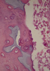
Scientists have found evidence to suggest that a genetic alteration in osteoblasts can induce acute myeloid leukemia (AML).
And this provides a potential therapeutic target for AML and myelodysplastic syndromes (MDS).
Stavroula Kousteni, PhD, of Columbia University Medical Center in New York, and her colleagues described these findings in Nature.
The researchers discovered that an activating mutation of beta-catenin in mouse osteoblasts induces AML.
This mutation leads to cancer in adjacent hematopoietic stem cells (HSCs) through a series of events. First, the mutated beta-catenin protein moves from its normal location on the exterior of the osteoblast to the cell’s nucleus, where it activates production of the protein jagged1.
Jagged1 proteins are then shipped to the osteoblast’s exterior membrane, where they can bind to Notch proteins—which activate signaling pathways—on neighboring HSCs. When this happens, Notch transmits signals inside the HSCs that enable leukemic transformation.
To confirm the role of jagged1 in AML development, the investigators removed 1 allele of jagged1 in osteoblasts. This decreased Notch signaling in Lin-Sca+c-Kit+ cells, rescued anemia and deregulation of HSC lineage differentiation, and prevented AML development.
The researchers then evaluated the effects of blocking Notch signaling using a gamma-secretase inhibitor. The treatment reversed hematopoietic deregulation and myeloid expansion in the blood, marrow, and spleens of the mice and reversed their AML.
“If the [process] works the same way in humans, our study suggests practical ways that we may be able to intervene with a drug or an antibody,” Dr Kousteni said.
With this in mind, she and her colleagues analyzed cells from 107 patients with AML or MDS. About 38% of the patients had changes in beta-catenin, jagged1, and Notch signaling that mirrored the changes in the mice. But none of the 56 healthy control subjects studied had these changes.
The investigators therefore concluded that these findings provide new insight into AML/MDS pathogenesis and may have implications for treatment.
Scientists have found evidence to suggest that a genetic alteration in osteoblasts can induce acute myeloid leukemia (AML).
And this provides a potential therapeutic target for AML and myelodysplastic syndromes (MDS).
Stavroula Kousteni, PhD, of Columbia University Medical Center in New York, and her colleagues described these findings in Nature.
The researchers discovered that an activating mutation of beta-catenin in mouse osteoblasts induces AML.
This mutation leads to cancer in adjacent hematopoietic stem cells (HSCs) through a series of events. First, the mutated beta-catenin protein moves from its normal location on the exterior of the osteoblast to the cell’s nucleus, where it activates production of the protein jagged1.
Jagged1 proteins are then shipped to the osteoblast’s exterior membrane, where they can bind to Notch proteins—which activate signaling pathways—on neighboring HSCs. When this happens, Notch transmits signals inside the HSCs that enable leukemic transformation.
To confirm the role of jagged1 in AML development, the investigators removed 1 allele of jagged1 in osteoblasts. This decreased Notch signaling in Lin-Sca+c-Kit+ cells, rescued anemia and deregulation of HSC lineage differentiation, and prevented AML development.
The researchers then evaluated the effects of blocking Notch signaling using a gamma-secretase inhibitor. The treatment reversed hematopoietic deregulation and myeloid expansion in the blood, marrow, and spleens of the mice and reversed their AML.
“If the [process] works the same way in humans, our study suggests practical ways that we may be able to intervene with a drug or an antibody,” Dr Kousteni said.
With this in mind, she and her colleagues analyzed cells from 107 patients with AML or MDS. About 38% of the patients had changes in beta-catenin, jagged1, and Notch signaling that mirrored the changes in the mice. But none of the 56 healthy control subjects studied had these changes.
The investigators therefore concluded that these findings provide new insight into AML/MDS pathogenesis and may have implications for treatment.
Scientists have found evidence to suggest that a genetic alteration in osteoblasts can induce acute myeloid leukemia (AML).
And this provides a potential therapeutic target for AML and myelodysplastic syndromes (MDS).
Stavroula Kousteni, PhD, of Columbia University Medical Center in New York, and her colleagues described these findings in Nature.
The researchers discovered that an activating mutation of beta-catenin in mouse osteoblasts induces AML.
This mutation leads to cancer in adjacent hematopoietic stem cells (HSCs) through a series of events. First, the mutated beta-catenin protein moves from its normal location on the exterior of the osteoblast to the cell’s nucleus, where it activates production of the protein jagged1.
Jagged1 proteins are then shipped to the osteoblast’s exterior membrane, where they can bind to Notch proteins—which activate signaling pathways—on neighboring HSCs. When this happens, Notch transmits signals inside the HSCs that enable leukemic transformation.
To confirm the role of jagged1 in AML development, the investigators removed 1 allele of jagged1 in osteoblasts. This decreased Notch signaling in Lin-Sca+c-Kit+ cells, rescued anemia and deregulation of HSC lineage differentiation, and prevented AML development.
The researchers then evaluated the effects of blocking Notch signaling using a gamma-secretase inhibitor. The treatment reversed hematopoietic deregulation and myeloid expansion in the blood, marrow, and spleens of the mice and reversed their AML.
“If the [process] works the same way in humans, our study suggests practical ways that we may be able to intervene with a drug or an antibody,” Dr Kousteni said.
With this in mind, she and her colleagues analyzed cells from 107 patients with AML or MDS. About 38% of the patients had changes in beta-catenin, jagged1, and Notch signaling that mirrored the changes in the mice. But none of the 56 healthy control subjects studied had these changes.
The investigators therefore concluded that these findings provide new insight into AML/MDS pathogenesis and may have implications for treatment.
Sickle cell trait affects need for ESAs
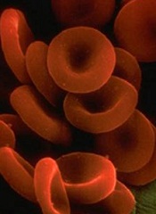
Researchers may have discovered why African Americans on dialysis sometimes require higher doses of erythropoietin-stimulating agents (ESAs) than dialysis patients of other ethnicities.
The team found that sickle cell trait was more common in African Americans on dialysis than in the general African American population.
And patients with sickle cell trait required higher ESA doses than other African American dialysis patients to reach the same hemoglobin level.
The researchers reported these findings in the Journal of the American Society of Nephrology.
The team noted that kidney abnormalities have been reported in some individuals with sickle cell trait. And studies have shown that African Americans with kidney failure require higher doses of ESAs to treat anemia during dialysis. So researchers wondered if there was a correlation.
To find out, Vimal Derebail, MD, of the University of North Carolina at Chapel Hill, and his colleagues examined laboratory and clinical data concerning 5319 adult African American hemodialysis patients.
But the researchers looked at the presence of hemoglobin C trait as well as sickle cell trait.
In the entire study cohort, 542 (10.2%) patients had sickle cell trait, and 129 (2.4%) had hemoglobin C trait. There were no other hemoglobinopathy traits present.
Sickle cell trait was more common among dialysis patients than the general African American population—10.2% and 6.5%-8.7%, respectively (P<0.05).
Among the 5002 patients receiving ESAs, 10.3% had sickle cell trait and 2.4% had hemoglobin C trait.
The patients with hemoglobinopathy traits received higher median ESA doses than patients with normal traits—4737.4 units/treatment and 4364.1 units/treatment, respectively (P=0.02).
Having either hemoglobinopathy trait was associated with a 13.2% increase in ESA dose (P=0.001). And patients with either trait had a 30% increased risk of falling into the highest quartile of ESA dosing.
There was no significant difference in the dose increase according to trait type (P=0.10).
The researchers therefore said these findings suggest the presence of hemoglobinopathy traits, particularly sickle cell trait, may explain why greater ESA doses are administered to African American dialysis patients relative to Caucasian patients.
“While we don’t know whether there are any adverse consequences to this higher dose of medication yet, further policies and decisions regarding management of anemia in dialysis patients should take into account these findings,” Dr Derebail said.
He added that future research should also explore whether sickle cell trait is more common in dialysis patients because it contributes to kidney disease.
Researchers may have discovered why African Americans on dialysis sometimes require higher doses of erythropoietin-stimulating agents (ESAs) than dialysis patients of other ethnicities.
The team found that sickle cell trait was more common in African Americans on dialysis than in the general African American population.
And patients with sickle cell trait required higher ESA doses than other African American dialysis patients to reach the same hemoglobin level.
The researchers reported these findings in the Journal of the American Society of Nephrology.
The team noted that kidney abnormalities have been reported in some individuals with sickle cell trait. And studies have shown that African Americans with kidney failure require higher doses of ESAs to treat anemia during dialysis. So researchers wondered if there was a correlation.
To find out, Vimal Derebail, MD, of the University of North Carolina at Chapel Hill, and his colleagues examined laboratory and clinical data concerning 5319 adult African American hemodialysis patients.
But the researchers looked at the presence of hemoglobin C trait as well as sickle cell trait.
In the entire study cohort, 542 (10.2%) patients had sickle cell trait, and 129 (2.4%) had hemoglobin C trait. There were no other hemoglobinopathy traits present.
Sickle cell trait was more common among dialysis patients than the general African American population—10.2% and 6.5%-8.7%, respectively (P<0.05).
Among the 5002 patients receiving ESAs, 10.3% had sickle cell trait and 2.4% had hemoglobin C trait.
The patients with hemoglobinopathy traits received higher median ESA doses than patients with normal traits—4737.4 units/treatment and 4364.1 units/treatment, respectively (P=0.02).
Having either hemoglobinopathy trait was associated with a 13.2% increase in ESA dose (P=0.001). And patients with either trait had a 30% increased risk of falling into the highest quartile of ESA dosing.
There was no significant difference in the dose increase according to trait type (P=0.10).
The researchers therefore said these findings suggest the presence of hemoglobinopathy traits, particularly sickle cell trait, may explain why greater ESA doses are administered to African American dialysis patients relative to Caucasian patients.
“While we don’t know whether there are any adverse consequences to this higher dose of medication yet, further policies and decisions regarding management of anemia in dialysis patients should take into account these findings,” Dr Derebail said.
He added that future research should also explore whether sickle cell trait is more common in dialysis patients because it contributes to kidney disease.
Researchers may have discovered why African Americans on dialysis sometimes require higher doses of erythropoietin-stimulating agents (ESAs) than dialysis patients of other ethnicities.
The team found that sickle cell trait was more common in African Americans on dialysis than in the general African American population.
And patients with sickle cell trait required higher ESA doses than other African American dialysis patients to reach the same hemoglobin level.
The researchers reported these findings in the Journal of the American Society of Nephrology.
The team noted that kidney abnormalities have been reported in some individuals with sickle cell trait. And studies have shown that African Americans with kidney failure require higher doses of ESAs to treat anemia during dialysis. So researchers wondered if there was a correlation.
To find out, Vimal Derebail, MD, of the University of North Carolina at Chapel Hill, and his colleagues examined laboratory and clinical data concerning 5319 adult African American hemodialysis patients.
But the researchers looked at the presence of hemoglobin C trait as well as sickle cell trait.
In the entire study cohort, 542 (10.2%) patients had sickle cell trait, and 129 (2.4%) had hemoglobin C trait. There were no other hemoglobinopathy traits present.
Sickle cell trait was more common among dialysis patients than the general African American population—10.2% and 6.5%-8.7%, respectively (P<0.05).
Among the 5002 patients receiving ESAs, 10.3% had sickle cell trait and 2.4% had hemoglobin C trait.
The patients with hemoglobinopathy traits received higher median ESA doses than patients with normal traits—4737.4 units/treatment and 4364.1 units/treatment, respectively (P=0.02).
Having either hemoglobinopathy trait was associated with a 13.2% increase in ESA dose (P=0.001). And patients with either trait had a 30% increased risk of falling into the highest quartile of ESA dosing.
There was no significant difference in the dose increase according to trait type (P=0.10).
The researchers therefore said these findings suggest the presence of hemoglobinopathy traits, particularly sickle cell trait, may explain why greater ESA doses are administered to African American dialysis patients relative to Caucasian patients.
“While we don’t know whether there are any adverse consequences to this higher dose of medication yet, further policies and decisions regarding management of anemia in dialysis patients should take into account these findings,” Dr Derebail said.
He added that future research should also explore whether sickle cell trait is more common in dialysis patients because it contributes to kidney disease.
High-volume centers better for severe sepsis patients

Credit: CDC
A new study suggests that “practice makes perfect” when it comes to caring for patients with severe sepsis.
Researchers found that patients admitted to academic medical centers with a higher volume of severe sepsis patients had significantly lower mortality rates than patients treated at centers with lower volumes of sepsis patients.
And the superior outcomes did not come at a greater cost.
Allan J. Walkey, MD, of the Boston University School of Medicine in Massachusetts, and his colleagues reported these findings in the American Journal of Respiratory and Critical Care Medicine.
The researchers noted that processes of care can influence outcomes in patients with severe sepsis. However, it hasn’t been clear whether a hospital’s level of experience in caring for patients with severe sepsis affects patient outcomes.
So Dr Walkey and his colleagues conducted a large, retrospective study to find out. The team analyzed data from academic hospitals across the US, provided by the University HealthSystem Consortium.
They identified 56,997 patients with severe sepsis who were admitted to 124 academic medical centers in 2011.
The patients’ median length of stay was 12.5 days, the median direct cost for each patient was $26,304, and the average hospital mortality was 25.6 ± 5.3%.
Hospitals caring for more sepsis patients had a 7% lower mortality rate than hospitals with lower volumes.
The high-volume medical centers (604-977 cases) had a 22.2% adjusted mortality rate, and the lower-volume hospitals (30-317) had a 29.2% adjusted mortality rate (P<0.01).
There was no significant difference in direct costs between the low-volume and high-volume centers (P=0.79).
“Given the lack of new drugs to treat severe sepsis, medical professionals must look at other ways to increase patient safety and positive outcomes, including the process of how we deliver care,” Dr Walkey said.
“Our study results demonstrate that hospitals with more experience caring for patients with severe sepsis were able to achieve better outcomes than hospitals with less experience with sepsis, possibly due to better processes of care for patients with sepsis.”
Credit: CDC
A new study suggests that “practice makes perfect” when it comes to caring for patients with severe sepsis.
Researchers found that patients admitted to academic medical centers with a higher volume of severe sepsis patients had significantly lower mortality rates than patients treated at centers with lower volumes of sepsis patients.
And the superior outcomes did not come at a greater cost.
Allan J. Walkey, MD, of the Boston University School of Medicine in Massachusetts, and his colleagues reported these findings in the American Journal of Respiratory and Critical Care Medicine.
The researchers noted that processes of care can influence outcomes in patients with severe sepsis. However, it hasn’t been clear whether a hospital’s level of experience in caring for patients with severe sepsis affects patient outcomes.
So Dr Walkey and his colleagues conducted a large, retrospective study to find out. The team analyzed data from academic hospitals across the US, provided by the University HealthSystem Consortium.
They identified 56,997 patients with severe sepsis who were admitted to 124 academic medical centers in 2011.
The patients’ median length of stay was 12.5 days, the median direct cost for each patient was $26,304, and the average hospital mortality was 25.6 ± 5.3%.
Hospitals caring for more sepsis patients had a 7% lower mortality rate than hospitals with lower volumes.
The high-volume medical centers (604-977 cases) had a 22.2% adjusted mortality rate, and the lower-volume hospitals (30-317) had a 29.2% adjusted mortality rate (P<0.01).
There was no significant difference in direct costs between the low-volume and high-volume centers (P=0.79).
“Given the lack of new drugs to treat severe sepsis, medical professionals must look at other ways to increase patient safety and positive outcomes, including the process of how we deliver care,” Dr Walkey said.
“Our study results demonstrate that hospitals with more experience caring for patients with severe sepsis were able to achieve better outcomes than hospitals with less experience with sepsis, possibly due to better processes of care for patients with sepsis.”
Credit: CDC
A new study suggests that “practice makes perfect” when it comes to caring for patients with severe sepsis.
Researchers found that patients admitted to academic medical centers with a higher volume of severe sepsis patients had significantly lower mortality rates than patients treated at centers with lower volumes of sepsis patients.
And the superior outcomes did not come at a greater cost.
Allan J. Walkey, MD, of the Boston University School of Medicine in Massachusetts, and his colleagues reported these findings in the American Journal of Respiratory and Critical Care Medicine.
The researchers noted that processes of care can influence outcomes in patients with severe sepsis. However, it hasn’t been clear whether a hospital’s level of experience in caring for patients with severe sepsis affects patient outcomes.
So Dr Walkey and his colleagues conducted a large, retrospective study to find out. The team analyzed data from academic hospitals across the US, provided by the University HealthSystem Consortium.
They identified 56,997 patients with severe sepsis who were admitted to 124 academic medical centers in 2011.
The patients’ median length of stay was 12.5 days, the median direct cost for each patient was $26,304, and the average hospital mortality was 25.6 ± 5.3%.
Hospitals caring for more sepsis patients had a 7% lower mortality rate than hospitals with lower volumes.
The high-volume medical centers (604-977 cases) had a 22.2% adjusted mortality rate, and the lower-volume hospitals (30-317) had a 29.2% adjusted mortality rate (P<0.01).
There was no significant difference in direct costs between the low-volume and high-volume centers (P=0.79).
“Given the lack of new drugs to treat severe sepsis, medical professionals must look at other ways to increase patient safety and positive outcomes, including the process of how we deliver care,” Dr Walkey said.
“Our study results demonstrate that hospitals with more experience caring for patients with severe sepsis were able to achieve better outcomes than hospitals with less experience with sepsis, possibly due to better processes of care for patients with sepsis.”
Group simulates blood vessel growth

Louis Heiser & Robert Ackland
Bioengineers say they’ve found a way to accurately predict blood vessel growth, and this finding has implications for cancers and other diseases.
The team discovered that tiny blood vessels grow better in the lab if the tissue surrounding them is less dense.
And this discovery allowed them to create a computer simulation that can accurately predict such growth.
“Better understanding of the processes that regulate the growth of blood vessels puts us in a position, ultimately, to develop new treatments for diseases related to blood vessel growth,” said study author Jeff Weiss, PhD, of the University of Utah in Salt Lake City.
Dr Weiss and his colleagues described their research in PLOS ONE.
Like some previous studies, the group’s research showed that capillaries grow, branch, and interconnect best when the density of the surrounding tissue, the extracellular matrix, is lower rather than higher. But unlike earlier research, Dr Weiss and his colleagues used pieces of real blood vessels from rats (rather than single cells).
Earlier work also focused on how the extracellular matrix, made mostly of collagen, sends chemical signals to promote capillary growth. The current study focused more on how the collagen’s mechanical or physical properties—specifically, the density or stiffness of the matrix—affect blood vessel growth.
Both the lab experiments and computer simulations showed that the denser or stiffer this collagen matrix, the more difficult it is for blood vessels to form a network necessary to supply blood to living tissue.
Growing blood vessels
To grow a network of blood vessels, the researchers extracted blood vessel fragments from the fat tissues of rats and suspended them in liquid. This extract contained 35,000 of those blood-vessel fragments per mL of solution.
The blood vessel fragments were grown in plastic plates with tiny mold-like wells filled with gel-like collagen as the extracellular matrix. The team cultured the fragments for 6 days with 3 densities of collagen: 2 mg, 3 mg, and 4 mg of collagen per mL of solution.
Vessels in the lower-density collagen grew and branched more, had fewer dead ends, and interconnected with each other better than the vessels growing in the higher-density collagen. These blood vessel networks mirrored those found in living mammals.
Simulating growth
The vessels grown in the lab provided data on total length of the vessels, the degree to which they connected into a network of vessels, and the number of vessels branches and dead ends.
And these data allowed the researchers to program a 3-D computer simulation that accurately predicted blood vessel network formation based on collagen matrix density.
“Now, we can answer all sorts of ‘what if’ questions about the geometry of these tissues, their shape, boundaries, initial densities, and mechanical properties,” Dr Weiss said. “We can use the computer to predict the influence that these factors have in the layout of a vascular network structure.”
The 3-D computer simulation also enabled the researchers to “conduct” experiments that couldn’t be done in the lab. One simulation showed blood vessels grow easily from denser toward less-dense collagen, but not the other way around.
A second simulation showed that vessels grew in collagen, except where a dense piece of collagen was placed in the center of less-dense collagen.
The third simulation showed that when researchers simulated 2 bands of less-dense collagen surrounded by bands of stiffer collagen, the nerve vessels grew along the bands of lower density.
Applications for cancer, other diseases
The researchers said these findings could ultimately be applied to aid the development of treatments for patients with cancer or diabetes, as well as patients who have had a heart attack and those who require tissue implants.
By better understanding the role that density of surrounding tissue plays in vessel formation, bioengineers could prepare “prevascularized” implantable tissues already equipped with blood vessels that match a patient’s blood vessel structure.
Prevascularized tissues might also help diabetes patients suffering from wounds that heal slowly—if at all—due to impaired blood microcirculation. Implanted skin grafts with their own blood vessels could stimulate blood flow to promote healing of diabetic ulcers.
Dr Weiss said he envisions prevascularized patches rehabilitating heart muscle that is damaged when a heart attack cuts off part of the heart’s oxygen supply, turning some of the heart into stiff scar tissue. A tissue patch implanted on the scar tissue could encourage blood vessel regrowth to repair the damaged, oxygen-deprived heart muscle.
As for cancer metastasis, most tumors begin as dense, blood-free masses. To grow and spread, the tumor tricks the body into fueling it with oxygenated blood vessels.
“The vessels grow in and then provide a pathway for the tumor to spread,” Dr Weiss noted. “This research will help us understand the physical parameters that control whether blood vessels reach the tumor.”
Louis Heiser & Robert Ackland
Bioengineers say they’ve found a way to accurately predict blood vessel growth, and this finding has implications for cancers and other diseases.
The team discovered that tiny blood vessels grow better in the lab if the tissue surrounding them is less dense.
And this discovery allowed them to create a computer simulation that can accurately predict such growth.
“Better understanding of the processes that regulate the growth of blood vessels puts us in a position, ultimately, to develop new treatments for diseases related to blood vessel growth,” said study author Jeff Weiss, PhD, of the University of Utah in Salt Lake City.
Dr Weiss and his colleagues described their research in PLOS ONE.
Like some previous studies, the group’s research showed that capillaries grow, branch, and interconnect best when the density of the surrounding tissue, the extracellular matrix, is lower rather than higher. But unlike earlier research, Dr Weiss and his colleagues used pieces of real blood vessels from rats (rather than single cells).
Earlier work also focused on how the extracellular matrix, made mostly of collagen, sends chemical signals to promote capillary growth. The current study focused more on how the collagen’s mechanical or physical properties—specifically, the density or stiffness of the matrix—affect blood vessel growth.
Both the lab experiments and computer simulations showed that the denser or stiffer this collagen matrix, the more difficult it is for blood vessels to form a network necessary to supply blood to living tissue.
Growing blood vessels
To grow a network of blood vessels, the researchers extracted blood vessel fragments from the fat tissues of rats and suspended them in liquid. This extract contained 35,000 of those blood-vessel fragments per mL of solution.
The blood vessel fragments were grown in plastic plates with tiny mold-like wells filled with gel-like collagen as the extracellular matrix. The team cultured the fragments for 6 days with 3 densities of collagen: 2 mg, 3 mg, and 4 mg of collagen per mL of solution.
Vessels in the lower-density collagen grew and branched more, had fewer dead ends, and interconnected with each other better than the vessels growing in the higher-density collagen. These blood vessel networks mirrored those found in living mammals.
Simulating growth
The vessels grown in the lab provided data on total length of the vessels, the degree to which they connected into a network of vessels, and the number of vessels branches and dead ends.
And these data allowed the researchers to program a 3-D computer simulation that accurately predicted blood vessel network formation based on collagen matrix density.
“Now, we can answer all sorts of ‘what if’ questions about the geometry of these tissues, their shape, boundaries, initial densities, and mechanical properties,” Dr Weiss said. “We can use the computer to predict the influence that these factors have in the layout of a vascular network structure.”
The 3-D computer simulation also enabled the researchers to “conduct” experiments that couldn’t be done in the lab. One simulation showed blood vessels grow easily from denser toward less-dense collagen, but not the other way around.
A second simulation showed that vessels grew in collagen, except where a dense piece of collagen was placed in the center of less-dense collagen.
The third simulation showed that when researchers simulated 2 bands of less-dense collagen surrounded by bands of stiffer collagen, the nerve vessels grew along the bands of lower density.
Applications for cancer, other diseases
The researchers said these findings could ultimately be applied to aid the development of treatments for patients with cancer or diabetes, as well as patients who have had a heart attack and those who require tissue implants.
By better understanding the role that density of surrounding tissue plays in vessel formation, bioengineers could prepare “prevascularized” implantable tissues already equipped with blood vessels that match a patient’s blood vessel structure.
Prevascularized tissues might also help diabetes patients suffering from wounds that heal slowly—if at all—due to impaired blood microcirculation. Implanted skin grafts with their own blood vessels could stimulate blood flow to promote healing of diabetic ulcers.
Dr Weiss said he envisions prevascularized patches rehabilitating heart muscle that is damaged when a heart attack cuts off part of the heart’s oxygen supply, turning some of the heart into stiff scar tissue. A tissue patch implanted on the scar tissue could encourage blood vessel regrowth to repair the damaged, oxygen-deprived heart muscle.
As for cancer metastasis, most tumors begin as dense, blood-free masses. To grow and spread, the tumor tricks the body into fueling it with oxygenated blood vessels.
“The vessels grow in and then provide a pathway for the tumor to spread,” Dr Weiss noted. “This research will help us understand the physical parameters that control whether blood vessels reach the tumor.”
Louis Heiser & Robert Ackland
Bioengineers say they’ve found a way to accurately predict blood vessel growth, and this finding has implications for cancers and other diseases.
The team discovered that tiny blood vessels grow better in the lab if the tissue surrounding them is less dense.
And this discovery allowed them to create a computer simulation that can accurately predict such growth.
“Better understanding of the processes that regulate the growth of blood vessels puts us in a position, ultimately, to develop new treatments for diseases related to blood vessel growth,” said study author Jeff Weiss, PhD, of the University of Utah in Salt Lake City.
Dr Weiss and his colleagues described their research in PLOS ONE.
Like some previous studies, the group’s research showed that capillaries grow, branch, and interconnect best when the density of the surrounding tissue, the extracellular matrix, is lower rather than higher. But unlike earlier research, Dr Weiss and his colleagues used pieces of real blood vessels from rats (rather than single cells).
Earlier work also focused on how the extracellular matrix, made mostly of collagen, sends chemical signals to promote capillary growth. The current study focused more on how the collagen’s mechanical or physical properties—specifically, the density or stiffness of the matrix—affect blood vessel growth.
Both the lab experiments and computer simulations showed that the denser or stiffer this collagen matrix, the more difficult it is for blood vessels to form a network necessary to supply blood to living tissue.
Growing blood vessels
To grow a network of blood vessels, the researchers extracted blood vessel fragments from the fat tissues of rats and suspended them in liquid. This extract contained 35,000 of those blood-vessel fragments per mL of solution.
The blood vessel fragments were grown in plastic plates with tiny mold-like wells filled with gel-like collagen as the extracellular matrix. The team cultured the fragments for 6 days with 3 densities of collagen: 2 mg, 3 mg, and 4 mg of collagen per mL of solution.
Vessels in the lower-density collagen grew and branched more, had fewer dead ends, and interconnected with each other better than the vessels growing in the higher-density collagen. These blood vessel networks mirrored those found in living mammals.
Simulating growth
The vessels grown in the lab provided data on total length of the vessels, the degree to which they connected into a network of vessels, and the number of vessels branches and dead ends.
And these data allowed the researchers to program a 3-D computer simulation that accurately predicted blood vessel network formation based on collagen matrix density.
“Now, we can answer all sorts of ‘what if’ questions about the geometry of these tissues, their shape, boundaries, initial densities, and mechanical properties,” Dr Weiss said. “We can use the computer to predict the influence that these factors have in the layout of a vascular network structure.”
The 3-D computer simulation also enabled the researchers to “conduct” experiments that couldn’t be done in the lab. One simulation showed blood vessels grow easily from denser toward less-dense collagen, but not the other way around.
A second simulation showed that vessels grew in collagen, except where a dense piece of collagen was placed in the center of less-dense collagen.
The third simulation showed that when researchers simulated 2 bands of less-dense collagen surrounded by bands of stiffer collagen, the nerve vessels grew along the bands of lower density.
Applications for cancer, other diseases
The researchers said these findings could ultimately be applied to aid the development of treatments for patients with cancer or diabetes, as well as patients who have had a heart attack and those who require tissue implants.
By better understanding the role that density of surrounding tissue plays in vessel formation, bioengineers could prepare “prevascularized” implantable tissues already equipped with blood vessels that match a patient’s blood vessel structure.
Prevascularized tissues might also help diabetes patients suffering from wounds that heal slowly—if at all—due to impaired blood microcirculation. Implanted skin grafts with their own blood vessels could stimulate blood flow to promote healing of diabetic ulcers.
Dr Weiss said he envisions prevascularized patches rehabilitating heart muscle that is damaged when a heart attack cuts off part of the heart’s oxygen supply, turning some of the heart into stiff scar tissue. A tissue patch implanted on the scar tissue could encourage blood vessel regrowth to repair the damaged, oxygen-deprived heart muscle.
As for cancer metastasis, most tumors begin as dense, blood-free masses. To grow and spread, the tumor tricks the body into fueling it with oxygenated blood vessels.
“The vessels grow in and then provide a pathway for the tumor to spread,” Dr Weiss noted. “This research will help us understand the physical parameters that control whether blood vessels reach the tumor.”