User login
MRI method appears comparable to PET/CT
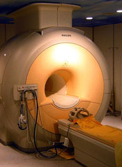
A modified MRI technique can effectively detect tumors in young cancer patients without exposing them to radiation, according to a small study published in The Lancet Oncology.
The method, called whole-body diffusion-weighted MRI, employs a contrast agent consisting of iron oxide nanoparticles.
This technique proved roughly as effective as 18F-FDG-PET/CT scans for detecting lymphoma and sarcoma in pediatric and young adult patients.
Researchers also noted that, as the MRI technique does not employ ionizing radiation, it might help prevent some of the adverse effects typically observed in patients who have undergone radiographic staging, particularly, secondary malignancies.
“I’m excited about having an imaging test for cancer patients that requires zero radiation exposure,” said senior study author Heike Daldrup-Link, MD, of the Stanford University School of Medicine in California.
She and her colleagues pointed out that, in the past, certain obstacles prevented physicians from using whole-body MRIs. For one, the scans take up to 2 hours, whereas a whole-body PET/CT takes only a few minutes.
In addition, in many organs, MRI does not distinguish healthy tissue from cancerous tissue. And existing contrast agents leave the tissues too quickly to be used in a lengthy, whole-body MRI.
In an attempt to overcome these obstacles, Dr Daldrup-Link and her colleagues used a contrast agent consisting of ferumoxytol nanoparticles. Injections of these iron oxide nanoparticles are approved by the US Food and Drug Administration (FDA) to treat anemia, and the researchers obtained FDA permission for use in their study.
The nanoparticles are retained in the body for days. On MRIs, they cause blood vessels to appear brighter, providing anatomic landmarks. The nanoparticles also cause healthy bone marrow, lymph nodes, livers, and spleens to appear darker, which makes tumors stand out.
The researchers compared the whole-body diffusion-weighted MRI method to PET/CTs in 22 patients, ages 8 to 33, who had lymphoma or sarcoma. Fourteen of the patients had Hodgkin lymphoma, 5 had non-Hodgkin lymphoma, 1 had Burkitt leukemia, 1 had Ewing’s sarcoma, and 1 had osteosarcoma.
The team found the MRI scans and PET/CT scans provided comparable information, although tumor detection was slightly better with PET/CT. The PET/CTs detected 163 of the 174 total tumors, and the MRIs detected 158.
The two methods had similar levels of sensitivity, specificity, and diagnostic accuracy. Sensitivity was 93.7% with PET/CT and 90.8% with MRI. Specificity was 97.7% with PET/CT and 99.5% with MRI. And diagnostic accuracy was 97.2% with PET/CT and 98.3% with MRI.
The researchers also noted that none of the patients experienced adverse reactions to the ferumoxytol nanoparticles, although the FDA previously observed a small risk of allergic reaction to the nanoparticles’ coating.
Dr Daldrup-Link said future research will aim to validate the MRI method in larger, more diverse groups of cancer patients, as well as examine its possible use for monitoring tumors over the course of cancer treatment. The technique also holds promise for scanning patients after their treatment is complete. 
A modified MRI technique can effectively detect tumors in young cancer patients without exposing them to radiation, according to a small study published in The Lancet Oncology.
The method, called whole-body diffusion-weighted MRI, employs a contrast agent consisting of iron oxide nanoparticles.
This technique proved roughly as effective as 18F-FDG-PET/CT scans for detecting lymphoma and sarcoma in pediatric and young adult patients.
Researchers also noted that, as the MRI technique does not employ ionizing radiation, it might help prevent some of the adverse effects typically observed in patients who have undergone radiographic staging, particularly, secondary malignancies.
“I’m excited about having an imaging test for cancer patients that requires zero radiation exposure,” said senior study author Heike Daldrup-Link, MD, of the Stanford University School of Medicine in California.
She and her colleagues pointed out that, in the past, certain obstacles prevented physicians from using whole-body MRIs. For one, the scans take up to 2 hours, whereas a whole-body PET/CT takes only a few minutes.
In addition, in many organs, MRI does not distinguish healthy tissue from cancerous tissue. And existing contrast agents leave the tissues too quickly to be used in a lengthy, whole-body MRI.
In an attempt to overcome these obstacles, Dr Daldrup-Link and her colleagues used a contrast agent consisting of ferumoxytol nanoparticles. Injections of these iron oxide nanoparticles are approved by the US Food and Drug Administration (FDA) to treat anemia, and the researchers obtained FDA permission for use in their study.
The nanoparticles are retained in the body for days. On MRIs, they cause blood vessels to appear brighter, providing anatomic landmarks. The nanoparticles also cause healthy bone marrow, lymph nodes, livers, and spleens to appear darker, which makes tumors stand out.
The researchers compared the whole-body diffusion-weighted MRI method to PET/CTs in 22 patients, ages 8 to 33, who had lymphoma or sarcoma. Fourteen of the patients had Hodgkin lymphoma, 5 had non-Hodgkin lymphoma, 1 had Burkitt leukemia, 1 had Ewing’s sarcoma, and 1 had osteosarcoma.
The team found the MRI scans and PET/CT scans provided comparable information, although tumor detection was slightly better with PET/CT. The PET/CTs detected 163 of the 174 total tumors, and the MRIs detected 158.
The two methods had similar levels of sensitivity, specificity, and diagnostic accuracy. Sensitivity was 93.7% with PET/CT and 90.8% with MRI. Specificity was 97.7% with PET/CT and 99.5% with MRI. And diagnostic accuracy was 97.2% with PET/CT and 98.3% with MRI.
The researchers also noted that none of the patients experienced adverse reactions to the ferumoxytol nanoparticles, although the FDA previously observed a small risk of allergic reaction to the nanoparticles’ coating.
Dr Daldrup-Link said future research will aim to validate the MRI method in larger, more diverse groups of cancer patients, as well as examine its possible use for monitoring tumors over the course of cancer treatment. The technique also holds promise for scanning patients after their treatment is complete.
A modified MRI technique can effectively detect tumors in young cancer patients without exposing them to radiation, according to a small study published in The Lancet Oncology.
The method, called whole-body diffusion-weighted MRI, employs a contrast agent consisting of iron oxide nanoparticles.
This technique proved roughly as effective as 18F-FDG-PET/CT scans for detecting lymphoma and sarcoma in pediatric and young adult patients.
Researchers also noted that, as the MRI technique does not employ ionizing radiation, it might help prevent some of the adverse effects typically observed in patients who have undergone radiographic staging, particularly, secondary malignancies.
“I’m excited about having an imaging test for cancer patients that requires zero radiation exposure,” said senior study author Heike Daldrup-Link, MD, of the Stanford University School of Medicine in California.
She and her colleagues pointed out that, in the past, certain obstacles prevented physicians from using whole-body MRIs. For one, the scans take up to 2 hours, whereas a whole-body PET/CT takes only a few minutes.
In addition, in many organs, MRI does not distinguish healthy tissue from cancerous tissue. And existing contrast agents leave the tissues too quickly to be used in a lengthy, whole-body MRI.
In an attempt to overcome these obstacles, Dr Daldrup-Link and her colleagues used a contrast agent consisting of ferumoxytol nanoparticles. Injections of these iron oxide nanoparticles are approved by the US Food and Drug Administration (FDA) to treat anemia, and the researchers obtained FDA permission for use in their study.
The nanoparticles are retained in the body for days. On MRIs, they cause blood vessels to appear brighter, providing anatomic landmarks. The nanoparticles also cause healthy bone marrow, lymph nodes, livers, and spleens to appear darker, which makes tumors stand out.
The researchers compared the whole-body diffusion-weighted MRI method to PET/CTs in 22 patients, ages 8 to 33, who had lymphoma or sarcoma. Fourteen of the patients had Hodgkin lymphoma, 5 had non-Hodgkin lymphoma, 1 had Burkitt leukemia, 1 had Ewing’s sarcoma, and 1 had osteosarcoma.
The team found the MRI scans and PET/CT scans provided comparable information, although tumor detection was slightly better with PET/CT. The PET/CTs detected 163 of the 174 total tumors, and the MRIs detected 158.
The two methods had similar levels of sensitivity, specificity, and diagnostic accuracy. Sensitivity was 93.7% with PET/CT and 90.8% with MRI. Specificity was 97.7% with PET/CT and 99.5% with MRI. And diagnostic accuracy was 97.2% with PET/CT and 98.3% with MRI.
The researchers also noted that none of the patients experienced adverse reactions to the ferumoxytol nanoparticles, although the FDA previously observed a small risk of allergic reaction to the nanoparticles’ coating.
Dr Daldrup-Link said future research will aim to validate the MRI method in larger, more diverse groups of cancer patients, as well as examine its possible use for monitoring tumors over the course of cancer treatment. The technique also holds promise for scanning patients after their treatment is complete.
Compression device can prevent VTE after surgery

Credit: Piotr Bodzek
A mobile compression device can prevent venous thromboembolism (VTE) after joint replacement surgery, according to research published in the Journal of Bone and Joint Surgery.
The device, called ActiveCare+S.F.T., delivers compressions to the leg that coordinate with a patient’s respiration rate, and this improves blood flow.
Of more than 3000 patients who used the device, with or without aspirin, less than 1% developed VTE.
When the researchers compared this rate to VTE rates observed in previous studies of warfarin, enoxaparin, rivaroxaban, and dabigatran, they found the device to be noninferior to anticoagulant therapy.
“Blood thinners have long been considered the standard of care to prevent blood clots after orthopedic surgery, but they can have side effects that are concerning for many patients,” said study author Clifford Colwell, MD, of the Scripps Clinic in La Jolla, California.
“Through this research, we have found and established an equally effective means of accomplishing the same goal, with an added layer of safety for patients.”
Dr Colwell and his colleagues established a registry of 3060 patients to determine the rate of symptomatic VTE after primary knee arthroplasty (n=1551) or hip arthroplasty (n=1509) performed at 10 different sites.
All of the patients enrolled were 18 years of age or older. They had no known history of VTE, coagulation disorders, or solid tumor malignancies.
Patients wore the ActiveCare+S.F.T device both during and after surgery, for a minimum of 10 days. The researchers evaluated patients at 3 months after their surgery to document evidence of deep vein thrombosis (DVT) or pulmonary embolism (PE).
In all, 28 patients (0.92%) developed VTE. Twenty patients had distal DVT, 3 had proximal DVT, and 5 had PE. One patient died of coronary failure, but there was no autopsy, so it is not clear if the patient developed a PE.
Overall, the rate of VTE with the compression device—0.92%—was considered noninferior to rates previously observed with anticoagulants—2.2% for warfarin, 1.1% for enoxaparin, 0.64% for rivaroxaban, and 1.2% for dabigatran.
However, among patients who underwent knee arthroplasty, the device fell short of the noninferiority margin (1.0%) for rivaroxaban by 0.06%.
The device’s manufacturer, Medical Compression Systems Inc., funded the registry used in this study but did not have a role in the registry design or protocol. And the researchers did not receive compensation from the manufacturer.
Credit: Piotr Bodzek
A mobile compression device can prevent venous thromboembolism (VTE) after joint replacement surgery, according to research published in the Journal of Bone and Joint Surgery.
The device, called ActiveCare+S.F.T., delivers compressions to the leg that coordinate with a patient’s respiration rate, and this improves blood flow.
Of more than 3000 patients who used the device, with or without aspirin, less than 1% developed VTE.
When the researchers compared this rate to VTE rates observed in previous studies of warfarin, enoxaparin, rivaroxaban, and dabigatran, they found the device to be noninferior to anticoagulant therapy.
“Blood thinners have long been considered the standard of care to prevent blood clots after orthopedic surgery, but they can have side effects that are concerning for many patients,” said study author Clifford Colwell, MD, of the Scripps Clinic in La Jolla, California.
“Through this research, we have found and established an equally effective means of accomplishing the same goal, with an added layer of safety for patients.”
Dr Colwell and his colleagues established a registry of 3060 patients to determine the rate of symptomatic VTE after primary knee arthroplasty (n=1551) or hip arthroplasty (n=1509) performed at 10 different sites.
All of the patients enrolled were 18 years of age or older. They had no known history of VTE, coagulation disorders, or solid tumor malignancies.
Patients wore the ActiveCare+S.F.T device both during and after surgery, for a minimum of 10 days. The researchers evaluated patients at 3 months after their surgery to document evidence of deep vein thrombosis (DVT) or pulmonary embolism (PE).
In all, 28 patients (0.92%) developed VTE. Twenty patients had distal DVT, 3 had proximal DVT, and 5 had PE. One patient died of coronary failure, but there was no autopsy, so it is not clear if the patient developed a PE.
Overall, the rate of VTE with the compression device—0.92%—was considered noninferior to rates previously observed with anticoagulants—2.2% for warfarin, 1.1% for enoxaparin, 0.64% for rivaroxaban, and 1.2% for dabigatran.
However, among patients who underwent knee arthroplasty, the device fell short of the noninferiority margin (1.0%) for rivaroxaban by 0.06%.
The device’s manufacturer, Medical Compression Systems Inc., funded the registry used in this study but did not have a role in the registry design or protocol. And the researchers did not receive compensation from the manufacturer.
Credit: Piotr Bodzek
A mobile compression device can prevent venous thromboembolism (VTE) after joint replacement surgery, according to research published in the Journal of Bone and Joint Surgery.
The device, called ActiveCare+S.F.T., delivers compressions to the leg that coordinate with a patient’s respiration rate, and this improves blood flow.
Of more than 3000 patients who used the device, with or without aspirin, less than 1% developed VTE.
When the researchers compared this rate to VTE rates observed in previous studies of warfarin, enoxaparin, rivaroxaban, and dabigatran, they found the device to be noninferior to anticoagulant therapy.
“Blood thinners have long been considered the standard of care to prevent blood clots after orthopedic surgery, but they can have side effects that are concerning for many patients,” said study author Clifford Colwell, MD, of the Scripps Clinic in La Jolla, California.
“Through this research, we have found and established an equally effective means of accomplishing the same goal, with an added layer of safety for patients.”
Dr Colwell and his colleagues established a registry of 3060 patients to determine the rate of symptomatic VTE after primary knee arthroplasty (n=1551) or hip arthroplasty (n=1509) performed at 10 different sites.
All of the patients enrolled were 18 years of age or older. They had no known history of VTE, coagulation disorders, or solid tumor malignancies.
Patients wore the ActiveCare+S.F.T device both during and after surgery, for a minimum of 10 days. The researchers evaluated patients at 3 months after their surgery to document evidence of deep vein thrombosis (DVT) or pulmonary embolism (PE).
In all, 28 patients (0.92%) developed VTE. Twenty patients had distal DVT, 3 had proximal DVT, and 5 had PE. One patient died of coronary failure, but there was no autopsy, so it is not clear if the patient developed a PE.
Overall, the rate of VTE with the compression device—0.92%—was considered noninferior to rates previously observed with anticoagulants—2.2% for warfarin, 1.1% for enoxaparin, 0.64% for rivaroxaban, and 1.2% for dabigatran.
However, among patients who underwent knee arthroplasty, the device fell short of the noninferiority margin (1.0%) for rivaroxaban by 0.06%.
The device’s manufacturer, Medical Compression Systems Inc., funded the registry used in this study but did not have a role in the registry design or protocol. And the researchers did not receive compensation from the manufacturer.
Environment may play role in malaria transmission

the wall of a mosquito midgut
Credit: Krijn Paaijmans
Researchers have found the environment can significantly influence whether or not Wolbachia bacteria will prevent mosquitoes from transmitting malaria.
“Bacteria in the genus Wolbachia represent a promising new tool for controlling malaria due to their demonstrated ability to block the development of the pathogen within Anopheles mosquitoes,” said study investigator Courtney Murdock, PhD, of Pennsylvania State University.
“However, much of the work on the Wolbachia-malaria interaction has been conducted under highly simplified laboratory conditions. In this study, we investigated the ability of Wolbachia to block transmission of malaria—Plasmodium—parasites across variable environmental conditions, which are more reflective of conditions in the field.”
Dr Murdock and her colleagues described this research in Nature Scientific Reports.
The researchers used the malaria parasite Plasmodium yoelii, which affects rodents, and the mosquito Anopheles stephensi as a model system to investigate whether Wolbachia would block the ability of the malaria parasite to infect the mosquitoes.
The team divided the mosquitoes into an uninfected control group and a group infected with Wolbachia. Next, they raised all groups of mosquitoes in incubators set to different experimental temperatures—68, 72, 75, 79, and 82 degrees Fahrenheit.
At 82 degrees, Wolbachia reduced the number of mosquitoes infected by malaria parasites, the number of malaria parasites within each mosquito, and the intensity of oocysts.
At 75 degrees, Wolbachia had no effect on the prevalence of malaria parasites but increased oocyst intensity. At 68 degrees, Wolbachia had no effect on the prevalence of parasites or the intensity of oocysts.
The researchers also identified a previously undiscovered effect of Wolbachia. Infection with the bacterium reduced the development of sporozoites across all temperatures. This suggests that Wolbachia and malaria parasites may compete for similar hosts.
“Typically, the more oocysts a mosquito has on its midgut, the more sporozoites it produces,” Dr Murdock said. “So, depending on the environmental temperature, Wolbachia either reduced, enhanced, or had no effect on the number of oocysts.”
“At 75 degrees Fahrenheit, Wolbachia-infected mosquitos had 3 times the numbers of oocysts relative to uninfected mosquitoes. Thus, we would predict these mosquitoes to produce more sporozoites. But instead, we see that this is not the case, and that is because Wolbachia infection significantly reduces the number of sporozoites produced per oocyst, regardless of the environmental temperature.”
“This effect counteracts the enhancement we see at 75 degrees Fahrenheit. How the influence of Wolbachia on parasite establishment and the production of sporozoites under different temperatures plays out to ultimately affect transmission remains to be determined.”
Dr Murdock and her colleagues plan to duplicate their experiment using a species of malaria parasite that affects humans to determine whether or not the temperature effects they observed occur in humans as well.
The team also intends to explore the effects of additional environmental variation—such as daily temperature fluctuation and differential access to food resources in the mosquito larval and adult environments—on the transmission-blocking ability of Wolbachia.
the wall of a mosquito midgut
Credit: Krijn Paaijmans
Researchers have found the environment can significantly influence whether or not Wolbachia bacteria will prevent mosquitoes from transmitting malaria.
“Bacteria in the genus Wolbachia represent a promising new tool for controlling malaria due to their demonstrated ability to block the development of the pathogen within Anopheles mosquitoes,” said study investigator Courtney Murdock, PhD, of Pennsylvania State University.
“However, much of the work on the Wolbachia-malaria interaction has been conducted under highly simplified laboratory conditions. In this study, we investigated the ability of Wolbachia to block transmission of malaria—Plasmodium—parasites across variable environmental conditions, which are more reflective of conditions in the field.”
Dr Murdock and her colleagues described this research in Nature Scientific Reports.
The researchers used the malaria parasite Plasmodium yoelii, which affects rodents, and the mosquito Anopheles stephensi as a model system to investigate whether Wolbachia would block the ability of the malaria parasite to infect the mosquitoes.
The team divided the mosquitoes into an uninfected control group and a group infected with Wolbachia. Next, they raised all groups of mosquitoes in incubators set to different experimental temperatures—68, 72, 75, 79, and 82 degrees Fahrenheit.
At 82 degrees, Wolbachia reduced the number of mosquitoes infected by malaria parasites, the number of malaria parasites within each mosquito, and the intensity of oocysts.
At 75 degrees, Wolbachia had no effect on the prevalence of malaria parasites but increased oocyst intensity. At 68 degrees, Wolbachia had no effect on the prevalence of parasites or the intensity of oocysts.
The researchers also identified a previously undiscovered effect of Wolbachia. Infection with the bacterium reduced the development of sporozoites across all temperatures. This suggests that Wolbachia and malaria parasites may compete for similar hosts.
“Typically, the more oocysts a mosquito has on its midgut, the more sporozoites it produces,” Dr Murdock said. “So, depending on the environmental temperature, Wolbachia either reduced, enhanced, or had no effect on the number of oocysts.”
“At 75 degrees Fahrenheit, Wolbachia-infected mosquitos had 3 times the numbers of oocysts relative to uninfected mosquitoes. Thus, we would predict these mosquitoes to produce more sporozoites. But instead, we see that this is not the case, and that is because Wolbachia infection significantly reduces the number of sporozoites produced per oocyst, regardless of the environmental temperature.”
“This effect counteracts the enhancement we see at 75 degrees Fahrenheit. How the influence of Wolbachia on parasite establishment and the production of sporozoites under different temperatures plays out to ultimately affect transmission remains to be determined.”
Dr Murdock and her colleagues plan to duplicate their experiment using a species of malaria parasite that affects humans to determine whether or not the temperature effects they observed occur in humans as well.
The team also intends to explore the effects of additional environmental variation—such as daily temperature fluctuation and differential access to food resources in the mosquito larval and adult environments—on the transmission-blocking ability of Wolbachia.
the wall of a mosquito midgut
Credit: Krijn Paaijmans
Researchers have found the environment can significantly influence whether or not Wolbachia bacteria will prevent mosquitoes from transmitting malaria.
“Bacteria in the genus Wolbachia represent a promising new tool for controlling malaria due to their demonstrated ability to block the development of the pathogen within Anopheles mosquitoes,” said study investigator Courtney Murdock, PhD, of Pennsylvania State University.
“However, much of the work on the Wolbachia-malaria interaction has been conducted under highly simplified laboratory conditions. In this study, we investigated the ability of Wolbachia to block transmission of malaria—Plasmodium—parasites across variable environmental conditions, which are more reflective of conditions in the field.”
Dr Murdock and her colleagues described this research in Nature Scientific Reports.
The researchers used the malaria parasite Plasmodium yoelii, which affects rodents, and the mosquito Anopheles stephensi as a model system to investigate whether Wolbachia would block the ability of the malaria parasite to infect the mosquitoes.
The team divided the mosquitoes into an uninfected control group and a group infected with Wolbachia. Next, they raised all groups of mosquitoes in incubators set to different experimental temperatures—68, 72, 75, 79, and 82 degrees Fahrenheit.
At 82 degrees, Wolbachia reduced the number of mosquitoes infected by malaria parasites, the number of malaria parasites within each mosquito, and the intensity of oocysts.
At 75 degrees, Wolbachia had no effect on the prevalence of malaria parasites but increased oocyst intensity. At 68 degrees, Wolbachia had no effect on the prevalence of parasites or the intensity of oocysts.
The researchers also identified a previously undiscovered effect of Wolbachia. Infection with the bacterium reduced the development of sporozoites across all temperatures. This suggests that Wolbachia and malaria parasites may compete for similar hosts.
“Typically, the more oocysts a mosquito has on its midgut, the more sporozoites it produces,” Dr Murdock said. “So, depending on the environmental temperature, Wolbachia either reduced, enhanced, or had no effect on the number of oocysts.”
“At 75 degrees Fahrenheit, Wolbachia-infected mosquitos had 3 times the numbers of oocysts relative to uninfected mosquitoes. Thus, we would predict these mosquitoes to produce more sporozoites. But instead, we see that this is not the case, and that is because Wolbachia infection significantly reduces the number of sporozoites produced per oocyst, regardless of the environmental temperature.”
“This effect counteracts the enhancement we see at 75 degrees Fahrenheit. How the influence of Wolbachia on parasite establishment and the production of sporozoites under different temperatures plays out to ultimately affect transmission remains to be determined.”
Dr Murdock and her colleagues plan to duplicate their experiment using a species of malaria parasite that affects humans to determine whether or not the temperature effects they observed occur in humans as well.
The team also intends to explore the effects of additional environmental variation—such as daily temperature fluctuation and differential access to food resources in the mosquito larval and adult environments—on the transmission-blocking ability of Wolbachia.
Group finds progenitors of ILCs

with ILCs (green), epithelial
cells (red), and nuclei (blue)
University of Pennsylvania
Scientists say they’ve discovered the progenitors of innate lymphoid cells (ILCs) in the liver of fetal mice and the bone marrow of adult mice.
ILCs are among the first components of the immune system to confront certain pathogens, yet the cells went undetected by researchers for a century.
“Scientists tend to look for immune cells in the blood, lymph nodes, or spleen,” said Albert Bendelac, PhD, of the University of Chicago in Illinois.
“That is precisely where you would not find these cells. Once they mature, they directly go to tissues, such as the gut or the skin. You seldom see them in blood.”
To understand how ILCs fit into the ecosystem of cells that fight off infections and cancers, Dr Bendelac’s team focused on finding ILCs’ source.
And they reported their findings in a letter to Nature.
The team noted that ILCs, which were first recognized 5 years ago, are rare. A mouse might have 200 million lymphocytes and only a few thousand ILCs.
But previous work on natural killer (NK) cells showed that ILCs express the transcription factor PLZF during their development.
So Dr Bendelac and his colleagues created mice with the gene for green fluorescent protein inserted into mouse DNA, just downstream from the PLZF gene. As a result, cells from mice that expressed PLZF appeared bright green under the microscope.
Nevertheless, finding the precursors to ILCs was not easy. The precursors are not in the blood, and, by the time they migrate to the lungs or gut, they have already matured into ILCs.
The researchers eventually found the precursor cells—known as ILCPs—in the liver of fetal mice and in the bone marrow of adult mice.
When the team purified the ILCPs, which still contained the GFP gene, and transferred them into mice that lacked ILCs, the precursors were able to reconstitute the 3 known types of ILCs—ILCs 1, 2, and 3.
“There were no B cells or T cells or myeloid cells—no other immune cells, just these,” Dr Bendelac said. “So we think the ILCP really is a committed precursor to innate lymphoid cells.”
To confirm their finding, the researchers designed mice in which PLZF gene expression was tied to the gene for diphtheria toxin. When the cells expressed PLZF, they also produced the toxin, which was lethal for those cells. The result was a mouse that had a normal immune system except that it completely lacked ILCs.
“ILCs are found in the most exposed tissues,” Dr Bendelac noted. “They are one of your first lines of defense. We now suspect they may also influence the ensuing adaptive immune response, priming the pump, influencing how T-helper cells respond.”
Each of the 3 types of ILCs has different properties and serves different functions. ILC1 cells help prevent viral infections and can detect and remove some cancerous cells. They are similar to NK cells, except that NK cells circulate in the blood, and ILC1s live in the gut and the liver.
ILC2s are found in the lungs, where they can detect and respond to parasites. But they can also initiate an allergic reaction and mucus hyper-secretion.
ILC3 cells cluster in the gut, where they help mediate interactions between the bowel and bacteria. When that balance is disturbed, they can accelerate inflammation and may play a role in inflammatory bowel disease.
Dr Bendelac said his group’s research provides “one more tool for understanding this complex system,” and it could help generate a “powerful new way to assess the function of innate lymphocytes.”
with ILCs (green), epithelial
cells (red), and nuclei (blue)
University of Pennsylvania
Scientists say they’ve discovered the progenitors of innate lymphoid cells (ILCs) in the liver of fetal mice and the bone marrow of adult mice.
ILCs are among the first components of the immune system to confront certain pathogens, yet the cells went undetected by researchers for a century.
“Scientists tend to look for immune cells in the blood, lymph nodes, or spleen,” said Albert Bendelac, PhD, of the University of Chicago in Illinois.
“That is precisely where you would not find these cells. Once they mature, they directly go to tissues, such as the gut or the skin. You seldom see them in blood.”
To understand how ILCs fit into the ecosystem of cells that fight off infections and cancers, Dr Bendelac’s team focused on finding ILCs’ source.
And they reported their findings in a letter to Nature.
The team noted that ILCs, which were first recognized 5 years ago, are rare. A mouse might have 200 million lymphocytes and only a few thousand ILCs.
But previous work on natural killer (NK) cells showed that ILCs express the transcription factor PLZF during their development.
So Dr Bendelac and his colleagues created mice with the gene for green fluorescent protein inserted into mouse DNA, just downstream from the PLZF gene. As a result, cells from mice that expressed PLZF appeared bright green under the microscope.
Nevertheless, finding the precursors to ILCs was not easy. The precursors are not in the blood, and, by the time they migrate to the lungs or gut, they have already matured into ILCs.
The researchers eventually found the precursor cells—known as ILCPs—in the liver of fetal mice and in the bone marrow of adult mice.
When the team purified the ILCPs, which still contained the GFP gene, and transferred them into mice that lacked ILCs, the precursors were able to reconstitute the 3 known types of ILCs—ILCs 1, 2, and 3.
“There were no B cells or T cells or myeloid cells—no other immune cells, just these,” Dr Bendelac said. “So we think the ILCP really is a committed precursor to innate lymphoid cells.”
To confirm their finding, the researchers designed mice in which PLZF gene expression was tied to the gene for diphtheria toxin. When the cells expressed PLZF, they also produced the toxin, which was lethal for those cells. The result was a mouse that had a normal immune system except that it completely lacked ILCs.
“ILCs are found in the most exposed tissues,” Dr Bendelac noted. “They are one of your first lines of defense. We now suspect they may also influence the ensuing adaptive immune response, priming the pump, influencing how T-helper cells respond.”
Each of the 3 types of ILCs has different properties and serves different functions. ILC1 cells help prevent viral infections and can detect and remove some cancerous cells. They are similar to NK cells, except that NK cells circulate in the blood, and ILC1s live in the gut and the liver.
ILC2s are found in the lungs, where they can detect and respond to parasites. But they can also initiate an allergic reaction and mucus hyper-secretion.
ILC3 cells cluster in the gut, where they help mediate interactions between the bowel and bacteria. When that balance is disturbed, they can accelerate inflammation and may play a role in inflammatory bowel disease.
Dr Bendelac said his group’s research provides “one more tool for understanding this complex system,” and it could help generate a “powerful new way to assess the function of innate lymphocytes.”
with ILCs (green), epithelial
cells (red), and nuclei (blue)
University of Pennsylvania
Scientists say they’ve discovered the progenitors of innate lymphoid cells (ILCs) in the liver of fetal mice and the bone marrow of adult mice.
ILCs are among the first components of the immune system to confront certain pathogens, yet the cells went undetected by researchers for a century.
“Scientists tend to look for immune cells in the blood, lymph nodes, or spleen,” said Albert Bendelac, PhD, of the University of Chicago in Illinois.
“That is precisely where you would not find these cells. Once they mature, they directly go to tissues, such as the gut or the skin. You seldom see them in blood.”
To understand how ILCs fit into the ecosystem of cells that fight off infections and cancers, Dr Bendelac’s team focused on finding ILCs’ source.
And they reported their findings in a letter to Nature.
The team noted that ILCs, which were first recognized 5 years ago, are rare. A mouse might have 200 million lymphocytes and only a few thousand ILCs.
But previous work on natural killer (NK) cells showed that ILCs express the transcription factor PLZF during their development.
So Dr Bendelac and his colleagues created mice with the gene for green fluorescent protein inserted into mouse DNA, just downstream from the PLZF gene. As a result, cells from mice that expressed PLZF appeared bright green under the microscope.
Nevertheless, finding the precursors to ILCs was not easy. The precursors are not in the blood, and, by the time they migrate to the lungs or gut, they have already matured into ILCs.
The researchers eventually found the precursor cells—known as ILCPs—in the liver of fetal mice and in the bone marrow of adult mice.
When the team purified the ILCPs, which still contained the GFP gene, and transferred them into mice that lacked ILCs, the precursors were able to reconstitute the 3 known types of ILCs—ILCs 1, 2, and 3.
“There were no B cells or T cells or myeloid cells—no other immune cells, just these,” Dr Bendelac said. “So we think the ILCP really is a committed precursor to innate lymphoid cells.”
To confirm their finding, the researchers designed mice in which PLZF gene expression was tied to the gene for diphtheria toxin. When the cells expressed PLZF, they also produced the toxin, which was lethal for those cells. The result was a mouse that had a normal immune system except that it completely lacked ILCs.
“ILCs are found in the most exposed tissues,” Dr Bendelac noted. “They are one of your first lines of defense. We now suspect they may also influence the ensuing adaptive immune response, priming the pump, influencing how T-helper cells respond.”
Each of the 3 types of ILCs has different properties and serves different functions. ILC1 cells help prevent viral infections and can detect and remove some cancerous cells. They are similar to NK cells, except that NK cells circulate in the blood, and ILC1s live in the gut and the liver.
ILC2s are found in the lungs, where they can detect and respond to parasites. But they can also initiate an allergic reaction and mucus hyper-secretion.
ILC3 cells cluster in the gut, where they help mediate interactions between the bowel and bacteria. When that balance is disturbed, they can accelerate inflammation and may play a role in inflammatory bowel disease.
Dr Bendelac said his group’s research provides “one more tool for understanding this complex system,” and it could help generate a “powerful new way to assess the function of innate lymphocytes.”
Protein may be target for AML treatment

Credit: Rhoda Baer
The protein WTAP could play an important role in the development of acute myeloid leukemia (AML), according to new research.
Investigators discovered that AML cells have higher-than-normal levels of WTAP.
But silencing WTAP expression in leukemic cells can suppress proliferation and induce differentiation.
And, in mouse models of AML, knocking down WTAP can reduce tumor growth.
The researchers recounted these findings in a letter to Leukemia.
The team first uncovered high levels of WTAP in AML cells compared to normal peripheral blood mononuclear cells. And they found evidence to suggest that this contributes to abnormal cell behavior.
WTAP levels were not associated with individual cytogenetic abnormalities, but FLT3-ITD and NPM1 mutations were significantly correlated with WTAP expression. And WTAP levels were positively correlated with levels of proliferation-related proteins, anti-apoptotic proteins, oncoproteins, and proteins important for stem cell function.
To gain more insight into the importance of WTAP, the investigators silenced its expression in K562 cells, HL-60 cells, OCI-AML3 cells, and primary AML cells.
“Knocking down this protein, WTAP, greatly suppressed proliferation and induced differentiation,” said study author Hima Bansal, PhD, of The University of Texas Health Science Center at San Antonio.
WTAP knockdown alone did not induce apoptosis, but it did enhance the apoptosis that occurred after the administration of etoposide.
The researchers also examined the role of WTAP in AML using mouse models. They found that tumors derived from WTAP-knockdown cells were significantly smaller and grew significantly slower than tumors derived from cells that expressed WTAP.
Finally, the investigators set out to determine why WTAP is overexpressed in AML. They noted that the Wilms’ tumor 1 (WT1) gene has an oncogenic role in leukemogenesis, and WTAP partners with WT1 to function as a switch gene, regulating the balance between cell quiescence and proliferation.
So the researchers decided to investigate Hsp90, a molecular chaperone that helps stabilize many oncoproteins, including WT1. And they found a direct interaction between Hsp90 and WTAP.
The Hsp90 inhibitor ganetespib promoted the degradation of WTAP in K562, MV4-11, and Kasumi-1 cell lines, as well as in leukemic blasts. In mice, ganetespib inhibited tumor growth.
And experiments suggested that ganetespib-mediated WTAP degradation is dependent on the ubiquitin-proteasome pathway. But the investigators said further research is needed to clarify WTAP’s mechanism of action.
Nevertheless, they believe the results of this research indicate that WTAP could be a promising therapeutic target for AML.
Credit: Rhoda Baer
The protein WTAP could play an important role in the development of acute myeloid leukemia (AML), according to new research.
Investigators discovered that AML cells have higher-than-normal levels of WTAP.
But silencing WTAP expression in leukemic cells can suppress proliferation and induce differentiation.
And, in mouse models of AML, knocking down WTAP can reduce tumor growth.
The researchers recounted these findings in a letter to Leukemia.
The team first uncovered high levels of WTAP in AML cells compared to normal peripheral blood mononuclear cells. And they found evidence to suggest that this contributes to abnormal cell behavior.
WTAP levels were not associated with individual cytogenetic abnormalities, but FLT3-ITD and NPM1 mutations were significantly correlated with WTAP expression. And WTAP levels were positively correlated with levels of proliferation-related proteins, anti-apoptotic proteins, oncoproteins, and proteins important for stem cell function.
To gain more insight into the importance of WTAP, the investigators silenced its expression in K562 cells, HL-60 cells, OCI-AML3 cells, and primary AML cells.
“Knocking down this protein, WTAP, greatly suppressed proliferation and induced differentiation,” said study author Hima Bansal, PhD, of The University of Texas Health Science Center at San Antonio.
WTAP knockdown alone did not induce apoptosis, but it did enhance the apoptosis that occurred after the administration of etoposide.
The researchers also examined the role of WTAP in AML using mouse models. They found that tumors derived from WTAP-knockdown cells were significantly smaller and grew significantly slower than tumors derived from cells that expressed WTAP.
Finally, the investigators set out to determine why WTAP is overexpressed in AML. They noted that the Wilms’ tumor 1 (WT1) gene has an oncogenic role in leukemogenesis, and WTAP partners with WT1 to function as a switch gene, regulating the balance between cell quiescence and proliferation.
So the researchers decided to investigate Hsp90, a molecular chaperone that helps stabilize many oncoproteins, including WT1. And they found a direct interaction between Hsp90 and WTAP.
The Hsp90 inhibitor ganetespib promoted the degradation of WTAP in K562, MV4-11, and Kasumi-1 cell lines, as well as in leukemic blasts. In mice, ganetespib inhibited tumor growth.
And experiments suggested that ganetespib-mediated WTAP degradation is dependent on the ubiquitin-proteasome pathway. But the investigators said further research is needed to clarify WTAP’s mechanism of action.
Nevertheless, they believe the results of this research indicate that WTAP could be a promising therapeutic target for AML.
Credit: Rhoda Baer
The protein WTAP could play an important role in the development of acute myeloid leukemia (AML), according to new research.
Investigators discovered that AML cells have higher-than-normal levels of WTAP.
But silencing WTAP expression in leukemic cells can suppress proliferation and induce differentiation.
And, in mouse models of AML, knocking down WTAP can reduce tumor growth.
The researchers recounted these findings in a letter to Leukemia.
The team first uncovered high levels of WTAP in AML cells compared to normal peripheral blood mononuclear cells. And they found evidence to suggest that this contributes to abnormal cell behavior.
WTAP levels were not associated with individual cytogenetic abnormalities, but FLT3-ITD and NPM1 mutations were significantly correlated with WTAP expression. And WTAP levels were positively correlated with levels of proliferation-related proteins, anti-apoptotic proteins, oncoproteins, and proteins important for stem cell function.
To gain more insight into the importance of WTAP, the investigators silenced its expression in K562 cells, HL-60 cells, OCI-AML3 cells, and primary AML cells.
“Knocking down this protein, WTAP, greatly suppressed proliferation and induced differentiation,” said study author Hima Bansal, PhD, of The University of Texas Health Science Center at San Antonio.
WTAP knockdown alone did not induce apoptosis, but it did enhance the apoptosis that occurred after the administration of etoposide.
The researchers also examined the role of WTAP in AML using mouse models. They found that tumors derived from WTAP-knockdown cells were significantly smaller and grew significantly slower than tumors derived from cells that expressed WTAP.
Finally, the investigators set out to determine why WTAP is overexpressed in AML. They noted that the Wilms’ tumor 1 (WT1) gene has an oncogenic role in leukemogenesis, and WTAP partners with WT1 to function as a switch gene, regulating the balance between cell quiescence and proliferation.
So the researchers decided to investigate Hsp90, a molecular chaperone that helps stabilize many oncoproteins, including WT1. And they found a direct interaction between Hsp90 and WTAP.
The Hsp90 inhibitor ganetespib promoted the degradation of WTAP in K562, MV4-11, and Kasumi-1 cell lines, as well as in leukemic blasts. In mice, ganetespib inhibited tumor growth.
And experiments suggested that ganetespib-mediated WTAP degradation is dependent on the ubiquitin-proteasome pathway. But the investigators said further research is needed to clarify WTAP’s mechanism of action.
Nevertheless, they believe the results of this research indicate that WTAP could be a promising therapeutic target for AML.
Creating an ‘inexhaustible’ supply of platelets

Scientists say they’ve discovered a way to create a potentially inexhaustible supply of functional platelets.
The researchers used human induced pluripotent stem cells (iPSCs) to create immortalized megakaryocyte progenitor cell lines (imMKCLs). And by manipulating the cell lines, the team produced platelets.
These imMKCL-derived platelets were functional, although not as functional as donor-derived platelets.
On the other hand, the imMKCL-derived cells offer an advantage over donated platelets—namely, the imMKCLs can be expanded in culture for up to 5 months, even after cryopreservation.
“[W]e established a method to achieve the long-term self-replication of megakaryocyte progenitors as an immortalized cell line, which could eventually contribute to large-scale cultivation and production of platelets,” said senior study author Koji Eto, MD, PhD, of Kyoto University and the University of Tokyo in Japan.
He and his colleagues believe this work, published in Cell Stem Cell, could eventually help us eliminate platelet shortages. The supply of donated platelets, which have a short shelf-life and must be kept at room temperature, is often insufficient to meet clinical needs.
With that in mind, Dr Eto’s team set out to create large quantities of functional platelets. They first generated stable imMKCLs from iPSC-derived hematopoietic progenitors.
They accomplished this by inducing overexpression of BMI1 and BCL-XL to suppress senescence and apoptosis. They also induced constrained overexpression of c-MYC to promote proliferation, as they found too-high c-MYC expression led to caspase-dependent MKCL apoptosis.
When the researchers turned off expression of c-MYC, BMI1, and BCL-XL, they saw an increase in CD42b+ platelet yield from the imMKCLs and upregulated CD42b expression in CD41a+ platelets. They noted that expression of CD42b is required for clotting initiation and bacterial clearance in vivo.
The team then conducted in vitro and in vivo experiments to test the functionality of their platelets. Most of the in vitro functional parameters indicated that imMKCL-derived platelets produced less robust responses than donor platelets.
But the imMKCL-derived platelets were functional enough to be useful and produced promising results in vivo. In mouse models of thrombocytopenia, the imMKCL-derived platelets contributed to thrombi development better than human endogenous pooled platelets.
Scientists say they’ve discovered a way to create a potentially inexhaustible supply of functional platelets.
The researchers used human induced pluripotent stem cells (iPSCs) to create immortalized megakaryocyte progenitor cell lines (imMKCLs). And by manipulating the cell lines, the team produced platelets.
These imMKCL-derived platelets were functional, although not as functional as donor-derived platelets.
On the other hand, the imMKCL-derived cells offer an advantage over donated platelets—namely, the imMKCLs can be expanded in culture for up to 5 months, even after cryopreservation.
“[W]e established a method to achieve the long-term self-replication of megakaryocyte progenitors as an immortalized cell line, which could eventually contribute to large-scale cultivation and production of platelets,” said senior study author Koji Eto, MD, PhD, of Kyoto University and the University of Tokyo in Japan.
He and his colleagues believe this work, published in Cell Stem Cell, could eventually help us eliminate platelet shortages. The supply of donated platelets, which have a short shelf-life and must be kept at room temperature, is often insufficient to meet clinical needs.
With that in mind, Dr Eto’s team set out to create large quantities of functional platelets. They first generated stable imMKCLs from iPSC-derived hematopoietic progenitors.
They accomplished this by inducing overexpression of BMI1 and BCL-XL to suppress senescence and apoptosis. They also induced constrained overexpression of c-MYC to promote proliferation, as they found too-high c-MYC expression led to caspase-dependent MKCL apoptosis.
When the researchers turned off expression of c-MYC, BMI1, and BCL-XL, they saw an increase in CD42b+ platelet yield from the imMKCLs and upregulated CD42b expression in CD41a+ platelets. They noted that expression of CD42b is required for clotting initiation and bacterial clearance in vivo.
The team then conducted in vitro and in vivo experiments to test the functionality of their platelets. Most of the in vitro functional parameters indicated that imMKCL-derived platelets produced less robust responses than donor platelets.
But the imMKCL-derived platelets were functional enough to be useful and produced promising results in vivo. In mouse models of thrombocytopenia, the imMKCL-derived platelets contributed to thrombi development better than human endogenous pooled platelets.
Scientists say they’ve discovered a way to create a potentially inexhaustible supply of functional platelets.
The researchers used human induced pluripotent stem cells (iPSCs) to create immortalized megakaryocyte progenitor cell lines (imMKCLs). And by manipulating the cell lines, the team produced platelets.
These imMKCL-derived platelets were functional, although not as functional as donor-derived platelets.
On the other hand, the imMKCL-derived cells offer an advantage over donated platelets—namely, the imMKCLs can be expanded in culture for up to 5 months, even after cryopreservation.
“[W]e established a method to achieve the long-term self-replication of megakaryocyte progenitors as an immortalized cell line, which could eventually contribute to large-scale cultivation and production of platelets,” said senior study author Koji Eto, MD, PhD, of Kyoto University and the University of Tokyo in Japan.
He and his colleagues believe this work, published in Cell Stem Cell, could eventually help us eliminate platelet shortages. The supply of donated platelets, which have a short shelf-life and must be kept at room temperature, is often insufficient to meet clinical needs.
With that in mind, Dr Eto’s team set out to create large quantities of functional platelets. They first generated stable imMKCLs from iPSC-derived hematopoietic progenitors.
They accomplished this by inducing overexpression of BMI1 and BCL-XL to suppress senescence and apoptosis. They also induced constrained overexpression of c-MYC to promote proliferation, as they found too-high c-MYC expression led to caspase-dependent MKCL apoptosis.
When the researchers turned off expression of c-MYC, BMI1, and BCL-XL, they saw an increase in CD42b+ platelet yield from the imMKCLs and upregulated CD42b expression in CD41a+ platelets. They noted that expression of CD42b is required for clotting initiation and bacterial clearance in vivo.
The team then conducted in vitro and in vivo experiments to test the functionality of their platelets. Most of the in vitro functional parameters indicated that imMKCL-derived platelets produced less robust responses than donor platelets.
But the imMKCL-derived platelets were functional enough to be useful and produced promising results in vivo. In mouse models of thrombocytopenia, the imMKCL-derived platelets contributed to thrombi development better than human endogenous pooled platelets.
Omacetaxine mepesuccinate gets full FDA approval

Credit: Bill Branson
The US Food and Drug Administration (FDA) has granted full approval of omacetaxine mepesuccinate (Synribo) for the treatment of chronic myeloid leukemia (CML).
The drug received accelerated approval in October 2012 to treat adults with chronic phase (CP) or accelerated phase (AP) CML who were resistant to or could not tolerate 2 or more tyrosine kinase inhibitors (TKIs).
But additional clinical trial data were required before the FDA could grant the drug full approval.
Now, the agency has granted that approval based on the final analysis of two phase 2 trials.
The original approval of omacetaxine mepesuccinate was based on an analysis of combined data subsets from these trials. The pooled analysis included patients who had received 2 or more approved TKIs and, at a minimum, had evidence of resistance or intolerance to dasatinib and/or nilotinib.
Forty-seven percent of patients with CP CML and 63% of patients with AP CML had failed treatment with 3 TKIs—imatinib, dasatinib, and nilotinib. The majority of patients had also received other treatments, including hydroxyurea, interferon, and cytarabine.
Among CP patients, 18% (14/76) achieved a major cytogenetic response (MCyR). The mean time to MCyR onset was 3.5 months, and the median duration of MCyR was 12.5 months.
Among AP Patients, 14% (5/35) achieved a major hematologic response (MaHR). The mean time to MaHR onset was 2.3 months, and the median duration of MaHR was 4.7 months.
The most common adverse events for AP and CP patients (occurring in 20% or more) were thrombocytopenia, anemia, neutropenia, diarrhea, nausea, fatigue, asthenia, injection site reaction, pyrexia, infection, and lymphopenia.
Omacetaxine mepesuccinate is the first protein synthesis inhibitor for CML. Although the drug’s mechanism of action is not fully understood, it is known to prevent the production of Bcr-Abl and Mcl-1, which help drive CML.
For more details on omacetaxine mepesuccinate, see the full prescribing information.
Credit: Bill Branson
The US Food and Drug Administration (FDA) has granted full approval of omacetaxine mepesuccinate (Synribo) for the treatment of chronic myeloid leukemia (CML).
The drug received accelerated approval in October 2012 to treat adults with chronic phase (CP) or accelerated phase (AP) CML who were resistant to or could not tolerate 2 or more tyrosine kinase inhibitors (TKIs).
But additional clinical trial data were required before the FDA could grant the drug full approval.
Now, the agency has granted that approval based on the final analysis of two phase 2 trials.
The original approval of omacetaxine mepesuccinate was based on an analysis of combined data subsets from these trials. The pooled analysis included patients who had received 2 or more approved TKIs and, at a minimum, had evidence of resistance or intolerance to dasatinib and/or nilotinib.
Forty-seven percent of patients with CP CML and 63% of patients with AP CML had failed treatment with 3 TKIs—imatinib, dasatinib, and nilotinib. The majority of patients had also received other treatments, including hydroxyurea, interferon, and cytarabine.
Among CP patients, 18% (14/76) achieved a major cytogenetic response (MCyR). The mean time to MCyR onset was 3.5 months, and the median duration of MCyR was 12.5 months.
Among AP Patients, 14% (5/35) achieved a major hematologic response (MaHR). The mean time to MaHR onset was 2.3 months, and the median duration of MaHR was 4.7 months.
The most common adverse events for AP and CP patients (occurring in 20% or more) were thrombocytopenia, anemia, neutropenia, diarrhea, nausea, fatigue, asthenia, injection site reaction, pyrexia, infection, and lymphopenia.
Omacetaxine mepesuccinate is the first protein synthesis inhibitor for CML. Although the drug’s mechanism of action is not fully understood, it is known to prevent the production of Bcr-Abl and Mcl-1, which help drive CML.
For more details on omacetaxine mepesuccinate, see the full prescribing information.
Credit: Bill Branson
The US Food and Drug Administration (FDA) has granted full approval of omacetaxine mepesuccinate (Synribo) for the treatment of chronic myeloid leukemia (CML).
The drug received accelerated approval in October 2012 to treat adults with chronic phase (CP) or accelerated phase (AP) CML who were resistant to or could not tolerate 2 or more tyrosine kinase inhibitors (TKIs).
But additional clinical trial data were required before the FDA could grant the drug full approval.
Now, the agency has granted that approval based on the final analysis of two phase 2 trials.
The original approval of omacetaxine mepesuccinate was based on an analysis of combined data subsets from these trials. The pooled analysis included patients who had received 2 or more approved TKIs and, at a minimum, had evidence of resistance or intolerance to dasatinib and/or nilotinib.
Forty-seven percent of patients with CP CML and 63% of patients with AP CML had failed treatment with 3 TKIs—imatinib, dasatinib, and nilotinib. The majority of patients had also received other treatments, including hydroxyurea, interferon, and cytarabine.
Among CP patients, 18% (14/76) achieved a major cytogenetic response (MCyR). The mean time to MCyR onset was 3.5 months, and the median duration of MCyR was 12.5 months.
Among AP Patients, 14% (5/35) achieved a major hematologic response (MaHR). The mean time to MaHR onset was 2.3 months, and the median duration of MaHR was 4.7 months.
The most common adverse events for AP and CP patients (occurring in 20% or more) were thrombocytopenia, anemia, neutropenia, diarrhea, nausea, fatigue, asthenia, injection site reaction, pyrexia, infection, and lymphopenia.
Omacetaxine mepesuccinate is the first protein synthesis inhibitor for CML. Although the drug’s mechanism of action is not fully understood, it is known to prevent the production of Bcr-Abl and Mcl-1, which help drive CML.
For more details on omacetaxine mepesuccinate, see the full prescribing information.
FDA again rejects rivaroxaban for use in ACS patients

Credit: Andre E.X. Brown
The US Food and Drug Administration (FDA) has again decided not to approve the anticoagulant rivaroxaban (Xarelto) for use in patients with acute coronary syndromes (ACS).
The drug’s developers are seeking approval of rivaroxaban to reduce the risk of secondary cardiovascular events—heart attack, stroke, or death—and to reduce the risk of stent thrombosis in ACS patients.
For both indications, the drug would be given in combination with standard antiplatelet therapy.
This is not the first time the FDA has decided against approving rivaroxaban for use in ACS patients. The agency rejected the drug as prophylaxis for cardiovascular events in June 2012 and March 2013. And the drug was denied approval for stent thrombosis in June 2013.
Nevertheless, it seems the companies developing rivaroxaban—Janssen Research & Development, LLC and Bayer HealthCare—plan to continue pursuing approvals for these indications.
“We remain committed to providing patients who have suffered from acute coronary syndrome with additional protection against stent thrombosis and secondary, life-threatening cardiovascular events,” said Paul Burton, MD, PhD, Vice President, Clinical Development, Janssen Research & Development.
“We are evaluating the contents of the [FDA’s complete response] letters and will determine the appropriate next steps.”
Both applications for expanding rivaroxaban use were based on results from the phase 3 ATLAS ACS 2 TIMI 51 trial, which were published in NEJM in November 2011.
The study showed that rivaroxaban, when given in combination with standard antiplatelet therapy, reduced the composite endpoint of cardiovascular death, myocardial infarction, and stroke in ACS patients, compared to placebo. But rivaroxaban also increased the risk of major bleeding and intracranial hemorrhage.
In past evaluations of rivaroxaban, the FDA and its advisors expressed concerns about data from this trial, particularly the risk of bleeding associated with rivaroxaban and some gaps in trial data.
Although Janssen submitted the missing data, the FDA still had reservations about rivaroxaban’s safety and efficacy in ACS patients. The FDA recently suggested the company limit the proposed duration of rivaroxaban treatment, as the drug might be safer and more effective when given for a shorter period.
So Janssen changed the suggested treatment duration to 90 days. But last month, an FDA advisory committee still recommended against expanding the drug’s indication. And the agency seems to have taken that recommendation to heart.
Rivaroxaban is currently FDA-approved to treat patients with venous thromboembolism (VTE) and to reduce the risk of VTE recurrence following an initial 6-month treatment for acute VTE. The drug is also approved for use as thromboprophylaxis for patients with non-valvular atrial fibrillation, those who have undergone knee replacement surgery, and patients who have had hip replacement surgery.
Credit: Andre E.X. Brown
The US Food and Drug Administration (FDA) has again decided not to approve the anticoagulant rivaroxaban (Xarelto) for use in patients with acute coronary syndromes (ACS).
The drug’s developers are seeking approval of rivaroxaban to reduce the risk of secondary cardiovascular events—heart attack, stroke, or death—and to reduce the risk of stent thrombosis in ACS patients.
For both indications, the drug would be given in combination with standard antiplatelet therapy.
This is not the first time the FDA has decided against approving rivaroxaban for use in ACS patients. The agency rejected the drug as prophylaxis for cardiovascular events in June 2012 and March 2013. And the drug was denied approval for stent thrombosis in June 2013.
Nevertheless, it seems the companies developing rivaroxaban—Janssen Research & Development, LLC and Bayer HealthCare—plan to continue pursuing approvals for these indications.
“We remain committed to providing patients who have suffered from acute coronary syndrome with additional protection against stent thrombosis and secondary, life-threatening cardiovascular events,” said Paul Burton, MD, PhD, Vice President, Clinical Development, Janssen Research & Development.
“We are evaluating the contents of the [FDA’s complete response] letters and will determine the appropriate next steps.”
Both applications for expanding rivaroxaban use were based on results from the phase 3 ATLAS ACS 2 TIMI 51 trial, which were published in NEJM in November 2011.
The study showed that rivaroxaban, when given in combination with standard antiplatelet therapy, reduced the composite endpoint of cardiovascular death, myocardial infarction, and stroke in ACS patients, compared to placebo. But rivaroxaban also increased the risk of major bleeding and intracranial hemorrhage.
In past evaluations of rivaroxaban, the FDA and its advisors expressed concerns about data from this trial, particularly the risk of bleeding associated with rivaroxaban and some gaps in trial data.
Although Janssen submitted the missing data, the FDA still had reservations about rivaroxaban’s safety and efficacy in ACS patients. The FDA recently suggested the company limit the proposed duration of rivaroxaban treatment, as the drug might be safer and more effective when given for a shorter period.
So Janssen changed the suggested treatment duration to 90 days. But last month, an FDA advisory committee still recommended against expanding the drug’s indication. And the agency seems to have taken that recommendation to heart.
Rivaroxaban is currently FDA-approved to treat patients with venous thromboembolism (VTE) and to reduce the risk of VTE recurrence following an initial 6-month treatment for acute VTE. The drug is also approved for use as thromboprophylaxis for patients with non-valvular atrial fibrillation, those who have undergone knee replacement surgery, and patients who have had hip replacement surgery.
Credit: Andre E.X. Brown
The US Food and Drug Administration (FDA) has again decided not to approve the anticoagulant rivaroxaban (Xarelto) for use in patients with acute coronary syndromes (ACS).
The drug’s developers are seeking approval of rivaroxaban to reduce the risk of secondary cardiovascular events—heart attack, stroke, or death—and to reduce the risk of stent thrombosis in ACS patients.
For both indications, the drug would be given in combination with standard antiplatelet therapy.
This is not the first time the FDA has decided against approving rivaroxaban for use in ACS patients. The agency rejected the drug as prophylaxis for cardiovascular events in June 2012 and March 2013. And the drug was denied approval for stent thrombosis in June 2013.
Nevertheless, it seems the companies developing rivaroxaban—Janssen Research & Development, LLC and Bayer HealthCare—plan to continue pursuing approvals for these indications.
“We remain committed to providing patients who have suffered from acute coronary syndrome with additional protection against stent thrombosis and secondary, life-threatening cardiovascular events,” said Paul Burton, MD, PhD, Vice President, Clinical Development, Janssen Research & Development.
“We are evaluating the contents of the [FDA’s complete response] letters and will determine the appropriate next steps.”
Both applications for expanding rivaroxaban use were based on results from the phase 3 ATLAS ACS 2 TIMI 51 trial, which were published in NEJM in November 2011.
The study showed that rivaroxaban, when given in combination with standard antiplatelet therapy, reduced the composite endpoint of cardiovascular death, myocardial infarction, and stroke in ACS patients, compared to placebo. But rivaroxaban also increased the risk of major bleeding and intracranial hemorrhage.
In past evaluations of rivaroxaban, the FDA and its advisors expressed concerns about data from this trial, particularly the risk of bleeding associated with rivaroxaban and some gaps in trial data.
Although Janssen submitted the missing data, the FDA still had reservations about rivaroxaban’s safety and efficacy in ACS patients. The FDA recently suggested the company limit the proposed duration of rivaroxaban treatment, as the drug might be safer and more effective when given for a shorter period.
So Janssen changed the suggested treatment duration to 90 days. But last month, an FDA advisory committee still recommended against expanding the drug’s indication. And the agency seems to have taken that recommendation to heart.
Rivaroxaban is currently FDA-approved to treat patients with venous thromboembolism (VTE) and to reduce the risk of VTE recurrence following an initial 6-month treatment for acute VTE. The drug is also approved for use as thromboprophylaxis for patients with non-valvular atrial fibrillation, those who have undergone knee replacement surgery, and patients who have had hip replacement surgery.
Risk of thrombosis remains high weeks after delivery

Credit: Nina Matthews
SAN DIEGO—A woman’s risk of thrombosis remains significantly elevated for 12 weeks after delivering a baby, according to research presented at the International Stroke Conference 2014.
The study suggested that pregnant and postpartum women have a low absolute risk of experiencing thrombotic events.
However, their risk is nearly 11 times higher than normal for the first 6 weeks after delivery. And they have roughly twice the normal risk of thrombosis in the following 6 weeks.
Hooman Kamel, MD, of Weill Cornell Medical College in New York, presented these findings at the meeting as abstract 216.*
Dr Kamel and his colleagues had analyzed data on 1,687,930 women who were admitted for labor and delivery at California hospitals from 2005 through 2010.
The researchers compared the risk of thrombosis during sequential 6-week periods after delivery to the same 6-week period 1 year later (0-6 weeks, 7-12 weeks, 13-18 weeks, and 19-24 weeks).
In all, 1015 women had a thrombotic event after delivery, including 720 cases of venous thromboembolism, 248 strokes, and 47 cases of myocardial infarction.
In the first 6 weeks after delivery, a woman’s risk of thrombosis was 10.8 times higher than normal. There were 24.4 thrombotic events per 100,000 deliveries in the first 6 weeks after delivery, compared to 2.3 events during the same period 1 year later.
From week 7 to 12 after delivery, the risk of thrombosis was 2.2 times higher than normal. There were 5.6 thrombotic events per 100,000 deliveries in the 7 to 12 weeks after delivery, compared to 2.6 events during the same period 1 year later.
The risk of thrombosis was 1.4 times higher than normal from 13 to 18 weeks after delivery, although this was not a significant increase. And by weeks 19 through 24, the risk of thrombosis had returned to normal.
“While rare, blood clots are a serious cause of disability and death in pregnant and postpartum women . . . ,” Dr Kamel said. “Clinicians should consider our results when caring for high-risk postpartum patients, such as those with previous clots, or postpartum patients with symptoms concerning for thrombosis.”
*Information in the abstract differs from that presented at the meeting.
Credit: Nina Matthews
SAN DIEGO—A woman’s risk of thrombosis remains significantly elevated for 12 weeks after delivering a baby, according to research presented at the International Stroke Conference 2014.
The study suggested that pregnant and postpartum women have a low absolute risk of experiencing thrombotic events.
However, their risk is nearly 11 times higher than normal for the first 6 weeks after delivery. And they have roughly twice the normal risk of thrombosis in the following 6 weeks.
Hooman Kamel, MD, of Weill Cornell Medical College in New York, presented these findings at the meeting as abstract 216.*
Dr Kamel and his colleagues had analyzed data on 1,687,930 women who were admitted for labor and delivery at California hospitals from 2005 through 2010.
The researchers compared the risk of thrombosis during sequential 6-week periods after delivery to the same 6-week period 1 year later (0-6 weeks, 7-12 weeks, 13-18 weeks, and 19-24 weeks).
In all, 1015 women had a thrombotic event after delivery, including 720 cases of venous thromboembolism, 248 strokes, and 47 cases of myocardial infarction.
In the first 6 weeks after delivery, a woman’s risk of thrombosis was 10.8 times higher than normal. There were 24.4 thrombotic events per 100,000 deliveries in the first 6 weeks after delivery, compared to 2.3 events during the same period 1 year later.
From week 7 to 12 after delivery, the risk of thrombosis was 2.2 times higher than normal. There were 5.6 thrombotic events per 100,000 deliveries in the 7 to 12 weeks after delivery, compared to 2.6 events during the same period 1 year later.
The risk of thrombosis was 1.4 times higher than normal from 13 to 18 weeks after delivery, although this was not a significant increase. And by weeks 19 through 24, the risk of thrombosis had returned to normal.
“While rare, blood clots are a serious cause of disability and death in pregnant and postpartum women . . . ,” Dr Kamel said. “Clinicians should consider our results when caring for high-risk postpartum patients, such as those with previous clots, or postpartum patients with symptoms concerning for thrombosis.”
*Information in the abstract differs from that presented at the meeting.
Credit: Nina Matthews
SAN DIEGO—A woman’s risk of thrombosis remains significantly elevated for 12 weeks after delivering a baby, according to research presented at the International Stroke Conference 2014.
The study suggested that pregnant and postpartum women have a low absolute risk of experiencing thrombotic events.
However, their risk is nearly 11 times higher than normal for the first 6 weeks after delivery. And they have roughly twice the normal risk of thrombosis in the following 6 weeks.
Hooman Kamel, MD, of Weill Cornell Medical College in New York, presented these findings at the meeting as abstract 216.*
Dr Kamel and his colleagues had analyzed data on 1,687,930 women who were admitted for labor and delivery at California hospitals from 2005 through 2010.
The researchers compared the risk of thrombosis during sequential 6-week periods after delivery to the same 6-week period 1 year later (0-6 weeks, 7-12 weeks, 13-18 weeks, and 19-24 weeks).
In all, 1015 women had a thrombotic event after delivery, including 720 cases of venous thromboembolism, 248 strokes, and 47 cases of myocardial infarction.
In the first 6 weeks after delivery, a woman’s risk of thrombosis was 10.8 times higher than normal. There were 24.4 thrombotic events per 100,000 deliveries in the first 6 weeks after delivery, compared to 2.3 events during the same period 1 year later.
From week 7 to 12 after delivery, the risk of thrombosis was 2.2 times higher than normal. There were 5.6 thrombotic events per 100,000 deliveries in the 7 to 12 weeks after delivery, compared to 2.6 events during the same period 1 year later.
The risk of thrombosis was 1.4 times higher than normal from 13 to 18 weeks after delivery, although this was not a significant increase. And by weeks 19 through 24, the risk of thrombosis had returned to normal.
“While rare, blood clots are a serious cause of disability and death in pregnant and postpartum women . . . ,” Dr Kamel said. “Clinicians should consider our results when caring for high-risk postpartum patients, such as those with previous clots, or postpartum patients with symptoms concerning for thrombosis.”
*Information in the abstract differs from that presented at the meeting.
Making K-Ras cancers druggable
Credit: PNAS
New findings suggest drugs can effectively fight K-Ras-mutant cancers—if they have a little help.
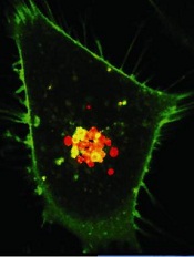
Experiments in human cancer cells showed that K-Ras-mutant tumor growth was highly dependent on the cells’ constant need to check and mend their DNA.
However, inhibiting the activity of H-Ras and N-Ras prevented the DNA damage response. And this made the cells more vulnerable to treatment.
“Our finding suggests that K-Ras cancers can be made more susceptible to existing therapies by interfering with their DNA repair mechanisms,” said Dafna Bar-Sagi, PhD, of the New York University School of Medicine.
“What some researchers have described as therapeutic ‘mission impossible’ may now become a ‘mission doable.’”
Dr Bar-Sagi and her colleagues reported this discovery in Cancer Cell.
The group’s research began with experiments to determine how Ras signaling leads to the uncontrolled growth of cancer cells. The team found that downregulation of wild-type H-Ras and N-Ras in mutant K-Ras cells caused the buildup of damaged DNA and slowed cell growth.
In the absence of H-Ras and N-Ras, K-Ras-mutant cancer cells failed to repair their DNA at the G2 phase of cell division. And this defect was caused by failure to properly activate Chk1.
With this in mind, the researchers decided to test the effects of H-Ras or N-Ras knockdown on treatment with DNA-damaging agents.
Knockdown of H-Ras or N-Ras sensitized K-Ras-mutant cancer cells to SN38 and oxaliplatin in vitro. But the same effect did not occur when H-Ras or N-Ras was knocked down in K-Ras-wild-type cancer cells.
K-Ras-mutant cancer cells were also sensitive to treatment with the Chk1/Chk2 inhibitor AZD7726 when H-Ras or N-Ras was knocked down in vitro.
To further support these findings, the researchers conducted experiments in mice with K-Ras-mutant tumors. Mice with H-Ras knockdown experienced tumor growth similar to controls.
But when the mice with H-Ras-suppressed tumors received the chemotherapy drug irinotecan, they experienced tumor regression that lasted up to 18 days post-treatment. On the other hand, mice without H-Ras suppression experienced modest tumor growth after treatment with irinotecan.
“Discovering more about how these different forms of Ras act on one another—including how they control DNA damage repair at Chk1 in combination with chemotherapy—could help us design drugs that greatly stall disease progression,” said study author Elda Grabocka, PhD, also of the New York University School of Medicine.
The researchers are now planning additional experiments on the biological interdependency of Ras proteins and what other chemotherapies might be involved in slowing cancer growth. Their goal is to map the Ras signaling pathways and identify as many therapeutic targets as possible.
Credit: PNAS
New findings suggest drugs can effectively fight K-Ras-mutant cancers—if they have a little help.
Experiments in human cancer cells showed that K-Ras-mutant tumor growth was highly dependent on the cells’ constant need to check and mend their DNA.
However, inhibiting the activity of H-Ras and N-Ras prevented the DNA damage response. And this made the cells more vulnerable to treatment.
“Our finding suggests that K-Ras cancers can be made more susceptible to existing therapies by interfering with their DNA repair mechanisms,” said Dafna Bar-Sagi, PhD, of the New York University School of Medicine.
“What some researchers have described as therapeutic ‘mission impossible’ may now become a ‘mission doable.’”
Dr Bar-Sagi and her colleagues reported this discovery in Cancer Cell.
The group’s research began with experiments to determine how Ras signaling leads to the uncontrolled growth of cancer cells. The team found that downregulation of wild-type H-Ras and N-Ras in mutant K-Ras cells caused the buildup of damaged DNA and slowed cell growth.
In the absence of H-Ras and N-Ras, K-Ras-mutant cancer cells failed to repair their DNA at the G2 phase of cell division. And this defect was caused by failure to properly activate Chk1.
With this in mind, the researchers decided to test the effects of H-Ras or N-Ras knockdown on treatment with DNA-damaging agents.
Knockdown of H-Ras or N-Ras sensitized K-Ras-mutant cancer cells to SN38 and oxaliplatin in vitro. But the same effect did not occur when H-Ras or N-Ras was knocked down in K-Ras-wild-type cancer cells.
K-Ras-mutant cancer cells were also sensitive to treatment with the Chk1/Chk2 inhibitor AZD7726 when H-Ras or N-Ras was knocked down in vitro.
To further support these findings, the researchers conducted experiments in mice with K-Ras-mutant tumors. Mice with H-Ras knockdown experienced tumor growth similar to controls.
But when the mice with H-Ras-suppressed tumors received the chemotherapy drug irinotecan, they experienced tumor regression that lasted up to 18 days post-treatment. On the other hand, mice without H-Ras suppression experienced modest tumor growth after treatment with irinotecan.
“Discovering more about how these different forms of Ras act on one another—including how they control DNA damage repair at Chk1 in combination with chemotherapy—could help us design drugs that greatly stall disease progression,” said study author Elda Grabocka, PhD, also of the New York University School of Medicine.
The researchers are now planning additional experiments on the biological interdependency of Ras proteins and what other chemotherapies might be involved in slowing cancer growth. Their goal is to map the Ras signaling pathways and identify as many therapeutic targets as possible.
Credit: PNAS
New findings suggest drugs can effectively fight K-Ras-mutant cancers—if they have a little help.
Experiments in human cancer cells showed that K-Ras-mutant tumor growth was highly dependent on the cells’ constant need to check and mend their DNA.
However, inhibiting the activity of H-Ras and N-Ras prevented the DNA damage response. And this made the cells more vulnerable to treatment.
“Our finding suggests that K-Ras cancers can be made more susceptible to existing therapies by interfering with their DNA repair mechanisms,” said Dafna Bar-Sagi, PhD, of the New York University School of Medicine.
“What some researchers have described as therapeutic ‘mission impossible’ may now become a ‘mission doable.’”
Dr Bar-Sagi and her colleagues reported this discovery in Cancer Cell.
The group’s research began with experiments to determine how Ras signaling leads to the uncontrolled growth of cancer cells. The team found that downregulation of wild-type H-Ras and N-Ras in mutant K-Ras cells caused the buildup of damaged DNA and slowed cell growth.
In the absence of H-Ras and N-Ras, K-Ras-mutant cancer cells failed to repair their DNA at the G2 phase of cell division. And this defect was caused by failure to properly activate Chk1.
With this in mind, the researchers decided to test the effects of H-Ras or N-Ras knockdown on treatment with DNA-damaging agents.
Knockdown of H-Ras or N-Ras sensitized K-Ras-mutant cancer cells to SN38 and oxaliplatin in vitro. But the same effect did not occur when H-Ras or N-Ras was knocked down in K-Ras-wild-type cancer cells.
K-Ras-mutant cancer cells were also sensitive to treatment with the Chk1/Chk2 inhibitor AZD7726 when H-Ras or N-Ras was knocked down in vitro.
To further support these findings, the researchers conducted experiments in mice with K-Ras-mutant tumors. Mice with H-Ras knockdown experienced tumor growth similar to controls.
But when the mice with H-Ras-suppressed tumors received the chemotherapy drug irinotecan, they experienced tumor regression that lasted up to 18 days post-treatment. On the other hand, mice without H-Ras suppression experienced modest tumor growth after treatment with irinotecan.
“Discovering more about how these different forms of Ras act on one another—including how they control DNA damage repair at Chk1 in combination with chemotherapy—could help us design drugs that greatly stall disease progression,” said study author Elda Grabocka, PhD, also of the New York University School of Medicine.
The researchers are now planning additional experiments on the biological interdependency of Ras proteins and what other chemotherapies might be involved in slowing cancer growth. Their goal is to map the Ras signaling pathways and identify as many therapeutic targets as possible.