User login
Study may explain why targeted treatment falls short in angiosarcoma

Heiser & Robert Ackland
Multiple mutations drive the development of angiosarcoma, according to a study published in Nature Genetics.
Researchers identified driver mutations in several genes associated with angiogenesis, including PTPRB and PLCG1.
They also found that PLCG1 mutations only occurred alongside mutations in PTPRB.
The investigators believe these findings may explain why angiosarcoma therapies directed at a single target fail to eradicate the disease.
Angiosarcoma is a rare cancer of the blood vessels that can occur spontaneously or develop after radiotherapy or chronic lymphedema.
Previous research indicated that aberrant angiogenesis, including somatic mutations in angiogenesis-signaling genes, drives angiosarcoma. So researchers developed drugs targeting pathways involved in angiogenesis, but these drugs have had little or no success.
“Because this cancer doesn’t respond well to traditional chemotherapy and radiotherapy, it makes sense to develop drugs that target pathways that control blood vessel formation,” said study author Peter Campbell, MD, PhD, of the Wellcome Trust Sanger Institute in the UK.
“We found 2 novel cancer genes that control blood vessel formation which are mutated in this cancer and which could be targeted for treatment of this highly aggressive cancer.”
To identify these genes, Dr Campbell and his colleagues performed whole-genome, whole-exome, and targeted sequencing in samples from patients with angiosarcoma.
Thirty-eight percent of the samples (15/39) carried mutations in genes that control angiogenesis, including PLCG1 and PTPRB.
The researchers identified 14 PTPRB mutations in 10 samples. This included 8 nonsense variants, 3 missense variants, 2 essential splice-site variants, and 1 frameshift insertion.
The investigators also discovered a recurrent mutation in PLCG1, a missense variant encoding p.Arg707Gln, which was present in 3 patient samples. All 3 PLCG1 mutations co-occurred with PTPRB mutations.
The researchers said this discovery may explain why drugs developed for a single target are ineffective in some angiosarcoma patients.
“Not only does our study change the way people view the biology of this tumor, it acts as a guide for future drug trials in angiosarcoma patients,” said study author Adrian Harris, MD, DPhil, of the University of Oxford in the UK.
He noted that researchers can use information from this study to determine if existing drugs could be effective against angiosarcoma. 
Heiser & Robert Ackland
Multiple mutations drive the development of angiosarcoma, according to a study published in Nature Genetics.
Researchers identified driver mutations in several genes associated with angiogenesis, including PTPRB and PLCG1.
They also found that PLCG1 mutations only occurred alongside mutations in PTPRB.
The investigators believe these findings may explain why angiosarcoma therapies directed at a single target fail to eradicate the disease.
Angiosarcoma is a rare cancer of the blood vessels that can occur spontaneously or develop after radiotherapy or chronic lymphedema.
Previous research indicated that aberrant angiogenesis, including somatic mutations in angiogenesis-signaling genes, drives angiosarcoma. So researchers developed drugs targeting pathways involved in angiogenesis, but these drugs have had little or no success.
“Because this cancer doesn’t respond well to traditional chemotherapy and radiotherapy, it makes sense to develop drugs that target pathways that control blood vessel formation,” said study author Peter Campbell, MD, PhD, of the Wellcome Trust Sanger Institute in the UK.
“We found 2 novel cancer genes that control blood vessel formation which are mutated in this cancer and which could be targeted for treatment of this highly aggressive cancer.”
To identify these genes, Dr Campbell and his colleagues performed whole-genome, whole-exome, and targeted sequencing in samples from patients with angiosarcoma.
Thirty-eight percent of the samples (15/39) carried mutations in genes that control angiogenesis, including PLCG1 and PTPRB.
The researchers identified 14 PTPRB mutations in 10 samples. This included 8 nonsense variants, 3 missense variants, 2 essential splice-site variants, and 1 frameshift insertion.
The investigators also discovered a recurrent mutation in PLCG1, a missense variant encoding p.Arg707Gln, which was present in 3 patient samples. All 3 PLCG1 mutations co-occurred with PTPRB mutations.
The researchers said this discovery may explain why drugs developed for a single target are ineffective in some angiosarcoma patients.
“Not only does our study change the way people view the biology of this tumor, it acts as a guide for future drug trials in angiosarcoma patients,” said study author Adrian Harris, MD, DPhil, of the University of Oxford in the UK.
He noted that researchers can use information from this study to determine if existing drugs could be effective against angiosarcoma.
Heiser & Robert Ackland
Multiple mutations drive the development of angiosarcoma, according to a study published in Nature Genetics.
Researchers identified driver mutations in several genes associated with angiogenesis, including PTPRB and PLCG1.
They also found that PLCG1 mutations only occurred alongside mutations in PTPRB.
The investigators believe these findings may explain why angiosarcoma therapies directed at a single target fail to eradicate the disease.
Angiosarcoma is a rare cancer of the blood vessels that can occur spontaneously or develop after radiotherapy or chronic lymphedema.
Previous research indicated that aberrant angiogenesis, including somatic mutations in angiogenesis-signaling genes, drives angiosarcoma. So researchers developed drugs targeting pathways involved in angiogenesis, but these drugs have had little or no success.
“Because this cancer doesn’t respond well to traditional chemotherapy and radiotherapy, it makes sense to develop drugs that target pathways that control blood vessel formation,” said study author Peter Campbell, MD, PhD, of the Wellcome Trust Sanger Institute in the UK.
“We found 2 novel cancer genes that control blood vessel formation which are mutated in this cancer and which could be targeted for treatment of this highly aggressive cancer.”
To identify these genes, Dr Campbell and his colleagues performed whole-genome, whole-exome, and targeted sequencing in samples from patients with angiosarcoma.
Thirty-eight percent of the samples (15/39) carried mutations in genes that control angiogenesis, including PLCG1 and PTPRB.
The researchers identified 14 PTPRB mutations in 10 samples. This included 8 nonsense variants, 3 missense variants, 2 essential splice-site variants, and 1 frameshift insertion.
The investigators also discovered a recurrent mutation in PLCG1, a missense variant encoding p.Arg707Gln, which was present in 3 patient samples. All 3 PLCG1 mutations co-occurred with PTPRB mutations.
The researchers said this discovery may explain why drugs developed for a single target are ineffective in some angiosarcoma patients.
“Not only does our study change the way people view the biology of this tumor, it acts as a guide for future drug trials in angiosarcoma patients,” said study author Adrian Harris, MD, DPhil, of the University of Oxford in the UK.
He noted that researchers can use information from this study to determine if existing drugs could be effective against angiosarcoma.
How diabetes drugs can fight hematologic malignancies
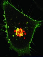
Credit: PNAS
Researchers say they’ve discovered how a class of diabetes drugs known as biguanides exerts anticancer properties in certain malignancies.
The team identified a mitochondrial pathway that imbues cancer cells with the ability to survive in low-glucose environments.
By finding cancer cells with defects in this pathway or impaired glucose utilization, the researchers found they could predict which cancers would be sensitive to drugs that inhibit this pathway.
And follow-up experiments confirmed that lymphoma, leukemia, and myeloma tumors were among those sensitive to treatment.
Kivanç Birsoy, PhD, of the Whitehead Institute for Biomedical Research in Cambridge, Massachusetts, and his colleagues reported these findings in Nature.
To study how cancer cells survive in the kind of low-glucose environment found within cancerous tumors, the researchers developed a system that circulates low-nutrient media continuously around cells.
Of the 30 cancer cell lines the team tested within this system, most appeared unaffected by a lack of glucose. However, a few of the cells lines thrived and reproduced rapidly, while others struggled.
Specifically, a low-glucose environment prompted an increase in proliferation for the Burkitt lymphoma cell line Raji, as well as in medulloblastoma, lung, and stomach cancer cell lines.
However, the lymphoma cell lines U-937 and MC116, as well as the myeloma cell lines NCI-H929 and KMS-26, saw significant decreases in proliferation in a low-glucose environment. The leukemia cell line Jurkat was moderately sensitive to a low-glucose environment.
“No one really understood why cancer cells had these responses or whether they were important for the formation of the tumor,” said study author Richard Possemato, PhD, also of the Whitehead Institute.
To gain more insight, the researchers screened overly distressed cells for genes whose suppression improved or further hindered the cells’ survival rates. The screen flagged genes involved in glucose transportation and oxidative phosphorylation.
The team hypothesized that cancer cells with mutations in these genes are over-taxing their mitochondria under normal conditions. When placed in a harsh, low-glucose environment, the mitochondria are maxed out, and the cells suffer.
If true, the hypothesis would suggest that further impairing mitochondrial function with biguanides, which are known oxidative phosphorylation inhibitors, could push the mitochondria beyond their limits, to the detriment of the cancer cells.
The researchers first tested this hypothesis in vitro on cell lines with glucose utilization defects (NCI-H929, KMS-26, LP-1, L-363, MOLP-8, D341Med, and KMS-28BM) or mitochondrial DNA (mtDNA) mutations (U-937, BxPC3, Cal-62, HCC-1438, HCC-827, and NU-DHL-1).
They found that, in a low-glucose environment, cell lines with mtDNA mutations or impaired glucose utilization were 5 to 20 times more susceptible to phenformin, a more potent biguanide than metformin, when compared to control cancer cell lines or an immortalized B-cell line.
The team then tested phenformin in mice implanted with tumors derived from low-glucose-sensitive cancer cells. The drug inhibited the growth of tumors derived from cancer cells with mtDNA mutations (Cal-62 and U-937) or poor glucose consumption (KMS-26 and NCI-H929) but not from cells lacking these defects (NCI-H2171 and NCI-H82).
“These results show that mitochondrial DNA mutations and glucose import defects can be used as biomarkers for biguanide sensitivity to determine if a cancer patient might benefit from these drugs,” Dr Birsoy said.
“And this is the first time that anyone has shown that the direct cytotoxic effects of this class of drugs, including metformin and phenformin, on cancer cells are mediated through their effect on mitochondria.”
To confirm the accuracy of their proposed biomarkers, the researchers now want to analyze previous clinical trials to see if cancer patients with the proposed biomarkers fared better with metformin treatment than patients without the biomarkers.
Credit: PNAS
Researchers say they’ve discovered how a class of diabetes drugs known as biguanides exerts anticancer properties in certain malignancies.
The team identified a mitochondrial pathway that imbues cancer cells with the ability to survive in low-glucose environments.
By finding cancer cells with defects in this pathway or impaired glucose utilization, the researchers found they could predict which cancers would be sensitive to drugs that inhibit this pathway.
And follow-up experiments confirmed that lymphoma, leukemia, and myeloma tumors were among those sensitive to treatment.
Kivanç Birsoy, PhD, of the Whitehead Institute for Biomedical Research in Cambridge, Massachusetts, and his colleagues reported these findings in Nature.
To study how cancer cells survive in the kind of low-glucose environment found within cancerous tumors, the researchers developed a system that circulates low-nutrient media continuously around cells.
Of the 30 cancer cell lines the team tested within this system, most appeared unaffected by a lack of glucose. However, a few of the cells lines thrived and reproduced rapidly, while others struggled.
Specifically, a low-glucose environment prompted an increase in proliferation for the Burkitt lymphoma cell line Raji, as well as in medulloblastoma, lung, and stomach cancer cell lines.
However, the lymphoma cell lines U-937 and MC116, as well as the myeloma cell lines NCI-H929 and KMS-26, saw significant decreases in proliferation in a low-glucose environment. The leukemia cell line Jurkat was moderately sensitive to a low-glucose environment.
“No one really understood why cancer cells had these responses or whether they were important for the formation of the tumor,” said study author Richard Possemato, PhD, also of the Whitehead Institute.
To gain more insight, the researchers screened overly distressed cells for genes whose suppression improved or further hindered the cells’ survival rates. The screen flagged genes involved in glucose transportation and oxidative phosphorylation.
The team hypothesized that cancer cells with mutations in these genes are over-taxing their mitochondria under normal conditions. When placed in a harsh, low-glucose environment, the mitochondria are maxed out, and the cells suffer.
If true, the hypothesis would suggest that further impairing mitochondrial function with biguanides, which are known oxidative phosphorylation inhibitors, could push the mitochondria beyond their limits, to the detriment of the cancer cells.
The researchers first tested this hypothesis in vitro on cell lines with glucose utilization defects (NCI-H929, KMS-26, LP-1, L-363, MOLP-8, D341Med, and KMS-28BM) or mitochondrial DNA (mtDNA) mutations (U-937, BxPC3, Cal-62, HCC-1438, HCC-827, and NU-DHL-1).
They found that, in a low-glucose environment, cell lines with mtDNA mutations or impaired glucose utilization were 5 to 20 times more susceptible to phenformin, a more potent biguanide than metformin, when compared to control cancer cell lines or an immortalized B-cell line.
The team then tested phenformin in mice implanted with tumors derived from low-glucose-sensitive cancer cells. The drug inhibited the growth of tumors derived from cancer cells with mtDNA mutations (Cal-62 and U-937) or poor glucose consumption (KMS-26 and NCI-H929) but not from cells lacking these defects (NCI-H2171 and NCI-H82).
“These results show that mitochondrial DNA mutations and glucose import defects can be used as biomarkers for biguanide sensitivity to determine if a cancer patient might benefit from these drugs,” Dr Birsoy said.
“And this is the first time that anyone has shown that the direct cytotoxic effects of this class of drugs, including metformin and phenformin, on cancer cells are mediated through their effect on mitochondria.”
To confirm the accuracy of their proposed biomarkers, the researchers now want to analyze previous clinical trials to see if cancer patients with the proposed biomarkers fared better with metformin treatment than patients without the biomarkers.
Credit: PNAS
Researchers say they’ve discovered how a class of diabetes drugs known as biguanides exerts anticancer properties in certain malignancies.
The team identified a mitochondrial pathway that imbues cancer cells with the ability to survive in low-glucose environments.
By finding cancer cells with defects in this pathway or impaired glucose utilization, the researchers found they could predict which cancers would be sensitive to drugs that inhibit this pathway.
And follow-up experiments confirmed that lymphoma, leukemia, and myeloma tumors were among those sensitive to treatment.
Kivanç Birsoy, PhD, of the Whitehead Institute for Biomedical Research in Cambridge, Massachusetts, and his colleagues reported these findings in Nature.
To study how cancer cells survive in the kind of low-glucose environment found within cancerous tumors, the researchers developed a system that circulates low-nutrient media continuously around cells.
Of the 30 cancer cell lines the team tested within this system, most appeared unaffected by a lack of glucose. However, a few of the cells lines thrived and reproduced rapidly, while others struggled.
Specifically, a low-glucose environment prompted an increase in proliferation for the Burkitt lymphoma cell line Raji, as well as in medulloblastoma, lung, and stomach cancer cell lines.
However, the lymphoma cell lines U-937 and MC116, as well as the myeloma cell lines NCI-H929 and KMS-26, saw significant decreases in proliferation in a low-glucose environment. The leukemia cell line Jurkat was moderately sensitive to a low-glucose environment.
“No one really understood why cancer cells had these responses or whether they were important for the formation of the tumor,” said study author Richard Possemato, PhD, also of the Whitehead Institute.
To gain more insight, the researchers screened overly distressed cells for genes whose suppression improved or further hindered the cells’ survival rates. The screen flagged genes involved in glucose transportation and oxidative phosphorylation.
The team hypothesized that cancer cells with mutations in these genes are over-taxing their mitochondria under normal conditions. When placed in a harsh, low-glucose environment, the mitochondria are maxed out, and the cells suffer.
If true, the hypothesis would suggest that further impairing mitochondrial function with biguanides, which are known oxidative phosphorylation inhibitors, could push the mitochondria beyond their limits, to the detriment of the cancer cells.
The researchers first tested this hypothesis in vitro on cell lines with glucose utilization defects (NCI-H929, KMS-26, LP-1, L-363, MOLP-8, D341Med, and KMS-28BM) or mitochondrial DNA (mtDNA) mutations (U-937, BxPC3, Cal-62, HCC-1438, HCC-827, and NU-DHL-1).
They found that, in a low-glucose environment, cell lines with mtDNA mutations or impaired glucose utilization were 5 to 20 times more susceptible to phenformin, a more potent biguanide than metformin, when compared to control cancer cell lines or an immortalized B-cell line.
The team then tested phenformin in mice implanted with tumors derived from low-glucose-sensitive cancer cells. The drug inhibited the growth of tumors derived from cancer cells with mtDNA mutations (Cal-62 and U-937) or poor glucose consumption (KMS-26 and NCI-H929) but not from cells lacking these defects (NCI-H2171 and NCI-H82).
“These results show that mitochondrial DNA mutations and glucose import defects can be used as biomarkers for biguanide sensitivity to determine if a cancer patient might benefit from these drugs,” Dr Birsoy said.
“And this is the first time that anyone has shown that the direct cytotoxic effects of this class of drugs, including metformin and phenformin, on cancer cells are mediated through their effect on mitochondria.”
To confirm the accuracy of their proposed biomarkers, the researchers now want to analyze previous clinical trials to see if cancer patients with the proposed biomarkers fared better with metformin treatment than patients without the biomarkers.
FDA approves apixaban to prevent DVT, PE

Credit: CDC
The US Food and Drug Administration (FDA) has approved apixaban (Eliquis) as prophylaxis for deep vein thrombosis (DVT), which may lead to pulmonary embolism (PE), in patients who have undergone hip or knee replacement surgery.
Apixaban is already FDA-approved to reduce the risk of stroke and systemic embolism in patients with nonvalvular atrial fibrillation.
The latest approval is supported by data from 3 trials comprising the ADVANCE clinical trial program.
Results of the first ADVANCE study suggested apixaban was roughly as effective as enoxaparin at preventing DVT and PE in patients who had undergone total knee replacement surgery. But apixaban posed a significantly lower risk of major and nonmajor bleeding.
The ADVANCE-2 study, on the other hand, indicated that apixaban was a more effective means of thromboprophylaxis than enoxaparin in this patient population. And there was no significant difference between the treatment arms in the frequency of major or clinically relevant bleeding.
The ADVANCE-3 study suggested apixaban was more effective than enoxaparin in preventing DVT and PE among patients undergoing hip replacement. And there was no significant difference between the groups with regard to major or clinically relevant bleeding.
The prescribing information for apixaban includes a boxed warning detailing the increased risk of stroke in patients with nonvalvular atrial fibrillation who discontinue the drug without adequate continuous anticoagulation.
The boxed warning also states that, in patients undergoing spinal epidural anesthesia or spinal puncture, apixaban poses an increased risk of epidural or spinal hematoma, which may cause long-term or permanent paralysis.
The risk of these events may be increased by the use of indwelling epidural catheters for the administration of analgesia or by the concomitant use of drugs affecting hemostasis, such as nonsteroidal anti-inflammatory drugs, platelet aggregation inhibitors, or other anticoagulants. The risk also appears to be increased by traumatic or repeated epidural or spinal puncture.
Healthcare professionals should monitor patients for signs and symptoms of neurologic impairment. If neurologic compromise is noted, urgent treatment is necessary.
For more information on adverse events and contraindications, see the full prescribing information for apixaban. The drug is under joint development by Pfizer and Bristol-Myers Squibb.
Credit: CDC
The US Food and Drug Administration (FDA) has approved apixaban (Eliquis) as prophylaxis for deep vein thrombosis (DVT), which may lead to pulmonary embolism (PE), in patients who have undergone hip or knee replacement surgery.
Apixaban is already FDA-approved to reduce the risk of stroke and systemic embolism in patients with nonvalvular atrial fibrillation.
The latest approval is supported by data from 3 trials comprising the ADVANCE clinical trial program.
Results of the first ADVANCE study suggested apixaban was roughly as effective as enoxaparin at preventing DVT and PE in patients who had undergone total knee replacement surgery. But apixaban posed a significantly lower risk of major and nonmajor bleeding.
The ADVANCE-2 study, on the other hand, indicated that apixaban was a more effective means of thromboprophylaxis than enoxaparin in this patient population. And there was no significant difference between the treatment arms in the frequency of major or clinically relevant bleeding.
The ADVANCE-3 study suggested apixaban was more effective than enoxaparin in preventing DVT and PE among patients undergoing hip replacement. And there was no significant difference between the groups with regard to major or clinically relevant bleeding.
The prescribing information for apixaban includes a boxed warning detailing the increased risk of stroke in patients with nonvalvular atrial fibrillation who discontinue the drug without adequate continuous anticoagulation.
The boxed warning also states that, in patients undergoing spinal epidural anesthesia or spinal puncture, apixaban poses an increased risk of epidural or spinal hematoma, which may cause long-term or permanent paralysis.
The risk of these events may be increased by the use of indwelling epidural catheters for the administration of analgesia or by the concomitant use of drugs affecting hemostasis, such as nonsteroidal anti-inflammatory drugs, platelet aggregation inhibitors, or other anticoagulants. The risk also appears to be increased by traumatic or repeated epidural or spinal puncture.
Healthcare professionals should monitor patients for signs and symptoms of neurologic impairment. If neurologic compromise is noted, urgent treatment is necessary.
For more information on adverse events and contraindications, see the full prescribing information for apixaban. The drug is under joint development by Pfizer and Bristol-Myers Squibb.
Credit: CDC
The US Food and Drug Administration (FDA) has approved apixaban (Eliquis) as prophylaxis for deep vein thrombosis (DVT), which may lead to pulmonary embolism (PE), in patients who have undergone hip or knee replacement surgery.
Apixaban is already FDA-approved to reduce the risk of stroke and systemic embolism in patients with nonvalvular atrial fibrillation.
The latest approval is supported by data from 3 trials comprising the ADVANCE clinical trial program.
Results of the first ADVANCE study suggested apixaban was roughly as effective as enoxaparin at preventing DVT and PE in patients who had undergone total knee replacement surgery. But apixaban posed a significantly lower risk of major and nonmajor bleeding.
The ADVANCE-2 study, on the other hand, indicated that apixaban was a more effective means of thromboprophylaxis than enoxaparin in this patient population. And there was no significant difference between the treatment arms in the frequency of major or clinically relevant bleeding.
The ADVANCE-3 study suggested apixaban was more effective than enoxaparin in preventing DVT and PE among patients undergoing hip replacement. And there was no significant difference between the groups with regard to major or clinically relevant bleeding.
The prescribing information for apixaban includes a boxed warning detailing the increased risk of stroke in patients with nonvalvular atrial fibrillation who discontinue the drug without adequate continuous anticoagulation.
The boxed warning also states that, in patients undergoing spinal epidural anesthesia or spinal puncture, apixaban poses an increased risk of epidural or spinal hematoma, which may cause long-term or permanent paralysis.
The risk of these events may be increased by the use of indwelling epidural catheters for the administration of analgesia or by the concomitant use of drugs affecting hemostasis, such as nonsteroidal anti-inflammatory drugs, platelet aggregation inhibitors, or other anticoagulants. The risk also appears to be increased by traumatic or repeated epidural or spinal puncture.
Healthcare professionals should monitor patients for signs and symptoms of neurologic impairment. If neurologic compromise is noted, urgent treatment is necessary.
For more information on adverse events and contraindications, see the full prescribing information for apixaban. The drug is under joint development by Pfizer and Bristol-Myers Squibb.
New guidelines for managing sickle cell patients

(right) and a normal one
Credit: Betty Pace
The American Thoracic Society has developed clinical practice guidelines to help clinicians identify and manage patients with sickle cell disease who are at an increased risk for mortality from pulmonary hypertension.
“With the development of new treatments, many patients with sickle cell disease are now surviving long enough to develop pulmonary hypertension, with an estimated prevalence of 6% to 11%,” said guideline author Elizabeth S. Klings, MD, of the Boston University School of Medicine in Massachusetts.
“Although pulmonary hypertension and elevated tricuspid jet velocity [TRV, an indicator of pulmonary hypertension measured by echocardiography] are both associated with an increased mortality risk, there is currently no standardized approach for identifying and managing these patients.”
So Dr Klings and her colleagues decided to offer some guidance. Their recommendations for managing these patients appear in the American Journal of Respiratory and Critical Care Medicine.
The guideline authors point out that clinicians can evaluate patient mortality noninvasively by measuring the TRV with Doppler echocardiography or by measuring serum N-terminal pro–brain natriuretic peptide (NT-pro-BNP) levels.
An invasive method is taking direct hemodynamic measurements via right heart catheterization (RHC).
An increased risk for mortality is defined as a TRV ≥ 2.5 m/second, an NT-pro-BNP level ≥ 160 pg/mL, or RHC-confirmed pulmonary hypertension.
Patients who meet these criteria should receive hydroxyurea. Patients who do not respond to or are not candidates for hydroxyurea treatment can be considered for chronic transfusion therapy.
For patients with RHC-confirmed pulmonary hypertension, venous thromboembolism, and no additional risk factors for hemorrhage, the guidelines recommend indefinite anticoagulant therapy rather than a limited duration of therapy.
Patients with elevated TRV alone or elevated NT-pro-BNP alone should not be treated with targeted pulmonary arterial hypertension therapies, including prostanoid, endothelin receptor antagonist, and phosphodiesterase-5 inhibitor therapy.
Most patients with RHC-confirmed pulmonary hypertension should not receive targeted therapy.
For select patents with RHC-confirmed marked elevation of pulmonary vascular resistance, normal pulmonary capillary wedge pressure, and related symptoms, the guidelines suggest a trial of either a prostanoid or an endothelin receptor antagonist.
Patients with RHC-confirmed marked elevation of pulmonary vascular resistance, normal pulmonary capillary wedge pressure, and related symptoms should not receive phosphodiesterase-5 inhibitor therapy as first-line treatment.
“Most of our current recommendations are limited by a lack of large-scale clinical trials in this population,” Dr Klings noted. “We need to continue our research efforts into this disease and its management to understand what the optimal treatment regimen for these patients is.”
“Management of patients with sickle cell disease with an increased risk for mortality and pulmonary hypertension will ultimately be a collaborative effort, including adult and pediatric pulmonologists, cardiologists, and hematologists.”
(right) and a normal one
Credit: Betty Pace
The American Thoracic Society has developed clinical practice guidelines to help clinicians identify and manage patients with sickle cell disease who are at an increased risk for mortality from pulmonary hypertension.
“With the development of new treatments, many patients with sickle cell disease are now surviving long enough to develop pulmonary hypertension, with an estimated prevalence of 6% to 11%,” said guideline author Elizabeth S. Klings, MD, of the Boston University School of Medicine in Massachusetts.
“Although pulmonary hypertension and elevated tricuspid jet velocity [TRV, an indicator of pulmonary hypertension measured by echocardiography] are both associated with an increased mortality risk, there is currently no standardized approach for identifying and managing these patients.”
So Dr Klings and her colleagues decided to offer some guidance. Their recommendations for managing these patients appear in the American Journal of Respiratory and Critical Care Medicine.
The guideline authors point out that clinicians can evaluate patient mortality noninvasively by measuring the TRV with Doppler echocardiography or by measuring serum N-terminal pro–brain natriuretic peptide (NT-pro-BNP) levels.
An invasive method is taking direct hemodynamic measurements via right heart catheterization (RHC).
An increased risk for mortality is defined as a TRV ≥ 2.5 m/second, an NT-pro-BNP level ≥ 160 pg/mL, or RHC-confirmed pulmonary hypertension.
Patients who meet these criteria should receive hydroxyurea. Patients who do not respond to or are not candidates for hydroxyurea treatment can be considered for chronic transfusion therapy.
For patients with RHC-confirmed pulmonary hypertension, venous thromboembolism, and no additional risk factors for hemorrhage, the guidelines recommend indefinite anticoagulant therapy rather than a limited duration of therapy.
Patients with elevated TRV alone or elevated NT-pro-BNP alone should not be treated with targeted pulmonary arterial hypertension therapies, including prostanoid, endothelin receptor antagonist, and phosphodiesterase-5 inhibitor therapy.
Most patients with RHC-confirmed pulmonary hypertension should not receive targeted therapy.
For select patents with RHC-confirmed marked elevation of pulmonary vascular resistance, normal pulmonary capillary wedge pressure, and related symptoms, the guidelines suggest a trial of either a prostanoid or an endothelin receptor antagonist.
Patients with RHC-confirmed marked elevation of pulmonary vascular resistance, normal pulmonary capillary wedge pressure, and related symptoms should not receive phosphodiesterase-5 inhibitor therapy as first-line treatment.
“Most of our current recommendations are limited by a lack of large-scale clinical trials in this population,” Dr Klings noted. “We need to continue our research efforts into this disease and its management to understand what the optimal treatment regimen for these patients is.”
“Management of patients with sickle cell disease with an increased risk for mortality and pulmonary hypertension will ultimately be a collaborative effort, including adult and pediatric pulmonologists, cardiologists, and hematologists.”
(right) and a normal one
Credit: Betty Pace
The American Thoracic Society has developed clinical practice guidelines to help clinicians identify and manage patients with sickle cell disease who are at an increased risk for mortality from pulmonary hypertension.
“With the development of new treatments, many patients with sickle cell disease are now surviving long enough to develop pulmonary hypertension, with an estimated prevalence of 6% to 11%,” said guideline author Elizabeth S. Klings, MD, of the Boston University School of Medicine in Massachusetts.
“Although pulmonary hypertension and elevated tricuspid jet velocity [TRV, an indicator of pulmonary hypertension measured by echocardiography] are both associated with an increased mortality risk, there is currently no standardized approach for identifying and managing these patients.”
So Dr Klings and her colleagues decided to offer some guidance. Their recommendations for managing these patients appear in the American Journal of Respiratory and Critical Care Medicine.
The guideline authors point out that clinicians can evaluate patient mortality noninvasively by measuring the TRV with Doppler echocardiography or by measuring serum N-terminal pro–brain natriuretic peptide (NT-pro-BNP) levels.
An invasive method is taking direct hemodynamic measurements via right heart catheterization (RHC).
An increased risk for mortality is defined as a TRV ≥ 2.5 m/second, an NT-pro-BNP level ≥ 160 pg/mL, or RHC-confirmed pulmonary hypertension.
Patients who meet these criteria should receive hydroxyurea. Patients who do not respond to or are not candidates for hydroxyurea treatment can be considered for chronic transfusion therapy.
For patients with RHC-confirmed pulmonary hypertension, venous thromboembolism, and no additional risk factors for hemorrhage, the guidelines recommend indefinite anticoagulant therapy rather than a limited duration of therapy.
Patients with elevated TRV alone or elevated NT-pro-BNP alone should not be treated with targeted pulmonary arterial hypertension therapies, including prostanoid, endothelin receptor antagonist, and phosphodiesterase-5 inhibitor therapy.
Most patients with RHC-confirmed pulmonary hypertension should not receive targeted therapy.
For select patents with RHC-confirmed marked elevation of pulmonary vascular resistance, normal pulmonary capillary wedge pressure, and related symptoms, the guidelines suggest a trial of either a prostanoid or an endothelin receptor antagonist.
Patients with RHC-confirmed marked elevation of pulmonary vascular resistance, normal pulmonary capillary wedge pressure, and related symptoms should not receive phosphodiesterase-5 inhibitor therapy as first-line treatment.
“Most of our current recommendations are limited by a lack of large-scale clinical trials in this population,” Dr Klings noted. “We need to continue our research efforts into this disease and its management to understand what the optimal treatment regimen for these patients is.”
“Management of patients with sickle cell disease with an increased risk for mortality and pulmonary hypertension will ultimately be a collaborative effort, including adult and pediatric pulmonologists, cardiologists, and hematologists.”
Investigation reveals ‘inappropriate data handling’ but no misconduct
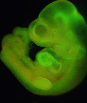
generated from STAP cells
Credit: Haruko Obokata
The Japanese research institute RIKEN has released early results of its investigation into allegations of misconduct leveled against the creators of STAP cells (stimulus-triggered acquisition of pluripotency cells).
RIKEN has confirmed 2 cases of “inappropriate data handling” but said the circumstances did not constitute misconduct.
The investigation is ongoing, with 4 issues—including charges of plagiarism and doctored figures—still to be resolved.
Research prompts questions, criticism
The investigation began shortly after a group of RIKEN scientists and colleagues from a few other institutions announced their creation of STAP cells.
The researchers said they could induce pluripotency in somatic cells by introducing the cells to a low-pH environment, and they reported this discovery in an article and a letter to Nature.
Not long after the papers were published, members of the scientific community began questioning the validity of the research, citing issues with images, possible plagiarism, and an inability to replicate the experiments described.
In light of these issues, one of the study authors recently called for the research to be retracted.
Teruhiko Wakayama, PhD, formerly of RIKEN but now a professor at the University of Yamanashi, said there are “too many uncertainties” surrounding the research at this point. After a retraction, the researchers could collect new data and images to ensure their accuracy and resubmit the research for publication.
On the other hand, fellow study author Charles Vacanti, MD, of Brigham and Women’s Hospital in Boston, has said a retraction is unnecessary.
“I firmly believe that the questions and concerns raised about our STAP cell paper published in Nature do not affect our findings or conclusions,” Dr Vacanti said.
Investigation launched
In response to the questions and allegations, RIKEN formed a committee to investigate the possibility of misconduct.
The investigation is focusing on 4 of the researchers involved: Haruko Obokata, PhD; Yoshiki Sasai, MD, PhD; Hitoshi Niwa, MD, PhD; and Dr Wakayama.
The committee is also looking into 6 issues with the research, 2 of which have been resolved.
Resolved issues
(1) Critics have questioned the “unnatural appearance of colored cell parts shown by arrows in d2 and d3 images of Figure 1f” in the article.
RIKEN concluded that the process of preparing these images did not constitute fabrication within the context of research misconduct.
(2) Questions have been raised about a “strong resemblance between the rightmost panel in Figure 1b and the lower panel in Figure 2g, both showing a fluorescence image of mice placenta” in the letter.
There is no reference to the figures in the figure legends or the main body of text, and RIKEN does define this sort of discrepancy as fabrication. However, the researchers claimed they had intended to delete one of the figures prior to publication but forgot, and there is no evidence to contradict that explanation. So RIKEN concluded that no malice was intended, and this should not be considered misconduct.
Issues under investigation
(1) In Figure 1i of the article, lane 3 appears to have been inserted.
(2) A part of the article’s “Methods” section on karyotyping analysis appears to have been copied from another paper.
(3) Some of the description of karyotyping in the “Methods” section of the article is different from the procedure the researchers followed.
(4) In the article, the image of differentiated cells for Figures 2d and 2e and the image of chimera mouse immunostaining data are incorrect, and investigation revealed that these images closely resemble images Dr Obokata used in her doctoral dissertation.
Next steps
RIKEN said it will continue with the investigation and issue a full report upon its completion. The institute also aims to determine whether the STAP cell experiments can be reproduced.
“The reproducibility and credibility of the STAP phenomenon must be rigorously validated, not only by RIKEN scientists, but also by others,” said RIKEN President Ryoji Noyori, PhD.
“I have instructed our people to cooperate fully with researchers at outside institutions in their efforts to replicate the STAP cell results.”
Dr Noyori added that RIKEN is prepared to withdraw the Nature papers and take “strict disciplinary action” against the researchers involved if the investigation reveals deliberate misconduct.
generated from STAP cells
Credit: Haruko Obokata
The Japanese research institute RIKEN has released early results of its investigation into allegations of misconduct leveled against the creators of STAP cells (stimulus-triggered acquisition of pluripotency cells).
RIKEN has confirmed 2 cases of “inappropriate data handling” but said the circumstances did not constitute misconduct.
The investigation is ongoing, with 4 issues—including charges of plagiarism and doctored figures—still to be resolved.
Research prompts questions, criticism
The investigation began shortly after a group of RIKEN scientists and colleagues from a few other institutions announced their creation of STAP cells.
The researchers said they could induce pluripotency in somatic cells by introducing the cells to a low-pH environment, and they reported this discovery in an article and a letter to Nature.
Not long after the papers were published, members of the scientific community began questioning the validity of the research, citing issues with images, possible plagiarism, and an inability to replicate the experiments described.
In light of these issues, one of the study authors recently called for the research to be retracted.
Teruhiko Wakayama, PhD, formerly of RIKEN but now a professor at the University of Yamanashi, said there are “too many uncertainties” surrounding the research at this point. After a retraction, the researchers could collect new data and images to ensure their accuracy and resubmit the research for publication.
On the other hand, fellow study author Charles Vacanti, MD, of Brigham and Women’s Hospital in Boston, has said a retraction is unnecessary.
“I firmly believe that the questions and concerns raised about our STAP cell paper published in Nature do not affect our findings or conclusions,” Dr Vacanti said.
Investigation launched
In response to the questions and allegations, RIKEN formed a committee to investigate the possibility of misconduct.
The investigation is focusing on 4 of the researchers involved: Haruko Obokata, PhD; Yoshiki Sasai, MD, PhD; Hitoshi Niwa, MD, PhD; and Dr Wakayama.
The committee is also looking into 6 issues with the research, 2 of which have been resolved.
Resolved issues
(1) Critics have questioned the “unnatural appearance of colored cell parts shown by arrows in d2 and d3 images of Figure 1f” in the article.
RIKEN concluded that the process of preparing these images did not constitute fabrication within the context of research misconduct.
(2) Questions have been raised about a “strong resemblance between the rightmost panel in Figure 1b and the lower panel in Figure 2g, both showing a fluorescence image of mice placenta” in the letter.
There is no reference to the figures in the figure legends or the main body of text, and RIKEN does define this sort of discrepancy as fabrication. However, the researchers claimed they had intended to delete one of the figures prior to publication but forgot, and there is no evidence to contradict that explanation. So RIKEN concluded that no malice was intended, and this should not be considered misconduct.
Issues under investigation
(1) In Figure 1i of the article, lane 3 appears to have been inserted.
(2) A part of the article’s “Methods” section on karyotyping analysis appears to have been copied from another paper.
(3) Some of the description of karyotyping in the “Methods” section of the article is different from the procedure the researchers followed.
(4) In the article, the image of differentiated cells for Figures 2d and 2e and the image of chimera mouse immunostaining data are incorrect, and investigation revealed that these images closely resemble images Dr Obokata used in her doctoral dissertation.
Next steps
RIKEN said it will continue with the investigation and issue a full report upon its completion. The institute also aims to determine whether the STAP cell experiments can be reproduced.
“The reproducibility and credibility of the STAP phenomenon must be rigorously validated, not only by RIKEN scientists, but also by others,” said RIKEN President Ryoji Noyori, PhD.
“I have instructed our people to cooperate fully with researchers at outside institutions in their efforts to replicate the STAP cell results.”
Dr Noyori added that RIKEN is prepared to withdraw the Nature papers and take “strict disciplinary action” against the researchers involved if the investigation reveals deliberate misconduct.
generated from STAP cells
Credit: Haruko Obokata
The Japanese research institute RIKEN has released early results of its investigation into allegations of misconduct leveled against the creators of STAP cells (stimulus-triggered acquisition of pluripotency cells).
RIKEN has confirmed 2 cases of “inappropriate data handling” but said the circumstances did not constitute misconduct.
The investigation is ongoing, with 4 issues—including charges of plagiarism and doctored figures—still to be resolved.
Research prompts questions, criticism
The investigation began shortly after a group of RIKEN scientists and colleagues from a few other institutions announced their creation of STAP cells.
The researchers said they could induce pluripotency in somatic cells by introducing the cells to a low-pH environment, and they reported this discovery in an article and a letter to Nature.
Not long after the papers were published, members of the scientific community began questioning the validity of the research, citing issues with images, possible plagiarism, and an inability to replicate the experiments described.
In light of these issues, one of the study authors recently called for the research to be retracted.
Teruhiko Wakayama, PhD, formerly of RIKEN but now a professor at the University of Yamanashi, said there are “too many uncertainties” surrounding the research at this point. After a retraction, the researchers could collect new data and images to ensure their accuracy and resubmit the research for publication.
On the other hand, fellow study author Charles Vacanti, MD, of Brigham and Women’s Hospital in Boston, has said a retraction is unnecessary.
“I firmly believe that the questions and concerns raised about our STAP cell paper published in Nature do not affect our findings or conclusions,” Dr Vacanti said.
Investigation launched
In response to the questions and allegations, RIKEN formed a committee to investigate the possibility of misconduct.
The investigation is focusing on 4 of the researchers involved: Haruko Obokata, PhD; Yoshiki Sasai, MD, PhD; Hitoshi Niwa, MD, PhD; and Dr Wakayama.
The committee is also looking into 6 issues with the research, 2 of which have been resolved.
Resolved issues
(1) Critics have questioned the “unnatural appearance of colored cell parts shown by arrows in d2 and d3 images of Figure 1f” in the article.
RIKEN concluded that the process of preparing these images did not constitute fabrication within the context of research misconduct.
(2) Questions have been raised about a “strong resemblance between the rightmost panel in Figure 1b and the lower panel in Figure 2g, both showing a fluorescence image of mice placenta” in the letter.
There is no reference to the figures in the figure legends or the main body of text, and RIKEN does define this sort of discrepancy as fabrication. However, the researchers claimed they had intended to delete one of the figures prior to publication but forgot, and there is no evidence to contradict that explanation. So RIKEN concluded that no malice was intended, and this should not be considered misconduct.
Issues under investigation
(1) In Figure 1i of the article, lane 3 appears to have been inserted.
(2) A part of the article’s “Methods” section on karyotyping analysis appears to have been copied from another paper.
(3) Some of the description of karyotyping in the “Methods” section of the article is different from the procedure the researchers followed.
(4) In the article, the image of differentiated cells for Figures 2d and 2e and the image of chimera mouse immunostaining data are incorrect, and investigation revealed that these images closely resemble images Dr Obokata used in her doctoral dissertation.
Next steps
RIKEN said it will continue with the investigation and issue a full report upon its completion. The institute also aims to determine whether the STAP cell experiments can be reproduced.
“The reproducibility and credibility of the STAP phenomenon must be rigorously validated, not only by RIKEN scientists, but also by others,” said RIKEN President Ryoji Noyori, PhD.
“I have instructed our people to cooperate fully with researchers at outside institutions in their efforts to replicate the STAP cell results.”
Dr Noyori added that RIKEN is prepared to withdraw the Nature papers and take “strict disciplinary action” against the researchers involved if the investigation reveals deliberate misconduct.
Cell adherence linked to treatment resistance in CML
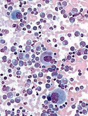
Credit: UC San Diego
Preclinical research in chronic myeloid leukemia (CML) has pointed to a relationship between cell adherence and treatment resistance.
Investigators found that a population of plastic-adherent K562 cells with increased expression of BCR-ABL exhibited greater resistance to the tyrosine kinase inhibitor imatinib than nonadherent K562 cells.
“Previous studies have linked high levels of the BCR-ABL mutation with drug resistance,” said Richard Byers, PhD, of The University of Manchester in the UK.
“We wanted to see how expression of BCR-ABL differed across groups of CML cells and, in particular, whether there were differences between adherent and nonadherent populations.”
Dr Byers and his colleagues described this investigation in Experimental Hematology.
The researchers evaluated the heterogeneity of BCR-ABL expression at DNA, messenger RNA, and protein levels, using the CML-derived K562 cell line.
They grew cells in suspension and found that some cells adhered to the plastic dish. The investigators then separated the plastic-adherent and nonadherent cell populations and studied them as single cells and in bulk.
The first discovery was that adherent and nonadherent cells had similar BCR-ABL fusion gene copy numbers.
In bulk-cell analysis, the mean relative normalized ratio for genomic ABL DNA copy number was 47.73 for adherent cells and 53.40 for nonadherent cells (P=0.11). In single-cell analysis, the mean copy numbers were 13.83 and 14.22, respectively (P=0.63).
On the other hand, there was a significant difference in BCR-ABL messenger RNA expression between adherent and nonadherent cells.
In bulk cells, the level of BCR-ABL messenger RNA transcripts was 11-fold higher in adherent cells than in nondherent cells (P=0.022). And single-cell analysis revealed the mean BCR-ABL copy number was 53.11 for adherent cells and 14.06 for nonadherent cells (P=0.0013).
Adherent cells also exhibited significantly upregulated phosphorylation of BCR protein compared to nonadherent cells.
Flow cytometry showed that a mean of 61.9% of adherent cells were positive for phosphor-BCR, compared to 14.5% of nonadherent cells (P=0.0074). And single-cell analysis revealed a mean signal number per cell of 8.23 among adherent cells and 3.02 among nonadherent cells (P<0.0001).
In addition, whole-genome microRNA profiling showed that adherent and nonadherent cell populations expressed significantly different microRNA species.
Finally, the researchers found that treatment with imatinib reduced cell viability more rapidly in nonadherent cells than in adherent cells (P<0.005). The adherent cells showed a decrease in cell viability at 24 hours, compared to 4 hours for nonadherent cells.
The investigators said this research suggests that CML patients may have a similar adherent cell population that mediates resistance to imatinib. And the study highlights the importance of single-cell analysis.
“The small number of cells that show high levels of BCR-ABL may not be detectable through bulk analysis of large samples,” Dr Byers said. “It looks like it is important to look at protein levels in single cells.”
“In future, it may be possible to measure BCR-ABL levels in individual cells in the clinic. This will help us identify the resistant, high-BCR-ABL cells and better understand how patients develop resistance to imatinib treatment, with the aim of combating this resistance to make response more durable and the treatment more effective.”
Credit: UC San Diego
Preclinical research in chronic myeloid leukemia (CML) has pointed to a relationship between cell adherence and treatment resistance.
Investigators found that a population of plastic-adherent K562 cells with increased expression of BCR-ABL exhibited greater resistance to the tyrosine kinase inhibitor imatinib than nonadherent K562 cells.
“Previous studies have linked high levels of the BCR-ABL mutation with drug resistance,” said Richard Byers, PhD, of The University of Manchester in the UK.
“We wanted to see how expression of BCR-ABL differed across groups of CML cells and, in particular, whether there were differences between adherent and nonadherent populations.”
Dr Byers and his colleagues described this investigation in Experimental Hematology.
The researchers evaluated the heterogeneity of BCR-ABL expression at DNA, messenger RNA, and protein levels, using the CML-derived K562 cell line.
They grew cells in suspension and found that some cells adhered to the plastic dish. The investigators then separated the plastic-adherent and nonadherent cell populations and studied them as single cells and in bulk.
The first discovery was that adherent and nonadherent cells had similar BCR-ABL fusion gene copy numbers.
In bulk-cell analysis, the mean relative normalized ratio for genomic ABL DNA copy number was 47.73 for adherent cells and 53.40 for nonadherent cells (P=0.11). In single-cell analysis, the mean copy numbers were 13.83 and 14.22, respectively (P=0.63).
On the other hand, there was a significant difference in BCR-ABL messenger RNA expression between adherent and nonadherent cells.
In bulk cells, the level of BCR-ABL messenger RNA transcripts was 11-fold higher in adherent cells than in nondherent cells (P=0.022). And single-cell analysis revealed the mean BCR-ABL copy number was 53.11 for adherent cells and 14.06 for nonadherent cells (P=0.0013).
Adherent cells also exhibited significantly upregulated phosphorylation of BCR protein compared to nonadherent cells.
Flow cytometry showed that a mean of 61.9% of adherent cells were positive for phosphor-BCR, compared to 14.5% of nonadherent cells (P=0.0074). And single-cell analysis revealed a mean signal number per cell of 8.23 among adherent cells and 3.02 among nonadherent cells (P<0.0001).
In addition, whole-genome microRNA profiling showed that adherent and nonadherent cell populations expressed significantly different microRNA species.
Finally, the researchers found that treatment with imatinib reduced cell viability more rapidly in nonadherent cells than in adherent cells (P<0.005). The adherent cells showed a decrease in cell viability at 24 hours, compared to 4 hours for nonadherent cells.
The investigators said this research suggests that CML patients may have a similar adherent cell population that mediates resistance to imatinib. And the study highlights the importance of single-cell analysis.
“The small number of cells that show high levels of BCR-ABL may not be detectable through bulk analysis of large samples,” Dr Byers said. “It looks like it is important to look at protein levels in single cells.”
“In future, it may be possible to measure BCR-ABL levels in individual cells in the clinic. This will help us identify the resistant, high-BCR-ABL cells and better understand how patients develop resistance to imatinib treatment, with the aim of combating this resistance to make response more durable and the treatment more effective.”
Credit: UC San Diego
Preclinical research in chronic myeloid leukemia (CML) has pointed to a relationship between cell adherence and treatment resistance.
Investigators found that a population of plastic-adherent K562 cells with increased expression of BCR-ABL exhibited greater resistance to the tyrosine kinase inhibitor imatinib than nonadherent K562 cells.
“Previous studies have linked high levels of the BCR-ABL mutation with drug resistance,” said Richard Byers, PhD, of The University of Manchester in the UK.
“We wanted to see how expression of BCR-ABL differed across groups of CML cells and, in particular, whether there were differences between adherent and nonadherent populations.”
Dr Byers and his colleagues described this investigation in Experimental Hematology.
The researchers evaluated the heterogeneity of BCR-ABL expression at DNA, messenger RNA, and protein levels, using the CML-derived K562 cell line.
They grew cells in suspension and found that some cells adhered to the plastic dish. The investigators then separated the plastic-adherent and nonadherent cell populations and studied them as single cells and in bulk.
The first discovery was that adherent and nonadherent cells had similar BCR-ABL fusion gene copy numbers.
In bulk-cell analysis, the mean relative normalized ratio for genomic ABL DNA copy number was 47.73 for adherent cells and 53.40 for nonadherent cells (P=0.11). In single-cell analysis, the mean copy numbers were 13.83 and 14.22, respectively (P=0.63).
On the other hand, there was a significant difference in BCR-ABL messenger RNA expression between adherent and nonadherent cells.
In bulk cells, the level of BCR-ABL messenger RNA transcripts was 11-fold higher in adherent cells than in nondherent cells (P=0.022). And single-cell analysis revealed the mean BCR-ABL copy number was 53.11 for adherent cells and 14.06 for nonadherent cells (P=0.0013).
Adherent cells also exhibited significantly upregulated phosphorylation of BCR protein compared to nonadherent cells.
Flow cytometry showed that a mean of 61.9% of adherent cells were positive for phosphor-BCR, compared to 14.5% of nonadherent cells (P=0.0074). And single-cell analysis revealed a mean signal number per cell of 8.23 among adherent cells and 3.02 among nonadherent cells (P<0.0001).
In addition, whole-genome microRNA profiling showed that adherent and nonadherent cell populations expressed significantly different microRNA species.
Finally, the researchers found that treatment with imatinib reduced cell viability more rapidly in nonadherent cells than in adherent cells (P<0.005). The adherent cells showed a decrease in cell viability at 24 hours, compared to 4 hours for nonadherent cells.
The investigators said this research suggests that CML patients may have a similar adherent cell population that mediates resistance to imatinib. And the study highlights the importance of single-cell analysis.
“The small number of cells that show high levels of BCR-ABL may not be detectable through bulk analysis of large samples,” Dr Byers said. “It looks like it is important to look at protein levels in single cells.”
“In future, it may be possible to measure BCR-ABL levels in individual cells in the clinic. This will help us identify the resistant, high-BCR-ABL cells and better understand how patients develop resistance to imatinib treatment, with the aim of combating this resistance to make response more durable and the treatment more effective.”
Team finds hidden reservoir of HCMV
Credit: Chad McNeeley
Researchers have found evidence suggesting that perivascular mesenchymal stromal cells (MSCs) are a reservoir of human cytomegalovirus (HCMV).
This opens up the possibility of therapeutically targeting these cells, which surround blood vessels in the organs and can be found in the bone marrow.
If effective, such a treatment method could prove life-saving for individuals who experience HCMV reactivation, such as transplant recipients and patients receiving chemotherapy.
“There are antiviral medications designed to prevent HCMV from re-activating, but HCMV infection remains one of the major complications after both organ and bone marrow transplants,” said study author Graca Almeida-Porada, MD, PhD, of Wake Forest Baptist Medical Center in Winston-Salem, North Carolina.
“The question scientists have been asking for years is, ‘Where does the virus hide when it is latent?’ Maybe if we knew, we could target it.”
Previous research showed that hematopoietic stem cells can harbor HCMV. Dr Almeida-Porada and her colleagues hypothesized that other cell populations may also harbor the virus, and they suspected that perivascular MSCs were a likely culprit.
The team’s suspicions were confirmed when testing revealed that perivascular MSCs are susceptible to HCMV infection and that the virus can grow within these cells.
The researchers also compared the susceptibility of perivascular MSCs from the liver, brain, lung, and bone marrow. And they found the highest rate of HCMV infection in cells from the lung.
“This may explain why pneumonia is the primary manifestation of the HCMV infection in bone marrow transplant recipients,” Dr Almeida-Porada said.
To expand upon these findings, she and her colleagues analyzed bone marrow samples from 19 healthy individuals who had tested positive for HCMV. Quantitative PCR revealed HCMV DNA in perivascular MSCs from 7 of the subjects.
This suggests bone marrow-derived perivascular MSCs may be a natural HCMV reservoir, according to the researchers.
“We have found another source of cells that can harbor HCMV virus,” Dr Almeida-Porada concluded. “Knowing the identity of the cells opens the possibility of targeting treatments to stop its re-activation.”
Dr Almeida-Porada and her colleagues recounted their discoveries in the American Journal of Transplantation.
Credit: Chad McNeeley
Researchers have found evidence suggesting that perivascular mesenchymal stromal cells (MSCs) are a reservoir of human cytomegalovirus (HCMV).
This opens up the possibility of therapeutically targeting these cells, which surround blood vessels in the organs and can be found in the bone marrow.
If effective, such a treatment method could prove life-saving for individuals who experience HCMV reactivation, such as transplant recipients and patients receiving chemotherapy.
“There are antiviral medications designed to prevent HCMV from re-activating, but HCMV infection remains one of the major complications after both organ and bone marrow transplants,” said study author Graca Almeida-Porada, MD, PhD, of Wake Forest Baptist Medical Center in Winston-Salem, North Carolina.
“The question scientists have been asking for years is, ‘Where does the virus hide when it is latent?’ Maybe if we knew, we could target it.”
Previous research showed that hematopoietic stem cells can harbor HCMV. Dr Almeida-Porada and her colleagues hypothesized that other cell populations may also harbor the virus, and they suspected that perivascular MSCs were a likely culprit.
The team’s suspicions were confirmed when testing revealed that perivascular MSCs are susceptible to HCMV infection and that the virus can grow within these cells.
The researchers also compared the susceptibility of perivascular MSCs from the liver, brain, lung, and bone marrow. And they found the highest rate of HCMV infection in cells from the lung.
“This may explain why pneumonia is the primary manifestation of the HCMV infection in bone marrow transplant recipients,” Dr Almeida-Porada said.
To expand upon these findings, she and her colleagues analyzed bone marrow samples from 19 healthy individuals who had tested positive for HCMV. Quantitative PCR revealed HCMV DNA in perivascular MSCs from 7 of the subjects.
This suggests bone marrow-derived perivascular MSCs may be a natural HCMV reservoir, according to the researchers.
“We have found another source of cells that can harbor HCMV virus,” Dr Almeida-Porada concluded. “Knowing the identity of the cells opens the possibility of targeting treatments to stop its re-activation.”
Dr Almeida-Porada and her colleagues recounted their discoveries in the American Journal of Transplantation.
Credit: Chad McNeeley
Researchers have found evidence suggesting that perivascular mesenchymal stromal cells (MSCs) are a reservoir of human cytomegalovirus (HCMV).
This opens up the possibility of therapeutically targeting these cells, which surround blood vessels in the organs and can be found in the bone marrow.
If effective, such a treatment method could prove life-saving for individuals who experience HCMV reactivation, such as transplant recipients and patients receiving chemotherapy.
“There are antiviral medications designed to prevent HCMV from re-activating, but HCMV infection remains one of the major complications after both organ and bone marrow transplants,” said study author Graca Almeida-Porada, MD, PhD, of Wake Forest Baptist Medical Center in Winston-Salem, North Carolina.
“The question scientists have been asking for years is, ‘Where does the virus hide when it is latent?’ Maybe if we knew, we could target it.”
Previous research showed that hematopoietic stem cells can harbor HCMV. Dr Almeida-Porada and her colleagues hypothesized that other cell populations may also harbor the virus, and they suspected that perivascular MSCs were a likely culprit.
The team’s suspicions were confirmed when testing revealed that perivascular MSCs are susceptible to HCMV infection and that the virus can grow within these cells.
The researchers also compared the susceptibility of perivascular MSCs from the liver, brain, lung, and bone marrow. And they found the highest rate of HCMV infection in cells from the lung.
“This may explain why pneumonia is the primary manifestation of the HCMV infection in bone marrow transplant recipients,” Dr Almeida-Porada said.
To expand upon these findings, she and her colleagues analyzed bone marrow samples from 19 healthy individuals who had tested positive for HCMV. Quantitative PCR revealed HCMV DNA in perivascular MSCs from 7 of the subjects.
This suggests bone marrow-derived perivascular MSCs may be a natural HCMV reservoir, according to the researchers.
“We have found another source of cells that can harbor HCMV virus,” Dr Almeida-Porada concluded. “Knowing the identity of the cells opens the possibility of targeting treatments to stop its re-activation.”
Dr Almeida-Porada and her colleagues recounted their discoveries in the American Journal of Transplantation.
Mouse model allows tracking of cells, drug activity

Scientists have created a mouse that expresses a fluorescing biosensor in every cell of its body, allowing diseased cells and drugs to be tracked and evaluated in real time.
The biosensor is a single-molecule probe called Raichu-Rac, which was invented in 2002.
Although Raichu-Rac has been used by many research groups since then, this is the first time researchers have successfully modified a mouse to express the molecule throughout the body without affecting cell function.
The team described this work in Cell Reports.
Raichu-Rac mimics the action of the protein Rac, which drives cell movement and oscillates between active and inactive states. When Rac is active, Raichu-Rac picks up chemical cues and glows blue. When Rac is inactive, the molecule glows yellow.
Researchers found they could use imaging techniques to follow Rac activation in any organ at any time. They could watch moment-by-moment oscillation of Rac activity at the front or back of cells as they moved in the body. And they used the technology to monitor Rac activity in response to treatment.
“It allows us to watch and map, in real time, parts of a cell or organ where Rac is active and driving invasion,” said study author Paul Timpson, PhD, of the Garvan Institute of Medical Research in Darlinghurst, New South Wales, Australia.
“In cancers, a lot of blue indicates an aggressive tumor that is in the process of spreading. You can literally watch parts of a tumor turn from blue to yellow as a drug hits its target. This can be an hour or more after the drug is administered, and the effect can wane quickly or slowly. Drug companies need to know these details—specifically, how much, how often, and how long to administer drugs.”
The researchers said the mouse can be used to study any cancer type by crossing it with other models, limiting expression of Raichu-Rac to specific cell or tissue types. And the mouse can be adapted to study other diseases by expressing Raichu-Rac in different disease models.
“The great thing about this mouse is its flexibility and potential for looking at a broad range of diseases and molecular targets,” Dr Timpson said.
The mouse was created by Heidi Welch, PhD, of the Babraham Institute in Cambridge, UK, although she said she cannot take all the credit.
“The credit must go to Professor Miki Matsuda, the genius who invented [Raichu-Rac] in the first place 12 years ago,” Dr Welch said. “He made his discovery freely available to the scientific community and has been very open about his findings since.”
“Miki Matsuda was super helpful in suggesting the expression levels we should be looking for and in recommending the exact biosensor we should use, out of many he developed.”
Scientists have created a mouse that expresses a fluorescing biosensor in every cell of its body, allowing diseased cells and drugs to be tracked and evaluated in real time.
The biosensor is a single-molecule probe called Raichu-Rac, which was invented in 2002.
Although Raichu-Rac has been used by many research groups since then, this is the first time researchers have successfully modified a mouse to express the molecule throughout the body without affecting cell function.
The team described this work in Cell Reports.
Raichu-Rac mimics the action of the protein Rac, which drives cell movement and oscillates between active and inactive states. When Rac is active, Raichu-Rac picks up chemical cues and glows blue. When Rac is inactive, the molecule glows yellow.
Researchers found they could use imaging techniques to follow Rac activation in any organ at any time. They could watch moment-by-moment oscillation of Rac activity at the front or back of cells as they moved in the body. And they used the technology to monitor Rac activity in response to treatment.
“It allows us to watch and map, in real time, parts of a cell or organ where Rac is active and driving invasion,” said study author Paul Timpson, PhD, of the Garvan Institute of Medical Research in Darlinghurst, New South Wales, Australia.
“In cancers, a lot of blue indicates an aggressive tumor that is in the process of spreading. You can literally watch parts of a tumor turn from blue to yellow as a drug hits its target. This can be an hour or more after the drug is administered, and the effect can wane quickly or slowly. Drug companies need to know these details—specifically, how much, how often, and how long to administer drugs.”
The researchers said the mouse can be used to study any cancer type by crossing it with other models, limiting expression of Raichu-Rac to specific cell or tissue types. And the mouse can be adapted to study other diseases by expressing Raichu-Rac in different disease models.
“The great thing about this mouse is its flexibility and potential for looking at a broad range of diseases and molecular targets,” Dr Timpson said.
The mouse was created by Heidi Welch, PhD, of the Babraham Institute in Cambridge, UK, although she said she cannot take all the credit.
“The credit must go to Professor Miki Matsuda, the genius who invented [Raichu-Rac] in the first place 12 years ago,” Dr Welch said. “He made his discovery freely available to the scientific community and has been very open about his findings since.”
“Miki Matsuda was super helpful in suggesting the expression levels we should be looking for and in recommending the exact biosensor we should use, out of many he developed.”
Scientists have created a mouse that expresses a fluorescing biosensor in every cell of its body, allowing diseased cells and drugs to be tracked and evaluated in real time.
The biosensor is a single-molecule probe called Raichu-Rac, which was invented in 2002.
Although Raichu-Rac has been used by many research groups since then, this is the first time researchers have successfully modified a mouse to express the molecule throughout the body without affecting cell function.
The team described this work in Cell Reports.
Raichu-Rac mimics the action of the protein Rac, which drives cell movement and oscillates between active and inactive states. When Rac is active, Raichu-Rac picks up chemical cues and glows blue. When Rac is inactive, the molecule glows yellow.
Researchers found they could use imaging techniques to follow Rac activation in any organ at any time. They could watch moment-by-moment oscillation of Rac activity at the front or back of cells as they moved in the body. And they used the technology to monitor Rac activity in response to treatment.
“It allows us to watch and map, in real time, parts of a cell or organ where Rac is active and driving invasion,” said study author Paul Timpson, PhD, of the Garvan Institute of Medical Research in Darlinghurst, New South Wales, Australia.
“In cancers, a lot of blue indicates an aggressive tumor that is in the process of spreading. You can literally watch parts of a tumor turn from blue to yellow as a drug hits its target. This can be an hour or more after the drug is administered, and the effect can wane quickly or slowly. Drug companies need to know these details—specifically, how much, how often, and how long to administer drugs.”
The researchers said the mouse can be used to study any cancer type by crossing it with other models, limiting expression of Raichu-Rac to specific cell or tissue types. And the mouse can be adapted to study other diseases by expressing Raichu-Rac in different disease models.
“The great thing about this mouse is its flexibility and potential for looking at a broad range of diseases and molecular targets,” Dr Timpson said.
The mouse was created by Heidi Welch, PhD, of the Babraham Institute in Cambridge, UK, although she said she cannot take all the credit.
“The credit must go to Professor Miki Matsuda, the genius who invented [Raichu-Rac] in the first place 12 years ago,” Dr Welch said. “He made his discovery freely available to the scientific community and has been very open about his findings since.”
“Miki Matsuda was super helpful in suggesting the expression levels we should be looking for and in recommending the exact biosensor we should use, out of many he developed.”
FDA places imetelstat trials on hold
The US Food and Drug Administration (FDA) has issued a full clinical hold for the telomerase inhibitor imetelstat, citing concerns that long-term exposure to the drug may pose a risk of chronic liver injury.
The hold temporarily suspends all ongoing clinical trials of imetelstat sponsored by the drug’s maker, Geron Corporation.
It’s possible that other studies of imetelstat, such as ongoing investigator-sponsored trials, may be placed on hold as well.
At present, the hold affects the remaining 8 patients enrolled on Geron’s phase 2 study of imetelstat in essential thrombocythemia and polycythemia vera.
It also affects the remaining 2 patients in the company’s phase 2 study of the drug in previously treated multiple myeloma. And Geron believes its planned phase 2 trial of imetelstat in myelofibrosis will likely be delayed as well.
The FDA has not yet issued a written notice of the hold but has given Geron verbal notice.
The agency indicated that the hold is due to persistent low-grade liver function test abnormalities observed in the study of imetelstat in patients with essential thrombocythemia or polycythemia vera and the potential risk of chronic liver injury following long-term exposure to imetelstat.
The FDA expressed concerns about whether these abnormalities are reversible. Geron said it plans to work with the FDA to put an end to the hold.
The US Food and Drug Administration (FDA) has issued a full clinical hold for the telomerase inhibitor imetelstat, citing concerns that long-term exposure to the drug may pose a risk of chronic liver injury.
The hold temporarily suspends all ongoing clinical trials of imetelstat sponsored by the drug’s maker, Geron Corporation.
It’s possible that other studies of imetelstat, such as ongoing investigator-sponsored trials, may be placed on hold as well.
At present, the hold affects the remaining 8 patients enrolled on Geron’s phase 2 study of imetelstat in essential thrombocythemia and polycythemia vera.
It also affects the remaining 2 patients in the company’s phase 2 study of the drug in previously treated multiple myeloma. And Geron believes its planned phase 2 trial of imetelstat in myelofibrosis will likely be delayed as well.
The FDA has not yet issued a written notice of the hold but has given Geron verbal notice.
The agency indicated that the hold is due to persistent low-grade liver function test abnormalities observed in the study of imetelstat in patients with essential thrombocythemia or polycythemia vera and the potential risk of chronic liver injury following long-term exposure to imetelstat.
The FDA expressed concerns about whether these abnormalities are reversible. Geron said it plans to work with the FDA to put an end to the hold.
The US Food and Drug Administration (FDA) has issued a full clinical hold for the telomerase inhibitor imetelstat, citing concerns that long-term exposure to the drug may pose a risk of chronic liver injury.
The hold temporarily suspends all ongoing clinical trials of imetelstat sponsored by the drug’s maker, Geron Corporation.
It’s possible that other studies of imetelstat, such as ongoing investigator-sponsored trials, may be placed on hold as well.
At present, the hold affects the remaining 8 patients enrolled on Geron’s phase 2 study of imetelstat in essential thrombocythemia and polycythemia vera.
It also affects the remaining 2 patients in the company’s phase 2 study of the drug in previously treated multiple myeloma. And Geron believes its planned phase 2 trial of imetelstat in myelofibrosis will likely be delayed as well.
The FDA has not yet issued a written notice of the hold but has given Geron verbal notice.
The agency indicated that the hold is due to persistent low-grade liver function test abnormalities observed in the study of imetelstat in patients with essential thrombocythemia or polycythemia vera and the potential risk of chronic liver injury following long-term exposure to imetelstat.
The FDA expressed concerns about whether these abnormalities are reversible. Geron said it plans to work with the FDA to put an end to the hold.
ALL maintenance phase still stressful, studies show

Credit: Bill Branson
The stress and lifestyle changes mothers experience after a child’s leukemia diagnosis do not disappear once the disease is in the maintenance phase, new research suggests.
One study showed that mothers of patients with acute lymphoblastic leukemia (ALL) continued to exhibit signs of stress, anxiety, and depression when their child’s disease was in the maintenance phase.
And another study showed that a family’s daily schedule and sleeping arrangements did not return to the way they were before the child’s diagnosis.
“Even though these mothers were in the maintenance phase of their child’s illness, and the prognosis was good, we heard them say over and over that things could never go back to what they were before,” said study author Madalynn Neu, PhD, RN, of the Colorado University College of Nursing.
Stress, depression, and anxiety
In the Journal of Pediatric Oncology Nursing, Dr Neu and her colleagues reported their analysis of stress, anxiety, and depression among the mothers of patients with ALL. The team evaluated 26 mothers of ALL patients and 26 mothers of healthy children, who were matched according to the child’s age and gender.
To assess anxiety, the researchers collected salivary cortisol from the mothers 4 times a day for 3 consecutive days. The mothers also completed questionnaires—the Hospital Anxiety and Depression Scale and the Perceived Stress Scale.
More mothers in the ALL group had questionnaire scores indicating clinical anxiety (46%) and depressive symptoms (27%). And there was a trend toward increased stress in the mothers of ALL patients.
However, the researchers were surprised to find that the mothers’ anxiety levels—as measured by salivary cortisol—were similar to the mothers of well children.
“This may have been affected by the fact that even the control group wasn’t without anxiety,” said study author Ellen Matthews, PhD, RN, of the Colorado University College of Nursing.
“Financial, marital, social, and career concerns mean that parents of young children experience anxiety even without ALL.”
The sleep-wake experience
In the Journal of Pediatric Nursing, the researchers described their assessment of the sleep-wake experience for mothers of ALL patients. The team conducted interviews with 20 mothers, using open-ended, semi-structured questions, and the answers were transcribed verbatim.
Two main themes emerged during these interviews. The first was dubbed, “It’s a whole new cancer world” and contained 4 subthemes: losing normality, being off-balance/insecure, juggling duties, and making transitions.
“Many [of the mothers] had lost their normal lives—lost jobs, houses, friends,” Dr Matthews said. “Some were juggling their time around their child’s needs, and they had fears about many things—fear of recurrence, fear of making a mistake with medication, fear their kids might get sick with an infection.”
The second theme that emerged in interviews was, “I don’t remember what it’s like to have sleep.” This also contained 4 subthemes: sleeping trouble before and after ALL, the child feeling sick at night, worrying, and coping with exhaustion.
The mothers also noted that once sleep arrangements had changed, they often did not return to their pretreatment “normal.”
“Mothers talked about the difficulty of sleep while giving steroid medication,” Dr Neu said. “And if the ill child got to stay up late watching movies, the siblings wanted to stay up too.”
“The same was true of sleeping in a parent’s room. If an ill child wanted to sleep close to a parent (or if a parent wanted to sleep close to an ill child), siblings tended to move in as well. Sleep can be challenging for parents of well children, and our studies show it’s even more so for parents of children who have experienced ALL.”
The researchers hope these studies increase awareness of maternal concerns after a child’s leukemia diagnosis and that leads to interventions to help mothers manage these lifestyle issues.
Credit: Bill Branson
The stress and lifestyle changes mothers experience after a child’s leukemia diagnosis do not disappear once the disease is in the maintenance phase, new research suggests.
One study showed that mothers of patients with acute lymphoblastic leukemia (ALL) continued to exhibit signs of stress, anxiety, and depression when their child’s disease was in the maintenance phase.
And another study showed that a family’s daily schedule and sleeping arrangements did not return to the way they were before the child’s diagnosis.
“Even though these mothers were in the maintenance phase of their child’s illness, and the prognosis was good, we heard them say over and over that things could never go back to what they were before,” said study author Madalynn Neu, PhD, RN, of the Colorado University College of Nursing.
Stress, depression, and anxiety
In the Journal of Pediatric Oncology Nursing, Dr Neu and her colleagues reported their analysis of stress, anxiety, and depression among the mothers of patients with ALL. The team evaluated 26 mothers of ALL patients and 26 mothers of healthy children, who were matched according to the child’s age and gender.
To assess anxiety, the researchers collected salivary cortisol from the mothers 4 times a day for 3 consecutive days. The mothers also completed questionnaires—the Hospital Anxiety and Depression Scale and the Perceived Stress Scale.
More mothers in the ALL group had questionnaire scores indicating clinical anxiety (46%) and depressive symptoms (27%). And there was a trend toward increased stress in the mothers of ALL patients.
However, the researchers were surprised to find that the mothers’ anxiety levels—as measured by salivary cortisol—were similar to the mothers of well children.
“This may have been affected by the fact that even the control group wasn’t without anxiety,” said study author Ellen Matthews, PhD, RN, of the Colorado University College of Nursing.
“Financial, marital, social, and career concerns mean that parents of young children experience anxiety even without ALL.”
The sleep-wake experience
In the Journal of Pediatric Nursing, the researchers described their assessment of the sleep-wake experience for mothers of ALL patients. The team conducted interviews with 20 mothers, using open-ended, semi-structured questions, and the answers were transcribed verbatim.
Two main themes emerged during these interviews. The first was dubbed, “It’s a whole new cancer world” and contained 4 subthemes: losing normality, being off-balance/insecure, juggling duties, and making transitions.
“Many [of the mothers] had lost their normal lives—lost jobs, houses, friends,” Dr Matthews said. “Some were juggling their time around their child’s needs, and they had fears about many things—fear of recurrence, fear of making a mistake with medication, fear their kids might get sick with an infection.”
The second theme that emerged in interviews was, “I don’t remember what it’s like to have sleep.” This also contained 4 subthemes: sleeping trouble before and after ALL, the child feeling sick at night, worrying, and coping with exhaustion.
The mothers also noted that once sleep arrangements had changed, they often did not return to their pretreatment “normal.”
“Mothers talked about the difficulty of sleep while giving steroid medication,” Dr Neu said. “And if the ill child got to stay up late watching movies, the siblings wanted to stay up too.”
“The same was true of sleeping in a parent’s room. If an ill child wanted to sleep close to a parent (or if a parent wanted to sleep close to an ill child), siblings tended to move in as well. Sleep can be challenging for parents of well children, and our studies show it’s even more so for parents of children who have experienced ALL.”
The researchers hope these studies increase awareness of maternal concerns after a child’s leukemia diagnosis and that leads to interventions to help mothers manage these lifestyle issues.
Credit: Bill Branson
The stress and lifestyle changes mothers experience after a child’s leukemia diagnosis do not disappear once the disease is in the maintenance phase, new research suggests.
One study showed that mothers of patients with acute lymphoblastic leukemia (ALL) continued to exhibit signs of stress, anxiety, and depression when their child’s disease was in the maintenance phase.
And another study showed that a family’s daily schedule and sleeping arrangements did not return to the way they were before the child’s diagnosis.
“Even though these mothers were in the maintenance phase of their child’s illness, and the prognosis was good, we heard them say over and over that things could never go back to what they were before,” said study author Madalynn Neu, PhD, RN, of the Colorado University College of Nursing.
Stress, depression, and anxiety
In the Journal of Pediatric Oncology Nursing, Dr Neu and her colleagues reported their analysis of stress, anxiety, and depression among the mothers of patients with ALL. The team evaluated 26 mothers of ALL patients and 26 mothers of healthy children, who were matched according to the child’s age and gender.
To assess anxiety, the researchers collected salivary cortisol from the mothers 4 times a day for 3 consecutive days. The mothers also completed questionnaires—the Hospital Anxiety and Depression Scale and the Perceived Stress Scale.
More mothers in the ALL group had questionnaire scores indicating clinical anxiety (46%) and depressive symptoms (27%). And there was a trend toward increased stress in the mothers of ALL patients.
However, the researchers were surprised to find that the mothers’ anxiety levels—as measured by salivary cortisol—were similar to the mothers of well children.
“This may have been affected by the fact that even the control group wasn’t without anxiety,” said study author Ellen Matthews, PhD, RN, of the Colorado University College of Nursing.
“Financial, marital, social, and career concerns mean that parents of young children experience anxiety even without ALL.”
The sleep-wake experience
In the Journal of Pediatric Nursing, the researchers described their assessment of the sleep-wake experience for mothers of ALL patients. The team conducted interviews with 20 mothers, using open-ended, semi-structured questions, and the answers were transcribed verbatim.
Two main themes emerged during these interviews. The first was dubbed, “It’s a whole new cancer world” and contained 4 subthemes: losing normality, being off-balance/insecure, juggling duties, and making transitions.
“Many [of the mothers] had lost their normal lives—lost jobs, houses, friends,” Dr Matthews said. “Some were juggling their time around their child’s needs, and they had fears about many things—fear of recurrence, fear of making a mistake with medication, fear their kids might get sick with an infection.”
The second theme that emerged in interviews was, “I don’t remember what it’s like to have sleep.” This also contained 4 subthemes: sleeping trouble before and after ALL, the child feeling sick at night, worrying, and coping with exhaustion.
The mothers also noted that once sleep arrangements had changed, they often did not return to their pretreatment “normal.”
“Mothers talked about the difficulty of sleep while giving steroid medication,” Dr Neu said. “And if the ill child got to stay up late watching movies, the siblings wanted to stay up too.”
“The same was true of sleeping in a parent’s room. If an ill child wanted to sleep close to a parent (or if a parent wanted to sleep close to an ill child), siblings tended to move in as well. Sleep can be challenging for parents of well children, and our studies show it’s even more so for parents of children who have experienced ALL.”
The researchers hope these studies increase awareness of maternal concerns after a child’s leukemia diagnosis and that leads to interventions to help mothers manage these lifestyle issues.