User login
LMWH more cost-effective than UFH for VTE

Credit: Petr Kratochvil
New research suggests the low-molecular-weight heparin (LMWH) dalteparin is more cost-effective than unfractionated heparin (UFH) for preventing venous thromboembolism (VTE) in critically ill patients.
The study showed that using dalteparin was the most effective and least costly strategy to prevent all thrombotic events, pulmonary embolism (PE), deep-vein thrombosis (DVT), major bleeding, and heparin-induced thrombocytopenia (HIT).
These results were published in JAMA and presented at the Critical Care Canada Forum in Toronto.
For this study, Robert A. Fowler, MDCM, of the Sunnybrook Health Sciences Centre at the University of Toronto in Ontario, and his colleagues conducted an economic evaluation concurrent with the PROTECT trial.
For PROTECT, researchers compared the effectiveness of dalteparin and UFH as VTE prophylaxis in critically ill patients. The results revealed no difference in the rate of DVT between the two treatment groups, but patients who received dalteparin had lower rates of PE and HIT.
To evaluate the cost-effectiveness of LMWH and UFH, Dr Fowler and his colleagues assessed costs among 2344 patients enrolled on PROTECT. The team evaluated costs in the context of resource use and patient outcomes.
The median post-randomization hospital cost of care was greater for patients who received UFH than for those who received dalteparin—$40,805 vs $39,508—but the difference was not statistically significant (P=0.41).
Subgroup analyses (assessing patients according to such factors as illness severity and body mass index) indicated that dalteparin was the most effective and least costly strategy to prevent all thrombotic events, PE, DVT, major bleeding, and HIT.
Sensitivity analyses indicated that a strategy using LMWH was most effective, least costly 78% of the time, and would remain less costly unless the drug acquisition cost of dalteparin was to increase by more than 20-fold. There was no threshold in which lowering the acquisition cost of UFH favored VTE prophylaxis with UFH.
The researchers said these findings are important for the care of critically ill patients because they provide a cost-minimization rationale that complements clinical effectiveness knowledge from PROTECT.
For example, if an intensive care unit with 1000 medical-surgical admissions per year uses UFH instead of LMWH for VTE prophylaxis, the annual incremental cost would be between $1,000,000 and $1,500,000 with similar or worse clinical outcomes, despite the individual drug cost of UFH being $4 to $5 less per day.
The researchers noted that these findings were driven by lower rates of PE and HIT and the corresponding lower overall use of resources with LMWH. 
Credit: Petr Kratochvil
New research suggests the low-molecular-weight heparin (LMWH) dalteparin is more cost-effective than unfractionated heparin (UFH) for preventing venous thromboembolism (VTE) in critically ill patients.
The study showed that using dalteparin was the most effective and least costly strategy to prevent all thrombotic events, pulmonary embolism (PE), deep-vein thrombosis (DVT), major bleeding, and heparin-induced thrombocytopenia (HIT).
These results were published in JAMA and presented at the Critical Care Canada Forum in Toronto.
For this study, Robert A. Fowler, MDCM, of the Sunnybrook Health Sciences Centre at the University of Toronto in Ontario, and his colleagues conducted an economic evaluation concurrent with the PROTECT trial.
For PROTECT, researchers compared the effectiveness of dalteparin and UFH as VTE prophylaxis in critically ill patients. The results revealed no difference in the rate of DVT between the two treatment groups, but patients who received dalteparin had lower rates of PE and HIT.
To evaluate the cost-effectiveness of LMWH and UFH, Dr Fowler and his colleagues assessed costs among 2344 patients enrolled on PROTECT. The team evaluated costs in the context of resource use and patient outcomes.
The median post-randomization hospital cost of care was greater for patients who received UFH than for those who received dalteparin—$40,805 vs $39,508—but the difference was not statistically significant (P=0.41).
Subgroup analyses (assessing patients according to such factors as illness severity and body mass index) indicated that dalteparin was the most effective and least costly strategy to prevent all thrombotic events, PE, DVT, major bleeding, and HIT.
Sensitivity analyses indicated that a strategy using LMWH was most effective, least costly 78% of the time, and would remain less costly unless the drug acquisition cost of dalteparin was to increase by more than 20-fold. There was no threshold in which lowering the acquisition cost of UFH favored VTE prophylaxis with UFH.
The researchers said these findings are important for the care of critically ill patients because they provide a cost-minimization rationale that complements clinical effectiveness knowledge from PROTECT.
For example, if an intensive care unit with 1000 medical-surgical admissions per year uses UFH instead of LMWH for VTE prophylaxis, the annual incremental cost would be between $1,000,000 and $1,500,000 with similar or worse clinical outcomes, despite the individual drug cost of UFH being $4 to $5 less per day.
The researchers noted that these findings were driven by lower rates of PE and HIT and the corresponding lower overall use of resources with LMWH.
Credit: Petr Kratochvil
New research suggests the low-molecular-weight heparin (LMWH) dalteparin is more cost-effective than unfractionated heparin (UFH) for preventing venous thromboembolism (VTE) in critically ill patients.
The study showed that using dalteparin was the most effective and least costly strategy to prevent all thrombotic events, pulmonary embolism (PE), deep-vein thrombosis (DVT), major bleeding, and heparin-induced thrombocytopenia (HIT).
These results were published in JAMA and presented at the Critical Care Canada Forum in Toronto.
For this study, Robert A. Fowler, MDCM, of the Sunnybrook Health Sciences Centre at the University of Toronto in Ontario, and his colleagues conducted an economic evaluation concurrent with the PROTECT trial.
For PROTECT, researchers compared the effectiveness of dalteparin and UFH as VTE prophylaxis in critically ill patients. The results revealed no difference in the rate of DVT between the two treatment groups, but patients who received dalteparin had lower rates of PE and HIT.
To evaluate the cost-effectiveness of LMWH and UFH, Dr Fowler and his colleagues assessed costs among 2344 patients enrolled on PROTECT. The team evaluated costs in the context of resource use and patient outcomes.
The median post-randomization hospital cost of care was greater for patients who received UFH than for those who received dalteparin—$40,805 vs $39,508—but the difference was not statistically significant (P=0.41).
Subgroup analyses (assessing patients according to such factors as illness severity and body mass index) indicated that dalteparin was the most effective and least costly strategy to prevent all thrombotic events, PE, DVT, major bleeding, and HIT.
Sensitivity analyses indicated that a strategy using LMWH was most effective, least costly 78% of the time, and would remain less costly unless the drug acquisition cost of dalteparin was to increase by more than 20-fold. There was no threshold in which lowering the acquisition cost of UFH favored VTE prophylaxis with UFH.
The researchers said these findings are important for the care of critically ill patients because they provide a cost-minimization rationale that complements clinical effectiveness knowledge from PROTECT.
For example, if an intensive care unit with 1000 medical-surgical admissions per year uses UFH instead of LMWH for VTE prophylaxis, the annual incremental cost would be between $1,000,000 and $1,500,000 with similar or worse clinical outcomes, despite the individual drug cost of UFH being $4 to $5 less per day.
The researchers noted that these findings were driven by lower rates of PE and HIT and the corresponding lower overall use of resources with LMWH.
NICE recommends dabigatran for DVT, PE
Credit: NHS
The UK’s National Institute for Health and Care Excellence (NICE) has issued a draft guidance recommending dabigatran etexilate (Pradaxa) as an option for treating and preventing recurrent deep-vein thrombosis (DVT) and pulmonary embolism (PE) in adults.
A committee advising NICE concluded that dabigatran is a cost-effective use of resources and a convenient alternative to warfarin, especially for patients who require longer-term anticoagulant therapy.
“For many people, using warfarin can be difficult because of the need for frequent tests to see if the blood is clotting properly and having to adjust the dose of the drug if it is not,” said Carole Longson, NICE Health Technology Evaluation Centre Director.
“The appraisal committee felt that dabigatran represents a potential benefit for many people who have had a DVT or PE, particularly those who have risk factors for recurrence of a blood clot and who therefore need longer-term treatment. We are pleased, therefore, to be able to recommend dabigatran as a cost-effective option for treating DVT and PE and preventing further episodes in adults.”
Dabigatran, made by Boehringer Ingelheim, costs £65.90 for a 60-capsule pack of the 150 mg or 110 mg doses (excluding value-added tax) and costs £2.20 per day of treatment. However, costs may vary in different settings because of negotiated procurement discounts.
The most plausible incremental cost-effectiveness ratio (ICER) for dabigatran compared with warfarin for acute treatment of venous thromboembolism (VTE) was uncertain.
However, both Boehringer Ingelheim’s ICER and an evidence review group’s ICER remained in the range that could be considered a cost-effective use of National Health Service resources. Both were under £20,000 per quality-adjusted life-year (QALY) gained.
Neither Boehringer Ingelheim nor the evidence review group found any significant difference in efficacy between dabigatran and rivaroxaban for acute treatment of VTE in their indirect comparisons, and the costs were very similar between these two treatments.
For combined treatment and secondary prevention of VTE, the committee noted that Boehringer Ingelheim’s base-case ICER for dabigatran compared with warfarin was likely too low (£9973 per QALY gained).
But the evidence review group’s base-case for dabigatran compared with warfarin may have overestimated the ICER (£35,786 per QALY gained). So the committee said the ICER probably falls somewhere between the two estimates.
The committee also noted that dabigatran and rivaroxaban appear to have similar efficacy for combined treatment and secondary prevention of VTE, and their costs are very similar.
For more details, see the draft guidance.
Credit: NHS
The UK’s National Institute for Health and Care Excellence (NICE) has issued a draft guidance recommending dabigatran etexilate (Pradaxa) as an option for treating and preventing recurrent deep-vein thrombosis (DVT) and pulmonary embolism (PE) in adults.
A committee advising NICE concluded that dabigatran is a cost-effective use of resources and a convenient alternative to warfarin, especially for patients who require longer-term anticoagulant therapy.
“For many people, using warfarin can be difficult because of the need for frequent tests to see if the blood is clotting properly and having to adjust the dose of the drug if it is not,” said Carole Longson, NICE Health Technology Evaluation Centre Director.
“The appraisal committee felt that dabigatran represents a potential benefit for many people who have had a DVT or PE, particularly those who have risk factors for recurrence of a blood clot and who therefore need longer-term treatment. We are pleased, therefore, to be able to recommend dabigatran as a cost-effective option for treating DVT and PE and preventing further episodes in adults.”
Dabigatran, made by Boehringer Ingelheim, costs £65.90 for a 60-capsule pack of the 150 mg or 110 mg doses (excluding value-added tax) and costs £2.20 per day of treatment. However, costs may vary in different settings because of negotiated procurement discounts.
The most plausible incremental cost-effectiveness ratio (ICER) for dabigatran compared with warfarin for acute treatment of venous thromboembolism (VTE) was uncertain.
However, both Boehringer Ingelheim’s ICER and an evidence review group’s ICER remained in the range that could be considered a cost-effective use of National Health Service resources. Both were under £20,000 per quality-adjusted life-year (QALY) gained.
Neither Boehringer Ingelheim nor the evidence review group found any significant difference in efficacy between dabigatran and rivaroxaban for acute treatment of VTE in their indirect comparisons, and the costs were very similar between these two treatments.
For combined treatment and secondary prevention of VTE, the committee noted that Boehringer Ingelheim’s base-case ICER for dabigatran compared with warfarin was likely too low (£9973 per QALY gained).
But the evidence review group’s base-case for dabigatran compared with warfarin may have overestimated the ICER (£35,786 per QALY gained). So the committee said the ICER probably falls somewhere between the two estimates.
The committee also noted that dabigatran and rivaroxaban appear to have similar efficacy for combined treatment and secondary prevention of VTE, and their costs are very similar.
For more details, see the draft guidance.
Credit: NHS
The UK’s National Institute for Health and Care Excellence (NICE) has issued a draft guidance recommending dabigatran etexilate (Pradaxa) as an option for treating and preventing recurrent deep-vein thrombosis (DVT) and pulmonary embolism (PE) in adults.
A committee advising NICE concluded that dabigatran is a cost-effective use of resources and a convenient alternative to warfarin, especially for patients who require longer-term anticoagulant therapy.
“For many people, using warfarin can be difficult because of the need for frequent tests to see if the blood is clotting properly and having to adjust the dose of the drug if it is not,” said Carole Longson, NICE Health Technology Evaluation Centre Director.
“The appraisal committee felt that dabigatran represents a potential benefit for many people who have had a DVT or PE, particularly those who have risk factors for recurrence of a blood clot and who therefore need longer-term treatment. We are pleased, therefore, to be able to recommend dabigatran as a cost-effective option for treating DVT and PE and preventing further episodes in adults.”
Dabigatran, made by Boehringer Ingelheim, costs £65.90 for a 60-capsule pack of the 150 mg or 110 mg doses (excluding value-added tax) and costs £2.20 per day of treatment. However, costs may vary in different settings because of negotiated procurement discounts.
The most plausible incremental cost-effectiveness ratio (ICER) for dabigatran compared with warfarin for acute treatment of venous thromboembolism (VTE) was uncertain.
However, both Boehringer Ingelheim’s ICER and an evidence review group’s ICER remained in the range that could be considered a cost-effective use of National Health Service resources. Both were under £20,000 per quality-adjusted life-year (QALY) gained.
Neither Boehringer Ingelheim nor the evidence review group found any significant difference in efficacy between dabigatran and rivaroxaban for acute treatment of VTE in their indirect comparisons, and the costs were very similar between these two treatments.
For combined treatment and secondary prevention of VTE, the committee noted that Boehringer Ingelheim’s base-case ICER for dabigatran compared with warfarin was likely too low (£9973 per QALY gained).
But the evidence review group’s base-case for dabigatran compared with warfarin may have overestimated the ICER (£35,786 per QALY gained). So the committee said the ICER probably falls somewhere between the two estimates.
The committee also noted that dabigatran and rivaroxaban appear to have similar efficacy for combined treatment and secondary prevention of VTE, and their costs are very similar.
For more details, see the draft guidance.
Air pollution not to blame for childhood leukemia, study suggests

The increased risk of leukemia reported among children born close to overhead power lines is likely not a result of alterations in air pollution, researchers have reported in the Journal of Radiological Protection.
The group found little evidence to support the “corona-ion hypothesis” which has been cited as a possible explanation for the excess of childhood leukemia cases close to high-voltage overhead power lines in the UK prior to the 1980s.
The hypothesis is based on the fact that high-voltage overhead power lines create charged particles in the surrounding air.
These ionized particles, known as corona ions, can be blown away by the wind and attach to air pollutants, such as those from traffic or smoking.
The corona-ion hypothesis suggests these electrically charged pollutants are more likely to be retained in the airways or lungs, and this could lead to serious health effects, including childhood leukemia.
The researchers previously showed that, on average, there has been no increased risk of leukemia among children born near high-voltage power lines in recent decades. However, the same piece of research confirmed an increased risk prior to the 1980s, which has yet to be explained.
To investigate this theory, John Swanson, of National Grid in London, and his colleagues used data from 7347 children in England and Wales who were born and diagnosed with leukemia between 1968 and 2008, and who lived within 600 m of a high-voltage overhead power line.
The researchers calculated the exposure of each of the subjects to corona ions using a model based on: the voltage of the power line; the distance from the line; how the concentration of corona ions varied with distance from the power lines; and, using data from various meteorological stations, the amount of time and speed that wind blew in each direction around the power lines.
The results did not suggest that exposure to corona ions explained the pattern of increased leukemia rates close to high-voltage overhead power lines previously found in earlier decades.
“We found in earlier studies that, for previous decades, childhood leukemia rates were higher near power lines,” said Kathryn Bunch, of the University of Oxford.
“This new paper seems to show that this wasn’t caused by corona ions, but it leaves us still searching for the true cause, and we are undertaking further investigations of the variation in risk over time.”
The increased risk of leukemia reported among children born close to overhead power lines is likely not a result of alterations in air pollution, researchers have reported in the Journal of Radiological Protection.
The group found little evidence to support the “corona-ion hypothesis” which has been cited as a possible explanation for the excess of childhood leukemia cases close to high-voltage overhead power lines in the UK prior to the 1980s.
The hypothesis is based on the fact that high-voltage overhead power lines create charged particles in the surrounding air.
These ionized particles, known as corona ions, can be blown away by the wind and attach to air pollutants, such as those from traffic or smoking.
The corona-ion hypothesis suggests these electrically charged pollutants are more likely to be retained in the airways or lungs, and this could lead to serious health effects, including childhood leukemia.
The researchers previously showed that, on average, there has been no increased risk of leukemia among children born near high-voltage power lines in recent decades. However, the same piece of research confirmed an increased risk prior to the 1980s, which has yet to be explained.
To investigate this theory, John Swanson, of National Grid in London, and his colleagues used data from 7347 children in England and Wales who were born and diagnosed with leukemia between 1968 and 2008, and who lived within 600 m of a high-voltage overhead power line.
The researchers calculated the exposure of each of the subjects to corona ions using a model based on: the voltage of the power line; the distance from the line; how the concentration of corona ions varied with distance from the power lines; and, using data from various meteorological stations, the amount of time and speed that wind blew in each direction around the power lines.
The results did not suggest that exposure to corona ions explained the pattern of increased leukemia rates close to high-voltage overhead power lines previously found in earlier decades.
“We found in earlier studies that, for previous decades, childhood leukemia rates were higher near power lines,” said Kathryn Bunch, of the University of Oxford.
“This new paper seems to show that this wasn’t caused by corona ions, but it leaves us still searching for the true cause, and we are undertaking further investigations of the variation in risk over time.”
The increased risk of leukemia reported among children born close to overhead power lines is likely not a result of alterations in air pollution, researchers have reported in the Journal of Radiological Protection.
The group found little evidence to support the “corona-ion hypothesis” which has been cited as a possible explanation for the excess of childhood leukemia cases close to high-voltage overhead power lines in the UK prior to the 1980s.
The hypothesis is based on the fact that high-voltage overhead power lines create charged particles in the surrounding air.
These ionized particles, known as corona ions, can be blown away by the wind and attach to air pollutants, such as those from traffic or smoking.
The corona-ion hypothesis suggests these electrically charged pollutants are more likely to be retained in the airways or lungs, and this could lead to serious health effects, including childhood leukemia.
The researchers previously showed that, on average, there has been no increased risk of leukemia among children born near high-voltage power lines in recent decades. However, the same piece of research confirmed an increased risk prior to the 1980s, which has yet to be explained.
To investigate this theory, John Swanson, of National Grid in London, and his colleagues used data from 7347 children in England and Wales who were born and diagnosed with leukemia between 1968 and 2008, and who lived within 600 m of a high-voltage overhead power line.
The researchers calculated the exposure of each of the subjects to corona ions using a model based on: the voltage of the power line; the distance from the line; how the concentration of corona ions varied with distance from the power lines; and, using data from various meteorological stations, the amount of time and speed that wind blew in each direction around the power lines.
The results did not suggest that exposure to corona ions explained the pattern of increased leukemia rates close to high-voltage overhead power lines previously found in earlier decades.
“We found in earlier studies that, for previous decades, childhood leukemia rates were higher near power lines,” said Kathryn Bunch, of the University of Oxford.
“This new paper seems to show that this wasn’t caused by corona ions, but it leaves us still searching for the true cause, and we are undertaking further investigations of the variation in risk over time.”
Team creates universal heparin antidote
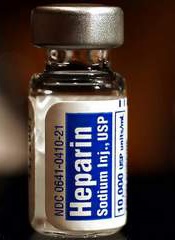
Researchers say they’ve developed a synthetic molecule that can reverse the effects of all heparin products, both in vitro and in vivo.
Finding a drug to reverse the anticoagulant effect of heparin is complicated because there are about a dozen approved heparin products on the market.
None of the available, synthetically made reversal drugs work with all varieties of heparins, and they are relatively toxic, with toxicity varying from person to person.
With all this in mind, Jayachandran Kizhakkedathu, PhD, of the University of British Columbia in Vancouver, and his colleagues set out to create a safe, synthetic antidote that works with all heparins used in clinics today.
They described their results in Science Translational Medicine.
The team created a range of fully synthetic dendritic polymer-based universal heparin reversal agents (UHRAs). These UHRA molecules completely neutralized the activity of unfractionated heparin, tinzaparin, semuloparin, and fondaparinux in vitro.
In comparison, the agent protamine was able to reverse the effects of unfractionated heparin but could only partially reverse the activity of tinzaparin, fondaparinux, and semuloparin.
The UHRA lead molecule, UHRA-7, completely and rapidly neutralized the effects of unfractionated heparin and enoxaparin in rats. Protamine reversed the effects of unfractionated heparin completely but could only reverse 60% of enoxaparin activity.
In mice, UHRA-7 arrested bleeding induced by all the heparins. Mice that received 10 mg/kg of UHRA-7 five minutes after unfractionated or low-molecular-weight heparins had significantly reduced bleeding times and hemoglobin loss compared to mice that received heparin alone (P<0.0001). UHRA-7 at 20 mg/kg arrested the bleeding induced by fondaparinux.
The researchers also assessed the safety of UHRA-7 in mice, monitoring them for 29 days after administration. The team saw no changes in body weight and no signs of acute toxicity in these mice.
Aditionally, there were no changes in lactate dehydrogenase levels in serum and no abnormalities detected by necropsy analysis of the organs.
These results suggest UHRA-7 could benefit patients undergoing high-risk surgical procedures and those requiring treatment for bleeding complications, Dr Kizhakkedathu said.
He noted that a synthetic drug offers consistency in health effects and performance. It also avoids possible immunological reactions sometimes associated with antidotes of biological origin.
Dr Kizhakkedathu and his colleagues plan to continue investigating UHRA-7 in the lab, with the goal of conducting human testing in 3 to 5 years.
Researchers say they’ve developed a synthetic molecule that can reverse the effects of all heparin products, both in vitro and in vivo.
Finding a drug to reverse the anticoagulant effect of heparin is complicated because there are about a dozen approved heparin products on the market.
None of the available, synthetically made reversal drugs work with all varieties of heparins, and they are relatively toxic, with toxicity varying from person to person.
With all this in mind, Jayachandran Kizhakkedathu, PhD, of the University of British Columbia in Vancouver, and his colleagues set out to create a safe, synthetic antidote that works with all heparins used in clinics today.
They described their results in Science Translational Medicine.
The team created a range of fully synthetic dendritic polymer-based universal heparin reversal agents (UHRAs). These UHRA molecules completely neutralized the activity of unfractionated heparin, tinzaparin, semuloparin, and fondaparinux in vitro.
In comparison, the agent protamine was able to reverse the effects of unfractionated heparin but could only partially reverse the activity of tinzaparin, fondaparinux, and semuloparin.
The UHRA lead molecule, UHRA-7, completely and rapidly neutralized the effects of unfractionated heparin and enoxaparin in rats. Protamine reversed the effects of unfractionated heparin completely but could only reverse 60% of enoxaparin activity.
In mice, UHRA-7 arrested bleeding induced by all the heparins. Mice that received 10 mg/kg of UHRA-7 five minutes after unfractionated or low-molecular-weight heparins had significantly reduced bleeding times and hemoglobin loss compared to mice that received heparin alone (P<0.0001). UHRA-7 at 20 mg/kg arrested the bleeding induced by fondaparinux.
The researchers also assessed the safety of UHRA-7 in mice, monitoring them for 29 days after administration. The team saw no changes in body weight and no signs of acute toxicity in these mice.
Aditionally, there were no changes in lactate dehydrogenase levels in serum and no abnormalities detected by necropsy analysis of the organs.
These results suggest UHRA-7 could benefit patients undergoing high-risk surgical procedures and those requiring treatment for bleeding complications, Dr Kizhakkedathu said.
He noted that a synthetic drug offers consistency in health effects and performance. It also avoids possible immunological reactions sometimes associated with antidotes of biological origin.
Dr Kizhakkedathu and his colleagues plan to continue investigating UHRA-7 in the lab, with the goal of conducting human testing in 3 to 5 years.
Researchers say they’ve developed a synthetic molecule that can reverse the effects of all heparin products, both in vitro and in vivo.
Finding a drug to reverse the anticoagulant effect of heparin is complicated because there are about a dozen approved heparin products on the market.
None of the available, synthetically made reversal drugs work with all varieties of heparins, and they are relatively toxic, with toxicity varying from person to person.
With all this in mind, Jayachandran Kizhakkedathu, PhD, of the University of British Columbia in Vancouver, and his colleagues set out to create a safe, synthetic antidote that works with all heparins used in clinics today.
They described their results in Science Translational Medicine.
The team created a range of fully synthetic dendritic polymer-based universal heparin reversal agents (UHRAs). These UHRA molecules completely neutralized the activity of unfractionated heparin, tinzaparin, semuloparin, and fondaparinux in vitro.
In comparison, the agent protamine was able to reverse the effects of unfractionated heparin but could only partially reverse the activity of tinzaparin, fondaparinux, and semuloparin.
The UHRA lead molecule, UHRA-7, completely and rapidly neutralized the effects of unfractionated heparin and enoxaparin in rats. Protamine reversed the effects of unfractionated heparin completely but could only reverse 60% of enoxaparin activity.
In mice, UHRA-7 arrested bleeding induced by all the heparins. Mice that received 10 mg/kg of UHRA-7 five minutes after unfractionated or low-molecular-weight heparins had significantly reduced bleeding times and hemoglobin loss compared to mice that received heparin alone (P<0.0001). UHRA-7 at 20 mg/kg arrested the bleeding induced by fondaparinux.
The researchers also assessed the safety of UHRA-7 in mice, monitoring them for 29 days after administration. The team saw no changes in body weight and no signs of acute toxicity in these mice.
Aditionally, there were no changes in lactate dehydrogenase levels in serum and no abnormalities detected by necropsy analysis of the organs.
These results suggest UHRA-7 could benefit patients undergoing high-risk surgical procedures and those requiring treatment for bleeding complications, Dr Kizhakkedathu said.
He noted that a synthetic drug offers consistency in health effects and performance. It also avoids possible immunological reactions sometimes associated with antidotes of biological origin.
Dr Kizhakkedathu and his colleagues plan to continue investigating UHRA-7 in the lab, with the goal of conducting human testing in 3 to 5 years.
EMA grants product orphan status for AML
The European Medicines Agency (EMA) has granted orphan status to Atir, a product consisting of T-cell-depleted donor immune cells, for the treatment of acute myeloid leukemia (AML).
The EMA and the US Food and Drug Administration previously granted Atir orphan status for the prevention of acute graft-vs-host-disease (GVHD) following hematopoietic stem cell transplant (HSCT).
The EMA’s orphan designation provides incentives to support drug development. This includes fee reductions and a 10-year period of market exclusivity in the European Union after product approval.
About Atir
Atir consists of donor immune cells from which the alloreactive T-cells that would otherwise attack the patient’s body have been selectively eliminated.
The product is produced using a molecule known as TH9402 to selectively remove those T cells from the donor graft, while preserving other immune cells. To activate patient-reactive T cells, the graft is mixed (ex vivo) with patient cells.
Then, TH9402 is added. As this phototoxic compound selectively accumulates in activated T cells, the cells can be eliminated by exposing the cell mixture to light of a specific wavelength. The resulting Atir product can be frozen and stored and is infused into the patient in a scheduled procedure.
Trial data
Researchers said Atir proved safe and effective in a phase 1/2 study in which high-risk leukemia patients with very poor prognosis were treated with escalating doses of Atir after a haploidentical HSCT.
The overall survival of 19 patients who received an optimal dose of Atir was 78% at 1 year and 67% at 5 years, rates that compare favorably to outcomes of HSCTs from fully matched donors. The data also suggest that immune cells responsible for the graft-vs-leukemia effect are retained in Atir.
Five-year follow-up data show that none of the 19 patients developed acute grade 3/4 GVHD, compared to an incidence of 30% in matched unrelated HSCTs. In the 9 patients who received an optimal dose of Atir, there was no transplant-related mortality.
Researchers are currently testing Atir in a phase 2 study of patients with AML, acute lymphoblastic leukemia, and myelodysplastic syndrome, to corroborate and extend the safety and efficacy results from the phase 1/2 study. Data from this trial are expected in the second half of 2014.
Atir is under development by Kiadis Pharma. For more information, visit the company’s website.
The European Medicines Agency (EMA) has granted orphan status to Atir, a product consisting of T-cell-depleted donor immune cells, for the treatment of acute myeloid leukemia (AML).
The EMA and the US Food and Drug Administration previously granted Atir orphan status for the prevention of acute graft-vs-host-disease (GVHD) following hematopoietic stem cell transplant (HSCT).
The EMA’s orphan designation provides incentives to support drug development. This includes fee reductions and a 10-year period of market exclusivity in the European Union after product approval.
About Atir
Atir consists of donor immune cells from which the alloreactive T-cells that would otherwise attack the patient’s body have been selectively eliminated.
The product is produced using a molecule known as TH9402 to selectively remove those T cells from the donor graft, while preserving other immune cells. To activate patient-reactive T cells, the graft is mixed (ex vivo) with patient cells.
Then, TH9402 is added. As this phototoxic compound selectively accumulates in activated T cells, the cells can be eliminated by exposing the cell mixture to light of a specific wavelength. The resulting Atir product can be frozen and stored and is infused into the patient in a scheduled procedure.
Trial data
Researchers said Atir proved safe and effective in a phase 1/2 study in which high-risk leukemia patients with very poor prognosis were treated with escalating doses of Atir after a haploidentical HSCT.
The overall survival of 19 patients who received an optimal dose of Atir was 78% at 1 year and 67% at 5 years, rates that compare favorably to outcomes of HSCTs from fully matched donors. The data also suggest that immune cells responsible for the graft-vs-leukemia effect are retained in Atir.
Five-year follow-up data show that none of the 19 patients developed acute grade 3/4 GVHD, compared to an incidence of 30% in matched unrelated HSCTs. In the 9 patients who received an optimal dose of Atir, there was no transplant-related mortality.
Researchers are currently testing Atir in a phase 2 study of patients with AML, acute lymphoblastic leukemia, and myelodysplastic syndrome, to corroborate and extend the safety and efficacy results from the phase 1/2 study. Data from this trial are expected in the second half of 2014.
Atir is under development by Kiadis Pharma. For more information, visit the company’s website.
The European Medicines Agency (EMA) has granted orphan status to Atir, a product consisting of T-cell-depleted donor immune cells, for the treatment of acute myeloid leukemia (AML).
The EMA and the US Food and Drug Administration previously granted Atir orphan status for the prevention of acute graft-vs-host-disease (GVHD) following hematopoietic stem cell transplant (HSCT).
The EMA’s orphan designation provides incentives to support drug development. This includes fee reductions and a 10-year period of market exclusivity in the European Union after product approval.
About Atir
Atir consists of donor immune cells from which the alloreactive T-cells that would otherwise attack the patient’s body have been selectively eliminated.
The product is produced using a molecule known as TH9402 to selectively remove those T cells from the donor graft, while preserving other immune cells. To activate patient-reactive T cells, the graft is mixed (ex vivo) with patient cells.
Then, TH9402 is added. As this phototoxic compound selectively accumulates in activated T cells, the cells can be eliminated by exposing the cell mixture to light of a specific wavelength. The resulting Atir product can be frozen and stored and is infused into the patient in a scheduled procedure.
Trial data
Researchers said Atir proved safe and effective in a phase 1/2 study in which high-risk leukemia patients with very poor prognosis were treated with escalating doses of Atir after a haploidentical HSCT.
The overall survival of 19 patients who received an optimal dose of Atir was 78% at 1 year and 67% at 5 years, rates that compare favorably to outcomes of HSCTs from fully matched donors. The data also suggest that immune cells responsible for the graft-vs-leukemia effect are retained in Atir.
Five-year follow-up data show that none of the 19 patients developed acute grade 3/4 GVHD, compared to an incidence of 30% in matched unrelated HSCTs. In the 9 patients who received an optimal dose of Atir, there was no transplant-related mortality.
Researchers are currently testing Atir in a phase 2 study of patients with AML, acute lymphoblastic leukemia, and myelodysplastic syndrome, to corroborate and extend the safety and efficacy results from the phase 1/2 study. Data from this trial are expected in the second half of 2014.
Atir is under development by Kiadis Pharma. For more information, visit the company’s website.
Number of cord blood units doesn’t affect survival
Credit: NHS
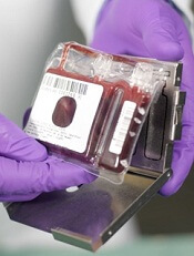
Single and double cord blood transplants produce similar outcomes, according to a study of young patients with hematologic disorders.
Researchers found that rates of overall and disease-free survival were not significantly different in patients who received a single unit of cord blood and those who received two units.
Other outcome measures, such as neutrophil recovery, relapse, and transplant-related death, were similar between the two groups as well.
However, patients who received a single cord blood unit showed improved platelet recovery, a lower incidence of grade 3-4 acute graft-vs-host disease (GVHD), and a lower rate of extensive chronic GVHD.
John Wagner, Jr, MD, of the University of Minnesota in Minneapolis, and his colleagues reported these results in NEJM. Dr Wagner previously presented results from this study at ASH 2012.
“Based on promising early studies using two cord blood units in adults for whom one unit is often not sufficient, we designed this study in order to determine if the higher number of blood-forming stem cells in two cord blood units might improve survival,” Dr Wagner said. “What we found, however, was that both treatment arms performed very well, with similar rates of white blood cell recovery and survival.”
The researchers enrolled 224 patients, ages 1 to 21 years, with hematologic disorders, including acute and chronic leukemias as well as myelodysplastic syndromes.
Patients were randomized to receive double-unit (n=111) or single-unit (n=113) cord blood transplants after a uniform myeloablative conditioning regimen and immunoprophylaxis for GVHD.
The researchers matched the treatment arms for age, sex, self-reported race, performance status, degree of donor-recipient HLA matching, disease type, and disease status at transplant.
Survival and relapse
The study’s primary endpoint was 1-year survival, which was 65% in the double-unit arm and 73% in the single-unit arm (P=0.17). In a multivariate analysis, the risk of death did not differ significantly between the arms (hazard ratio=1.34, P=0.20).
Similarly, there was no significant difference in 1-year disease-free survival between the double- and single-unit arms—64% and 70%, respectively (P=0.11). In a multivariate analysis, the risk of relapse or death did not differ significantly between arms (hazard ratio=1.48, P=0.08).
It therefore follows that rates of relapse and transplant-related death were similar at 1 year as well. The incidence of relapse was 14% in the double-unit arm and 12% in the single-unit arm (P=0.12). And rates of transplant-related death were 22% and 19%, respectively (P=0.43).
Recovery and GVHD
The incidence of neutrophil recovery was similar between treatment arms—88% in the double-unit arm and 89% in the single-unit arm (P=0.29) at a median of 23 days (range, 11 to 133) and 21 days (range, 11 to 62) after transplant, respectively.
However, the rate of platelet recovery was significantly higher in the single-unit arm—76% vs 65% (P=0.04). Furthermore, the median time to platelet recovery was 58 days (range, 28 to 295) in the single-unit arm and 84 days (range, 22 to 716) in the double-unit arm.
The rate of grade 2-4 acute GVHD was similar between the treatment arms (P=0.78), but patients in the double-unit arm had a higher incidence of grade 3-4 acute GVHD—23% vs 13% (P=0.02).
There was no difference in the incidence of any chronic GVHD at 1 year after transplant—32% in the double-unit arm and 30% in the single-unit arm (P=0.51). But there was a higher incidence of extensive chronic GVHD after double-unit transplant—15% vs 9% (P=0.05).
“This is helpful news for physicians considering the best treatment options for their patients,” said Joanne Kurtzberg, MD, of Duke University Medical Center in Durham, North Carolina.
“We found children who have a cord blood unit with an adequate number of cells do not benefit from receiving two units. This reduces the cost of a cord blood transplant for the majority of pediatric patients needing the procedure. However, for larger children without an adequately dosed single cord blood unit, using two units will provide access to a potentially life-saving transplant.”
Credit: NHS
Single and double cord blood transplants produce similar outcomes, according to a study of young patients with hematologic disorders.
Researchers found that rates of overall and disease-free survival were not significantly different in patients who received a single unit of cord blood and those who received two units.
Other outcome measures, such as neutrophil recovery, relapse, and transplant-related death, were similar between the two groups as well.
However, patients who received a single cord blood unit showed improved platelet recovery, a lower incidence of grade 3-4 acute graft-vs-host disease (GVHD), and a lower rate of extensive chronic GVHD.
John Wagner, Jr, MD, of the University of Minnesota in Minneapolis, and his colleagues reported these results in NEJM. Dr Wagner previously presented results from this study at ASH 2012.
“Based on promising early studies using two cord blood units in adults for whom one unit is often not sufficient, we designed this study in order to determine if the higher number of blood-forming stem cells in two cord blood units might improve survival,” Dr Wagner said. “What we found, however, was that both treatment arms performed very well, with similar rates of white blood cell recovery and survival.”
The researchers enrolled 224 patients, ages 1 to 21 years, with hematologic disorders, including acute and chronic leukemias as well as myelodysplastic syndromes.
Patients were randomized to receive double-unit (n=111) or single-unit (n=113) cord blood transplants after a uniform myeloablative conditioning regimen and immunoprophylaxis for GVHD.
The researchers matched the treatment arms for age, sex, self-reported race, performance status, degree of donor-recipient HLA matching, disease type, and disease status at transplant.
Survival and relapse
The study’s primary endpoint was 1-year survival, which was 65% in the double-unit arm and 73% in the single-unit arm (P=0.17). In a multivariate analysis, the risk of death did not differ significantly between the arms (hazard ratio=1.34, P=0.20).
Similarly, there was no significant difference in 1-year disease-free survival between the double- and single-unit arms—64% and 70%, respectively (P=0.11). In a multivariate analysis, the risk of relapse or death did not differ significantly between arms (hazard ratio=1.48, P=0.08).
It therefore follows that rates of relapse and transplant-related death were similar at 1 year as well. The incidence of relapse was 14% in the double-unit arm and 12% in the single-unit arm (P=0.12). And rates of transplant-related death were 22% and 19%, respectively (P=0.43).
Recovery and GVHD
The incidence of neutrophil recovery was similar between treatment arms—88% in the double-unit arm and 89% in the single-unit arm (P=0.29) at a median of 23 days (range, 11 to 133) and 21 days (range, 11 to 62) after transplant, respectively.
However, the rate of platelet recovery was significantly higher in the single-unit arm—76% vs 65% (P=0.04). Furthermore, the median time to platelet recovery was 58 days (range, 28 to 295) in the single-unit arm and 84 days (range, 22 to 716) in the double-unit arm.
The rate of grade 2-4 acute GVHD was similar between the treatment arms (P=0.78), but patients in the double-unit arm had a higher incidence of grade 3-4 acute GVHD—23% vs 13% (P=0.02).
There was no difference in the incidence of any chronic GVHD at 1 year after transplant—32% in the double-unit arm and 30% in the single-unit arm (P=0.51). But there was a higher incidence of extensive chronic GVHD after double-unit transplant—15% vs 9% (P=0.05).
“This is helpful news for physicians considering the best treatment options for their patients,” said Joanne Kurtzberg, MD, of Duke University Medical Center in Durham, North Carolina.
“We found children who have a cord blood unit with an adequate number of cells do not benefit from receiving two units. This reduces the cost of a cord blood transplant for the majority of pediatric patients needing the procedure. However, for larger children without an adequately dosed single cord blood unit, using two units will provide access to a potentially life-saving transplant.”
Credit: NHS
Single and double cord blood transplants produce similar outcomes, according to a study of young patients with hematologic disorders.
Researchers found that rates of overall and disease-free survival were not significantly different in patients who received a single unit of cord blood and those who received two units.
Other outcome measures, such as neutrophil recovery, relapse, and transplant-related death, were similar between the two groups as well.
However, patients who received a single cord blood unit showed improved platelet recovery, a lower incidence of grade 3-4 acute graft-vs-host disease (GVHD), and a lower rate of extensive chronic GVHD.
John Wagner, Jr, MD, of the University of Minnesota in Minneapolis, and his colleagues reported these results in NEJM. Dr Wagner previously presented results from this study at ASH 2012.
“Based on promising early studies using two cord blood units in adults for whom one unit is often not sufficient, we designed this study in order to determine if the higher number of blood-forming stem cells in two cord blood units might improve survival,” Dr Wagner said. “What we found, however, was that both treatment arms performed very well, with similar rates of white blood cell recovery and survival.”
The researchers enrolled 224 patients, ages 1 to 21 years, with hematologic disorders, including acute and chronic leukemias as well as myelodysplastic syndromes.
Patients were randomized to receive double-unit (n=111) or single-unit (n=113) cord blood transplants after a uniform myeloablative conditioning regimen and immunoprophylaxis for GVHD.
The researchers matched the treatment arms for age, sex, self-reported race, performance status, degree of donor-recipient HLA matching, disease type, and disease status at transplant.
Survival and relapse
The study’s primary endpoint was 1-year survival, which was 65% in the double-unit arm and 73% in the single-unit arm (P=0.17). In a multivariate analysis, the risk of death did not differ significantly between the arms (hazard ratio=1.34, P=0.20).
Similarly, there was no significant difference in 1-year disease-free survival between the double- and single-unit arms—64% and 70%, respectively (P=0.11). In a multivariate analysis, the risk of relapse or death did not differ significantly between arms (hazard ratio=1.48, P=0.08).
It therefore follows that rates of relapse and transplant-related death were similar at 1 year as well. The incidence of relapse was 14% in the double-unit arm and 12% in the single-unit arm (P=0.12). And rates of transplant-related death were 22% and 19%, respectively (P=0.43).
Recovery and GVHD
The incidence of neutrophil recovery was similar between treatment arms—88% in the double-unit arm and 89% in the single-unit arm (P=0.29) at a median of 23 days (range, 11 to 133) and 21 days (range, 11 to 62) after transplant, respectively.
However, the rate of platelet recovery was significantly higher in the single-unit arm—76% vs 65% (P=0.04). Furthermore, the median time to platelet recovery was 58 days (range, 28 to 295) in the single-unit arm and 84 days (range, 22 to 716) in the double-unit arm.
The rate of grade 2-4 acute GVHD was similar between the treatment arms (P=0.78), but patients in the double-unit arm had a higher incidence of grade 3-4 acute GVHD—23% vs 13% (P=0.02).
There was no difference in the incidence of any chronic GVHD at 1 year after transplant—32% in the double-unit arm and 30% in the single-unit arm (P=0.51). But there was a higher incidence of extensive chronic GVHD after double-unit transplant—15% vs 9% (P=0.05).
“This is helpful news for physicians considering the best treatment options for their patients,” said Joanne Kurtzberg, MD, of Duke University Medical Center in Durham, North Carolina.
“We found children who have a cord blood unit with an adequate number of cells do not benefit from receiving two units. This reduces the cost of a cord blood transplant for the majority of pediatric patients needing the procedure. However, for larger children without an adequately dosed single cord blood unit, using two units will provide access to a potentially life-saving transplant.”
FDA grants drug orphan designation for GVHD

Credit: Bill Branson
The US Food and Drug Administration (FDA) has granted orphan designation for a human alpha-1 antitrypsin (AAT) product known as Glassia to treat graft-vs-host disease (GVHD).
Orphan drug designation carries multiple benefits, including the availability of grant money, certain tax credits, and 7 years of market exclusivity, as well as the possibility of an expedited regulatory process.
Glassia is the first available ready-to-infuse liquid alpha1-proteinase inhibitor.
The product is already approved by the FDA to treat adults with clinically evident emphysema due to severe congenital AAT deficiency. Glassia is given intravenously once a week to augment the levels of AAT, a protein derived from human plasma, in the blood.
In recent years, researchers have discovered that AAT has anti-inflammatory, tissue protective, immunomodulatory, and anti-apoptotic properties in direct or indirect consequence of its underlying antiprotease capabilities.
These properties may attenuate inflammation by lowering levels of proinflammatory mediators such as cytokines, chemokines, and proteases that are associated with GVHD.
Preliminary human and animal studies indicate that Glassia may be able to treat and reduce the severity of GVHD occurring after allogeneic stem cell transplant.
Researchers are now evaluating Glassia in a phase 1/2 study of 24 GVHD patients with inadequate responses to steroid treatment following allogeneic stem cell transplant. The patients are enrolled in 4 dose cohorts, in which they receive up to 8 doses of Glassia. Interim data from this study is expected by the end of this year.
“Results from this phase 1/2 study in GVHD may support global clinical development activities and may serve as a platform to apply for an expansion of the AAT indications to include general organ transplantation, based on a similar mechanism of action,“ said David Tsur, Co-founder and Chief Executive Officer of Kamada, makers of Glassia.
“GVHD is a disease of significant unmet medical need, and both the disease and current therapy options carry considerable side effects. Given the favorable safety profile of Glassia, there is a strong rationale to support the development of this new indication and an increased likelihood of it becoming an effective therapy for this potentially life-threatening disease.”
“We will pursue discussion with the US and European regulators with regard to our development pathway and with an aim to move forward with a more advanced study of Glassia to treat GVHD.”
For more information on Glassia, see the full prescribing information.
Credit: Bill Branson
The US Food and Drug Administration (FDA) has granted orphan designation for a human alpha-1 antitrypsin (AAT) product known as Glassia to treat graft-vs-host disease (GVHD).
Orphan drug designation carries multiple benefits, including the availability of grant money, certain tax credits, and 7 years of market exclusivity, as well as the possibility of an expedited regulatory process.
Glassia is the first available ready-to-infuse liquid alpha1-proteinase inhibitor.
The product is already approved by the FDA to treat adults with clinically evident emphysema due to severe congenital AAT deficiency. Glassia is given intravenously once a week to augment the levels of AAT, a protein derived from human plasma, in the blood.
In recent years, researchers have discovered that AAT has anti-inflammatory, tissue protective, immunomodulatory, and anti-apoptotic properties in direct or indirect consequence of its underlying antiprotease capabilities.
These properties may attenuate inflammation by lowering levels of proinflammatory mediators such as cytokines, chemokines, and proteases that are associated with GVHD.
Preliminary human and animal studies indicate that Glassia may be able to treat and reduce the severity of GVHD occurring after allogeneic stem cell transplant.
Researchers are now evaluating Glassia in a phase 1/2 study of 24 GVHD patients with inadequate responses to steroid treatment following allogeneic stem cell transplant. The patients are enrolled in 4 dose cohorts, in which they receive up to 8 doses of Glassia. Interim data from this study is expected by the end of this year.
“Results from this phase 1/2 study in GVHD may support global clinical development activities and may serve as a platform to apply for an expansion of the AAT indications to include general organ transplantation, based on a similar mechanism of action,“ said David Tsur, Co-founder and Chief Executive Officer of Kamada, makers of Glassia.
“GVHD is a disease of significant unmet medical need, and both the disease and current therapy options carry considerable side effects. Given the favorable safety profile of Glassia, there is a strong rationale to support the development of this new indication and an increased likelihood of it becoming an effective therapy for this potentially life-threatening disease.”
“We will pursue discussion with the US and European regulators with regard to our development pathway and with an aim to move forward with a more advanced study of Glassia to treat GVHD.”
For more information on Glassia, see the full prescribing information.
Credit: Bill Branson
The US Food and Drug Administration (FDA) has granted orphan designation for a human alpha-1 antitrypsin (AAT) product known as Glassia to treat graft-vs-host disease (GVHD).
Orphan drug designation carries multiple benefits, including the availability of grant money, certain tax credits, and 7 years of market exclusivity, as well as the possibility of an expedited regulatory process.
Glassia is the first available ready-to-infuse liquid alpha1-proteinase inhibitor.
The product is already approved by the FDA to treat adults with clinically evident emphysema due to severe congenital AAT deficiency. Glassia is given intravenously once a week to augment the levels of AAT, a protein derived from human plasma, in the blood.
In recent years, researchers have discovered that AAT has anti-inflammatory, tissue protective, immunomodulatory, and anti-apoptotic properties in direct or indirect consequence of its underlying antiprotease capabilities.
These properties may attenuate inflammation by lowering levels of proinflammatory mediators such as cytokines, chemokines, and proteases that are associated with GVHD.
Preliminary human and animal studies indicate that Glassia may be able to treat and reduce the severity of GVHD occurring after allogeneic stem cell transplant.
Researchers are now evaluating Glassia in a phase 1/2 study of 24 GVHD patients with inadequate responses to steroid treatment following allogeneic stem cell transplant. The patients are enrolled in 4 dose cohorts, in which they receive up to 8 doses of Glassia. Interim data from this study is expected by the end of this year.
“Results from this phase 1/2 study in GVHD may support global clinical development activities and may serve as a platform to apply for an expansion of the AAT indications to include general organ transplantation, based on a similar mechanism of action,“ said David Tsur, Co-founder and Chief Executive Officer of Kamada, makers of Glassia.
“GVHD is a disease of significant unmet medical need, and both the disease and current therapy options carry considerable side effects. Given the favorable safety profile of Glassia, there is a strong rationale to support the development of this new indication and an increased likelihood of it becoming an effective therapy for this potentially life-threatening disease.”
“We will pursue discussion with the US and European regulators with regard to our development pathway and with an aim to move forward with a more advanced study of Glassia to treat GVHD.”
For more information on Glassia, see the full prescribing information.
Technique cures hemophilia in mice
Credit: Aaron Logan
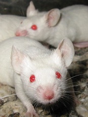
A new method of genome editing can cure hemophilia B in mice, researchers have reported in Nature.
This new technique doesn’t require the co-delivery of an endonuclease to clip the recipient’s DNA at specific locations, and it doesn’t rely on the co-insertion of genetic promoters to activate the new gene’s expression.
These differences may make the new approach both safer and longer-lasting than other genome editing methods, according to researchers.
“It appears that we may be able to achieve lifelong expression of the inserted gene, which is particularly important when treating genetic diseases like hemophilia and severe combined immunodeficiency,” said study author Mark Kay, MD, PhD, of the Stanford University School of Medicine in California.
“We’re able to do this without using promoters or nucleases, which significantly reduces the chances of cancers that can result if the new gene inserts itself at random places in the genome.”
Using their new technique, Dr Kay and his colleagues were able to insert a working copy of the human coagulation factor IX gene into the DNA of mice with hemophilia B. Although the insertion was accomplished in only about 1% of liver cells, those cells made enough factor IX to ameliorate the disorder.
Instead of using nucleases to cut the DNA or a promoter to drive expression of the factor IX gene, the researchers hitched the expression of the new gene to that of albumin.
They used a modified version of adeno-associated virus and relied on homologous recombination to insert the factor IX gene near the albumin gene.
Using a special DNA linker between the genes, the researchers were able to ensure that the clotting factor protein was made hand-in-hand with the highly expressed albumin protein.
During homologous recombination, the cell takes advantage of the fact that it has two copies of every chromosome. By lining up the damaged and undamaged chromosomes, the cell can “crib” off the intact copy to repair the damage without losing vital genetic information.
The researchers used this natural process to copy sequences from the viral vector into the genome at places they chose—in this case, after the albumin gene.
When they tested their approach in newborn lab mice with hemophilia, the team found the animals began to express levels of factor IX that were between 7% and 20% of normal. That amount of clotting factor has been shown in previous studies to be therapeutic in mice.
The researchers further showed that the technique worked as well in adult animals, even though the gene was successfully inserted in fewer than 1 in every 100 liver cells.
“We expected this approach to work best in newborn animals because the liver is still growing,” Dr Kay said. “However, because homologous recombination has been thought to occur mostly in proliferating cells, we didn’t expect it to work as well as it did in adult animals.”
The researchers are now planning to test the technique in mice with livers composed of human and mouse cells, a model that may be a good surrogate to further predict what will happen in humans.
Credit: Aaron Logan
A new method of genome editing can cure hemophilia B in mice, researchers have reported in Nature.
This new technique doesn’t require the co-delivery of an endonuclease to clip the recipient’s DNA at specific locations, and it doesn’t rely on the co-insertion of genetic promoters to activate the new gene’s expression.
These differences may make the new approach both safer and longer-lasting than other genome editing methods, according to researchers.
“It appears that we may be able to achieve lifelong expression of the inserted gene, which is particularly important when treating genetic diseases like hemophilia and severe combined immunodeficiency,” said study author Mark Kay, MD, PhD, of the Stanford University School of Medicine in California.
“We’re able to do this without using promoters or nucleases, which significantly reduces the chances of cancers that can result if the new gene inserts itself at random places in the genome.”
Using their new technique, Dr Kay and his colleagues were able to insert a working copy of the human coagulation factor IX gene into the DNA of mice with hemophilia B. Although the insertion was accomplished in only about 1% of liver cells, those cells made enough factor IX to ameliorate the disorder.
Instead of using nucleases to cut the DNA or a promoter to drive expression of the factor IX gene, the researchers hitched the expression of the new gene to that of albumin.
They used a modified version of adeno-associated virus and relied on homologous recombination to insert the factor IX gene near the albumin gene.
Using a special DNA linker between the genes, the researchers were able to ensure that the clotting factor protein was made hand-in-hand with the highly expressed albumin protein.
During homologous recombination, the cell takes advantage of the fact that it has two copies of every chromosome. By lining up the damaged and undamaged chromosomes, the cell can “crib” off the intact copy to repair the damage without losing vital genetic information.
The researchers used this natural process to copy sequences from the viral vector into the genome at places they chose—in this case, after the albumin gene.
When they tested their approach in newborn lab mice with hemophilia, the team found the animals began to express levels of factor IX that were between 7% and 20% of normal. That amount of clotting factor has been shown in previous studies to be therapeutic in mice.
The researchers further showed that the technique worked as well in adult animals, even though the gene was successfully inserted in fewer than 1 in every 100 liver cells.
“We expected this approach to work best in newborn animals because the liver is still growing,” Dr Kay said. “However, because homologous recombination has been thought to occur mostly in proliferating cells, we didn’t expect it to work as well as it did in adult animals.”
The researchers are now planning to test the technique in mice with livers composed of human and mouse cells, a model that may be a good surrogate to further predict what will happen in humans.
Credit: Aaron Logan
A new method of genome editing can cure hemophilia B in mice, researchers have reported in Nature.
This new technique doesn’t require the co-delivery of an endonuclease to clip the recipient’s DNA at specific locations, and it doesn’t rely on the co-insertion of genetic promoters to activate the new gene’s expression.
These differences may make the new approach both safer and longer-lasting than other genome editing methods, according to researchers.
“It appears that we may be able to achieve lifelong expression of the inserted gene, which is particularly important when treating genetic diseases like hemophilia and severe combined immunodeficiency,” said study author Mark Kay, MD, PhD, of the Stanford University School of Medicine in California.
“We’re able to do this without using promoters or nucleases, which significantly reduces the chances of cancers that can result if the new gene inserts itself at random places in the genome.”
Using their new technique, Dr Kay and his colleagues were able to insert a working copy of the human coagulation factor IX gene into the DNA of mice with hemophilia B. Although the insertion was accomplished in only about 1% of liver cells, those cells made enough factor IX to ameliorate the disorder.
Instead of using nucleases to cut the DNA or a promoter to drive expression of the factor IX gene, the researchers hitched the expression of the new gene to that of albumin.
They used a modified version of adeno-associated virus and relied on homologous recombination to insert the factor IX gene near the albumin gene.
Using a special DNA linker between the genes, the researchers were able to ensure that the clotting factor protein was made hand-in-hand with the highly expressed albumin protein.
During homologous recombination, the cell takes advantage of the fact that it has two copies of every chromosome. By lining up the damaged and undamaged chromosomes, the cell can “crib” off the intact copy to repair the damage without losing vital genetic information.
The researchers used this natural process to copy sequences from the viral vector into the genome at places they chose—in this case, after the albumin gene.
When they tested their approach in newborn lab mice with hemophilia, the team found the animals began to express levels of factor IX that were between 7% and 20% of normal. That amount of clotting factor has been shown in previous studies to be therapeutic in mice.
The researchers further showed that the technique worked as well in adult animals, even though the gene was successfully inserted in fewer than 1 in every 100 liver cells.
“We expected this approach to work best in newborn animals because the liver is still growing,” Dr Kay said. “However, because homologous recombination has been thought to occur mostly in proliferating cells, we didn’t expect it to work as well as it did in adult animals.”
The researchers are now planning to test the technique in mice with livers composed of human and mouse cells, a model that may be a good surrogate to further predict what will happen in humans.
Paperwork consumes docs’ time, erodes morale

A survey of nearly 5000 US physicians showed that the average doctor spent 16.6% of his or her working hours on non-patient-related paperwork.
This includes tasks such as billing, obtaining insurance approvals, financial and personnel management, and negotiating contracts.
The more time doctors spent on such tasks, the less satisfied they were with medicine as a career.
Researchers detailed these findings in the International Journal of Health Services.
“Our crazy health financing system is demoralizing doctors and wasting vast resources,” said study author David Himmelstein, MD, a professor at Hunter College of the City University of New York.
“Turning healthcare into a business means we spend more and more time on billing, insurance paperwork, and the bottom line. We need to move to a simple, nonprofit national health insurance system that lets doctors and hospitals focus on patients, not finances.”
Dr Himmelstein and colleague Steffie Woolhandler, MD, analyzed confidential data from the 2008 Health Tracking Physician Survey (the most recent data available). The survey included information from a nationally representative sample of 4720 physicians who practiced at least 20 hours per week.
The data showed that the average doctor spent 8.7 hours per week, or 16.6% of his or her working time, on non-patient-related administration. This excludes tasks such as writing chart notes, communicating with other doctors, and ordering lab tests.
In total, patient-care physicians spent 168.4 million hours on non-patient-related administrative tasks in 2008. Drs Himmelstein and Woolhandler estimate that the total cost of physician time spent on administration in 2014 will amount to $102 billion.
Career satisfaction was lower for physicians who spent more time on administration. “Very satisfied” doctors spent, on average, 16.1% of their time on administration. “Very dissatisfied” doctors spent 20.6% of their time on such tasks.
Among various specialties, psychiatrists spent the most time on administration (20.3%), followed by internists (17.3%) and family/general practitioners (17.3%). Pediatricians spent the least amount of time (14.1%) on non-patient-related administrative tasks and were the most satisfied group of doctors.
Solo practice was associated with more administrative work, but small group practice was not. Doctors practicing in groups of 100 or more actually spent more time (19.7%) on such tasks than those in small groups (16.3%).
The researchers were surprised to find that physicians who used electronic health records spent more time (17.2% for those using entirely electronic records, 18% for those using a mix of paper and electronic) on administration than those who used only paper records (15.5%).
The pair noted that physicians in Canada spend far less time on administration than US doctors, and they attributed the difference to Canada’s single-payer system, which has greatly simplified billing and reduced bureaucracy.
The researchers pointed out that the only previous nationally representative survey of this kind was carried out in 1995, and that study showed that administration and insurance-related matters accounted for 13.5% of physicians’ total work time. Other, less representative studies also suggest the bureaucratic burden on physicians has grown in the past two decades.
“American doctors are drowning in paperwork,” Dr Woolhandler said. “Our study almost certainly understates physicians’ current administrative burden.”
“Since 2008, when the survey we analyzed was collected, tens of thousands of doctors have moved from small private practices with minimal bureaucracy into giant group practices where bureaucracy is rampant. And under the accountable care organizations favored by insurers, more doctors are facing HMO-type incentives to deny care to their patients, a move that our data shows drives up administrative work.”
A survey of nearly 5000 US physicians showed that the average doctor spent 16.6% of his or her working hours on non-patient-related paperwork.
This includes tasks such as billing, obtaining insurance approvals, financial and personnel management, and negotiating contracts.
The more time doctors spent on such tasks, the less satisfied they were with medicine as a career.
Researchers detailed these findings in the International Journal of Health Services.
“Our crazy health financing system is demoralizing doctors and wasting vast resources,” said study author David Himmelstein, MD, a professor at Hunter College of the City University of New York.
“Turning healthcare into a business means we spend more and more time on billing, insurance paperwork, and the bottom line. We need to move to a simple, nonprofit national health insurance system that lets doctors and hospitals focus on patients, not finances.”
Dr Himmelstein and colleague Steffie Woolhandler, MD, analyzed confidential data from the 2008 Health Tracking Physician Survey (the most recent data available). The survey included information from a nationally representative sample of 4720 physicians who practiced at least 20 hours per week.
The data showed that the average doctor spent 8.7 hours per week, or 16.6% of his or her working time, on non-patient-related administration. This excludes tasks such as writing chart notes, communicating with other doctors, and ordering lab tests.
In total, patient-care physicians spent 168.4 million hours on non-patient-related administrative tasks in 2008. Drs Himmelstein and Woolhandler estimate that the total cost of physician time spent on administration in 2014 will amount to $102 billion.
Career satisfaction was lower for physicians who spent more time on administration. “Very satisfied” doctors spent, on average, 16.1% of their time on administration. “Very dissatisfied” doctors spent 20.6% of their time on such tasks.
Among various specialties, psychiatrists spent the most time on administration (20.3%), followed by internists (17.3%) and family/general practitioners (17.3%). Pediatricians spent the least amount of time (14.1%) on non-patient-related administrative tasks and were the most satisfied group of doctors.
Solo practice was associated with more administrative work, but small group practice was not. Doctors practicing in groups of 100 or more actually spent more time (19.7%) on such tasks than those in small groups (16.3%).
The researchers were surprised to find that physicians who used electronic health records spent more time (17.2% for those using entirely electronic records, 18% for those using a mix of paper and electronic) on administration than those who used only paper records (15.5%).
The pair noted that physicians in Canada spend far less time on administration than US doctors, and they attributed the difference to Canada’s single-payer system, which has greatly simplified billing and reduced bureaucracy.
The researchers pointed out that the only previous nationally representative survey of this kind was carried out in 1995, and that study showed that administration and insurance-related matters accounted for 13.5% of physicians’ total work time. Other, less representative studies also suggest the bureaucratic burden on physicians has grown in the past two decades.
“American doctors are drowning in paperwork,” Dr Woolhandler said. “Our study almost certainly understates physicians’ current administrative burden.”
“Since 2008, when the survey we analyzed was collected, tens of thousands of doctors have moved from small private practices with minimal bureaucracy into giant group practices where bureaucracy is rampant. And under the accountable care organizations favored by insurers, more doctors are facing HMO-type incentives to deny care to their patients, a move that our data shows drives up administrative work.”
A survey of nearly 5000 US physicians showed that the average doctor spent 16.6% of his or her working hours on non-patient-related paperwork.
This includes tasks such as billing, obtaining insurance approvals, financial and personnel management, and negotiating contracts.
The more time doctors spent on such tasks, the less satisfied they were with medicine as a career.
Researchers detailed these findings in the International Journal of Health Services.
“Our crazy health financing system is demoralizing doctors and wasting vast resources,” said study author David Himmelstein, MD, a professor at Hunter College of the City University of New York.
“Turning healthcare into a business means we spend more and more time on billing, insurance paperwork, and the bottom line. We need to move to a simple, nonprofit national health insurance system that lets doctors and hospitals focus on patients, not finances.”
Dr Himmelstein and colleague Steffie Woolhandler, MD, analyzed confidential data from the 2008 Health Tracking Physician Survey (the most recent data available). The survey included information from a nationally representative sample of 4720 physicians who practiced at least 20 hours per week.
The data showed that the average doctor spent 8.7 hours per week, or 16.6% of his or her working time, on non-patient-related administration. This excludes tasks such as writing chart notes, communicating with other doctors, and ordering lab tests.
In total, patient-care physicians spent 168.4 million hours on non-patient-related administrative tasks in 2008. Drs Himmelstein and Woolhandler estimate that the total cost of physician time spent on administration in 2014 will amount to $102 billion.
Career satisfaction was lower for physicians who spent more time on administration. “Very satisfied” doctors spent, on average, 16.1% of their time on administration. “Very dissatisfied” doctors spent 20.6% of their time on such tasks.
Among various specialties, psychiatrists spent the most time on administration (20.3%), followed by internists (17.3%) and family/general practitioners (17.3%). Pediatricians spent the least amount of time (14.1%) on non-patient-related administrative tasks and were the most satisfied group of doctors.
Solo practice was associated with more administrative work, but small group practice was not. Doctors practicing in groups of 100 or more actually spent more time (19.7%) on such tasks than those in small groups (16.3%).
The researchers were surprised to find that physicians who used electronic health records spent more time (17.2% for those using entirely electronic records, 18% for those using a mix of paper and electronic) on administration than those who used only paper records (15.5%).
The pair noted that physicians in Canada spend far less time on administration than US doctors, and they attributed the difference to Canada’s single-payer system, which has greatly simplified billing and reduced bureaucracy.
The researchers pointed out that the only previous nationally representative survey of this kind was carried out in 1995, and that study showed that administration and insurance-related matters accounted for 13.5% of physicians’ total work time. Other, less representative studies also suggest the bureaucratic burden on physicians has grown in the past two decades.
“American doctors are drowning in paperwork,” Dr Woolhandler said. “Our study almost certainly understates physicians’ current administrative burden.”
“Since 2008, when the survey we analyzed was collected, tens of thousands of doctors have moved from small private practices with minimal bureaucracy into giant group practices where bureaucracy is rampant. And under the accountable care organizations favored by insurers, more doctors are facing HMO-type incentives to deny care to their patients, a move that our data shows drives up administrative work.”
New criteria for diagnosing MM could prevent organ damage
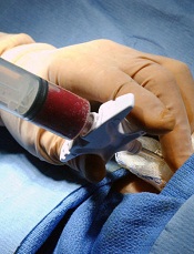
Credit: Chad McNeeley
The International Myeloma Working Group (IMWG) has published new criteria for diagnosing multiple myeloma (MM) in The Lancet Oncology.
The group has added validated biomarkers to the current clinical symptoms used for MM diagnosis—hypercalcemia, renal failure, anemia, and bone lesions.
This addition will allow physicians to diagnose MM before patients become symptomatic and, therefore, before organ damage occurs, according to the IMWG.
Lead author S. Vincent Rajkumar, MD, of the Mayo Clinic in Rochester, Minnesota, noted that MM is always preceded sequentially by two conditions—monoclonal gammopathy of undetermined significance and smoldering MM. Since both are asymptomatic, most MM patients are not diagnosed until organ damage occurs.
“The new IMWG criteria allow for the diagnosis of myeloma to be made in patients without symptoms and before organ damage occurs, using validated biomarkers that identify patients with [smoldering] MM who have an ‘ultra-high’ risk of progression to multiple myeloma,” Dr Rajkumar said.
“These biomarkers are associated with the near-inevitable development of clinical symptoms and are important for early diagnosis and treatment, which is very important for patients.”
Other updates to the criteria used to diagnose MM include the use of CT and PET-CT scans to identify bone lesions. According to the authors, this will enable more accurate diagnosis and intervention before fractures or other serious problems arise.
“We believe that the new criteria will rectify the situation where we were unable to use the considerable advances in multiple myeloma therapy prior to organ damage,” Dr Rajkumar said. “We can now initiate therapy in some patients early on in the course of their disease.”
The IMWG’s revised diagnostic criteria for MM and smoldering MM are as follows.
Definition of MM
Clonal bone marrow plasma cells ≥10% or biopsy-proven bony or extramedullary plasmacytoma* and one or more of the following myeloma defining events:
- Evidence of end organ damage that can be attributed to the underlying plasma cell proliferative disorder, specifically:
- Hypercalcemia: serum calcium >0.25 mmol/L (>1 mg/dL) higher than the upper limit of normal or >2.75 mmol/L (>11 mg/dL).
- Renal insufficiency: creatinine clearance <40 mL per min (measured or estimated by validated equations) or serum creatinine >177 μmol/L (>2 mg/dL).
- Anemia: hemoglobin value of >20 g/L below the lower limit of normal or a hemoglobin value <100 g/L.
- Bone lesions: one or more osteolytic lesions on skeletal radiography, CT, or PET-CT. If the bone marrow has less than 10% clonal plasma cells, more than one bone lesion is required to distinguish from solitary plasmacytoma with minimal marrow involvement.
- One or more of the following biomarkers:
- Clonal bone marrow plasma cell percentage ≥60%.
- Involved:uninvolved serum free light chain ratio ≥100. These values are based on the serum Freelite assay (The Binding Site Group, Birmingham, UK). The involved free light chain must be ≥100 mg/L.
- >1 focal lesions on MRI studies. Each focal lesion must be 5 mm or more in size.
*The IMWG said clonality should be established by showing κ/λ-light-chain restriction on flow cytometry, immunohistochemistry, or immunofluorescence. Bone
marrow plasma cell percentage should preferably be estimated from a core biopsy specimen. In case of a disparity between the aspirate and core biopsy, the highest value should be used.
Definition of smoldering MM
Both of the following criteria must be met:
- Serum monoclonal protein (IgG or IgA) ≥30 g/L or urinary monoclonal protein ≥500 mg per 24 hours and/or clonal bone marrow plasma cells 10%–60%.
- Absence of myeloma defining events or amyloidosis.
Credit: Chad McNeeley
The International Myeloma Working Group (IMWG) has published new criteria for diagnosing multiple myeloma (MM) in The Lancet Oncology.
The group has added validated biomarkers to the current clinical symptoms used for MM diagnosis—hypercalcemia, renal failure, anemia, and bone lesions.
This addition will allow physicians to diagnose MM before patients become symptomatic and, therefore, before organ damage occurs, according to the IMWG.
Lead author S. Vincent Rajkumar, MD, of the Mayo Clinic in Rochester, Minnesota, noted that MM is always preceded sequentially by two conditions—monoclonal gammopathy of undetermined significance and smoldering MM. Since both are asymptomatic, most MM patients are not diagnosed until organ damage occurs.
“The new IMWG criteria allow for the diagnosis of myeloma to be made in patients without symptoms and before organ damage occurs, using validated biomarkers that identify patients with [smoldering] MM who have an ‘ultra-high’ risk of progression to multiple myeloma,” Dr Rajkumar said.
“These biomarkers are associated with the near-inevitable development of clinical symptoms and are important for early diagnosis and treatment, which is very important for patients.”
Other updates to the criteria used to diagnose MM include the use of CT and PET-CT scans to identify bone lesions. According to the authors, this will enable more accurate diagnosis and intervention before fractures or other serious problems arise.
“We believe that the new criteria will rectify the situation where we were unable to use the considerable advances in multiple myeloma therapy prior to organ damage,” Dr Rajkumar said. “We can now initiate therapy in some patients early on in the course of their disease.”
The IMWG’s revised diagnostic criteria for MM and smoldering MM are as follows.
Definition of MM
Clonal bone marrow plasma cells ≥10% or biopsy-proven bony or extramedullary plasmacytoma* and one or more of the following myeloma defining events:
- Evidence of end organ damage that can be attributed to the underlying plasma cell proliferative disorder, specifically:
- Hypercalcemia: serum calcium >0.25 mmol/L (>1 mg/dL) higher than the upper limit of normal or >2.75 mmol/L (>11 mg/dL).
- Renal insufficiency: creatinine clearance <40 mL per min (measured or estimated by validated equations) or serum creatinine >177 μmol/L (>2 mg/dL).
- Anemia: hemoglobin value of >20 g/L below the lower limit of normal or a hemoglobin value <100 g/L.
- Bone lesions: one or more osteolytic lesions on skeletal radiography, CT, or PET-CT. If the bone marrow has less than 10% clonal plasma cells, more than one bone lesion is required to distinguish from solitary plasmacytoma with minimal marrow involvement.
- One or more of the following biomarkers:
- Clonal bone marrow plasma cell percentage ≥60%.
- Involved:uninvolved serum free light chain ratio ≥100. These values are based on the serum Freelite assay (The Binding Site Group, Birmingham, UK). The involved free light chain must be ≥100 mg/L.
- >1 focal lesions on MRI studies. Each focal lesion must be 5 mm or more in size.
*The IMWG said clonality should be established by showing κ/λ-light-chain restriction on flow cytometry, immunohistochemistry, or immunofluorescence. Bone
marrow plasma cell percentage should preferably be estimated from a core biopsy specimen. In case of a disparity between the aspirate and core biopsy, the highest value should be used.
Definition of smoldering MM
Both of the following criteria must be met:
- Serum monoclonal protein (IgG or IgA) ≥30 g/L or urinary monoclonal protein ≥500 mg per 24 hours and/or clonal bone marrow plasma cells 10%–60%.
- Absence of myeloma defining events or amyloidosis.
Credit: Chad McNeeley
The International Myeloma Working Group (IMWG) has published new criteria for diagnosing multiple myeloma (MM) in The Lancet Oncology.
The group has added validated biomarkers to the current clinical symptoms used for MM diagnosis—hypercalcemia, renal failure, anemia, and bone lesions.
This addition will allow physicians to diagnose MM before patients become symptomatic and, therefore, before organ damage occurs, according to the IMWG.
Lead author S. Vincent Rajkumar, MD, of the Mayo Clinic in Rochester, Minnesota, noted that MM is always preceded sequentially by two conditions—monoclonal gammopathy of undetermined significance and smoldering MM. Since both are asymptomatic, most MM patients are not diagnosed until organ damage occurs.
“The new IMWG criteria allow for the diagnosis of myeloma to be made in patients without symptoms and before organ damage occurs, using validated biomarkers that identify patients with [smoldering] MM who have an ‘ultra-high’ risk of progression to multiple myeloma,” Dr Rajkumar said.
“These biomarkers are associated with the near-inevitable development of clinical symptoms and are important for early diagnosis and treatment, which is very important for patients.”
Other updates to the criteria used to diagnose MM include the use of CT and PET-CT scans to identify bone lesions. According to the authors, this will enable more accurate diagnosis and intervention before fractures or other serious problems arise.
“We believe that the new criteria will rectify the situation where we were unable to use the considerable advances in multiple myeloma therapy prior to organ damage,” Dr Rajkumar said. “We can now initiate therapy in some patients early on in the course of their disease.”
The IMWG’s revised diagnostic criteria for MM and smoldering MM are as follows.
Definition of MM
Clonal bone marrow plasma cells ≥10% or biopsy-proven bony or extramedullary plasmacytoma* and one or more of the following myeloma defining events:
- Evidence of end organ damage that can be attributed to the underlying plasma cell proliferative disorder, specifically:
- Hypercalcemia: serum calcium >0.25 mmol/L (>1 mg/dL) higher than the upper limit of normal or >2.75 mmol/L (>11 mg/dL).
- Renal insufficiency: creatinine clearance <40 mL per min (measured or estimated by validated equations) or serum creatinine >177 μmol/L (>2 mg/dL).
- Anemia: hemoglobin value of >20 g/L below the lower limit of normal or a hemoglobin value <100 g/L.
- Bone lesions: one or more osteolytic lesions on skeletal radiography, CT, or PET-CT. If the bone marrow has less than 10% clonal plasma cells, more than one bone lesion is required to distinguish from solitary plasmacytoma with minimal marrow involvement.
- One or more of the following biomarkers:
- Clonal bone marrow plasma cell percentage ≥60%.
- Involved:uninvolved serum free light chain ratio ≥100. These values are based on the serum Freelite assay (The Binding Site Group, Birmingham, UK). The involved free light chain must be ≥100 mg/L.
- >1 focal lesions on MRI studies. Each focal lesion must be 5 mm or more in size.
*The IMWG said clonality should be established by showing κ/λ-light-chain restriction on flow cytometry, immunohistochemistry, or immunofluorescence. Bone
marrow plasma cell percentage should preferably be estimated from a core biopsy specimen. In case of a disparity between the aspirate and core biopsy, the highest value should be used.
Definition of smoldering MM
Both of the following criteria must be met:
- Serum monoclonal protein (IgG or IgA) ≥30 g/L or urinary monoclonal protein ≥500 mg per 24 hours and/or clonal bone marrow plasma cells 10%–60%.
- Absence of myeloma defining events or amyloidosis.