User login
FDA approves pathogen inactivation system for platelets
The US Food and Drug Administration (FDA) has approved the INTERCEPT Blood System for platelets, the first system of its kind to be approved in the US.
It is used to inactivate viruses, bacteria, spirochetes, parasites, and leukocytes in apheresis platelet components.
This can reduce the risk of transfusion-transmitted infection and, potentially, transfusion-associated graft-vs-host disease, although the system cannot inactivate all pathogens.
Certain non-enveloped viruses (such as HAV, HEV, B19, and poliovirus) and Bacillus cereus spores have demonstrated resistance to the INTERCEPT process.
Earlier this week, the FDA approved the INTERCEPT Blood System for plasma (also the first system of its kind to gain FDA approval).
The platelet and plasma systems use the same illumination device, the same active compound (amotosalen), and very similar production steps.
The INTERCEPT systems target a basic biological difference between the therapeutic components of blood. Platelets, plasma, and red blood cells do not require functional DNA or RNA for therapeutic efficacy. But pathogens and white blood cells do, in order to transmit infection.
The INTERCEPT systems use a proprietary molecule that, when activated by UVA light, binds to and blocks the replication of DNA and RNA, preventing nucleic acid replication and rendering the pathogen inactive.
The INTERCEPT Blood System for platelets has been approved in Europe since 2002 and is currently used in 20 countries.
The system was recently made available in the US and its territories under an investigational device exemption study to reduce the risk of transfusion-transmitted dengue and Chikungunya viruses, both of which are epidemic in the Caribbean region, including Puerto Rico, as well as sporadically in the southern US. No approved blood bank screening tests are available for either virus.
Researchers have evaluated INTERCEPT-processed platelets in 10 controlled clinical trials. Details on these trials can be found in the package insert. 
The US Food and Drug Administration (FDA) has approved the INTERCEPT Blood System for platelets, the first system of its kind to be approved in the US.
It is used to inactivate viruses, bacteria, spirochetes, parasites, and leukocytes in apheresis platelet components.
This can reduce the risk of transfusion-transmitted infection and, potentially, transfusion-associated graft-vs-host disease, although the system cannot inactivate all pathogens.
Certain non-enveloped viruses (such as HAV, HEV, B19, and poliovirus) and Bacillus cereus spores have demonstrated resistance to the INTERCEPT process.
Earlier this week, the FDA approved the INTERCEPT Blood System for plasma (also the first system of its kind to gain FDA approval).
The platelet and plasma systems use the same illumination device, the same active compound (amotosalen), and very similar production steps.
The INTERCEPT systems target a basic biological difference between the therapeutic components of blood. Platelets, plasma, and red blood cells do not require functional DNA or RNA for therapeutic efficacy. But pathogens and white blood cells do, in order to transmit infection.
The INTERCEPT systems use a proprietary molecule that, when activated by UVA light, binds to and blocks the replication of DNA and RNA, preventing nucleic acid replication and rendering the pathogen inactive.
The INTERCEPT Blood System for platelets has been approved in Europe since 2002 and is currently used in 20 countries.
The system was recently made available in the US and its territories under an investigational device exemption study to reduce the risk of transfusion-transmitted dengue and Chikungunya viruses, both of which are epidemic in the Caribbean region, including Puerto Rico, as well as sporadically in the southern US. No approved blood bank screening tests are available for either virus.
Researchers have evaluated INTERCEPT-processed platelets in 10 controlled clinical trials. Details on these trials can be found in the package insert.
The US Food and Drug Administration (FDA) has approved the INTERCEPT Blood System for platelets, the first system of its kind to be approved in the US.
It is used to inactivate viruses, bacteria, spirochetes, parasites, and leukocytes in apheresis platelet components.
This can reduce the risk of transfusion-transmitted infection and, potentially, transfusion-associated graft-vs-host disease, although the system cannot inactivate all pathogens.
Certain non-enveloped viruses (such as HAV, HEV, B19, and poliovirus) and Bacillus cereus spores have demonstrated resistance to the INTERCEPT process.
Earlier this week, the FDA approved the INTERCEPT Blood System for plasma (also the first system of its kind to gain FDA approval).
The platelet and plasma systems use the same illumination device, the same active compound (amotosalen), and very similar production steps.
The INTERCEPT systems target a basic biological difference between the therapeutic components of blood. Platelets, plasma, and red blood cells do not require functional DNA or RNA for therapeutic efficacy. But pathogens and white blood cells do, in order to transmit infection.
The INTERCEPT systems use a proprietary molecule that, when activated by UVA light, binds to and blocks the replication of DNA and RNA, preventing nucleic acid replication and rendering the pathogen inactive.
The INTERCEPT Blood System for platelets has been approved in Europe since 2002 and is currently used in 20 countries.
The system was recently made available in the US and its territories under an investigational device exemption study to reduce the risk of transfusion-transmitted dengue and Chikungunya viruses, both of which are epidemic in the Caribbean region, including Puerto Rico, as well as sporadically in the southern US. No approved blood bank screening tests are available for either virus.
Researchers have evaluated INTERCEPT-processed platelets in 10 controlled clinical trials. Details on these trials can be found in the package insert.
Studies show TRALI underreported, TACO on the decline
Credit: Elise Amendola
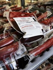
Two studies shed new light on the prevalence of transfusion-related acute lung injury (TRALI) and transfusion-associated circulatory overload (TACO) in the US.
The research showed that postoperative TRALI is significantly underreported and more common than previously thought, with an overall rate of 1.4%.
And the rate of TACO is on the decline, but the risk to surgical patients remains high, at 4%, similar to previous TACO estimates in non-surgical patients.
“An accurate understanding of the risks associated with blood transfusions is essential when determining the safety and appropriateness of transfusion therapies for patients,” said Daryl Kor, MD, senior author of both studies and an associate professor at Mayo Clinic in Rochester, Minnesota.
“Our research provides a greater awareness of the incidence of TRALI and TACO in surgical patients, a population that has been perhaps underrepresented in studies in this area. We believe this to be an important first step in our efforts to prevent these life-threatening transfusion complications.”
Dr Kor and his colleagues described this research in Anesthesiology alongside a related editorial.
In the two retrospective studies, the researchers examined the incidence of TRALI in 3379 patients and TACO in 4070 patients who received blood transfusions during non-cardiac surgery under general anesthesia in 2004 and 2011.
Using a novel algorithm, followed by a rigorous manual review, the team performed a detailed epidemiologic analysis for both complications.
The first study showed that TRALI occurred in 1.4% of surgical patients, with higher rates in specific surgical populations such as those having surgery inside the chest cavity, on major blood vessels, or having an organ transplant. Patients who received larger volumes of blood were also at increased risk.
Previous studies investigating TRALI rates have primarily focused on the critically ill and reported variable incidence rates. Many studies have reported incidences between 0.02% and 0.05%.
The second study showed that TACO occurs in 4.3% of surgical patients, with higher rates associated with increased volume of blood transfused, advanced age, and total intraoperative fluid balance. Again, patients having surgery inside the chest cavity, on major blood vessels, or organ transplants were at the greatest risk.
The study also revealed that the rate of TACO decreased significantly from 2004 to 2011—from 5.5% to 3%. This decline was not fully explained by any of the patient or transfusion characteristics evaluated in the study.
The researchers said future studies are needed to further explore which mechanisms and risk factors are responsible for TACO and TRALI.
“With improved understanding of the mechanisms underlying TRALI and TACO, we may be able to refine the novel electronic algorithms used to screen patients in these studies,” Dr Kor said. “Ultimately, we hope to develop a real-time prediction model for these complications so that we can identify those at greatest risk and perhaps implement strategies to reduce this risk.”
Credit: Elise Amendola
Two studies shed new light on the prevalence of transfusion-related acute lung injury (TRALI) and transfusion-associated circulatory overload (TACO) in the US.
The research showed that postoperative TRALI is significantly underreported and more common than previously thought, with an overall rate of 1.4%.
And the rate of TACO is on the decline, but the risk to surgical patients remains high, at 4%, similar to previous TACO estimates in non-surgical patients.
“An accurate understanding of the risks associated with blood transfusions is essential when determining the safety and appropriateness of transfusion therapies for patients,” said Daryl Kor, MD, senior author of both studies and an associate professor at Mayo Clinic in Rochester, Minnesota.
“Our research provides a greater awareness of the incidence of TRALI and TACO in surgical patients, a population that has been perhaps underrepresented in studies in this area. We believe this to be an important first step in our efforts to prevent these life-threatening transfusion complications.”
Dr Kor and his colleagues described this research in Anesthesiology alongside a related editorial.
In the two retrospective studies, the researchers examined the incidence of TRALI in 3379 patients and TACO in 4070 patients who received blood transfusions during non-cardiac surgery under general anesthesia in 2004 and 2011.
Using a novel algorithm, followed by a rigorous manual review, the team performed a detailed epidemiologic analysis for both complications.
The first study showed that TRALI occurred in 1.4% of surgical patients, with higher rates in specific surgical populations such as those having surgery inside the chest cavity, on major blood vessels, or having an organ transplant. Patients who received larger volumes of blood were also at increased risk.
Previous studies investigating TRALI rates have primarily focused on the critically ill and reported variable incidence rates. Many studies have reported incidences between 0.02% and 0.05%.
The second study showed that TACO occurs in 4.3% of surgical patients, with higher rates associated with increased volume of blood transfused, advanced age, and total intraoperative fluid balance. Again, patients having surgery inside the chest cavity, on major blood vessels, or organ transplants were at the greatest risk.
The study also revealed that the rate of TACO decreased significantly from 2004 to 2011—from 5.5% to 3%. This decline was not fully explained by any of the patient or transfusion characteristics evaluated in the study.
The researchers said future studies are needed to further explore which mechanisms and risk factors are responsible for TACO and TRALI.
“With improved understanding of the mechanisms underlying TRALI and TACO, we may be able to refine the novel electronic algorithms used to screen patients in these studies,” Dr Kor said. “Ultimately, we hope to develop a real-time prediction model for these complications so that we can identify those at greatest risk and perhaps implement strategies to reduce this risk.”
Credit: Elise Amendola
Two studies shed new light on the prevalence of transfusion-related acute lung injury (TRALI) and transfusion-associated circulatory overload (TACO) in the US.
The research showed that postoperative TRALI is significantly underreported and more common than previously thought, with an overall rate of 1.4%.
And the rate of TACO is on the decline, but the risk to surgical patients remains high, at 4%, similar to previous TACO estimates in non-surgical patients.
“An accurate understanding of the risks associated with blood transfusions is essential when determining the safety and appropriateness of transfusion therapies for patients,” said Daryl Kor, MD, senior author of both studies and an associate professor at Mayo Clinic in Rochester, Minnesota.
“Our research provides a greater awareness of the incidence of TRALI and TACO in surgical patients, a population that has been perhaps underrepresented in studies in this area. We believe this to be an important first step in our efforts to prevent these life-threatening transfusion complications.”
Dr Kor and his colleagues described this research in Anesthesiology alongside a related editorial.
In the two retrospective studies, the researchers examined the incidence of TRALI in 3379 patients and TACO in 4070 patients who received blood transfusions during non-cardiac surgery under general anesthesia in 2004 and 2011.
Using a novel algorithm, followed by a rigorous manual review, the team performed a detailed epidemiologic analysis for both complications.
The first study showed that TRALI occurred in 1.4% of surgical patients, with higher rates in specific surgical populations such as those having surgery inside the chest cavity, on major blood vessels, or having an organ transplant. Patients who received larger volumes of blood were also at increased risk.
Previous studies investigating TRALI rates have primarily focused on the critically ill and reported variable incidence rates. Many studies have reported incidences between 0.02% and 0.05%.
The second study showed that TACO occurs in 4.3% of surgical patients, with higher rates associated with increased volume of blood transfused, advanced age, and total intraoperative fluid balance. Again, patients having surgery inside the chest cavity, on major blood vessels, or organ transplants were at the greatest risk.
The study also revealed that the rate of TACO decreased significantly from 2004 to 2011—from 5.5% to 3%. This decline was not fully explained by any of the patient or transfusion characteristics evaluated in the study.
The researchers said future studies are needed to further explore which mechanisms and risk factors are responsible for TACO and TRALI.
“With improved understanding of the mechanisms underlying TRALI and TACO, we may be able to refine the novel electronic algorithms used to screen patients in these studies,” Dr Kor said. “Ultimately, we hope to develop a real-time prediction model for these complications so that we can identify those at greatest risk and perhaps implement strategies to reduce this risk.”
NICE backs dabigatran for VTE

Credit: CDC
The UK’s National Institute for Health and Care Excellence (NICE) has published a final guidance recommending the anticoagulant dabigatran (Pradaxa, Boehringer Ingelheim) as an option for treating and preventing recurrent deep vein thrombosis (DVT) and pulmonary embolism (PE) in adults.
The guidance says dabigatran can provide a benefit for these patients, with cost- and clinical-effectiveness similar to rivaroxaban and added convenience compared to warfarin.
“For many people, using warfarin can be difficult because of the need for frequent tests to see if the blood is clotting properly, and having to adjust the dose of the
drug if it is not,” said Carole Longson, NICE Health Technology Evaluation Centre Director.
“The appraisal committee felt that dabigatran represents a potential benefit for many people who have had a DVT or PE, particularly those who have risk factors for recurrence of a blood clot and who therefore need longer-term treatment. We are pleased, therefore, to be able to recommend dabigatran as a cost-effective option for treating DVT and PE and preventing further episodes in adults.”
NICE expects dabigatran to be available on the National Health Service within 3 months.
Cost considerations
Dabigatran costs £65.90 for a 60-capsule pack of the 150 mg or 110 mg doses (excluding tax) and £2.20 per day of treatment, although costs may vary in different settings.
The most plausible incremental cost-effectiveness ratio (ICER) for dabigatran compared with warfarin for acute treatment was uncertain.
However, both Boehringer Ingelheim’s and the evidence review group’s exploratory ICER remained in the range that could be considered a cost-effective use of National Health Service resources. That is, both were under £20,000 per quality-adjusted life-year gained (QALY).
Neither Boehringer Ingelheim nor the evidence review group found any significant difference in efficacy between dabigatran and rivaroxaban for acute treatment of venous thromboembolism (VTE) in their indirect comparisons, and the costs were also very similar between these two treatments.
For combined treatment and secondary prevention of VTE, the appraisal committee said the company’s base case ICER for dabigatran compared with warfarin was likely too low (£9973 per QALY gained).
But the evidence review group’s exploratory base case for dabigatran compared with warfarin may have overestimated the ICER (£35,786 per QALY gained). So the ICER probably lies somewhere between these estimates.
Clinical evidence
To assess the clinical effectiveness of dabigatran, the appraisal committee evaluated data from the RECOVER, RE-MEDY, and RESONATE trials.
In the first RE-COVER trial, dabigatran proved noninferior to warfarin for preventing VTE recurrence, and rates of major bleeding were similar between the treatment arms.
However, patients were more likely to discontinue dabigatran due to adverse events. Results from this trial were presented at ASH 2009 and published in NEJM.
The RE-COVER II trial also suggested that dabigatran was noninferior to warfarin for preventing VTE recurrence and related deaths, and dabigatran was associated with a lower rate of major bleeding.
Rates of death, adverse events, and acute coronary syndromes were similar between the treatment arms. Results from this trial were published in Circulation in 2013.
The RE-MEDY and RE-SONATE trials were designed to evaluate dabigatran as extended VTE prophylaxis. Results of both trials were reported in a single NEJM article published in 2013.
The RE-MEDY trial suggested that dabigatran was noninferior to warfarin as extended prophylaxis for recurrent VTE, and warfarin presented a significantly higher risk of bleeding.
Results of the RE-SONATE trial indicated that dabigatran was superior to placebo for preventing recurrent VTE, although the drug significantly increased the risk of major or clinically relevant bleeding.
Credit: CDC
The UK’s National Institute for Health and Care Excellence (NICE) has published a final guidance recommending the anticoagulant dabigatran (Pradaxa, Boehringer Ingelheim) as an option for treating and preventing recurrent deep vein thrombosis (DVT) and pulmonary embolism (PE) in adults.
The guidance says dabigatran can provide a benefit for these patients, with cost- and clinical-effectiveness similar to rivaroxaban and added convenience compared to warfarin.
“For many people, using warfarin can be difficult because of the need for frequent tests to see if the blood is clotting properly, and having to adjust the dose of the
drug if it is not,” said Carole Longson, NICE Health Technology Evaluation Centre Director.
“The appraisal committee felt that dabigatran represents a potential benefit for many people who have had a DVT or PE, particularly those who have risk factors for recurrence of a blood clot and who therefore need longer-term treatment. We are pleased, therefore, to be able to recommend dabigatran as a cost-effective option for treating DVT and PE and preventing further episodes in adults.”
NICE expects dabigatran to be available on the National Health Service within 3 months.
Cost considerations
Dabigatran costs £65.90 for a 60-capsule pack of the 150 mg or 110 mg doses (excluding tax) and £2.20 per day of treatment, although costs may vary in different settings.
The most plausible incremental cost-effectiveness ratio (ICER) for dabigatran compared with warfarin for acute treatment was uncertain.
However, both Boehringer Ingelheim’s and the evidence review group’s exploratory ICER remained in the range that could be considered a cost-effective use of National Health Service resources. That is, both were under £20,000 per quality-adjusted life-year gained (QALY).
Neither Boehringer Ingelheim nor the evidence review group found any significant difference in efficacy between dabigatran and rivaroxaban for acute treatment of venous thromboembolism (VTE) in their indirect comparisons, and the costs were also very similar between these two treatments.
For combined treatment and secondary prevention of VTE, the appraisal committee said the company’s base case ICER for dabigatran compared with warfarin was likely too low (£9973 per QALY gained).
But the evidence review group’s exploratory base case for dabigatran compared with warfarin may have overestimated the ICER (£35,786 per QALY gained). So the ICER probably lies somewhere between these estimates.
Clinical evidence
To assess the clinical effectiveness of dabigatran, the appraisal committee evaluated data from the RECOVER, RE-MEDY, and RESONATE trials.
In the first RE-COVER trial, dabigatran proved noninferior to warfarin for preventing VTE recurrence, and rates of major bleeding were similar between the treatment arms.
However, patients were more likely to discontinue dabigatran due to adverse events. Results from this trial were presented at ASH 2009 and published in NEJM.
The RE-COVER II trial also suggested that dabigatran was noninferior to warfarin for preventing VTE recurrence and related deaths, and dabigatran was associated with a lower rate of major bleeding.
Rates of death, adverse events, and acute coronary syndromes were similar between the treatment arms. Results from this trial were published in Circulation in 2013.
The RE-MEDY and RE-SONATE trials were designed to evaluate dabigatran as extended VTE prophylaxis. Results of both trials were reported in a single NEJM article published in 2013.
The RE-MEDY trial suggested that dabigatran was noninferior to warfarin as extended prophylaxis for recurrent VTE, and warfarin presented a significantly higher risk of bleeding.
Results of the RE-SONATE trial indicated that dabigatran was superior to placebo for preventing recurrent VTE, although the drug significantly increased the risk of major or clinically relevant bleeding.
Credit: CDC
The UK’s National Institute for Health and Care Excellence (NICE) has published a final guidance recommending the anticoagulant dabigatran (Pradaxa, Boehringer Ingelheim) as an option for treating and preventing recurrent deep vein thrombosis (DVT) and pulmonary embolism (PE) in adults.
The guidance says dabigatran can provide a benefit for these patients, with cost- and clinical-effectiveness similar to rivaroxaban and added convenience compared to warfarin.
“For many people, using warfarin can be difficult because of the need for frequent tests to see if the blood is clotting properly, and having to adjust the dose of the
drug if it is not,” said Carole Longson, NICE Health Technology Evaluation Centre Director.
“The appraisal committee felt that dabigatran represents a potential benefit for many people who have had a DVT or PE, particularly those who have risk factors for recurrence of a blood clot and who therefore need longer-term treatment. We are pleased, therefore, to be able to recommend dabigatran as a cost-effective option for treating DVT and PE and preventing further episodes in adults.”
NICE expects dabigatran to be available on the National Health Service within 3 months.
Cost considerations
Dabigatran costs £65.90 for a 60-capsule pack of the 150 mg or 110 mg doses (excluding tax) and £2.20 per day of treatment, although costs may vary in different settings.
The most plausible incremental cost-effectiveness ratio (ICER) for dabigatran compared with warfarin for acute treatment was uncertain.
However, both Boehringer Ingelheim’s and the evidence review group’s exploratory ICER remained in the range that could be considered a cost-effective use of National Health Service resources. That is, both were under £20,000 per quality-adjusted life-year gained (QALY).
Neither Boehringer Ingelheim nor the evidence review group found any significant difference in efficacy between dabigatran and rivaroxaban for acute treatment of venous thromboembolism (VTE) in their indirect comparisons, and the costs were also very similar between these two treatments.
For combined treatment and secondary prevention of VTE, the appraisal committee said the company’s base case ICER for dabigatran compared with warfarin was likely too low (£9973 per QALY gained).
But the evidence review group’s exploratory base case for dabigatran compared with warfarin may have overestimated the ICER (£35,786 per QALY gained). So the ICER probably lies somewhere between these estimates.
Clinical evidence
To assess the clinical effectiveness of dabigatran, the appraisal committee evaluated data from the RECOVER, RE-MEDY, and RESONATE trials.
In the first RE-COVER trial, dabigatran proved noninferior to warfarin for preventing VTE recurrence, and rates of major bleeding were similar between the treatment arms.
However, patients were more likely to discontinue dabigatran due to adverse events. Results from this trial were presented at ASH 2009 and published in NEJM.
The RE-COVER II trial also suggested that dabigatran was noninferior to warfarin for preventing VTE recurrence and related deaths, and dabigatran was associated with a lower rate of major bleeding.
Rates of death, adverse events, and acute coronary syndromes were similar between the treatment arms. Results from this trial were published in Circulation in 2013.
The RE-MEDY and RE-SONATE trials were designed to evaluate dabigatran as extended VTE prophylaxis. Results of both trials were reported in a single NEJM article published in 2013.
The RE-MEDY trial suggested that dabigatran was noninferior to warfarin as extended prophylaxis for recurrent VTE, and warfarin presented a significantly higher risk of bleeding.
Results of the RE-SONATE trial indicated that dabigatran was superior to placebo for preventing recurrent VTE, although the drug significantly increased the risk of major or clinically relevant bleeding.
Amount of blood transfused doesn’t affect long-term mortality
Credit: UAB Hospital
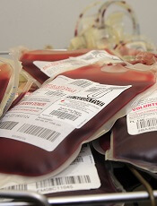
Long-term mortality is not affected by the amount of blood a patient receives following surgery, according to research published in The Lancet.
Investigators compared a liberal transfusion strategy, in which patients received blood to maintain a hemoglobin level at 100 g/L or higher, and a restrictive strategy, in which patients received blood when hemoglobin levels were lower than 80 g/L or if they had symptoms of anemia.
And, at about 3 years of follow-up, there was no difference in mortality between the two groups.
Jeffrey L. Carson, MD, of Robert Wood Johnson Medical School in New Jersey, and his colleagues conducted this research, analyzing data from the FOCUS trial, which included patients from 47 hospitals in the US and Canada.
The trial enrolled 2016 adults age 50 and older, with a history of or risk factors for cardiovascular disease, who had postoperative hemoglobin concentrations lower than 100 g/L within 3 days of surgery to repair a hip fracture.
Patients were randomized by a central telephone system to the liberal (n=1007) or restrictive (n=1009) transfusion groups. The investigators analyzed the long-term mortality of these patients, which was established by linking participants to national death registries in the US and Canada.
The results revealed no difference in mortality from cardiovascular disease, cancer, or severe infection due to the amount of the blood given after surgery.
This supports the initial results of the FOCUS trial, which Dr Carson and his colleagues published in NEJM in 2011 and which demonstrated the safety of fewer transfusions in the short-term.
For the current analysis, the median duration of follow-up was 3.1 years. Eight hundred and forty-one patients (42%) died during that time—432 in the liberal transfusion group and 409 in the restrictive transfusion group. This difference was not statistically significant, with a hazard ratio of 1.09 and a P value of 0.21.
“There has been a steady decline in the amount of blood in transfusions given to patients in the past 3 to 5 years,” Dr Carson noted. “I think it is very reassuring that we have found that using less blood is okay, not just from a short-term perspective, but also a long-term perspective.”
Medical experts had worried that larger amounts of transfused blood might suppress immune function—which could lead to death from infection or cancer—or that smaller transfusions might worsen a patient’s chronic heart disease by depriving the heart of oxygen and other nutrients that it might have absorbed by pumping more blood.
But in both instances, Dr Carson and his colleagues found no difference in long-term death rates, regardless of the number of transfusions.
Dr Carson noted that there are health conditions, such as heart attacks, where the effects of the two transfusion strategies are less certain. Preliminary evidence suggests heart attack patients need more blood, not less. But additional studies are needed to confirm that.
Credit: UAB Hospital
Long-term mortality is not affected by the amount of blood a patient receives following surgery, according to research published in The Lancet.
Investigators compared a liberal transfusion strategy, in which patients received blood to maintain a hemoglobin level at 100 g/L or higher, and a restrictive strategy, in which patients received blood when hemoglobin levels were lower than 80 g/L or if they had symptoms of anemia.
And, at about 3 years of follow-up, there was no difference in mortality between the two groups.
Jeffrey L. Carson, MD, of Robert Wood Johnson Medical School in New Jersey, and his colleagues conducted this research, analyzing data from the FOCUS trial, which included patients from 47 hospitals in the US and Canada.
The trial enrolled 2016 adults age 50 and older, with a history of or risk factors for cardiovascular disease, who had postoperative hemoglobin concentrations lower than 100 g/L within 3 days of surgery to repair a hip fracture.
Patients were randomized by a central telephone system to the liberal (n=1007) or restrictive (n=1009) transfusion groups. The investigators analyzed the long-term mortality of these patients, which was established by linking participants to national death registries in the US and Canada.
The results revealed no difference in mortality from cardiovascular disease, cancer, or severe infection due to the amount of the blood given after surgery.
This supports the initial results of the FOCUS trial, which Dr Carson and his colleagues published in NEJM in 2011 and which demonstrated the safety of fewer transfusions in the short-term.
For the current analysis, the median duration of follow-up was 3.1 years. Eight hundred and forty-one patients (42%) died during that time—432 in the liberal transfusion group and 409 in the restrictive transfusion group. This difference was not statistically significant, with a hazard ratio of 1.09 and a P value of 0.21.
“There has been a steady decline in the amount of blood in transfusions given to patients in the past 3 to 5 years,” Dr Carson noted. “I think it is very reassuring that we have found that using less blood is okay, not just from a short-term perspective, but also a long-term perspective.”
Medical experts had worried that larger amounts of transfused blood might suppress immune function—which could lead to death from infection or cancer—or that smaller transfusions might worsen a patient’s chronic heart disease by depriving the heart of oxygen and other nutrients that it might have absorbed by pumping more blood.
But in both instances, Dr Carson and his colleagues found no difference in long-term death rates, regardless of the number of transfusions.
Dr Carson noted that there are health conditions, such as heart attacks, where the effects of the two transfusion strategies are less certain. Preliminary evidence suggests heart attack patients need more blood, not less. But additional studies are needed to confirm that.
Credit: UAB Hospital
Long-term mortality is not affected by the amount of blood a patient receives following surgery, according to research published in The Lancet.
Investigators compared a liberal transfusion strategy, in which patients received blood to maintain a hemoglobin level at 100 g/L or higher, and a restrictive strategy, in which patients received blood when hemoglobin levels were lower than 80 g/L or if they had symptoms of anemia.
And, at about 3 years of follow-up, there was no difference in mortality between the two groups.
Jeffrey L. Carson, MD, of Robert Wood Johnson Medical School in New Jersey, and his colleagues conducted this research, analyzing data from the FOCUS trial, which included patients from 47 hospitals in the US and Canada.
The trial enrolled 2016 adults age 50 and older, with a history of or risk factors for cardiovascular disease, who had postoperative hemoglobin concentrations lower than 100 g/L within 3 days of surgery to repair a hip fracture.
Patients were randomized by a central telephone system to the liberal (n=1007) or restrictive (n=1009) transfusion groups. The investigators analyzed the long-term mortality of these patients, which was established by linking participants to national death registries in the US and Canada.
The results revealed no difference in mortality from cardiovascular disease, cancer, or severe infection due to the amount of the blood given after surgery.
This supports the initial results of the FOCUS trial, which Dr Carson and his colleagues published in NEJM in 2011 and which demonstrated the safety of fewer transfusions in the short-term.
For the current analysis, the median duration of follow-up was 3.1 years. Eight hundred and forty-one patients (42%) died during that time—432 in the liberal transfusion group and 409 in the restrictive transfusion group. This difference was not statistically significant, with a hazard ratio of 1.09 and a P value of 0.21.
“There has been a steady decline in the amount of blood in transfusions given to patients in the past 3 to 5 years,” Dr Carson noted. “I think it is very reassuring that we have found that using less blood is okay, not just from a short-term perspective, but also a long-term perspective.”
Medical experts had worried that larger amounts of transfused blood might suppress immune function—which could lead to death from infection or cancer—or that smaller transfusions might worsen a patient’s chronic heart disease by depriving the heart of oxygen and other nutrients that it might have absorbed by pumping more blood.
But in both instances, Dr Carson and his colleagues found no difference in long-term death rates, regardless of the number of transfusions.
Dr Carson noted that there are health conditions, such as heart attacks, where the effects of the two transfusion strategies are less certain. Preliminary evidence suggests heart attack patients need more blood, not less. But additional studies are needed to confirm that.
Team identifies prognostic markers for CN-AML
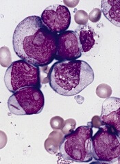
Researchers say they’ve identified novel prognostic markers for older patients with cytogenetically normal acute myeloid leukemia (CN-AML).
The team examined the expression of long noncoding RNAs (lncRNAs) in patients with CN-AML who were at least 60 years of age.
This revealed a pattern of 48 lncRNAs that predicted both response to standard chemotherapy and overall survival.
The researchers described this discovery in PNAS.
“[Our findings] strongly suggest that lncRNA expression profiles can predict which patients will respond to standard therapy,” said principal investigator Clara D. Bloomfield, MD, of The Ohio State University Comprehensive Cancer Center in Columbus.
“That’s important because it would spare these patients from the toxic side effects of experimental therapies. Patients who are classified in the unfavorable group would receive different therapy: stem cell transplant or a clinical trial using new therapeutic approaches. Thus, this research will help to tailor leukemia therapy to each individual.”
In addition, she said, the study revealed novel targets for the development of new therapies.
Dr Bloomfield and her colleagues began this research by analyzing bone marrow samples from 148 older patients with CN-AML who were treated on Cancer and Leukemia Group B clinical trials. All had received similar chemotherapy regimens.
The researchers first identified the 48 lncRNAs that were most associated with survival. Using these 48 lncRNAs, the team divided patients into two groups: those with a favorable prognostic score and those with an unfavorable score.
The researchers then validated the scores in an independent matched set of 71 similarly treated CN-AML patients and compared patients with an unfavorable score to those with a favorable score.
Results showed that patients with an unfavorable score had a lower complete response (CR) rate—54%, compared to 89% for patients with a favorable score.
Three years after CR, only 7% of patients with an unfavorable score were disease-free, compared with 39% of patients with a favorable score.
Overall survival at 3 years for those with an unfavorable score was 10%, compared to 43% for patients with a favorable score.
Distinct lncRNA profiles were associated with mutations in 6 genes—FLT3, NPM1, CEBPA, IDH2, ASXL1, and RUNX1.
The researchers concluded that lncRNA expression profiles are associated with recurrent mutations, clinical features, and outcome in AML. A fraction of these lncRNAs may have a functional role in leukemogenesis. Furthermore, lncRNAs could be used as biomarkers for outcome in AML.
Researchers say they’ve identified novel prognostic markers for older patients with cytogenetically normal acute myeloid leukemia (CN-AML).
The team examined the expression of long noncoding RNAs (lncRNAs) in patients with CN-AML who were at least 60 years of age.
This revealed a pattern of 48 lncRNAs that predicted both response to standard chemotherapy and overall survival.
The researchers described this discovery in PNAS.
“[Our findings] strongly suggest that lncRNA expression profiles can predict which patients will respond to standard therapy,” said principal investigator Clara D. Bloomfield, MD, of The Ohio State University Comprehensive Cancer Center in Columbus.
“That’s important because it would spare these patients from the toxic side effects of experimental therapies. Patients who are classified in the unfavorable group would receive different therapy: stem cell transplant or a clinical trial using new therapeutic approaches. Thus, this research will help to tailor leukemia therapy to each individual.”
In addition, she said, the study revealed novel targets for the development of new therapies.
Dr Bloomfield and her colleagues began this research by analyzing bone marrow samples from 148 older patients with CN-AML who were treated on Cancer and Leukemia Group B clinical trials. All had received similar chemotherapy regimens.
The researchers first identified the 48 lncRNAs that were most associated with survival. Using these 48 lncRNAs, the team divided patients into two groups: those with a favorable prognostic score and those with an unfavorable score.
The researchers then validated the scores in an independent matched set of 71 similarly treated CN-AML patients and compared patients with an unfavorable score to those with a favorable score.
Results showed that patients with an unfavorable score had a lower complete response (CR) rate—54%, compared to 89% for patients with a favorable score.
Three years after CR, only 7% of patients with an unfavorable score were disease-free, compared with 39% of patients with a favorable score.
Overall survival at 3 years for those with an unfavorable score was 10%, compared to 43% for patients with a favorable score.
Distinct lncRNA profiles were associated with mutations in 6 genes—FLT3, NPM1, CEBPA, IDH2, ASXL1, and RUNX1.
The researchers concluded that lncRNA expression profiles are associated with recurrent mutations, clinical features, and outcome in AML. A fraction of these lncRNAs may have a functional role in leukemogenesis. Furthermore, lncRNAs could be used as biomarkers for outcome in AML.
Researchers say they’ve identified novel prognostic markers for older patients with cytogenetically normal acute myeloid leukemia (CN-AML).
The team examined the expression of long noncoding RNAs (lncRNAs) in patients with CN-AML who were at least 60 years of age.
This revealed a pattern of 48 lncRNAs that predicted both response to standard chemotherapy and overall survival.
The researchers described this discovery in PNAS.
“[Our findings] strongly suggest that lncRNA expression profiles can predict which patients will respond to standard therapy,” said principal investigator Clara D. Bloomfield, MD, of The Ohio State University Comprehensive Cancer Center in Columbus.
“That’s important because it would spare these patients from the toxic side effects of experimental therapies. Patients who are classified in the unfavorable group would receive different therapy: stem cell transplant or a clinical trial using new therapeutic approaches. Thus, this research will help to tailor leukemia therapy to each individual.”
In addition, she said, the study revealed novel targets for the development of new therapies.
Dr Bloomfield and her colleagues began this research by analyzing bone marrow samples from 148 older patients with CN-AML who were treated on Cancer and Leukemia Group B clinical trials. All had received similar chemotherapy regimens.
The researchers first identified the 48 lncRNAs that were most associated with survival. Using these 48 lncRNAs, the team divided patients into two groups: those with a favorable prognostic score and those with an unfavorable score.
The researchers then validated the scores in an independent matched set of 71 similarly treated CN-AML patients and compared patients with an unfavorable score to those with a favorable score.
Results showed that patients with an unfavorable score had a lower complete response (CR) rate—54%, compared to 89% for patients with a favorable score.
Three years after CR, only 7% of patients with an unfavorable score were disease-free, compared with 39% of patients with a favorable score.
Overall survival at 3 years for those with an unfavorable score was 10%, compared to 43% for patients with a favorable score.
Distinct lncRNA profiles were associated with mutations in 6 genes—FLT3, NPM1, CEBPA, IDH2, ASXL1, and RUNX1.
The researchers concluded that lncRNA expression profiles are associated with recurrent mutations, clinical features, and outcome in AML. A fraction of these lncRNAs may have a functional role in leukemogenesis. Furthermore, lncRNAs could be used as biomarkers for outcome in AML.
FDA approves pathogen inactivation system for plasma
The US Food and Drug Administration (FDA) has approved the INTERCEPT Blood System for plasma, the first pathogen inactivation system for the preparation of plasma to reduce the risk of transfusion-transmitted infections.
The system inactivates pathogens through a photochemical process involving controlled exposure to ultraviolet light and the chemical amotosalen. The plasma is then purified to remove the chemical and its byproducts.
The INTERCEPT Blood System for plasma can be used to reduce pathogens in plasma derived from whole blood and plasma obtained by apheresis.
The system has proven effective in reducing a broad spectrum of viruses, bacteria, spirochetes, and parasites. However, there is no pathogen inactivation process that has been shown to eliminate all pathogens. Certain viruses (eg, human parvovirus B19) and spores formed by certain bacteria are known to be resistant to the INTERCEPT process.
“The approval of devices like the INTERCEPT Blood System allows blood establishments to prepare plasma that carries a lower risk of transmitting infectious pathogens through transfusion,” said Karen Midthun, MD, director of the FDA’s Center for Biologics Evaluation and Research.
The INTERCEPT Blood System for plasma, which is marketed by Cerus Corporation, has been approved in Europe since 2006 and was recently made available in the US under an Investigational Device Exemption study. The system is being used to prepare Ebola convalescent plasma for transfusion into acutely infected patients.
Clinical studies
Researchers investigated the safety and effectiveness of plasma prepared with the INTERCEPT Blood System in 6 clinical studies and 2 post-marketing studies.
The research showed no significant difference in coagulation factor activity between INTERCEPT-processed plasma and unprocessed fresh-frozen plasma (FFP) in healthy subjects.
There was no significant difference in prothrombin time or activated partial thromboplastin time between INTERCEPT-processed plasma and FFP in patients with coagulation factor deficiencies or in patients with liver disease.
In patients who underwent liver transplant, there was no significant difference in plasma use, hepatic artery thrombosis, or mortality whether patients received INTERCEPT-process plasma or FFP.
In a prospective, phase 3 trial and a retrospective study of patients with thrombotic thrombocytopenic purpura (TTP), there was no significant difference in the percentage of patients who achieved remission with INTERCEPT-treated plasma or FFP.
Adverse events
In a post-marketing study*, researchers analyzed acute transfusion reactions (ATRs) after 57,171 INTERCEPT-processed plasma components were transfused to 9669 patients. Thirty-two subjects (0.3%) experienced an ATR after 41 transfusion episodes (0.2%), including 5 subjects (0.05%) who experienced an ATR after more than 1 transfusion.
Six ATRs were considered serious and possibly or probably related to transfusion. There were 3 instances of allergic reaction or symptoms of allergic reaction (eg, rash, tachycardia, hypotension, respiratory symptoms, and chills), 2 cases of fluid overload, and 1 report of respiratory distress.
In another hemovigilance study, researchers compared the frequency of adverse events due to any of the 4 types of FFP prepared and delivered by the French Blood Establishment over a 10-year period—methylene blue, amotosalen, quarantine, and solvent-detergent.
The study showed that all types of FFP were associated with a low rate of adverse events. Per 10,000 deliveries, there were 7.14 events in the quarantine group, 4.86 in the solvent-detergent group, 4.16 in the amotosalen group, and 1.05 in the methylene blue group.
The package insert of the INTERCEPT Blood System for plasma warns that amotosalen-treated plasma has been associated with cardiac events.
In the aforementioned prospective trial of the system in patients with TTP, 5 patients treated with INTERCEPT-processed plasma and none with conventional plasma had adverse events in the cardiac system organ class.
This included angina pectoris (n=3), cardiac arrest (n=1), bradycardia (n=1), tachycardia (n=1), and sinus arrhythmia (n=1). None of the events resulted in documented myocardial infarction or death.
*Results have been updated since publication.
The US Food and Drug Administration (FDA) has approved the INTERCEPT Blood System for plasma, the first pathogen inactivation system for the preparation of plasma to reduce the risk of transfusion-transmitted infections.
The system inactivates pathogens through a photochemical process involving controlled exposure to ultraviolet light and the chemical amotosalen. The plasma is then purified to remove the chemical and its byproducts.
The INTERCEPT Blood System for plasma can be used to reduce pathogens in plasma derived from whole blood and plasma obtained by apheresis.
The system has proven effective in reducing a broad spectrum of viruses, bacteria, spirochetes, and parasites. However, there is no pathogen inactivation process that has been shown to eliminate all pathogens. Certain viruses (eg, human parvovirus B19) and spores formed by certain bacteria are known to be resistant to the INTERCEPT process.
“The approval of devices like the INTERCEPT Blood System allows blood establishments to prepare plasma that carries a lower risk of transmitting infectious pathogens through transfusion,” said Karen Midthun, MD, director of the FDA’s Center for Biologics Evaluation and Research.
The INTERCEPT Blood System for plasma, which is marketed by Cerus Corporation, has been approved in Europe since 2006 and was recently made available in the US under an Investigational Device Exemption study. The system is being used to prepare Ebola convalescent plasma for transfusion into acutely infected patients.
Clinical studies
Researchers investigated the safety and effectiveness of plasma prepared with the INTERCEPT Blood System in 6 clinical studies and 2 post-marketing studies.
The research showed no significant difference in coagulation factor activity between INTERCEPT-processed plasma and unprocessed fresh-frozen plasma (FFP) in healthy subjects.
There was no significant difference in prothrombin time or activated partial thromboplastin time between INTERCEPT-processed plasma and FFP in patients with coagulation factor deficiencies or in patients with liver disease.
In patients who underwent liver transplant, there was no significant difference in plasma use, hepatic artery thrombosis, or mortality whether patients received INTERCEPT-process plasma or FFP.
In a prospective, phase 3 trial and a retrospective study of patients with thrombotic thrombocytopenic purpura (TTP), there was no significant difference in the percentage of patients who achieved remission with INTERCEPT-treated plasma or FFP.
Adverse events
In a post-marketing study*, researchers analyzed acute transfusion reactions (ATRs) after 57,171 INTERCEPT-processed plasma components were transfused to 9669 patients. Thirty-two subjects (0.3%) experienced an ATR after 41 transfusion episodes (0.2%), including 5 subjects (0.05%) who experienced an ATR after more than 1 transfusion.
Six ATRs were considered serious and possibly or probably related to transfusion. There were 3 instances of allergic reaction or symptoms of allergic reaction (eg, rash, tachycardia, hypotension, respiratory symptoms, and chills), 2 cases of fluid overload, and 1 report of respiratory distress.
In another hemovigilance study, researchers compared the frequency of adverse events due to any of the 4 types of FFP prepared and delivered by the French Blood Establishment over a 10-year period—methylene blue, amotosalen, quarantine, and solvent-detergent.
The study showed that all types of FFP were associated with a low rate of adverse events. Per 10,000 deliveries, there were 7.14 events in the quarantine group, 4.86 in the solvent-detergent group, 4.16 in the amotosalen group, and 1.05 in the methylene blue group.
The package insert of the INTERCEPT Blood System for plasma warns that amotosalen-treated plasma has been associated with cardiac events.
In the aforementioned prospective trial of the system in patients with TTP, 5 patients treated with INTERCEPT-processed plasma and none with conventional plasma had adverse events in the cardiac system organ class.
This included angina pectoris (n=3), cardiac arrest (n=1), bradycardia (n=1), tachycardia (n=1), and sinus arrhythmia (n=1). None of the events resulted in documented myocardial infarction or death.
*Results have been updated since publication.
The US Food and Drug Administration (FDA) has approved the INTERCEPT Blood System for plasma, the first pathogen inactivation system for the preparation of plasma to reduce the risk of transfusion-transmitted infections.
The system inactivates pathogens through a photochemical process involving controlled exposure to ultraviolet light and the chemical amotosalen. The plasma is then purified to remove the chemical and its byproducts.
The INTERCEPT Blood System for plasma can be used to reduce pathogens in plasma derived from whole blood and plasma obtained by apheresis.
The system has proven effective in reducing a broad spectrum of viruses, bacteria, spirochetes, and parasites. However, there is no pathogen inactivation process that has been shown to eliminate all pathogens. Certain viruses (eg, human parvovirus B19) and spores formed by certain bacteria are known to be resistant to the INTERCEPT process.
“The approval of devices like the INTERCEPT Blood System allows blood establishments to prepare plasma that carries a lower risk of transmitting infectious pathogens through transfusion,” said Karen Midthun, MD, director of the FDA’s Center for Biologics Evaluation and Research.
The INTERCEPT Blood System for plasma, which is marketed by Cerus Corporation, has been approved in Europe since 2006 and was recently made available in the US under an Investigational Device Exemption study. The system is being used to prepare Ebola convalescent plasma for transfusion into acutely infected patients.
Clinical studies
Researchers investigated the safety and effectiveness of plasma prepared with the INTERCEPT Blood System in 6 clinical studies and 2 post-marketing studies.
The research showed no significant difference in coagulation factor activity between INTERCEPT-processed plasma and unprocessed fresh-frozen plasma (FFP) in healthy subjects.
There was no significant difference in prothrombin time or activated partial thromboplastin time between INTERCEPT-processed plasma and FFP in patients with coagulation factor deficiencies or in patients with liver disease.
In patients who underwent liver transplant, there was no significant difference in plasma use, hepatic artery thrombosis, or mortality whether patients received INTERCEPT-process plasma or FFP.
In a prospective, phase 3 trial and a retrospective study of patients with thrombotic thrombocytopenic purpura (TTP), there was no significant difference in the percentage of patients who achieved remission with INTERCEPT-treated plasma or FFP.
Adverse events
In a post-marketing study*, researchers analyzed acute transfusion reactions (ATRs) after 57,171 INTERCEPT-processed plasma components were transfused to 9669 patients. Thirty-two subjects (0.3%) experienced an ATR after 41 transfusion episodes (0.2%), including 5 subjects (0.05%) who experienced an ATR after more than 1 transfusion.
Six ATRs were considered serious and possibly or probably related to transfusion. There were 3 instances of allergic reaction or symptoms of allergic reaction (eg, rash, tachycardia, hypotension, respiratory symptoms, and chills), 2 cases of fluid overload, and 1 report of respiratory distress.
In another hemovigilance study, researchers compared the frequency of adverse events due to any of the 4 types of FFP prepared and delivered by the French Blood Establishment over a 10-year period—methylene blue, amotosalen, quarantine, and solvent-detergent.
The study showed that all types of FFP were associated with a low rate of adverse events. Per 10,000 deliveries, there were 7.14 events in the quarantine group, 4.86 in the solvent-detergent group, 4.16 in the amotosalen group, and 1.05 in the methylene blue group.
The package insert of the INTERCEPT Blood System for plasma warns that amotosalen-treated plasma has been associated with cardiac events.
In the aforementioned prospective trial of the system in patients with TTP, 5 patients treated with INTERCEPT-processed plasma and none with conventional plasma had adverse events in the cardiac system organ class.
This included angina pectoris (n=3), cardiac arrest (n=1), bradycardia (n=1), tachycardia (n=1), and sinus arrhythmia (n=1). None of the events resulted in documented myocardial infarction or death.
*Results have been updated since publication.
Newborn screening test for SCID gets FDA approval

Credit: Vera Kratochvil
The US Food and Drug Administration (FDA) has approved marketing of the EnLite Neonatal TREC Kit, the first screening test for severe combined immunodeficiency (SCID) in newborns.
Using a few drops of blood from the newborn’s heel, which is dried on filter paper, the EnLite Neonatal TREC Kit can determine whether T-cell receptor excision circles (TREC) DNA is low or missing from the newborn’s blood.
Newborns with SCID typically have no or low amounts of TREC DNA compared to healthy infants.
Additional testing is required to obtain a SCID diagnosis once the EnLite Neonatal TREC Kit has suggested a child may have SCID.
“SCID is a fatal disease that can be treated with early intervention, including screening,” said Alberto Gutierrez, PhD, director of the Office of In Vitro Diagnostics and Radiological Health in the FDA’s Center for Devices and Radiological Health.
“For the first time, the FDA is allowing the marketing of a newborn screening test that will enable states to incorporate an FDA-reviewed SCID test into their standard newborn screening panels and allow earlier identification for affected individuals.”
The FDA reviewed the EnLite Neonatal TREC Kit through its de novo classification process, a regulatory pathway for some novel, low- to moderate-risk medical devices that are not substantially equivalent to an already legally marketed device.
The FDA’s review included a clinical study of approximately 6400 blood spot specimens from routine screening of newborns, 17 of which had a confirmed SCID diagnosis. The EnLite Neonatal TREC Kit correctly identified all 17 SCID cases.
The agency also evaluated the test’s ability to accurately distinguish low TREC DNA numbers that would be observed in newborns with SCID from high TREC DNA numbers that would be present in healthy newborns. The FDA found the EnLite Neonatal TREC Kit could adequately detect very low TREC DNA values that are associated with SCID.
The EnLite Neonatal TREC Kit is not intended for use as a diagnostic test or to screen for SCID-like syndromes, such as DiGeorge syndrome or Omenn syndrome. Likewise, it is not intended to screen for less acute SCID syndromes, such as leaky SCID or variant SCID.
The EnLite Neonatal TREC Kit is manufactured by Wallac Oy, a subsidiary of PerkinElmer, at its facility in Turku, Finland. PerkinElmer is based in Waltham, Massachusetts.
Credit: Vera Kratochvil
The US Food and Drug Administration (FDA) has approved marketing of the EnLite Neonatal TREC Kit, the first screening test for severe combined immunodeficiency (SCID) in newborns.
Using a few drops of blood from the newborn’s heel, which is dried on filter paper, the EnLite Neonatal TREC Kit can determine whether T-cell receptor excision circles (TREC) DNA is low or missing from the newborn’s blood.
Newborns with SCID typically have no or low amounts of TREC DNA compared to healthy infants.
Additional testing is required to obtain a SCID diagnosis once the EnLite Neonatal TREC Kit has suggested a child may have SCID.
“SCID is a fatal disease that can be treated with early intervention, including screening,” said Alberto Gutierrez, PhD, director of the Office of In Vitro Diagnostics and Radiological Health in the FDA’s Center for Devices and Radiological Health.
“For the first time, the FDA is allowing the marketing of a newborn screening test that will enable states to incorporate an FDA-reviewed SCID test into their standard newborn screening panels and allow earlier identification for affected individuals.”
The FDA reviewed the EnLite Neonatal TREC Kit through its de novo classification process, a regulatory pathway for some novel, low- to moderate-risk medical devices that are not substantially equivalent to an already legally marketed device.
The FDA’s review included a clinical study of approximately 6400 blood spot specimens from routine screening of newborns, 17 of which had a confirmed SCID diagnosis. The EnLite Neonatal TREC Kit correctly identified all 17 SCID cases.
The agency also evaluated the test’s ability to accurately distinguish low TREC DNA numbers that would be observed in newborns with SCID from high TREC DNA numbers that would be present in healthy newborns. The FDA found the EnLite Neonatal TREC Kit could adequately detect very low TREC DNA values that are associated with SCID.
The EnLite Neonatal TREC Kit is not intended for use as a diagnostic test or to screen for SCID-like syndromes, such as DiGeorge syndrome or Omenn syndrome. Likewise, it is not intended to screen for less acute SCID syndromes, such as leaky SCID or variant SCID.
The EnLite Neonatal TREC Kit is manufactured by Wallac Oy, a subsidiary of PerkinElmer, at its facility in Turku, Finland. PerkinElmer is based in Waltham, Massachusetts.
Credit: Vera Kratochvil
The US Food and Drug Administration (FDA) has approved marketing of the EnLite Neonatal TREC Kit, the first screening test for severe combined immunodeficiency (SCID) in newborns.
Using a few drops of blood from the newborn’s heel, which is dried on filter paper, the EnLite Neonatal TREC Kit can determine whether T-cell receptor excision circles (TREC) DNA is low or missing from the newborn’s blood.
Newborns with SCID typically have no or low amounts of TREC DNA compared to healthy infants.
Additional testing is required to obtain a SCID diagnosis once the EnLite Neonatal TREC Kit has suggested a child may have SCID.
“SCID is a fatal disease that can be treated with early intervention, including screening,” said Alberto Gutierrez, PhD, director of the Office of In Vitro Diagnostics and Radiological Health in the FDA’s Center for Devices and Radiological Health.
“For the first time, the FDA is allowing the marketing of a newborn screening test that will enable states to incorporate an FDA-reviewed SCID test into their standard newborn screening panels and allow earlier identification for affected individuals.”
The FDA reviewed the EnLite Neonatal TREC Kit through its de novo classification process, a regulatory pathway for some novel, low- to moderate-risk medical devices that are not substantially equivalent to an already legally marketed device.
The FDA’s review included a clinical study of approximately 6400 blood spot specimens from routine screening of newborns, 17 of which had a confirmed SCID diagnosis. The EnLite Neonatal TREC Kit correctly identified all 17 SCID cases.
The agency also evaluated the test’s ability to accurately distinguish low TREC DNA numbers that would be observed in newborns with SCID from high TREC DNA numbers that would be present in healthy newborns. The FDA found the EnLite Neonatal TREC Kit could adequately detect very low TREC DNA values that are associated with SCID.
The EnLite Neonatal TREC Kit is not intended for use as a diagnostic test or to screen for SCID-like syndromes, such as DiGeorge syndrome or Omenn syndrome. Likewise, it is not intended to screen for less acute SCID syndromes, such as leaky SCID or variant SCID.
The EnLite Neonatal TREC Kit is manufactured by Wallac Oy, a subsidiary of PerkinElmer, at its facility in Turku, Finland. PerkinElmer is based in Waltham, Massachusetts.
Study reveals superior antiviral drug for lymphoma
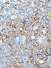
A study of lymphoma patients receiving R-CHOP chemotherapy showed that use of the antiviral drug entecavir resulted in a lower incidence of hepatitis B virus (HBV)-related hepatitis and HBV reactivation, when compared with the drug lamivudine.
HBV reactivation is a well-documented complication of chemotherapy that can result in life-threatening liver failure, as well as delays in chemotherapy or
premature termination of treatment.
However, researchers have not identified an optimal approach to prevent HBV reactivation.
So He Huang, MD, of the Sun Yat-sen University Cancer Center in Guangzhou, China, and colleagues set out to do that. They reported their findings in JAMA alongside a related editorial.
The team enrolled 121 patients with untreated diffuse large B-cell lymphoma who were receiving chemotherapy with rituximab, cyclophosphamide, doxorubicin, vincristine, and prednisone (R-CHOP) and were seropositive for the hepatitis B surface antigen.
Patients were randomized to receive entecavir (n=61) or lamivudine (n=60). They began taking these drugs 1 week before the initiation of R-CHOP and continued until 6 months after they completed chemotherapy.
The study was conducted from February 2008 through December 2012 at 10 medical centers in China. This trial was a substudy of a parent study designed to compare a 3-week R-CHOP regimen with a 2-week R-CHOP regimen.
Results showed that, compared to the lamivudine group, patients in the entecavir group had significantly lower rates of hepatitis (8.2% vs 23.3%, P=0.02), HBV-related hepatitis (0% vs 13.3%, P=0.003), HBV reactivation (6.6% vs 30%, P=0.001), delayed hepatitis B (0% vs 8.3%, P=0.03), and chemotherapy disruption (1.6% vs 18.3%, P=0.002).
The rate of treatment-related adverse events was not significantly different between the groups. Such events occurred in 24.6% of patients in the entecavir group and 30.0% of patients in the lamivudine group (P=0.50).
The researchers noted that entecavir is more expensive than lamivudine, so additional studies are needed to determine whether all patients who are seropositive for the hepatitis B surface antigen and are receiving rituximab-based immunosuppressive therapy should be given entecavir.
Additional studies could also help pinpoint which patients will benefit most from entecavir prophylaxis. However, if these findings are replicated, they support the use of entecavir in these patients.
A study of lymphoma patients receiving R-CHOP chemotherapy showed that use of the antiviral drug entecavir resulted in a lower incidence of hepatitis B virus (HBV)-related hepatitis and HBV reactivation, when compared with the drug lamivudine.
HBV reactivation is a well-documented complication of chemotherapy that can result in life-threatening liver failure, as well as delays in chemotherapy or
premature termination of treatment.
However, researchers have not identified an optimal approach to prevent HBV reactivation.
So He Huang, MD, of the Sun Yat-sen University Cancer Center in Guangzhou, China, and colleagues set out to do that. They reported their findings in JAMA alongside a related editorial.
The team enrolled 121 patients with untreated diffuse large B-cell lymphoma who were receiving chemotherapy with rituximab, cyclophosphamide, doxorubicin, vincristine, and prednisone (R-CHOP) and were seropositive for the hepatitis B surface antigen.
Patients were randomized to receive entecavir (n=61) or lamivudine (n=60). They began taking these drugs 1 week before the initiation of R-CHOP and continued until 6 months after they completed chemotherapy.
The study was conducted from February 2008 through December 2012 at 10 medical centers in China. This trial was a substudy of a parent study designed to compare a 3-week R-CHOP regimen with a 2-week R-CHOP regimen.
Results showed that, compared to the lamivudine group, patients in the entecavir group had significantly lower rates of hepatitis (8.2% vs 23.3%, P=0.02), HBV-related hepatitis (0% vs 13.3%, P=0.003), HBV reactivation (6.6% vs 30%, P=0.001), delayed hepatitis B (0% vs 8.3%, P=0.03), and chemotherapy disruption (1.6% vs 18.3%, P=0.002).
The rate of treatment-related adverse events was not significantly different between the groups. Such events occurred in 24.6% of patients in the entecavir group and 30.0% of patients in the lamivudine group (P=0.50).
The researchers noted that entecavir is more expensive than lamivudine, so additional studies are needed to determine whether all patients who are seropositive for the hepatitis B surface antigen and are receiving rituximab-based immunosuppressive therapy should be given entecavir.
Additional studies could also help pinpoint which patients will benefit most from entecavir prophylaxis. However, if these findings are replicated, they support the use of entecavir in these patients.
A study of lymphoma patients receiving R-CHOP chemotherapy showed that use of the antiviral drug entecavir resulted in a lower incidence of hepatitis B virus (HBV)-related hepatitis and HBV reactivation, when compared with the drug lamivudine.
HBV reactivation is a well-documented complication of chemotherapy that can result in life-threatening liver failure, as well as delays in chemotherapy or
premature termination of treatment.
However, researchers have not identified an optimal approach to prevent HBV reactivation.
So He Huang, MD, of the Sun Yat-sen University Cancer Center in Guangzhou, China, and colleagues set out to do that. They reported their findings in JAMA alongside a related editorial.
The team enrolled 121 patients with untreated diffuse large B-cell lymphoma who were receiving chemotherapy with rituximab, cyclophosphamide, doxorubicin, vincristine, and prednisone (R-CHOP) and were seropositive for the hepatitis B surface antigen.
Patients were randomized to receive entecavir (n=61) or lamivudine (n=60). They began taking these drugs 1 week before the initiation of R-CHOP and continued until 6 months after they completed chemotherapy.
The study was conducted from February 2008 through December 2012 at 10 medical centers in China. This trial was a substudy of a parent study designed to compare a 3-week R-CHOP regimen with a 2-week R-CHOP regimen.
Results showed that, compared to the lamivudine group, patients in the entecavir group had significantly lower rates of hepatitis (8.2% vs 23.3%, P=0.02), HBV-related hepatitis (0% vs 13.3%, P=0.003), HBV reactivation (6.6% vs 30%, P=0.001), delayed hepatitis B (0% vs 8.3%, P=0.03), and chemotherapy disruption (1.6% vs 18.3%, P=0.002).
The rate of treatment-related adverse events was not significantly different between the groups. Such events occurred in 24.6% of patients in the entecavir group and 30.0% of patients in the lamivudine group (P=0.50).
The researchers noted that entecavir is more expensive than lamivudine, so additional studies are needed to determine whether all patients who are seropositive for the hepatitis B surface antigen and are receiving rituximab-based immunosuppressive therapy should be given entecavir.
Additional studies could also help pinpoint which patients will benefit most from entecavir prophylaxis. However, if these findings are replicated, they support the use of entecavir in these patients.
TBI increases risk of cognitive decline in young kids
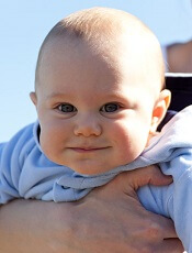
Credit: Petr Kratochvil
Young children who undergo total body irradiation (TBI) in preparation for hematopoietic stem cell transplant (HSCT) are at a higher risk for a decline in IQ, according to research published in the Journal of Clinical Oncology.
The study showed that most young patients don’t experience lasting effects on their IQ following HSCT.
However, patients who underwent HSCT at 3 years of age or younger and received TBI had a greater risk of intellectual decline after transplant.
“For the great majority of patients, these findings provide reassurance that transplantation will not have a significant negative impact on cognitive development,” said study author Sean Phipps, PhD, of St Jude Children’s Research Hospital in Memphis, Tennessee.
“We have also identified a high-risk group of younger patients who may benefit from more intensive interventions, including developmental stimulation and other rehabilitative therapies designed to prevent a decline in intellectual functioning and aid in recovery.”
Dr Phipps and his colleagues tracked the IQ scores of 170 St Jude patients before HSCT and for 5 years after the procedure. The patients were 4 months to 23 years of age when their transplants occurred.
Before HSCT, the average IQ scores of all patients were in the normal range. One year after transplant, average IQ scores of patients aged 5 and younger had declined sharply.
But the scores of most patients rebounded in subsequent years. Five years after the procedure, IQ scores for most patients, even the youngest survivors, had largely recovered and fell within the range of normal intelligence.
Patients in the high-risk group were the lone exception. The IQ scores of patients who were both aged 3 or younger when their transplants occurred and who received TBI failed to recover from the first-year decline.
Five years after HSCT, these survivors had average IQ scores in the low-normal range of intelligence. Their scores were more than 16 points lower than the scores of patients who were just as young when their transplants occurred but did not receive TBI.
Furthermore, of the 72 patients in this study whose transplants included TBI, there was a long-term impact on intellectual functioning only for patients who were 3 or younger at transplant.
“The significant first-year decline reflects the intensity of transplantation, which our results suggest leads to greater disruption in development in the youngest children than was previously recognized,” said study author Victoria Willard, PhD, also of St Jude.
The researchers said these findings are good news for most parents whose children must undergo HSCT and provide another reason for hope of good long-term outcomes.
For those whose children are in the newly recognized high-risk group, increased attention and activities designed to stimulate cognitive development may help to prevent reduced IQ following transplant, according to Dr Phipps.
Credit: Petr Kratochvil
Young children who undergo total body irradiation (TBI) in preparation for hematopoietic stem cell transplant (HSCT) are at a higher risk for a decline in IQ, according to research published in the Journal of Clinical Oncology.
The study showed that most young patients don’t experience lasting effects on their IQ following HSCT.
However, patients who underwent HSCT at 3 years of age or younger and received TBI had a greater risk of intellectual decline after transplant.
“For the great majority of patients, these findings provide reassurance that transplantation will not have a significant negative impact on cognitive development,” said study author Sean Phipps, PhD, of St Jude Children’s Research Hospital in Memphis, Tennessee.
“We have also identified a high-risk group of younger patients who may benefit from more intensive interventions, including developmental stimulation and other rehabilitative therapies designed to prevent a decline in intellectual functioning and aid in recovery.”
Dr Phipps and his colleagues tracked the IQ scores of 170 St Jude patients before HSCT and for 5 years after the procedure. The patients were 4 months to 23 years of age when their transplants occurred.
Before HSCT, the average IQ scores of all patients were in the normal range. One year after transplant, average IQ scores of patients aged 5 and younger had declined sharply.
But the scores of most patients rebounded in subsequent years. Five years after the procedure, IQ scores for most patients, even the youngest survivors, had largely recovered and fell within the range of normal intelligence.
Patients in the high-risk group were the lone exception. The IQ scores of patients who were both aged 3 or younger when their transplants occurred and who received TBI failed to recover from the first-year decline.
Five years after HSCT, these survivors had average IQ scores in the low-normal range of intelligence. Their scores were more than 16 points lower than the scores of patients who were just as young when their transplants occurred but did not receive TBI.
Furthermore, of the 72 patients in this study whose transplants included TBI, there was a long-term impact on intellectual functioning only for patients who were 3 or younger at transplant.
“The significant first-year decline reflects the intensity of transplantation, which our results suggest leads to greater disruption in development in the youngest children than was previously recognized,” said study author Victoria Willard, PhD, also of St Jude.
The researchers said these findings are good news for most parents whose children must undergo HSCT and provide another reason for hope of good long-term outcomes.
For those whose children are in the newly recognized high-risk group, increased attention and activities designed to stimulate cognitive development may help to prevent reduced IQ following transplant, according to Dr Phipps.
Credit: Petr Kratochvil
Young children who undergo total body irradiation (TBI) in preparation for hematopoietic stem cell transplant (HSCT) are at a higher risk for a decline in IQ, according to research published in the Journal of Clinical Oncology.
The study showed that most young patients don’t experience lasting effects on their IQ following HSCT.
However, patients who underwent HSCT at 3 years of age or younger and received TBI had a greater risk of intellectual decline after transplant.
“For the great majority of patients, these findings provide reassurance that transplantation will not have a significant negative impact on cognitive development,” said study author Sean Phipps, PhD, of St Jude Children’s Research Hospital in Memphis, Tennessee.
“We have also identified a high-risk group of younger patients who may benefit from more intensive interventions, including developmental stimulation and other rehabilitative therapies designed to prevent a decline in intellectual functioning and aid in recovery.”
Dr Phipps and his colleagues tracked the IQ scores of 170 St Jude patients before HSCT and for 5 years after the procedure. The patients were 4 months to 23 years of age when their transplants occurred.
Before HSCT, the average IQ scores of all patients were in the normal range. One year after transplant, average IQ scores of patients aged 5 and younger had declined sharply.
But the scores of most patients rebounded in subsequent years. Five years after the procedure, IQ scores for most patients, even the youngest survivors, had largely recovered and fell within the range of normal intelligence.
Patients in the high-risk group were the lone exception. The IQ scores of patients who were both aged 3 or younger when their transplants occurred and who received TBI failed to recover from the first-year decline.
Five years after HSCT, these survivors had average IQ scores in the low-normal range of intelligence. Their scores were more than 16 points lower than the scores of patients who were just as young when their transplants occurred but did not receive TBI.
Furthermore, of the 72 patients in this study whose transplants included TBI, there was a long-term impact on intellectual functioning only for patients who were 3 or younger at transplant.
“The significant first-year decline reflects the intensity of transplantation, which our results suggest leads to greater disruption in development in the youngest children than was previously recognized,” said study author Victoria Willard, PhD, also of St Jude.
The researchers said these findings are good news for most parents whose children must undergo HSCT and provide another reason for hope of good long-term outcomes.
For those whose children are in the newly recognized high-risk group, increased attention and activities designed to stimulate cognitive development may help to prevent reduced IQ following transplant, according to Dr Phipps.
‘Father of hematopoietic cytokines’ dies

Photo courtesy of The Walter
and Eliza Hall Institute
of Medical Research
Donald Metcalf, MD, an Australian researcher who has been called “the father of hematopoietic cytokines,” has died at the age of 85.
Dr Metcalf’s studies of blood production led to his speculation that there must be a biological mechanism—one or more hormones—that control white blood cell production.
These substances, which he termed colony-stimulating factors (CSFs), were the focus of more than 50 years of research.
Over this time, Dr Metcalf led researchers to characterize and purify 4 separate CSFs—granulocyte CSF (G-CSF), granulocyte-macrophage CSF (GM-CSF), macrophage CSF (M-CSF), and multi-CSF (now called interleukin-3).
Dr Metcalf recognized that CSFs had a potential role in clinical medicine, and his team was among the first in the world to discover the genes for CSFs.
Dr Metcalf was a central figure in the international clinical trials of CSFs in the 1980s, assessing whether CSFs could boost immune cell numbers in cancer patients whose immune system was weakened as a side effect of chemotherapy. On the basis of these studies, G-CSF (Neupogen) was approved for clinical use in 1991.
Now, an estimated 20 million people have been treated with CSFs. As well as boosting the immune system in patients who receive chemotherapy or have other immune deficiencies, CSFs are thought to have revolutionized hematopoietic stem cell transplantation.
A man with many achievements
Dr Metcalf was born in 1929 and started school at the age of 3, by which time he was already reading. He entered university at the age of 16, ultimately obtaining bachelor’s and medical degrees from the University of Sydney.
After an internship at the Royal Prince Alfred Hospital in Sydney, Dr Metcalf joined the staff of the Walter and Eliza Hall Institute of Medical Research (WEHI) in Melbourne in 1954. He was supported by Cancer Council Victoria’s Carden Fellowship, an award he held until his retirement in 2014. (Dr Metcalf officially retired in 1996 but continued to do research until 2014.)
Dr Metcalf spent his early years at WEHI studying vaccinia virus. In 1965, he began studying blood cell formation and, by association, leukemia. In 1966, he became deputy director of WEHI and the head of its Cancer Research Unit.
Dr Metcalf took several sabbaticals from WEHI, serving as a visiting scientist at Harvard Medical School in Boston, Massachusetts; the Roswell Park Memorial Institute in Buffalo, New York; the Swiss Institute for Experimental Cancer Research in Lausanne, Switzerland; the Radiobiological Institute in Rijswijk, The Netherlands; and the University of Cambridge in the UK.
Among Dr Metcalf’s many honors and awards are the Companion of the Order of Australia (1993), the Albert Lasker Award for Clinical Medical Research (1993), the Gairdner Foundation International Award (1994), the Royal Medal of the Royal Society (1995), the Victoria Prize (2000), and the Prime Minister’s Prize for Science (2001).
Dr Metcalf is survived by his wife Jo; daughters Kate, Johanna, Penelope, and Mary-Ann; grandchildren James, Martin, Patrick, Elizabeth, Rose, and Robert; and their extended families.
Photo courtesy of The Walter
and Eliza Hall Institute
of Medical Research
Donald Metcalf, MD, an Australian researcher who has been called “the father of hematopoietic cytokines,” has died at the age of 85.
Dr Metcalf’s studies of blood production led to his speculation that there must be a biological mechanism—one or more hormones—that control white blood cell production.
These substances, which he termed colony-stimulating factors (CSFs), were the focus of more than 50 years of research.
Over this time, Dr Metcalf led researchers to characterize and purify 4 separate CSFs—granulocyte CSF (G-CSF), granulocyte-macrophage CSF (GM-CSF), macrophage CSF (M-CSF), and multi-CSF (now called interleukin-3).
Dr Metcalf recognized that CSFs had a potential role in clinical medicine, and his team was among the first in the world to discover the genes for CSFs.
Dr Metcalf was a central figure in the international clinical trials of CSFs in the 1980s, assessing whether CSFs could boost immune cell numbers in cancer patients whose immune system was weakened as a side effect of chemotherapy. On the basis of these studies, G-CSF (Neupogen) was approved for clinical use in 1991.
Now, an estimated 20 million people have been treated with CSFs. As well as boosting the immune system in patients who receive chemotherapy or have other immune deficiencies, CSFs are thought to have revolutionized hematopoietic stem cell transplantation.
A man with many achievements
Dr Metcalf was born in 1929 and started school at the age of 3, by which time he was already reading. He entered university at the age of 16, ultimately obtaining bachelor’s and medical degrees from the University of Sydney.
After an internship at the Royal Prince Alfred Hospital in Sydney, Dr Metcalf joined the staff of the Walter and Eliza Hall Institute of Medical Research (WEHI) in Melbourne in 1954. He was supported by Cancer Council Victoria’s Carden Fellowship, an award he held until his retirement in 2014. (Dr Metcalf officially retired in 1996 but continued to do research until 2014.)
Dr Metcalf spent his early years at WEHI studying vaccinia virus. In 1965, he began studying blood cell formation and, by association, leukemia. In 1966, he became deputy director of WEHI and the head of its Cancer Research Unit.
Dr Metcalf took several sabbaticals from WEHI, serving as a visiting scientist at Harvard Medical School in Boston, Massachusetts; the Roswell Park Memorial Institute in Buffalo, New York; the Swiss Institute for Experimental Cancer Research in Lausanne, Switzerland; the Radiobiological Institute in Rijswijk, The Netherlands; and the University of Cambridge in the UK.
Among Dr Metcalf’s many honors and awards are the Companion of the Order of Australia (1993), the Albert Lasker Award for Clinical Medical Research (1993), the Gairdner Foundation International Award (1994), the Royal Medal of the Royal Society (1995), the Victoria Prize (2000), and the Prime Minister’s Prize for Science (2001).
Dr Metcalf is survived by his wife Jo; daughters Kate, Johanna, Penelope, and Mary-Ann; grandchildren James, Martin, Patrick, Elizabeth, Rose, and Robert; and their extended families.
Photo courtesy of The Walter
and Eliza Hall Institute
of Medical Research
Donald Metcalf, MD, an Australian researcher who has been called “the father of hematopoietic cytokines,” has died at the age of 85.
Dr Metcalf’s studies of blood production led to his speculation that there must be a biological mechanism—one or more hormones—that control white blood cell production.
These substances, which he termed colony-stimulating factors (CSFs), were the focus of more than 50 years of research.
Over this time, Dr Metcalf led researchers to characterize and purify 4 separate CSFs—granulocyte CSF (G-CSF), granulocyte-macrophage CSF (GM-CSF), macrophage CSF (M-CSF), and multi-CSF (now called interleukin-3).
Dr Metcalf recognized that CSFs had a potential role in clinical medicine, and his team was among the first in the world to discover the genes for CSFs.
Dr Metcalf was a central figure in the international clinical trials of CSFs in the 1980s, assessing whether CSFs could boost immune cell numbers in cancer patients whose immune system was weakened as a side effect of chemotherapy. On the basis of these studies, G-CSF (Neupogen) was approved for clinical use in 1991.
Now, an estimated 20 million people have been treated with CSFs. As well as boosting the immune system in patients who receive chemotherapy or have other immune deficiencies, CSFs are thought to have revolutionized hematopoietic stem cell transplantation.
A man with many achievements
Dr Metcalf was born in 1929 and started school at the age of 3, by which time he was already reading. He entered university at the age of 16, ultimately obtaining bachelor’s and medical degrees from the University of Sydney.
After an internship at the Royal Prince Alfred Hospital in Sydney, Dr Metcalf joined the staff of the Walter and Eliza Hall Institute of Medical Research (WEHI) in Melbourne in 1954. He was supported by Cancer Council Victoria’s Carden Fellowship, an award he held until his retirement in 2014. (Dr Metcalf officially retired in 1996 but continued to do research until 2014.)
Dr Metcalf spent his early years at WEHI studying vaccinia virus. In 1965, he began studying blood cell formation and, by association, leukemia. In 1966, he became deputy director of WEHI and the head of its Cancer Research Unit.
Dr Metcalf took several sabbaticals from WEHI, serving as a visiting scientist at Harvard Medical School in Boston, Massachusetts; the Roswell Park Memorial Institute in Buffalo, New York; the Swiss Institute for Experimental Cancer Research in Lausanne, Switzerland; the Radiobiological Institute in Rijswijk, The Netherlands; and the University of Cambridge in the UK.
Among Dr Metcalf’s many honors and awards are the Companion of the Order of Australia (1993), the Albert Lasker Award for Clinical Medical Research (1993), the Gairdner Foundation International Award (1994), the Royal Medal of the Royal Society (1995), the Victoria Prize (2000), and the Prime Minister’s Prize for Science (2001).
Dr Metcalf is survived by his wife Jo; daughters Kate, Johanna, Penelope, and Mary-Ann; grandchildren James, Martin, Patrick, Elizabeth, Rose, and Robert; and their extended families.