User login
Mice are suitable models for inflammatory conditions, study suggests

Results of a new study contradict previous research suggesting mice do not make suitable models for human inflammatory conditions.
The original study, published in PNAS in February 2013, indicated that genomic responses to different acute inflammatory stressors—trauma, burns, sepsis, and infection—are highly similar in humans but poorly reproduced in corresponding mouse models.
The new study, published in PNAS yesterday, suggests that is not the case.
The original study was conducted by Junhee Seok, PhD, of Northwestern University, and his colleagues. It garnered a lot of attention from the scientific community and the general public, reigniting the debate over mouse models’ suitability for medical research.
Tsuyoshi Miyakawa, PhD, of Fujita Health University in Japan, was among those who argued that mice are suitable models, and Dr Seok’s findings were likely incorrect.
So Dr Miyakawa and his colleague, Keizo Takao, PhD, of the National Institute for Physiological Sciences in Japan, reanalyzed the data from Dr Seok’s study using the bioinformatics tool NextBio.
Dr Seok’s group had compared the expression levels of genes that were altered in a particular human condition between humans and mice.
A comparison of the genomic response between humans and mice, including those genes altered in one species but not in another, obscures the correlation between homologous genes of humans and mice to nearly 0, as the team showed.
The group’s comparison of the gene expression patterns between human burn victims and mouse models of burns, trauma, sepsis, and infection revealed a Pearson’s correlation coefficient (R) that ranged from 0.14 to 0.28. And the percentage of genes whose expression changed in the same direction was 55% to 61%.
In the new analysis based on the same data, Drs Miyakawa and Takao found the R values ranged from 0.36 to 0.59. And 77% to 93% of the genes changed in the same directions between the human condition and the mouse models.
Non-parametric ranking analysis using NextBio showed the pattern of the gene expression changes in mouse models was highly similar to that in human burn conditions—a significant correlation (P = 6.5 x 10⁻¹¹ to 1.2 x 10⁻³⁵).
Drs Miyakawa and Takao noted that many molecular pathways are commonly dysregulated in human diseases and mouse models. And focusing on the commonalities between human diseases and mouse models will allow us to derive useful information for studying the pathophysiology and pathogenesis of human diseases, as well as aid treatment development. 
Results of a new study contradict previous research suggesting mice do not make suitable models for human inflammatory conditions.
The original study, published in PNAS in February 2013, indicated that genomic responses to different acute inflammatory stressors—trauma, burns, sepsis, and infection—are highly similar in humans but poorly reproduced in corresponding mouse models.
The new study, published in PNAS yesterday, suggests that is not the case.
The original study was conducted by Junhee Seok, PhD, of Northwestern University, and his colleagues. It garnered a lot of attention from the scientific community and the general public, reigniting the debate over mouse models’ suitability for medical research.
Tsuyoshi Miyakawa, PhD, of Fujita Health University in Japan, was among those who argued that mice are suitable models, and Dr Seok’s findings were likely incorrect.
So Dr Miyakawa and his colleague, Keizo Takao, PhD, of the National Institute for Physiological Sciences in Japan, reanalyzed the data from Dr Seok’s study using the bioinformatics tool NextBio.
Dr Seok’s group had compared the expression levels of genes that were altered in a particular human condition between humans and mice.
A comparison of the genomic response between humans and mice, including those genes altered in one species but not in another, obscures the correlation between homologous genes of humans and mice to nearly 0, as the team showed.
The group’s comparison of the gene expression patterns between human burn victims and mouse models of burns, trauma, sepsis, and infection revealed a Pearson’s correlation coefficient (R) that ranged from 0.14 to 0.28. And the percentage of genes whose expression changed in the same direction was 55% to 61%.
In the new analysis based on the same data, Drs Miyakawa and Takao found the R values ranged from 0.36 to 0.59. And 77% to 93% of the genes changed in the same directions between the human condition and the mouse models.
Non-parametric ranking analysis using NextBio showed the pattern of the gene expression changes in mouse models was highly similar to that in human burn conditions—a significant correlation (P = 6.5 x 10⁻¹¹ to 1.2 x 10⁻³⁵).
Drs Miyakawa and Takao noted that many molecular pathways are commonly dysregulated in human diseases and mouse models. And focusing on the commonalities between human diseases and mouse models will allow us to derive useful information for studying the pathophysiology and pathogenesis of human diseases, as well as aid treatment development.
Results of a new study contradict previous research suggesting mice do not make suitable models for human inflammatory conditions.
The original study, published in PNAS in February 2013, indicated that genomic responses to different acute inflammatory stressors—trauma, burns, sepsis, and infection—are highly similar in humans but poorly reproduced in corresponding mouse models.
The new study, published in PNAS yesterday, suggests that is not the case.
The original study was conducted by Junhee Seok, PhD, of Northwestern University, and his colleagues. It garnered a lot of attention from the scientific community and the general public, reigniting the debate over mouse models’ suitability for medical research.
Tsuyoshi Miyakawa, PhD, of Fujita Health University in Japan, was among those who argued that mice are suitable models, and Dr Seok’s findings were likely incorrect.
So Dr Miyakawa and his colleague, Keizo Takao, PhD, of the National Institute for Physiological Sciences in Japan, reanalyzed the data from Dr Seok’s study using the bioinformatics tool NextBio.
Dr Seok’s group had compared the expression levels of genes that were altered in a particular human condition between humans and mice.
A comparison of the genomic response between humans and mice, including those genes altered in one species but not in another, obscures the correlation between homologous genes of humans and mice to nearly 0, as the team showed.
The group’s comparison of the gene expression patterns between human burn victims and mouse models of burns, trauma, sepsis, and infection revealed a Pearson’s correlation coefficient (R) that ranged from 0.14 to 0.28. And the percentage of genes whose expression changed in the same direction was 55% to 61%.
In the new analysis based on the same data, Drs Miyakawa and Takao found the R values ranged from 0.36 to 0.59. And 77% to 93% of the genes changed in the same directions between the human condition and the mouse models.
Non-parametric ranking analysis using NextBio showed the pattern of the gene expression changes in mouse models was highly similar to that in human burn conditions—a significant correlation (P = 6.5 x 10⁻¹¹ to 1.2 x 10⁻³⁵).
Drs Miyakawa and Takao noted that many molecular pathways are commonly dysregulated in human diseases and mouse models. And focusing on the commonalities between human diseases and mouse models will allow us to derive useful information for studying the pathophysiology and pathogenesis of human diseases, as well as aid treatment development.
Pathologist Emmanuel Farber dies at 95

Emmanuel Farber, MD, PhD, a renowned pathologist who made fundamental contributions to our understanding of chemical carcinogenesis, passed away on August 3 at the age of 95.
Dr Farber’s studies in experimental pathology demonstrated that chemical carcinogens are capable of binding to nucleic acids, in turn generating specific DNA adducts.
These early studies led to the observation that chemical carcinogenesis is a sequential process.
Dr Farber later proved this theory by showing that cancer could be induced through a series of step-by-step chemical treatments in the liver. He served on the Surgeon General’s first Advisory Committee on Smoking and Health from 1961 to 1964.
The committee was responsible for issuing the 1964 Surgeon General’s Report, which has now done more to prevent tobacco-related disease than any other preventive measure.
Throughout his career, Dr Farber promoted the concept that to understand carcinogenesis, one must also understand the cellular, genetic, metabolic, and molecular changes that are occurring during the process. This mindset, along with Dr Farber’s energy and enthusiasm in exploring the nature of cancer, has served as a source of inspiration and guidance for cancer researchers worldwide.
Dr Farber was born in Toronto, Canada, on October 19, 1918. He obtained his medical degree from the Faculty of Medicine, University of Toronto in 1942.
After completing his residency training in pathology at the Hamilton General Hospital in Ontario, Canada, he served in the Royal Canadian Army Medical Corps and later obtained a doctorate in biochemistry from the University of California, Berkeley.
His academic career began at Tulane University in New Orleans, Louisiana. It continued with his appointment as Professor and Chairman of Pathology and Professor of Biochemistry at the University of Pittsburgh School of Medicine and at the Fels Research Institute, Temple University School of Medicine, in Philadelphia, Pennsylvania, where he was Professor of Pathology and Biochemistry and Director of the Institute.
In 1975, Dr Farber moved back to his native city to take the post of Professor and Chairman of the Department of Pathology and Professor in the Department of Biochemistry at the University of Toronto. At his death, he held the title of Chairman Emeritus and Professor in the Department of Pathology at the University of Toronto.
Dr Farber is survived by his daughter Naomi Farber, son-in-law Steven Grosby, and grandson Samuel Grosby, who wish to extend their sincere appreciation to those who enriched his personal and professional life and joined his tireless search for scientific truth.
Emmanuel Farber, MD, PhD, a renowned pathologist who made fundamental contributions to our understanding of chemical carcinogenesis, passed away on August 3 at the age of 95.
Dr Farber’s studies in experimental pathology demonstrated that chemical carcinogens are capable of binding to nucleic acids, in turn generating specific DNA adducts.
These early studies led to the observation that chemical carcinogenesis is a sequential process.
Dr Farber later proved this theory by showing that cancer could be induced through a series of step-by-step chemical treatments in the liver. He served on the Surgeon General’s first Advisory Committee on Smoking and Health from 1961 to 1964.
The committee was responsible for issuing the 1964 Surgeon General’s Report, which has now done more to prevent tobacco-related disease than any other preventive measure.
Throughout his career, Dr Farber promoted the concept that to understand carcinogenesis, one must also understand the cellular, genetic, metabolic, and molecular changes that are occurring during the process. This mindset, along with Dr Farber’s energy and enthusiasm in exploring the nature of cancer, has served as a source of inspiration and guidance for cancer researchers worldwide.
Dr Farber was born in Toronto, Canada, on October 19, 1918. He obtained his medical degree from the Faculty of Medicine, University of Toronto in 1942.
After completing his residency training in pathology at the Hamilton General Hospital in Ontario, Canada, he served in the Royal Canadian Army Medical Corps and later obtained a doctorate in biochemistry from the University of California, Berkeley.
His academic career began at Tulane University in New Orleans, Louisiana. It continued with his appointment as Professor and Chairman of Pathology and Professor of Biochemistry at the University of Pittsburgh School of Medicine and at the Fels Research Institute, Temple University School of Medicine, in Philadelphia, Pennsylvania, where he was Professor of Pathology and Biochemistry and Director of the Institute.
In 1975, Dr Farber moved back to his native city to take the post of Professor and Chairman of the Department of Pathology and Professor in the Department of Biochemistry at the University of Toronto. At his death, he held the title of Chairman Emeritus and Professor in the Department of Pathology at the University of Toronto.
Dr Farber is survived by his daughter Naomi Farber, son-in-law Steven Grosby, and grandson Samuel Grosby, who wish to extend their sincere appreciation to those who enriched his personal and professional life and joined his tireless search for scientific truth.
Emmanuel Farber, MD, PhD, a renowned pathologist who made fundamental contributions to our understanding of chemical carcinogenesis, passed away on August 3 at the age of 95.
Dr Farber’s studies in experimental pathology demonstrated that chemical carcinogens are capable of binding to nucleic acids, in turn generating specific DNA adducts.
These early studies led to the observation that chemical carcinogenesis is a sequential process.
Dr Farber later proved this theory by showing that cancer could be induced through a series of step-by-step chemical treatments in the liver. He served on the Surgeon General’s first Advisory Committee on Smoking and Health from 1961 to 1964.
The committee was responsible for issuing the 1964 Surgeon General’s Report, which has now done more to prevent tobacco-related disease than any other preventive measure.
Throughout his career, Dr Farber promoted the concept that to understand carcinogenesis, one must also understand the cellular, genetic, metabolic, and molecular changes that are occurring during the process. This mindset, along with Dr Farber’s energy and enthusiasm in exploring the nature of cancer, has served as a source of inspiration and guidance for cancer researchers worldwide.
Dr Farber was born in Toronto, Canada, on October 19, 1918. He obtained his medical degree from the Faculty of Medicine, University of Toronto in 1942.
After completing his residency training in pathology at the Hamilton General Hospital in Ontario, Canada, he served in the Royal Canadian Army Medical Corps and later obtained a doctorate in biochemistry from the University of California, Berkeley.
His academic career began at Tulane University in New Orleans, Louisiana. It continued with his appointment as Professor and Chairman of Pathology and Professor of Biochemistry at the University of Pittsburgh School of Medicine and at the Fels Research Institute, Temple University School of Medicine, in Philadelphia, Pennsylvania, where he was Professor of Pathology and Biochemistry and Director of the Institute.
In 1975, Dr Farber moved back to his native city to take the post of Professor and Chairman of the Department of Pathology and Professor in the Department of Biochemistry at the University of Toronto. At his death, he held the title of Chairman Emeritus and Professor in the Department of Pathology at the University of Toronto.
Dr Farber is survived by his daughter Naomi Farber, son-in-law Steven Grosby, and grandson Samuel Grosby, who wish to extend their sincere appreciation to those who enriched his personal and professional life and joined his tireless search for scientific truth.
Study reveals why HSCs falter with age

in the bone marrow
A new study helps explain how blood production declines with age and why older individuals are not suitable donors for hematopoietic stem cell (HSC) transplant.
The research also reveals a potential approach for mitigating
the negative effects of aging on the blood, which can lead to anemia,
bone marrow failure, and myeloid malignancies.
The study, conducted in mice, suggests HSCs falter with age because they lose the ability to replicate their DNA accurately and efficiently during cell division.
Emmanuelle Passegué, PhD, of the University of California San Francisco, and her colleagues reported this discovery in Nature.
The researchers analyzed old HSCs in mice and found a scarcity of protein components needed to form the mini-chromosome maintenance helicase. This molecular machine unwinds double-stranded DNA so the cell’s genetic material can be duplicated and allocated to daughter cells later in cell division.
The HSCs were stressed by the loss of this machine’s activity. As a result, they had an increased risk for DNA damage and death when forced to divide.
On the other hand, the cells tended to survive unless they were confronted with a “strong replication challenge” like transplantation.
The researchers also discovered that even after the stress associated with DNA replication, old HSCs retained molecular tags on histones, a feature often associated with DNA damage.
However, these old survivors could repair induced DNA damage as efficiently as young stem cells.
“Old stem cells are not just sitting there with damaged DNA ready to develop cancer, as it has long been postulated,” Dr Passegué said.
Of course, not all was well in the old, surviving HSCs. The molecular tags accumulated on genes needed to make ribosomes.
Dr Passegué said she will further explore the consequences of reduced protein production as part of her ongoing research. She hopes it might be possible to prevent declining stem cell populations by developing a drug to prevent the loss of the helicase components needed to unwind and replicate DNA, thereby avoiding immune system failure.
in the bone marrow
A new study helps explain how blood production declines with age and why older individuals are not suitable donors for hematopoietic stem cell (HSC) transplant.
The research also reveals a potential approach for mitigating
the negative effects of aging on the blood, which can lead to anemia,
bone marrow failure, and myeloid malignancies.
The study, conducted in mice, suggests HSCs falter with age because they lose the ability to replicate their DNA accurately and efficiently during cell division.
Emmanuelle Passegué, PhD, of the University of California San Francisco, and her colleagues reported this discovery in Nature.
The researchers analyzed old HSCs in mice and found a scarcity of protein components needed to form the mini-chromosome maintenance helicase. This molecular machine unwinds double-stranded DNA so the cell’s genetic material can be duplicated and allocated to daughter cells later in cell division.
The HSCs were stressed by the loss of this machine’s activity. As a result, they had an increased risk for DNA damage and death when forced to divide.
On the other hand, the cells tended to survive unless they were confronted with a “strong replication challenge” like transplantation.
The researchers also discovered that even after the stress associated with DNA replication, old HSCs retained molecular tags on histones, a feature often associated with DNA damage.
However, these old survivors could repair induced DNA damage as efficiently as young stem cells.
“Old stem cells are not just sitting there with damaged DNA ready to develop cancer, as it has long been postulated,” Dr Passegué said.
Of course, not all was well in the old, surviving HSCs. The molecular tags accumulated on genes needed to make ribosomes.
Dr Passegué said she will further explore the consequences of reduced protein production as part of her ongoing research. She hopes it might be possible to prevent declining stem cell populations by developing a drug to prevent the loss of the helicase components needed to unwind and replicate DNA, thereby avoiding immune system failure.
in the bone marrow
A new study helps explain how blood production declines with age and why older individuals are not suitable donors for hematopoietic stem cell (HSC) transplant.
The research also reveals a potential approach for mitigating
the negative effects of aging on the blood, which can lead to anemia,
bone marrow failure, and myeloid malignancies.
The study, conducted in mice, suggests HSCs falter with age because they lose the ability to replicate their DNA accurately and efficiently during cell division.
Emmanuelle Passegué, PhD, of the University of California San Francisco, and her colleagues reported this discovery in Nature.
The researchers analyzed old HSCs in mice and found a scarcity of protein components needed to form the mini-chromosome maintenance helicase. This molecular machine unwinds double-stranded DNA so the cell’s genetic material can be duplicated and allocated to daughter cells later in cell division.
The HSCs were stressed by the loss of this machine’s activity. As a result, they had an increased risk for DNA damage and death when forced to divide.
On the other hand, the cells tended to survive unless they were confronted with a “strong replication challenge” like transplantation.
The researchers also discovered that even after the stress associated with DNA replication, old HSCs retained molecular tags on histones, a feature often associated with DNA damage.
However, these old survivors could repair induced DNA damage as efficiently as young stem cells.
“Old stem cells are not just sitting there with damaged DNA ready to develop cancer, as it has long been postulated,” Dr Passegué said.
Of course, not all was well in the old, surviving HSCs. The molecular tags accumulated on genes needed to make ribosomes.
Dr Passegué said she will further explore the consequences of reduced protein production as part of her ongoing research. She hopes it might be possible to prevent declining stem cell populations by developing a drug to prevent the loss of the helicase components needed to unwind and replicate DNA, thereby avoiding immune system failure.
Group identifies targets for malaria vaccine
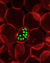
red blood cell; Credit: St Jude
Children’s Research Hospital
Scientists have uncovered a number of potential targets for a blood-stage malaria vaccine.
The team assessed how antibodies from a group of malaria-infected children responded to a library of proteins from the Plasmodium falciparum parasite.
This revealed antigens that had not previously been identified as possible vaccine targets and provided new insight into the ways antigens could be used in combination to increase protection from malaria.
“Resistance to malaria drugs is an increasing problem, so vaccines are desperately needed to battle the Plasmodium falciparum parasite before it has a chance to make people sick,” said Faith Osier, MBChB, of the Kenya Medical Research Institute in Nairobi City.
“This study presents us with a large number of new vaccine candidates that offer real hope for the future.”
Dr Osier and her colleagues described the study in Science Translational Medicine.
The researchers generated a library of correctly folded, full-length proteins from the P falciparum parasite. They then tested antibody reactivity against these proteins in a cohort of Kenyan children who were monitored for clinical episodes of malaria over 6 months.
This revealed antibodies that provide protection against clinical episodes of malaria. Some were equivalent or superior to current leading malaria vaccine candidates.
In fact, combinations consisting of 5 of the 10 top-ranked antigens (PF3D7_1136200, MSP2, RhopH3, P41, MSP11, MSP3, PF3D7_0606800, AMA1, Pf113, and MSRP1) could provide 100% protection against clinical episodes of malaria.
The researchers said these results add further weight to the theory that a successful blood-stage vaccine needs to target multiple antigens.
The team’s next step will be to generate antibodies against all of the proteins in the library and test them in different combinations to see whether combinations that appear to provide protection can directly prevent parasite invasion.
red blood cell; Credit: St Jude
Children’s Research Hospital
Scientists have uncovered a number of potential targets for a blood-stage malaria vaccine.
The team assessed how antibodies from a group of malaria-infected children responded to a library of proteins from the Plasmodium falciparum parasite.
This revealed antigens that had not previously been identified as possible vaccine targets and provided new insight into the ways antigens could be used in combination to increase protection from malaria.
“Resistance to malaria drugs is an increasing problem, so vaccines are desperately needed to battle the Plasmodium falciparum parasite before it has a chance to make people sick,” said Faith Osier, MBChB, of the Kenya Medical Research Institute in Nairobi City.
“This study presents us with a large number of new vaccine candidates that offer real hope for the future.”
Dr Osier and her colleagues described the study in Science Translational Medicine.
The researchers generated a library of correctly folded, full-length proteins from the P falciparum parasite. They then tested antibody reactivity against these proteins in a cohort of Kenyan children who were monitored for clinical episodes of malaria over 6 months.
This revealed antibodies that provide protection against clinical episodes of malaria. Some were equivalent or superior to current leading malaria vaccine candidates.
In fact, combinations consisting of 5 of the 10 top-ranked antigens (PF3D7_1136200, MSP2, RhopH3, P41, MSP11, MSP3, PF3D7_0606800, AMA1, Pf113, and MSRP1) could provide 100% protection against clinical episodes of malaria.
The researchers said these results add further weight to the theory that a successful blood-stage vaccine needs to target multiple antigens.
The team’s next step will be to generate antibodies against all of the proteins in the library and test them in different combinations to see whether combinations that appear to provide protection can directly prevent parasite invasion.
red blood cell; Credit: St Jude
Children’s Research Hospital
Scientists have uncovered a number of potential targets for a blood-stage malaria vaccine.
The team assessed how antibodies from a group of malaria-infected children responded to a library of proteins from the Plasmodium falciparum parasite.
This revealed antigens that had not previously been identified as possible vaccine targets and provided new insight into the ways antigens could be used in combination to increase protection from malaria.
“Resistance to malaria drugs is an increasing problem, so vaccines are desperately needed to battle the Plasmodium falciparum parasite before it has a chance to make people sick,” said Faith Osier, MBChB, of the Kenya Medical Research Institute in Nairobi City.
“This study presents us with a large number of new vaccine candidates that offer real hope for the future.”
Dr Osier and her colleagues described the study in Science Translational Medicine.
The researchers generated a library of correctly folded, full-length proteins from the P falciparum parasite. They then tested antibody reactivity against these proteins in a cohort of Kenyan children who were monitored for clinical episodes of malaria over 6 months.
This revealed antibodies that provide protection against clinical episodes of malaria. Some were equivalent or superior to current leading malaria vaccine candidates.
In fact, combinations consisting of 5 of the 10 top-ranked antigens (PF3D7_1136200, MSP2, RhopH3, P41, MSP11, MSP3, PF3D7_0606800, AMA1, Pf113, and MSRP1) could provide 100% protection against clinical episodes of malaria.
The researchers said these results add further weight to the theory that a successful blood-stage vaccine needs to target multiple antigens.
The team’s next step will be to generate antibodies against all of the proteins in the library and test them in different combinations to see whether combinations that appear to provide protection can directly prevent parasite invasion.
Tool aids analysis of genomic data
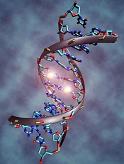
Credit: Christoph Bock
Computer scientists have developed a web-based tool that allows researchers to visualize and compare large amounts of genomic information from high-throughput sequencing experiments.
The group described the tool, called Epiviz, in Nature Methods.
“Prior tools limited visualization to presentation and dissemination, rather than a hybrid tool integrating interactive visualization with algorithmic analysis,” said Héctor Corrada Bravo, PhD, of the University of Maryland in College Park.
Dr Corrada Bravo and his colleagues developed Epiviz, a web-based genome browser that integrates with the widely used, open-source Bioconductor analysis software through its Epivizr Bioconductor package.
Epiviz supports many popular next-generation sequencing techniques, such as ChIP-seq, RNA-seq, and DNA methylation analyses.
The tool also implements multiple visualization methods for location-based data (such as genomic regions of interest) and feature-based data (such as gene expression).
For example, because display objects are mapped directly to data elements, Epiviz links data across different visualizations, giving users visual insights of the spatial relationships of multiple data sets. The tool is designed to allow biomedical scientists to easily incorporate their own visualizations.
In the Nature Methods paper, Dr Corrada Bravo and his colleagues describe how they used Epiviz to visualize and analyze DNA methylation and gene expression data in colon cancer.
Using Epiviz and Bioconductor, the team found consistent regions of DNA methylation changes in colon cancer samples generated by the Cancer Genome Atlas project and similar gene expression in these regions of DNA methylation changes in other cancer types.
The results were in agreement with previous experiments showing DNA methylation changes across large regions in the colon cancer genome.
Credit: Christoph Bock
Computer scientists have developed a web-based tool that allows researchers to visualize and compare large amounts of genomic information from high-throughput sequencing experiments.
The group described the tool, called Epiviz, in Nature Methods.
“Prior tools limited visualization to presentation and dissemination, rather than a hybrid tool integrating interactive visualization with algorithmic analysis,” said Héctor Corrada Bravo, PhD, of the University of Maryland in College Park.
Dr Corrada Bravo and his colleagues developed Epiviz, a web-based genome browser that integrates with the widely used, open-source Bioconductor analysis software through its Epivizr Bioconductor package.
Epiviz supports many popular next-generation sequencing techniques, such as ChIP-seq, RNA-seq, and DNA methylation analyses.
The tool also implements multiple visualization methods for location-based data (such as genomic regions of interest) and feature-based data (such as gene expression).
For example, because display objects are mapped directly to data elements, Epiviz links data across different visualizations, giving users visual insights of the spatial relationships of multiple data sets. The tool is designed to allow biomedical scientists to easily incorporate their own visualizations.
In the Nature Methods paper, Dr Corrada Bravo and his colleagues describe how they used Epiviz to visualize and analyze DNA methylation and gene expression data in colon cancer.
Using Epiviz and Bioconductor, the team found consistent regions of DNA methylation changes in colon cancer samples generated by the Cancer Genome Atlas project and similar gene expression in these regions of DNA methylation changes in other cancer types.
The results were in agreement with previous experiments showing DNA methylation changes across large regions in the colon cancer genome.
Credit: Christoph Bock
Computer scientists have developed a web-based tool that allows researchers to visualize and compare large amounts of genomic information from high-throughput sequencing experiments.
The group described the tool, called Epiviz, in Nature Methods.
“Prior tools limited visualization to presentation and dissemination, rather than a hybrid tool integrating interactive visualization with algorithmic analysis,” said Héctor Corrada Bravo, PhD, of the University of Maryland in College Park.
Dr Corrada Bravo and his colleagues developed Epiviz, a web-based genome browser that integrates with the widely used, open-source Bioconductor analysis software through its Epivizr Bioconductor package.
Epiviz supports many popular next-generation sequencing techniques, such as ChIP-seq, RNA-seq, and DNA methylation analyses.
The tool also implements multiple visualization methods for location-based data (such as genomic regions of interest) and feature-based data (such as gene expression).
For example, because display objects are mapped directly to data elements, Epiviz links data across different visualizations, giving users visual insights of the spatial relationships of multiple data sets. The tool is designed to allow biomedical scientists to easily incorporate their own visualizations.
In the Nature Methods paper, Dr Corrada Bravo and his colleagues describe how they used Epiviz to visualize and analyze DNA methylation and gene expression data in colon cancer.
Using Epiviz and Bioconductor, the team found consistent regions of DNA methylation changes in colon cancer samples generated by the Cancer Genome Atlas project and similar gene expression in these regions of DNA methylation changes in other cancer types.
The results were in agreement with previous experiments showing DNA methylation changes across large regions in the colon cancer genome.
System simplifies complex pipetting protocols

Credit: John Correa
Researchers say they’ve developed a simple system that can help scientists perform complex pipetting protocols efficiently and accurately.
The system, called iPipet, allows users to track the transfer of samples and reagents by illuminating well plates on a computer tablet.
In tests, iPipet proved more efficient than a liquid-handling robot.
The researchers have made information on iPipet available online so scientists can use the system in their own labs.
The team also described iPipet in a letter to Nature Methods.
They noted that experiments frequently rely on high-throughput methods that combine large numbers of samples with large-scale, complex pipetting designs. And pipetting errors can lead to experimental failure.
Although liquid-handling robots would seem to be a logical choice for such work, they are also extremely expensive, difficult to program, and require trained personnel. Moreover, they can be plagued by technical snafus, ranging from bent or clogged tips to an inability to capture liquids lying close to the bottoms of individual wells.
“We needed an alternative to costly robots that would allow us to execute complex pipetting protocols,” said Yaniv Erlich, PhD, of the Whitehead Institute in Cambridge, Massachusetts.
So Dr Erlich and his colleagues developed iPipet. The system illuminates individual wells of standard 96- or 384-well plates placed on top of a tablet screen, guiding users through the transfer of samples or reagents from source to destination plates according to specific designs.
Users create their own protocols in Microsoft Excel files in comma-separated format and upload them to the iPipet website, which generates a downloadable link for execution on a tablet computer. Included on the iPipet site are a variety of demos and an instructional video.
In a test of the tool against a liquid-handling robot, iPipet enabled nearly 3000 fixed-volume pipetting steps in approximately 7 hours. After significant time spent on calibration, the robot accomplished only half that number of steps in the same allotted time.
To date, one of the only challenges lab users have encountered is keeping well plates in a fixed position on the tablet screen. For that, Dr Erlich’s team provides a solution: a 3D printed plastic adaptor that users can create with a file accessible via the iPipet website.
“The entire iPipet system is open source,” Dr Erlich said. “We want to maximize the benefit for the community and allow them to further develop this new man-machine interface for biological experiments.”
Credit: John Correa
Researchers say they’ve developed a simple system that can help scientists perform complex pipetting protocols efficiently and accurately.
The system, called iPipet, allows users to track the transfer of samples and reagents by illuminating well plates on a computer tablet.
In tests, iPipet proved more efficient than a liquid-handling robot.
The researchers have made information on iPipet available online so scientists can use the system in their own labs.
The team also described iPipet in a letter to Nature Methods.
They noted that experiments frequently rely on high-throughput methods that combine large numbers of samples with large-scale, complex pipetting designs. And pipetting errors can lead to experimental failure.
Although liquid-handling robots would seem to be a logical choice for such work, they are also extremely expensive, difficult to program, and require trained personnel. Moreover, they can be plagued by technical snafus, ranging from bent or clogged tips to an inability to capture liquids lying close to the bottoms of individual wells.
“We needed an alternative to costly robots that would allow us to execute complex pipetting protocols,” said Yaniv Erlich, PhD, of the Whitehead Institute in Cambridge, Massachusetts.
So Dr Erlich and his colleagues developed iPipet. The system illuminates individual wells of standard 96- or 384-well plates placed on top of a tablet screen, guiding users through the transfer of samples or reagents from source to destination plates according to specific designs.
Users create their own protocols in Microsoft Excel files in comma-separated format and upload them to the iPipet website, which generates a downloadable link for execution on a tablet computer. Included on the iPipet site are a variety of demos and an instructional video.
In a test of the tool against a liquid-handling robot, iPipet enabled nearly 3000 fixed-volume pipetting steps in approximately 7 hours. After significant time spent on calibration, the robot accomplished only half that number of steps in the same allotted time.
To date, one of the only challenges lab users have encountered is keeping well plates in a fixed position on the tablet screen. For that, Dr Erlich’s team provides a solution: a 3D printed plastic adaptor that users can create with a file accessible via the iPipet website.
“The entire iPipet system is open source,” Dr Erlich said. “We want to maximize the benefit for the community and allow them to further develop this new man-machine interface for biological experiments.”
Credit: John Correa
Researchers say they’ve developed a simple system that can help scientists perform complex pipetting protocols efficiently and accurately.
The system, called iPipet, allows users to track the transfer of samples and reagents by illuminating well plates on a computer tablet.
In tests, iPipet proved more efficient than a liquid-handling robot.
The researchers have made information on iPipet available online so scientists can use the system in their own labs.
The team also described iPipet in a letter to Nature Methods.
They noted that experiments frequently rely on high-throughput methods that combine large numbers of samples with large-scale, complex pipetting designs. And pipetting errors can lead to experimental failure.
Although liquid-handling robots would seem to be a logical choice for such work, they are also extremely expensive, difficult to program, and require trained personnel. Moreover, they can be plagued by technical snafus, ranging from bent or clogged tips to an inability to capture liquids lying close to the bottoms of individual wells.
“We needed an alternative to costly robots that would allow us to execute complex pipetting protocols,” said Yaniv Erlich, PhD, of the Whitehead Institute in Cambridge, Massachusetts.
So Dr Erlich and his colleagues developed iPipet. The system illuminates individual wells of standard 96- or 384-well plates placed on top of a tablet screen, guiding users through the transfer of samples or reagents from source to destination plates according to specific designs.
Users create their own protocols in Microsoft Excel files in comma-separated format and upload them to the iPipet website, which generates a downloadable link for execution on a tablet computer. Included on the iPipet site are a variety of demos and an instructional video.
In a test of the tool against a liquid-handling robot, iPipet enabled nearly 3000 fixed-volume pipetting steps in approximately 7 hours. After significant time spent on calibration, the robot accomplished only half that number of steps in the same allotted time.
To date, one of the only challenges lab users have encountered is keeping well plates in a fixed position on the tablet screen. For that, Dr Erlich’s team provides a solution: a 3D printed plastic adaptor that users can create with a file accessible via the iPipet website.
“The entire iPipet system is open source,” Dr Erlich said. “We want to maximize the benefit for the community and allow them to further develop this new man-machine interface for biological experiments.”
Team pinpoints possible target for T-ALL

Researchers have discovered a potential therapeutic target for T-cell acute lymphoblastic leukemia (T-ALL), according to a paper published in Cell.
The team first identified long, non-coding strands of RNA (lncRNA) that were active in T cells from patients with T-ALL but not in the healthy T cells of subjects without the disease.
Further analysis revealed that inhibiting 1 of these lncRNAs, LUNAR1 (leukemia-induced non-coding activator RNA-1), stalled T-ALL growth in vitro and in vivo.
The study offers preliminary evidence that drugs targeting LUNAR1 could treat T-ALL, and LUNAR1 could aid in diagnosing the disease, said Iannis Aifantis, PhD, of NYU Langone Medical Center in New York.
“Our study shows that LUNAR1 is highly specific for T-cell acute lymphoblastic leukemia and plays a key role in how this cancer develops,” he noted, adding that overproduction of LUNAR1 was recorded in almost all (90%) of the leukemia patients analyzed.
To make these discoveries, Dr Aifantis and his colleagues performed ultra-high-depth RNA sequencing of human T-ALL cell lines and primary leukemia samples.
They used the resulting data to generate the most comprehensive T-ALL transcriptome assembly to date and then isolated putative lncRNA genes. This revealed 6023 lncRNAs that are active in T-ALL, 60% of which had not been identified before.
The researchers zeroed in on LUNAR1 by pinpointing the lncRNAs that were active in the NOTCH1 pathway, which is active in at least half of T-ALL patients. LUNAR1 stood out right away, the team said, as the most highly expressed lncRNA.
The researchers also found that LUNAR1 does not produce cancerous proteins on its own. However, its production proved essential to the cell-to-cell signaling action of another protein, IGF-1R (insulin-like growth factor 1 receptor), which is tied to many cancers, including leukemia.
Additional experiments showed that the gene coding for LUNAR1 is near the gene for IGF-1R and located toward the end of the chromosome. When activated, LUNAR1’s position allows it to chemically loop back and, in turn, bind to and activate IGF-1R.
According to Dr Aifantis, this research shows that T-ALL could simply be described as a condition of “too much errant signaling.” He noted that, in normal T cells, lncRNAs such as LUNAR1 are not transcribed, NOTCH1 is inactive, and there is no looping back of LUNAR1 to activate IGF-1R.
To confirm their findings, the researchers also transplanted human leukemia T cells into mice and inhibited LUNAR1 in some of the animals. Tumor growth stalled only in those mice in which LUNAR1 was inactivated.
The researchers said their next step is to develop a more effective inhibitor of LUNAR1, preferably something that would precisely target 1 or more of its 200-plus component nucleotides.
Researchers have discovered a potential therapeutic target for T-cell acute lymphoblastic leukemia (T-ALL), according to a paper published in Cell.
The team first identified long, non-coding strands of RNA (lncRNA) that were active in T cells from patients with T-ALL but not in the healthy T cells of subjects without the disease.
Further analysis revealed that inhibiting 1 of these lncRNAs, LUNAR1 (leukemia-induced non-coding activator RNA-1), stalled T-ALL growth in vitro and in vivo.
The study offers preliminary evidence that drugs targeting LUNAR1 could treat T-ALL, and LUNAR1 could aid in diagnosing the disease, said Iannis Aifantis, PhD, of NYU Langone Medical Center in New York.
“Our study shows that LUNAR1 is highly specific for T-cell acute lymphoblastic leukemia and plays a key role in how this cancer develops,” he noted, adding that overproduction of LUNAR1 was recorded in almost all (90%) of the leukemia patients analyzed.
To make these discoveries, Dr Aifantis and his colleagues performed ultra-high-depth RNA sequencing of human T-ALL cell lines and primary leukemia samples.
They used the resulting data to generate the most comprehensive T-ALL transcriptome assembly to date and then isolated putative lncRNA genes. This revealed 6023 lncRNAs that are active in T-ALL, 60% of which had not been identified before.
The researchers zeroed in on LUNAR1 by pinpointing the lncRNAs that were active in the NOTCH1 pathway, which is active in at least half of T-ALL patients. LUNAR1 stood out right away, the team said, as the most highly expressed lncRNA.
The researchers also found that LUNAR1 does not produce cancerous proteins on its own. However, its production proved essential to the cell-to-cell signaling action of another protein, IGF-1R (insulin-like growth factor 1 receptor), which is tied to many cancers, including leukemia.
Additional experiments showed that the gene coding for LUNAR1 is near the gene for IGF-1R and located toward the end of the chromosome. When activated, LUNAR1’s position allows it to chemically loop back and, in turn, bind to and activate IGF-1R.
According to Dr Aifantis, this research shows that T-ALL could simply be described as a condition of “too much errant signaling.” He noted that, in normal T cells, lncRNAs such as LUNAR1 are not transcribed, NOTCH1 is inactive, and there is no looping back of LUNAR1 to activate IGF-1R.
To confirm their findings, the researchers also transplanted human leukemia T cells into mice and inhibited LUNAR1 in some of the animals. Tumor growth stalled only in those mice in which LUNAR1 was inactivated.
The researchers said their next step is to develop a more effective inhibitor of LUNAR1, preferably something that would precisely target 1 or more of its 200-plus component nucleotides.
Researchers have discovered a potential therapeutic target for T-cell acute lymphoblastic leukemia (T-ALL), according to a paper published in Cell.
The team first identified long, non-coding strands of RNA (lncRNA) that were active in T cells from patients with T-ALL but not in the healthy T cells of subjects without the disease.
Further analysis revealed that inhibiting 1 of these lncRNAs, LUNAR1 (leukemia-induced non-coding activator RNA-1), stalled T-ALL growth in vitro and in vivo.
The study offers preliminary evidence that drugs targeting LUNAR1 could treat T-ALL, and LUNAR1 could aid in diagnosing the disease, said Iannis Aifantis, PhD, of NYU Langone Medical Center in New York.
“Our study shows that LUNAR1 is highly specific for T-cell acute lymphoblastic leukemia and plays a key role in how this cancer develops,” he noted, adding that overproduction of LUNAR1 was recorded in almost all (90%) of the leukemia patients analyzed.
To make these discoveries, Dr Aifantis and his colleagues performed ultra-high-depth RNA sequencing of human T-ALL cell lines and primary leukemia samples.
They used the resulting data to generate the most comprehensive T-ALL transcriptome assembly to date and then isolated putative lncRNA genes. This revealed 6023 lncRNAs that are active in T-ALL, 60% of which had not been identified before.
The researchers zeroed in on LUNAR1 by pinpointing the lncRNAs that were active in the NOTCH1 pathway, which is active in at least half of T-ALL patients. LUNAR1 stood out right away, the team said, as the most highly expressed lncRNA.
The researchers also found that LUNAR1 does not produce cancerous proteins on its own. However, its production proved essential to the cell-to-cell signaling action of another protein, IGF-1R (insulin-like growth factor 1 receptor), which is tied to many cancers, including leukemia.
Additional experiments showed that the gene coding for LUNAR1 is near the gene for IGF-1R and located toward the end of the chromosome. When activated, LUNAR1’s position allows it to chemically loop back and, in turn, bind to and activate IGF-1R.
According to Dr Aifantis, this research shows that T-ALL could simply be described as a condition of “too much errant signaling.” He noted that, in normal T cells, lncRNAs such as LUNAR1 are not transcribed, NOTCH1 is inactive, and there is no looping back of LUNAR1 to activate IGF-1R.
To confirm their findings, the researchers also transplanted human leukemia T cells into mice and inhibited LUNAR1 in some of the animals. Tumor growth stalled only in those mice in which LUNAR1 was inactivated.
The researchers said their next step is to develop a more effective inhibitor of LUNAR1, preferably something that would precisely target 1 or more of its 200-plus component nucleotides.
Approach could improve treatment of lymphoma, other cancers
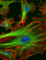
Credit: NIH
Targeting a molecule in endothelial cells could make cancer therapies significantly more effective, preclinical research suggests.
The researchers found that a molecule called focal adhesion kinase (FAK) can help protect cancer cells from the damaging effects of chemotherapy and radiotherapy.
But deleting FAK can enhance the effects of treatment directed against melanoma, lung cancer, and lymphoma.
The team recounted these findings in Nature.
“This work shows that sensitivity to cancer treatment is related to our own body mistakenly trying to shield the cancer from cell-killing effects caused by radiotherapy and chemotherapy,” said study author Bernardo Tavora, PhD, of Rockefeller University in New York.
“Although taking out FAK from blood vessels won’t destroy the cancer by itself, it can remove the barrier cancer uses to protect itself from treatment.”
Dr Tavora and his colleagues removed FAK from endothelial cells in mouse models of melanoma, lung cancer, and lymphoma. This had no effect on tumor growth in untreated mice.
However, the loss of endothelial-cell FAK did aid the effects of doxorubicin and radiotherapy. It increased apoptosis and decreased proliferation within perivascular tumor-cell compartments, thereby extending survival in the mice.
The researchers also studied samples from lymphoma patients. And they found that patients with low levels of FAK were more likely to achieve a complete remission after treatment.
Investigation into the mechanism behind these effects revealed that endothelial-cell FAK is required for the production of cytokines and for NF-κB activation induced by DNA damage.
So the loss of endothelial-cell FAK reduces DNA-damage-induced cytokine production, thereby increasing cancer cells’ sensitivity to DNA-damaging therapies in vitro and in vivo.
Taken together, these results suggest that developing drugs to target FAK could help improve the efficacy of cancer treatments and potentially prevent relapse in a number of malignancies.
Credit: NIH
Targeting a molecule in endothelial cells could make cancer therapies significantly more effective, preclinical research suggests.
The researchers found that a molecule called focal adhesion kinase (FAK) can help protect cancer cells from the damaging effects of chemotherapy and radiotherapy.
But deleting FAK can enhance the effects of treatment directed against melanoma, lung cancer, and lymphoma.
The team recounted these findings in Nature.
“This work shows that sensitivity to cancer treatment is related to our own body mistakenly trying to shield the cancer from cell-killing effects caused by radiotherapy and chemotherapy,” said study author Bernardo Tavora, PhD, of Rockefeller University in New York.
“Although taking out FAK from blood vessels won’t destroy the cancer by itself, it can remove the barrier cancer uses to protect itself from treatment.”
Dr Tavora and his colleagues removed FAK from endothelial cells in mouse models of melanoma, lung cancer, and lymphoma. This had no effect on tumor growth in untreated mice.
However, the loss of endothelial-cell FAK did aid the effects of doxorubicin and radiotherapy. It increased apoptosis and decreased proliferation within perivascular tumor-cell compartments, thereby extending survival in the mice.
The researchers also studied samples from lymphoma patients. And they found that patients with low levels of FAK were more likely to achieve a complete remission after treatment.
Investigation into the mechanism behind these effects revealed that endothelial-cell FAK is required for the production of cytokines and for NF-κB activation induced by DNA damage.
So the loss of endothelial-cell FAK reduces DNA-damage-induced cytokine production, thereby increasing cancer cells’ sensitivity to DNA-damaging therapies in vitro and in vivo.
Taken together, these results suggest that developing drugs to target FAK could help improve the efficacy of cancer treatments and potentially prevent relapse in a number of malignancies.
Credit: NIH
Targeting a molecule in endothelial cells could make cancer therapies significantly more effective, preclinical research suggests.
The researchers found that a molecule called focal adhesion kinase (FAK) can help protect cancer cells from the damaging effects of chemotherapy and radiotherapy.
But deleting FAK can enhance the effects of treatment directed against melanoma, lung cancer, and lymphoma.
The team recounted these findings in Nature.
“This work shows that sensitivity to cancer treatment is related to our own body mistakenly trying to shield the cancer from cell-killing effects caused by radiotherapy and chemotherapy,” said study author Bernardo Tavora, PhD, of Rockefeller University in New York.
“Although taking out FAK from blood vessels won’t destroy the cancer by itself, it can remove the barrier cancer uses to protect itself from treatment.”
Dr Tavora and his colleagues removed FAK from endothelial cells in mouse models of melanoma, lung cancer, and lymphoma. This had no effect on tumor growth in untreated mice.
However, the loss of endothelial-cell FAK did aid the effects of doxorubicin and radiotherapy. It increased apoptosis and decreased proliferation within perivascular tumor-cell compartments, thereby extending survival in the mice.
The researchers also studied samples from lymphoma patients. And they found that patients with low levels of FAK were more likely to achieve a complete remission after treatment.
Investigation into the mechanism behind these effects revealed that endothelial-cell FAK is required for the production of cytokines and for NF-κB activation induced by DNA damage.
So the loss of endothelial-cell FAK reduces DNA-damage-induced cytokine production, thereby increasing cancer cells’ sensitivity to DNA-damaging therapies in vitro and in vivo.
Taken together, these results suggest that developing drugs to target FAK could help improve the efficacy of cancer treatments and potentially prevent relapse in a number of malignancies.
Early HSCT best for infants with SCID, study shows

Credit: Chad McNeeley
Children with severe combined immune deficiency (SCID) have a good chance of survival if they undergo hematopoietic stem cell transplant (HSCT) within 3.5 months of birth, a new analysis suggests.
The risk of death is also lower if patients are free of infection at transplant and have a matched sibling donor.
“Survival is much, much better if infants undergo transplant before they turn 3.5 months old and before they contract any SCID-related infections,” said Sung-Yun Pai, MD, of the Dana-Farber Cancer Institute in Boston.
This underlines the importance of screening newborns for SCID, she added.
Dr Pai and her colleagues expressed this viewpoint, and detailed the research to support it, in The New England Journal of Medicine.
The team analyzed data on 240 children with SCID who were transplanted at 25 centers across North America between January 1, 2000, and December 31, 2009, (before the US Department of Health and Human Services recommended newborn screening for SCID in 2010).
The researchers assessed the patients’ outcomes according to age, infection status, donor source, and conditioning regimen.
Results revealed that children who underwent transplant before 3.5 months of age had excellent survival, regardless of donor source or infection status, as did patients with a matched sibling donor.
Children transplanted after 3.5 months also had a high survival rate regardless of donor source, as long as they did not have an active infection at the time of transplant.
Overall, 74% of patients survived at least 5 years. The 5-year survival rate was 97% in patients with a matched sibling donor, 94% among patients transplanted within 3.5 months of birth, 90% among patients who never had an infection, and 82% in patients whose infection resolved before transplant.
Survival was low—50%—among patients who were older than 3.5 months and had active infections at the time of transplant.
Actively infected infants who did not have a matched sibling donor and received immunosuppressive therapy or chemotherapy before transplant had particularly poor survival as well, ranging from 39% to 53%.
“This study accomplishes several things,” Dr Pai said. “First, it creates a baseline with which to compare patient outcomes since the advent of newborn screening for SCID. Second, it provides guidance for clinicians regarding the use of chemotherapy conditioning before transplantation.”
“Third, it highlights the relative impacts of infection status and patient age on transplant success. Lastly, it establishes the importance of early detection and transplantation, which points to the benefit of expanding newborn screening for SCID as broadly as possible.”
Credit: Chad McNeeley
Children with severe combined immune deficiency (SCID) have a good chance of survival if they undergo hematopoietic stem cell transplant (HSCT) within 3.5 months of birth, a new analysis suggests.
The risk of death is also lower if patients are free of infection at transplant and have a matched sibling donor.
“Survival is much, much better if infants undergo transplant before they turn 3.5 months old and before they contract any SCID-related infections,” said Sung-Yun Pai, MD, of the Dana-Farber Cancer Institute in Boston.
This underlines the importance of screening newborns for SCID, she added.
Dr Pai and her colleagues expressed this viewpoint, and detailed the research to support it, in The New England Journal of Medicine.
The team analyzed data on 240 children with SCID who were transplanted at 25 centers across North America between January 1, 2000, and December 31, 2009, (before the US Department of Health and Human Services recommended newborn screening for SCID in 2010).
The researchers assessed the patients’ outcomes according to age, infection status, donor source, and conditioning regimen.
Results revealed that children who underwent transplant before 3.5 months of age had excellent survival, regardless of donor source or infection status, as did patients with a matched sibling donor.
Children transplanted after 3.5 months also had a high survival rate regardless of donor source, as long as they did not have an active infection at the time of transplant.
Overall, 74% of patients survived at least 5 years. The 5-year survival rate was 97% in patients with a matched sibling donor, 94% among patients transplanted within 3.5 months of birth, 90% among patients who never had an infection, and 82% in patients whose infection resolved before transplant.
Survival was low—50%—among patients who were older than 3.5 months and had active infections at the time of transplant.
Actively infected infants who did not have a matched sibling donor and received immunosuppressive therapy or chemotherapy before transplant had particularly poor survival as well, ranging from 39% to 53%.
“This study accomplishes several things,” Dr Pai said. “First, it creates a baseline with which to compare patient outcomes since the advent of newborn screening for SCID. Second, it provides guidance for clinicians regarding the use of chemotherapy conditioning before transplantation.”
“Third, it highlights the relative impacts of infection status and patient age on transplant success. Lastly, it establishes the importance of early detection and transplantation, which points to the benefit of expanding newborn screening for SCID as broadly as possible.”
Credit: Chad McNeeley
Children with severe combined immune deficiency (SCID) have a good chance of survival if they undergo hematopoietic stem cell transplant (HSCT) within 3.5 months of birth, a new analysis suggests.
The risk of death is also lower if patients are free of infection at transplant and have a matched sibling donor.
“Survival is much, much better if infants undergo transplant before they turn 3.5 months old and before they contract any SCID-related infections,” said Sung-Yun Pai, MD, of the Dana-Farber Cancer Institute in Boston.
This underlines the importance of screening newborns for SCID, she added.
Dr Pai and her colleagues expressed this viewpoint, and detailed the research to support it, in The New England Journal of Medicine.
The team analyzed data on 240 children with SCID who were transplanted at 25 centers across North America between January 1, 2000, and December 31, 2009, (before the US Department of Health and Human Services recommended newborn screening for SCID in 2010).
The researchers assessed the patients’ outcomes according to age, infection status, donor source, and conditioning regimen.
Results revealed that children who underwent transplant before 3.5 months of age had excellent survival, regardless of donor source or infection status, as did patients with a matched sibling donor.
Children transplanted after 3.5 months also had a high survival rate regardless of donor source, as long as they did not have an active infection at the time of transplant.
Overall, 74% of patients survived at least 5 years. The 5-year survival rate was 97% in patients with a matched sibling donor, 94% among patients transplanted within 3.5 months of birth, 90% among patients who never had an infection, and 82% in patients whose infection resolved before transplant.
Survival was low—50%—among patients who were older than 3.5 months and had active infections at the time of transplant.
Actively infected infants who did not have a matched sibling donor and received immunosuppressive therapy or chemotherapy before transplant had particularly poor survival as well, ranging from 39% to 53%.
“This study accomplishes several things,” Dr Pai said. “First, it creates a baseline with which to compare patient outcomes since the advent of newborn screening for SCID. Second, it provides guidance for clinicians regarding the use of chemotherapy conditioning before transplantation.”
“Third, it highlights the relative impacts of infection status and patient age on transplant success. Lastly, it establishes the importance of early detection and transplantation, which points to the benefit of expanding newborn screening for SCID as broadly as possible.”
FDA aims for tighter regulation of diagnostic tests
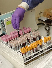
Credit: William Weinert
The US Food and Drug Administration (FDA) is taking steps to ensure better regulation of certain diagnostic tests.
The agency has issued a final guidance on the development, review, and approval of companion diagnostics.
The FDA has also notified Congress of its intention to publish a draft guidance outlining a plan for regulating laboratory-developed tests (LDTs).
The FDA is required to notify Congress before making the draft guidance public. This is mandated by the Food and Drug Administration Safety and Innovation Act of 2012 (FDASIA).
Companion diagnostics guidance
A companion diagnostic is a medical device that provides information essential for the safe and effective use of a corresponding drug or biological product. These tests are commonly used to detect certain types of gene-based cancers.
The FDA’s companion diagnostics guidance is intended to help companies identify the need for these tests during the earliest stages of drug development and to plan for the development of a drug and a companion test at the same time.
The ultimate goal of the final guidance is to stimulate early collaborations that will result in faster access to promising new treatments for patients living with serious and life-threatening diseases. This guidance finalizes and takes into consideration public comments on the draft guidance issued in 2011.
LDT oversight
An LDT is a type of in vitro diagnostic test that is designed, manufactured, and used within a single lab. LDTs include some genetic tests and tests used by healthcare professionals to guide patient treatment.
The FDA already oversees direct-to-consumer tests, regardless of whether they are LDTs or traditional diagnostics.
And while the FDA has historically exercised enforcement discretion over LDTs (generally not enforced applicable regulatory requirements), today, these tests may compete with FDA-approved tests without clinical studies to support their use.
The LDT notification to Congress provides the details of a draft guidance in which the FDA would propose to establish an LDT oversight framework. This would include pre-market review for higher-risk LDTs, such as those that have the same intended use as FDA-approved or cleared companion diagnostics currently on the market.
The draft guidance would also propose to phase in enforcement of pre-market review for other high-risk and moderate-risk LDTs over time.
In addition, the FDA intends to propose that it continue to exercise enforcement discretion for low-risk LDTs, LDTs for rare diseases and, under certain circumstances, LDTs for which there is no FDA-approved or cleared test.
“With today’s notification of the agency’s intent to issue the lab-developed test draft guidance, the FDA is seeking a better balanced approach for all diagnostics,” said Jeffrey Shuren, MD, director of the FDA’s Center for Devices and Radiological Health.
“The agency’s oversight would be based on a test’s level of risk to patients, not on whether it is made by a conventional manufacturer or in a single laboratory, while still providing flexibility to encourage innovation that addresses unmet medical needs.”
Finally, the FDA intends to publish a draft guidance outlining how labs can notify the FDA that they are manufacturing and using LDTs, how to provide information about their LDTs, and how they can comply with the medical device reporting requirements.
A provision in FDASIA requires the FDA to provide at least 60 days’ notice to Congress before the agency publishes for public comment any draft guidance on the regulation of LDTs.
As such, the comment period will open at a later date, when the draft guidances are published in the Federal Register and the public is alerted to the start of the comment period. The agency also intends to hold a public meeting during the comment period to collect additional input.
Credit: William Weinert
The US Food and Drug Administration (FDA) is taking steps to ensure better regulation of certain diagnostic tests.
The agency has issued a final guidance on the development, review, and approval of companion diagnostics.
The FDA has also notified Congress of its intention to publish a draft guidance outlining a plan for regulating laboratory-developed tests (LDTs).
The FDA is required to notify Congress before making the draft guidance public. This is mandated by the Food and Drug Administration Safety and Innovation Act of 2012 (FDASIA).
Companion diagnostics guidance
A companion diagnostic is a medical device that provides information essential for the safe and effective use of a corresponding drug or biological product. These tests are commonly used to detect certain types of gene-based cancers.
The FDA’s companion diagnostics guidance is intended to help companies identify the need for these tests during the earliest stages of drug development and to plan for the development of a drug and a companion test at the same time.
The ultimate goal of the final guidance is to stimulate early collaborations that will result in faster access to promising new treatments for patients living with serious and life-threatening diseases. This guidance finalizes and takes into consideration public comments on the draft guidance issued in 2011.
LDT oversight
An LDT is a type of in vitro diagnostic test that is designed, manufactured, and used within a single lab. LDTs include some genetic tests and tests used by healthcare professionals to guide patient treatment.
The FDA already oversees direct-to-consumer tests, regardless of whether they are LDTs or traditional diagnostics.
And while the FDA has historically exercised enforcement discretion over LDTs (generally not enforced applicable regulatory requirements), today, these tests may compete with FDA-approved tests without clinical studies to support their use.
The LDT notification to Congress provides the details of a draft guidance in which the FDA would propose to establish an LDT oversight framework. This would include pre-market review for higher-risk LDTs, such as those that have the same intended use as FDA-approved or cleared companion diagnostics currently on the market.
The draft guidance would also propose to phase in enforcement of pre-market review for other high-risk and moderate-risk LDTs over time.
In addition, the FDA intends to propose that it continue to exercise enforcement discretion for low-risk LDTs, LDTs for rare diseases and, under certain circumstances, LDTs for which there is no FDA-approved or cleared test.
“With today’s notification of the agency’s intent to issue the lab-developed test draft guidance, the FDA is seeking a better balanced approach for all diagnostics,” said Jeffrey Shuren, MD, director of the FDA’s Center for Devices and Radiological Health.
“The agency’s oversight would be based on a test’s level of risk to patients, not on whether it is made by a conventional manufacturer or in a single laboratory, while still providing flexibility to encourage innovation that addresses unmet medical needs.”
Finally, the FDA intends to publish a draft guidance outlining how labs can notify the FDA that they are manufacturing and using LDTs, how to provide information about their LDTs, and how they can comply with the medical device reporting requirements.
A provision in FDASIA requires the FDA to provide at least 60 days’ notice to Congress before the agency publishes for public comment any draft guidance on the regulation of LDTs.
As such, the comment period will open at a later date, when the draft guidances are published in the Federal Register and the public is alerted to the start of the comment period. The agency also intends to hold a public meeting during the comment period to collect additional input.
Credit: William Weinert
The US Food and Drug Administration (FDA) is taking steps to ensure better regulation of certain diagnostic tests.
The agency has issued a final guidance on the development, review, and approval of companion diagnostics.
The FDA has also notified Congress of its intention to publish a draft guidance outlining a plan for regulating laboratory-developed tests (LDTs).
The FDA is required to notify Congress before making the draft guidance public. This is mandated by the Food and Drug Administration Safety and Innovation Act of 2012 (FDASIA).
Companion diagnostics guidance
A companion diagnostic is a medical device that provides information essential for the safe and effective use of a corresponding drug or biological product. These tests are commonly used to detect certain types of gene-based cancers.
The FDA’s companion diagnostics guidance is intended to help companies identify the need for these tests during the earliest stages of drug development and to plan for the development of a drug and a companion test at the same time.
The ultimate goal of the final guidance is to stimulate early collaborations that will result in faster access to promising new treatments for patients living with serious and life-threatening diseases. This guidance finalizes and takes into consideration public comments on the draft guidance issued in 2011.
LDT oversight
An LDT is a type of in vitro diagnostic test that is designed, manufactured, and used within a single lab. LDTs include some genetic tests and tests used by healthcare professionals to guide patient treatment.
The FDA already oversees direct-to-consumer tests, regardless of whether they are LDTs or traditional diagnostics.
And while the FDA has historically exercised enforcement discretion over LDTs (generally not enforced applicable regulatory requirements), today, these tests may compete with FDA-approved tests without clinical studies to support their use.
The LDT notification to Congress provides the details of a draft guidance in which the FDA would propose to establish an LDT oversight framework. This would include pre-market review for higher-risk LDTs, such as those that have the same intended use as FDA-approved or cleared companion diagnostics currently on the market.
The draft guidance would also propose to phase in enforcement of pre-market review for other high-risk and moderate-risk LDTs over time.
In addition, the FDA intends to propose that it continue to exercise enforcement discretion for low-risk LDTs, LDTs for rare diseases and, under certain circumstances, LDTs for which there is no FDA-approved or cleared test.
“With today’s notification of the agency’s intent to issue the lab-developed test draft guidance, the FDA is seeking a better balanced approach for all diagnostics,” said Jeffrey Shuren, MD, director of the FDA’s Center for Devices and Radiological Health.
“The agency’s oversight would be based on a test’s level of risk to patients, not on whether it is made by a conventional manufacturer or in a single laboratory, while still providing flexibility to encourage innovation that addresses unmet medical needs.”
Finally, the FDA intends to publish a draft guidance outlining how labs can notify the FDA that they are manufacturing and using LDTs, how to provide information about their LDTs, and how they can comply with the medical device reporting requirements.
A provision in FDASIA requires the FDA to provide at least 60 days’ notice to Congress before the agency publishes for public comment any draft guidance on the regulation of LDTs.
As such, the comment period will open at a later date, when the draft guidances are published in the Federal Register and the public is alerted to the start of the comment period. The agency also intends to hold a public meeting during the comment period to collect additional input.