User login
AABB creates new guidelines for platelet transfusion
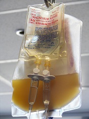
The AABB has developed new guidelines that specify situations
in which adults should receive platelet transfusions.
The group’s
recommendations are based on results of a systematic review.
The
suggestions cover several clinical situations in which platelet
transfusions might be an option, such as therapy-induced hypoproliferative thrombocytopenia, central venous catheter placement, diagnostic lumbar puncture, and cardiac surgery.
To formulate the guidelines, researchers conducted a review of randomized clinical trials and observational studies published from 1900 through September 2014. All studies reported clinical outcomes in patients receiving prophylactic or therapeutic platelet transfusions.
Based on the collected data, the AABB now strongly recommends prophylactic platelet transfusions to reduce the risk for spontaneous bleeding in hospitalized adults with therapy-induced hypoproliferative thrombocytopenia and a platelet count of 10 x 109 cells/L or less. The recommendation states that transfusing 0.5 to 1 apheresis unit is sufficient in these patients.
A weaker recommendation is that prophylactic platelet transfusions be given to patients undergoing elective central venous catheter placement who have a platelet count of less than 20 x 109 cells/L.
Likewise, the AABB says prophylactic transfusions should be given to patients having elective diagnostic lumbar puncture or major elective nonneuraxial surgery who have a platelet count less than 50 x 109 cells/L.
Another weak recommendation advises against routine prophylactic platelet transfusions in patients who are nonthrombocytopenic and have cardiac surgery with cardiopulmonary bypass.
The AABB also suggests (without recommending) that patients with perioperative bleeding and thrombocytopenia or platelet dysfunction may benefit from transfusions.
Finally, the AABB said it cannot recommend for or against platelet transfusions in patients receiving antiplatelet therapy who have intracranial hemorrhage (traumatic or spontaneous).
For more details, see the complete guidelines in Annals of Internal Medicine. 
The AABB has developed new guidelines that specify situations
in which adults should receive platelet transfusions.
The group’s
recommendations are based on results of a systematic review.
The
suggestions cover several clinical situations in which platelet
transfusions might be an option, such as therapy-induced hypoproliferative thrombocytopenia, central venous catheter placement, diagnostic lumbar puncture, and cardiac surgery.
To formulate the guidelines, researchers conducted a review of randomized clinical trials and observational studies published from 1900 through September 2014. All studies reported clinical outcomes in patients receiving prophylactic or therapeutic platelet transfusions.
Based on the collected data, the AABB now strongly recommends prophylactic platelet transfusions to reduce the risk for spontaneous bleeding in hospitalized adults with therapy-induced hypoproliferative thrombocytopenia and a platelet count of 10 x 109 cells/L or less. The recommendation states that transfusing 0.5 to 1 apheresis unit is sufficient in these patients.
A weaker recommendation is that prophylactic platelet transfusions be given to patients undergoing elective central venous catheter placement who have a platelet count of less than 20 x 109 cells/L.
Likewise, the AABB says prophylactic transfusions should be given to patients having elective diagnostic lumbar puncture or major elective nonneuraxial surgery who have a platelet count less than 50 x 109 cells/L.
Another weak recommendation advises against routine prophylactic platelet transfusions in patients who are nonthrombocytopenic and have cardiac surgery with cardiopulmonary bypass.
The AABB also suggests (without recommending) that patients with perioperative bleeding and thrombocytopenia or platelet dysfunction may benefit from transfusions.
Finally, the AABB said it cannot recommend for or against platelet transfusions in patients receiving antiplatelet therapy who have intracranial hemorrhage (traumatic or spontaneous).
For more details, see the complete guidelines in Annals of Internal Medicine.
The AABB has developed new guidelines that specify situations
in which adults should receive platelet transfusions.
The group’s
recommendations are based on results of a systematic review.
The
suggestions cover several clinical situations in which platelet
transfusions might be an option, such as therapy-induced hypoproliferative thrombocytopenia, central venous catheter placement, diagnostic lumbar puncture, and cardiac surgery.
To formulate the guidelines, researchers conducted a review of randomized clinical trials and observational studies published from 1900 through September 2014. All studies reported clinical outcomes in patients receiving prophylactic or therapeutic platelet transfusions.
Based on the collected data, the AABB now strongly recommends prophylactic platelet transfusions to reduce the risk for spontaneous bleeding in hospitalized adults with therapy-induced hypoproliferative thrombocytopenia and a platelet count of 10 x 109 cells/L or less. The recommendation states that transfusing 0.5 to 1 apheresis unit is sufficient in these patients.
A weaker recommendation is that prophylactic platelet transfusions be given to patients undergoing elective central venous catheter placement who have a platelet count of less than 20 x 109 cells/L.
Likewise, the AABB says prophylactic transfusions should be given to patients having elective diagnostic lumbar puncture or major elective nonneuraxial surgery who have a platelet count less than 50 x 109 cells/L.
Another weak recommendation advises against routine prophylactic platelet transfusions in patients who are nonthrombocytopenic and have cardiac surgery with cardiopulmonary bypass.
The AABB also suggests (without recommending) that patients with perioperative bleeding and thrombocytopenia or platelet dysfunction may benefit from transfusions.
Finally, the AABB said it cannot recommend for or against platelet transfusions in patients receiving antiplatelet therapy who have intracranial hemorrhage (traumatic or spontaneous).
For more details, see the complete guidelines in Annals of Internal Medicine.
Malaria prevents hospitals from meeting blood demands
PHILADELPHIA—The demand for blood transfusions increases substantially during high malaria transmission season, according to a study of hospitals in Tanzania.
On average, blood demand increased about 24% from low transmission season to high transmission season.
And some hospitals could not meet the increased demand. Unmet demand was highest in government hospitals, followed by faith-based institutions and private facilities.
Bakary Drammeh, DrPH, of the Centers for Disease Control and Prevention in Atlanta, Georgia, and his colleagues presented these results in a poster (SP356) at the AABB Annual Meeting 2014.
The researchers analyzed 14,706 blood prescriptions at 42 Tanzanian hospitals—21 government, 9 private, and 12 faith-based institutions.
The team assessed the number of blood prescriptions according to high and low malaria transmission periods—June-July vs August-September.
On average, there were 130 monthly blood prescriptions written per 100 beds during high malaria transmission season and 107 written during low transmission season.
There were 189 and 153 units of blood or blood components requested per 100 beds during high and low seasons, respectively. And there were 159 and 127 units issued, respectively.
Ultimately, an average of 145 units were transfused per 100 beds during high malaria transmission season, and 122 were transfused during low transmission season.
Across all 42 hospitals, total blood prescriptions increased 29% from low malaria transmission season to high transmission season.
The number of units requested increased 26%, the number of units issued increased 34%, and the number of units transfused increased 28%.
So, on average, blood demand increased 23.5% at these 42 hospitals during high malaria transmission season. And some hospitals did not have enough blood to meet demand.
The unmet blood demand was highest in government hospitals, at 25%, compared to faith-based hospitals, at 11%, and private hospitals, at 5%.
The researchers said these results suggest blood services should monitor malaria transmission surveillance reports and revise or project blood collection targets to meet the anticipated demand.
PHILADELPHIA—The demand for blood transfusions increases substantially during high malaria transmission season, according to a study of hospitals in Tanzania.
On average, blood demand increased about 24% from low transmission season to high transmission season.
And some hospitals could not meet the increased demand. Unmet demand was highest in government hospitals, followed by faith-based institutions and private facilities.
Bakary Drammeh, DrPH, of the Centers for Disease Control and Prevention in Atlanta, Georgia, and his colleagues presented these results in a poster (SP356) at the AABB Annual Meeting 2014.
The researchers analyzed 14,706 blood prescriptions at 42 Tanzanian hospitals—21 government, 9 private, and 12 faith-based institutions.
The team assessed the number of blood prescriptions according to high and low malaria transmission periods—June-July vs August-September.
On average, there were 130 monthly blood prescriptions written per 100 beds during high malaria transmission season and 107 written during low transmission season.
There were 189 and 153 units of blood or blood components requested per 100 beds during high and low seasons, respectively. And there were 159 and 127 units issued, respectively.
Ultimately, an average of 145 units were transfused per 100 beds during high malaria transmission season, and 122 were transfused during low transmission season.
Across all 42 hospitals, total blood prescriptions increased 29% from low malaria transmission season to high transmission season.
The number of units requested increased 26%, the number of units issued increased 34%, and the number of units transfused increased 28%.
So, on average, blood demand increased 23.5% at these 42 hospitals during high malaria transmission season. And some hospitals did not have enough blood to meet demand.
The unmet blood demand was highest in government hospitals, at 25%, compared to faith-based hospitals, at 11%, and private hospitals, at 5%.
The researchers said these results suggest blood services should monitor malaria transmission surveillance reports and revise or project blood collection targets to meet the anticipated demand.
PHILADELPHIA—The demand for blood transfusions increases substantially during high malaria transmission season, according to a study of hospitals in Tanzania.
On average, blood demand increased about 24% from low transmission season to high transmission season.
And some hospitals could not meet the increased demand. Unmet demand was highest in government hospitals, followed by faith-based institutions and private facilities.
Bakary Drammeh, DrPH, of the Centers for Disease Control and Prevention in Atlanta, Georgia, and his colleagues presented these results in a poster (SP356) at the AABB Annual Meeting 2014.
The researchers analyzed 14,706 blood prescriptions at 42 Tanzanian hospitals—21 government, 9 private, and 12 faith-based institutions.
The team assessed the number of blood prescriptions according to high and low malaria transmission periods—June-July vs August-September.
On average, there were 130 monthly blood prescriptions written per 100 beds during high malaria transmission season and 107 written during low transmission season.
There were 189 and 153 units of blood or blood components requested per 100 beds during high and low seasons, respectively. And there were 159 and 127 units issued, respectively.
Ultimately, an average of 145 units were transfused per 100 beds during high malaria transmission season, and 122 were transfused during low transmission season.
Across all 42 hospitals, total blood prescriptions increased 29% from low malaria transmission season to high transmission season.
The number of units requested increased 26%, the number of units issued increased 34%, and the number of units transfused increased 28%.
So, on average, blood demand increased 23.5% at these 42 hospitals during high malaria transmission season. And some hospitals did not have enough blood to meet demand.
The unmet blood demand was highest in government hospitals, at 25%, compared to faith-based hospitals, at 11%, and private hospitals, at 5%.
The researchers said these results suggest blood services should monitor malaria transmission surveillance reports and revise or project blood collection targets to meet the anticipated demand.
Sugar beets can be used to create hemoglobin

holding a sugar beet
Credit: Lund University
Biochemists have found evidence to suggest that sugar beets can be used to create a substitute for human hemoglobin.
While studying the genome of the sugar beet, the researchers identified 4 hemoglobin genes—3 non-symbiotic genes (BvHb1.1, BvHb1.2, and BvHb2) and 1 truncated gene (BvHb3).
The team then discovered they could extract hemoglobin from sugar beets using a process that’s about as simple as the one used to extract sugar.
The researchers cloned the hemoglobin genes and inserted them into bacteria, which facilitates their expression and purification.
The group described the gene discovery in Plant and Cell Physiology. Study author Nélida Leiva-Eriksson, a doctoral student at Lund University in Sweden, disclosed additional details in her dissertation. And a short video on the research is available on YouTube.
The researchers discovered that the hemoglobin extracted from sugar beets is almost identical to human hemoglobin, especially the form of hemoglobin in the brain.
“There is a difference in a small detail on the surface of the protein, but this simply appears to extend the lifespan of the hemoglobin from sugar beet, which is good news,” Leiva-Eriksson said.
On the other hand, sugar beet hemoglobin has a completely different function from human hemoglobin.
“We have found that the hemoglobin in the plant binds nitric oxide,” Leiva-Eriksson said. “It is probably needed to keep certain processes in check, for example, so that the nitric oxide doesn’t become toxic, and to ward off bacteria.”
The researchers are planning to start testing the sugar beet hemoglobin in animal experiments in just over a year.
The team said there is good reason to think hemoglobin derived from sugar beets and other crops could become a realistic alternative to human hemoglobin.
“From 1 hectare, we could produce 1 to 2 tons of hemoglobin,” said Leif Bülow, PhD, of Lund University, “which could save thousands of lives.”
holding a sugar beet
Credit: Lund University
Biochemists have found evidence to suggest that sugar beets can be used to create a substitute for human hemoglobin.
While studying the genome of the sugar beet, the researchers identified 4 hemoglobin genes—3 non-symbiotic genes (BvHb1.1, BvHb1.2, and BvHb2) and 1 truncated gene (BvHb3).
The team then discovered they could extract hemoglobin from sugar beets using a process that’s about as simple as the one used to extract sugar.
The researchers cloned the hemoglobin genes and inserted them into bacteria, which facilitates their expression and purification.
The group described the gene discovery in Plant and Cell Physiology. Study author Nélida Leiva-Eriksson, a doctoral student at Lund University in Sweden, disclosed additional details in her dissertation. And a short video on the research is available on YouTube.
The researchers discovered that the hemoglobin extracted from sugar beets is almost identical to human hemoglobin, especially the form of hemoglobin in the brain.
“There is a difference in a small detail on the surface of the protein, but this simply appears to extend the lifespan of the hemoglobin from sugar beet, which is good news,” Leiva-Eriksson said.
On the other hand, sugar beet hemoglobin has a completely different function from human hemoglobin.
“We have found that the hemoglobin in the plant binds nitric oxide,” Leiva-Eriksson said. “It is probably needed to keep certain processes in check, for example, so that the nitric oxide doesn’t become toxic, and to ward off bacteria.”
The researchers are planning to start testing the sugar beet hemoglobin in animal experiments in just over a year.
The team said there is good reason to think hemoglobin derived from sugar beets and other crops could become a realistic alternative to human hemoglobin.
“From 1 hectare, we could produce 1 to 2 tons of hemoglobin,” said Leif Bülow, PhD, of Lund University, “which could save thousands of lives.”
holding a sugar beet
Credit: Lund University
Biochemists have found evidence to suggest that sugar beets can be used to create a substitute for human hemoglobin.
While studying the genome of the sugar beet, the researchers identified 4 hemoglobin genes—3 non-symbiotic genes (BvHb1.1, BvHb1.2, and BvHb2) and 1 truncated gene (BvHb3).
The team then discovered they could extract hemoglobin from sugar beets using a process that’s about as simple as the one used to extract sugar.
The researchers cloned the hemoglobin genes and inserted them into bacteria, which facilitates their expression and purification.
The group described the gene discovery in Plant and Cell Physiology. Study author Nélida Leiva-Eriksson, a doctoral student at Lund University in Sweden, disclosed additional details in her dissertation. And a short video on the research is available on YouTube.
The researchers discovered that the hemoglobin extracted from sugar beets is almost identical to human hemoglobin, especially the form of hemoglobin in the brain.
“There is a difference in a small detail on the surface of the protein, but this simply appears to extend the lifespan of the hemoglobin from sugar beet, which is good news,” Leiva-Eriksson said.
On the other hand, sugar beet hemoglobin has a completely different function from human hemoglobin.
“We have found that the hemoglobin in the plant binds nitric oxide,” Leiva-Eriksson said. “It is probably needed to keep certain processes in check, for example, so that the nitric oxide doesn’t become toxic, and to ward off bacteria.”
The researchers are planning to start testing the sugar beet hemoglobin in animal experiments in just over a year.
The team said there is good reason to think hemoglobin derived from sugar beets and other crops could become a realistic alternative to human hemoglobin.
“From 1 hectare, we could produce 1 to 2 tons of hemoglobin,” said Leif Bülow, PhD, of Lund University, “which could save thousands of lives.”
Team pinpoints new target for MM therapy
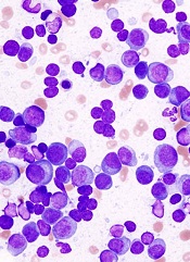
Researchers say they’ve identified a promising therapeutic target for multiple myeloma (MM).
The group first discovered that MM patients have higher expression of proline-rich tyrosine kinase 2 (PYK2), a member of the focal adhesion kinase (FAK) family, compared to healthy individuals.
Then, preclinical experiments revealed that PYK2 plays a role in tumor progression, and inhibiting this kinase can impede MM cell growth.
Yu Zhang, PhD, of the Dana-Farber Cancer Institute in Boston, and colleagues reported these findings in Blood.
Comparing samples from MM patients and healthy controls, the researchers found that PYK2 is highly expressed in MM. The team even detected higher levels of PYK2 in patients with monoclonal gammopathy of unknown significance and smoldering myeloma.
Additional experiments helped the group elucidate PYK2’s role in MM, showing that the kinase promotes tumor progression by modulating the Wnt/β-catenin signaling pathway.
When the researchers inhibited PYK2, they observed a reduction in MM tumor growth in vivo as well as decreased cell proliferation, cell cycle progression, and adhesion ability in vitro.
In contrast, overexpression of PYK2 promoted the malignant phenotype, as evidenced by enhanced tumor growth and reduced survival in mouse models.
Finally, the researchers showed that a FAK/PYK2 inhibitor, VS-4718, inhibited MM cell growth both in vitro and in vivo.
“In addition to this work recently published by our collaborators at the Dana-Farber Cancer Institute, we have been conducting further research and collaborating with scientific leaders to understand the potential of cancer stem cell inhibitors in hematological malignancies,” said Jonathan Pachter, PhD, head of research at Verastem, Inc., the company developing VS-4781.
“We believe that inhibitors of FAK and PYK2, such as VS-4718, may be useful in the treatment of many types of cancer, particularly where there is minimal residual disease with enrichment of cancer stem cells following chemotherapy. We and our collaborators will be presenting additional research at the upcoming American Society of Hematology in December.”
VS-4718 is being evaluated in a phase 1 dose-escalation trial in patients with advanced solid tumors. Another phase 1 trial in hematological malignancies is set to begin in the first quarter of 2015.
Researchers say they’ve identified a promising therapeutic target for multiple myeloma (MM).
The group first discovered that MM patients have higher expression of proline-rich tyrosine kinase 2 (PYK2), a member of the focal adhesion kinase (FAK) family, compared to healthy individuals.
Then, preclinical experiments revealed that PYK2 plays a role in tumor progression, and inhibiting this kinase can impede MM cell growth.
Yu Zhang, PhD, of the Dana-Farber Cancer Institute in Boston, and colleagues reported these findings in Blood.
Comparing samples from MM patients and healthy controls, the researchers found that PYK2 is highly expressed in MM. The team even detected higher levels of PYK2 in patients with monoclonal gammopathy of unknown significance and smoldering myeloma.
Additional experiments helped the group elucidate PYK2’s role in MM, showing that the kinase promotes tumor progression by modulating the Wnt/β-catenin signaling pathway.
When the researchers inhibited PYK2, they observed a reduction in MM tumor growth in vivo as well as decreased cell proliferation, cell cycle progression, and adhesion ability in vitro.
In contrast, overexpression of PYK2 promoted the malignant phenotype, as evidenced by enhanced tumor growth and reduced survival in mouse models.
Finally, the researchers showed that a FAK/PYK2 inhibitor, VS-4718, inhibited MM cell growth both in vitro and in vivo.
“In addition to this work recently published by our collaborators at the Dana-Farber Cancer Institute, we have been conducting further research and collaborating with scientific leaders to understand the potential of cancer stem cell inhibitors in hematological malignancies,” said Jonathan Pachter, PhD, head of research at Verastem, Inc., the company developing VS-4781.
“We believe that inhibitors of FAK and PYK2, such as VS-4718, may be useful in the treatment of many types of cancer, particularly where there is minimal residual disease with enrichment of cancer stem cells following chemotherapy. We and our collaborators will be presenting additional research at the upcoming American Society of Hematology in December.”
VS-4718 is being evaluated in a phase 1 dose-escalation trial in patients with advanced solid tumors. Another phase 1 trial in hematological malignancies is set to begin in the first quarter of 2015.
Researchers say they’ve identified a promising therapeutic target for multiple myeloma (MM).
The group first discovered that MM patients have higher expression of proline-rich tyrosine kinase 2 (PYK2), a member of the focal adhesion kinase (FAK) family, compared to healthy individuals.
Then, preclinical experiments revealed that PYK2 plays a role in tumor progression, and inhibiting this kinase can impede MM cell growth.
Yu Zhang, PhD, of the Dana-Farber Cancer Institute in Boston, and colleagues reported these findings in Blood.
Comparing samples from MM patients and healthy controls, the researchers found that PYK2 is highly expressed in MM. The team even detected higher levels of PYK2 in patients with monoclonal gammopathy of unknown significance and smoldering myeloma.
Additional experiments helped the group elucidate PYK2’s role in MM, showing that the kinase promotes tumor progression by modulating the Wnt/β-catenin signaling pathway.
When the researchers inhibited PYK2, they observed a reduction in MM tumor growth in vivo as well as decreased cell proliferation, cell cycle progression, and adhesion ability in vitro.
In contrast, overexpression of PYK2 promoted the malignant phenotype, as evidenced by enhanced tumor growth and reduced survival in mouse models.
Finally, the researchers showed that a FAK/PYK2 inhibitor, VS-4718, inhibited MM cell growth both in vitro and in vivo.
“In addition to this work recently published by our collaborators at the Dana-Farber Cancer Institute, we have been conducting further research and collaborating with scientific leaders to understand the potential of cancer stem cell inhibitors in hematological malignancies,” said Jonathan Pachter, PhD, head of research at Verastem, Inc., the company developing VS-4781.
“We believe that inhibitors of FAK and PYK2, such as VS-4718, may be useful in the treatment of many types of cancer, particularly where there is minimal residual disease with enrichment of cancer stem cells following chemotherapy. We and our collaborators will be presenting additional research at the upcoming American Society of Hematology in December.”
VS-4718 is being evaluated in a phase 1 dose-escalation trial in patients with advanced solid tumors. Another phase 1 trial in hematological malignancies is set to begin in the first quarter of 2015.
Hematology drugs on the fast track

Credit: Bill Branson
The US Food and Drug Administration (FDA) has granted fast track designation to two hematology drugs: the monoclonal antibody MOR208 to treat relapsed or refractory diffuse large B-cell lymphoma (DLBCL) and the antifibrotic agent PRM-151 to treat myelofibrosis (MF).
The FDA’s fast track program aims to expedite the development and review of drugs that have the potential to fill an unmet medical need in serious or life-threatening conditions.
MOR208
MOR208 is a humanized monoclonal antibody targeting CD19. It is under development by MorphoSys AG to treat B-cell malignancies. The program is in phase 2 clinical development in chronic lymphocytic leukemia (CLL), acute lymphoblastic leukemia (ALL), and non-Hodgkin lymphoma.
Preclinical research with MOR208 revealed that it can trigger natural killer cell-mediated lysis of ALL cells. The drug had lytic activity against ALL cells from both adult and pediatric patients.
In a phase 1 study, MOR208 exhibited preliminary efficacy in patients with high-risk, heavily pretreated CLL, prompting responses in 67% of patients. Researchers said toxicity was acceptable, but infusion reactions were common.
“First results of our ongoing phase 2 trial, which we will present at this year’s ASH conference in December, have helped to identify diffuse large B-cell lymphoma as a valuable development opportunity for MOR208,” said Arndt Schottelius, chief development officer of MorphoSys AG.
“We are therefore delighted to have received the fast track designation for further development of MOR208 in DLBCL. The more frequent interactions with the FDA that this enables will help us to expedite the development of MOR208 in this particular subset of non-Hodgkin’s lymphoma patients.”
PRM-151
PRM-151 is a recombinant form of an endogenous human protein, pentraxin-2, that is specifically active at the site of tissue damage. PRM-151 is an agonist that acts as a monocyte/macrophage differentiation factor to prevent and potentially reverse fibrosis.
The drug has shown broad anti-fibrotic activity in preclinical models of fibrotic disease, including pulmonary fibrosis, acute and chronic nephropathy, liver fibrosis, and age-related macular degeneration.
PRM-151 has orphan designation in the US for MF and in both the US and European Union for the treatment of idiopathic pulmonary fibrosis.
The FDA’s fast track designation for PRM-151 covers primary MF, post-polycythemia vera MF, and post-essential thrombocythemia MF.
“This designation validates our perspective that there is a clear and compelling need for a novel mechanism for the treatment of myelofibrosis that specifically targets the underlying fibrotic processes of the disease,” said Beth Tréhu, MD, FACP, chief medical officer of Promedior Inc., the company developing PRM-151.
“We will continue to work expeditiously to advance this program through the clinic and look forward to presenting the full data set from the first stage of our phase 2 study later this year.”
Preliminary data from the phase 2 study of PRM-151 demonstrated benefits across all clinically relevant measures of MF, including decreases in bone marrow fibrosis, symptom responses, improvements in hemoglobin and platelets, and reductions in spleen size.
The drug also appeared to be well-tolerated and did not prompt myelosuppression.
Credit: Bill Branson
The US Food and Drug Administration (FDA) has granted fast track designation to two hematology drugs: the monoclonal antibody MOR208 to treat relapsed or refractory diffuse large B-cell lymphoma (DLBCL) and the antifibrotic agent PRM-151 to treat myelofibrosis (MF).
The FDA’s fast track program aims to expedite the development and review of drugs that have the potential to fill an unmet medical need in serious or life-threatening conditions.
MOR208
MOR208 is a humanized monoclonal antibody targeting CD19. It is under development by MorphoSys AG to treat B-cell malignancies. The program is in phase 2 clinical development in chronic lymphocytic leukemia (CLL), acute lymphoblastic leukemia (ALL), and non-Hodgkin lymphoma.
Preclinical research with MOR208 revealed that it can trigger natural killer cell-mediated lysis of ALL cells. The drug had lytic activity against ALL cells from both adult and pediatric patients.
In a phase 1 study, MOR208 exhibited preliminary efficacy in patients with high-risk, heavily pretreated CLL, prompting responses in 67% of patients. Researchers said toxicity was acceptable, but infusion reactions were common.
“First results of our ongoing phase 2 trial, which we will present at this year’s ASH conference in December, have helped to identify diffuse large B-cell lymphoma as a valuable development opportunity for MOR208,” said Arndt Schottelius, chief development officer of MorphoSys AG.
“We are therefore delighted to have received the fast track designation for further development of MOR208 in DLBCL. The more frequent interactions with the FDA that this enables will help us to expedite the development of MOR208 in this particular subset of non-Hodgkin’s lymphoma patients.”
PRM-151
PRM-151 is a recombinant form of an endogenous human protein, pentraxin-2, that is specifically active at the site of tissue damage. PRM-151 is an agonist that acts as a monocyte/macrophage differentiation factor to prevent and potentially reverse fibrosis.
The drug has shown broad anti-fibrotic activity in preclinical models of fibrotic disease, including pulmonary fibrosis, acute and chronic nephropathy, liver fibrosis, and age-related macular degeneration.
PRM-151 has orphan designation in the US for MF and in both the US and European Union for the treatment of idiopathic pulmonary fibrosis.
The FDA’s fast track designation for PRM-151 covers primary MF, post-polycythemia vera MF, and post-essential thrombocythemia MF.
“This designation validates our perspective that there is a clear and compelling need for a novel mechanism for the treatment of myelofibrosis that specifically targets the underlying fibrotic processes of the disease,” said Beth Tréhu, MD, FACP, chief medical officer of Promedior Inc., the company developing PRM-151.
“We will continue to work expeditiously to advance this program through the clinic and look forward to presenting the full data set from the first stage of our phase 2 study later this year.”
Preliminary data from the phase 2 study of PRM-151 demonstrated benefits across all clinically relevant measures of MF, including decreases in bone marrow fibrosis, symptom responses, improvements in hemoglobin and platelets, and reductions in spleen size.
The drug also appeared to be well-tolerated and did not prompt myelosuppression.
Credit: Bill Branson
The US Food and Drug Administration (FDA) has granted fast track designation to two hematology drugs: the monoclonal antibody MOR208 to treat relapsed or refractory diffuse large B-cell lymphoma (DLBCL) and the antifibrotic agent PRM-151 to treat myelofibrosis (MF).
The FDA’s fast track program aims to expedite the development and review of drugs that have the potential to fill an unmet medical need in serious or life-threatening conditions.
MOR208
MOR208 is a humanized monoclonal antibody targeting CD19. It is under development by MorphoSys AG to treat B-cell malignancies. The program is in phase 2 clinical development in chronic lymphocytic leukemia (CLL), acute lymphoblastic leukemia (ALL), and non-Hodgkin lymphoma.
Preclinical research with MOR208 revealed that it can trigger natural killer cell-mediated lysis of ALL cells. The drug had lytic activity against ALL cells from both adult and pediatric patients.
In a phase 1 study, MOR208 exhibited preliminary efficacy in patients with high-risk, heavily pretreated CLL, prompting responses in 67% of patients. Researchers said toxicity was acceptable, but infusion reactions were common.
“First results of our ongoing phase 2 trial, which we will present at this year’s ASH conference in December, have helped to identify diffuse large B-cell lymphoma as a valuable development opportunity for MOR208,” said Arndt Schottelius, chief development officer of MorphoSys AG.
“We are therefore delighted to have received the fast track designation for further development of MOR208 in DLBCL. The more frequent interactions with the FDA that this enables will help us to expedite the development of MOR208 in this particular subset of non-Hodgkin’s lymphoma patients.”
PRM-151
PRM-151 is a recombinant form of an endogenous human protein, pentraxin-2, that is specifically active at the site of tissue damage. PRM-151 is an agonist that acts as a monocyte/macrophage differentiation factor to prevent and potentially reverse fibrosis.
The drug has shown broad anti-fibrotic activity in preclinical models of fibrotic disease, including pulmonary fibrosis, acute and chronic nephropathy, liver fibrosis, and age-related macular degeneration.
PRM-151 has orphan designation in the US for MF and in both the US and European Union for the treatment of idiopathic pulmonary fibrosis.
The FDA’s fast track designation for PRM-151 covers primary MF, post-polycythemia vera MF, and post-essential thrombocythemia MF.
“This designation validates our perspective that there is a clear and compelling need for a novel mechanism for the treatment of myelofibrosis that specifically targets the underlying fibrotic processes of the disease,” said Beth Tréhu, MD, FACP, chief medical officer of Promedior Inc., the company developing PRM-151.
“We will continue to work expeditiously to advance this program through the clinic and look forward to presenting the full data set from the first stage of our phase 2 study later this year.”
Preliminary data from the phase 2 study of PRM-151 demonstrated benefits across all clinically relevant measures of MF, including decreases in bone marrow fibrosis, symptom responses, improvements in hemoglobin and platelets, and reductions in spleen size.
The drug also appeared to be well-tolerated and did not prompt myelosuppression.
Artificial platelets halt bleeding faster

Credit: Andre E.X. Brown
Artificial platelet mimics can halt bleeding in mouse models 65% faster than nature can on its own, a new study has shown.
These platelet-like nanoparticles (PLNs) mimic the shape, size, flexibility, and surface chemistry of real blood platelets.
Researchers believe these design factors together are important for inducing clots to form faster at vascular injury sites while preventing harmful clots from forming indiscriminately elsewhere in the body.
The new technology is reported in ACS Nano.
Study investigator Anirban Sen Gupta, PhD, of Case Western Reserve in Cleveland, Ohio, previously designed peptide-based surface chemistries that mimic the clot-relevant activities of real platelets.
Building on this work, Dr Sen Gupta has now focused on incorporating morphological and mechanical cues that are naturally present in platelets to further refine the design.
“Morphological and mechanical factors influence the margination of natural platelets to the blood vessel wall, and only when they are near the wall can the critical clot-promoting chemical interactions take place,” he said.
These natural cues motivated Dr Sen Gupta to team up with Samir Mitragotri, PhD, of the University of California Santa Barbara, whose lab recently developed albumin-based technologies to make particles that mimic the geometry and mechanical properties of red blood cells and platelets.
Together, the researchers developed PLNs that combine morphological, mechanical, and surface chemical properties of natural platelets.
The team believes this refined design will be able to simulate natural platelet’s ability to collide effectively with larger and softer red blood cells in systemic blood flow. The collisions cause margination—pushing the platelets out of the main flow and closer to the blood vessel wall—increasing the probability of interacting with an injury site.
The surface coatings enable the PLNs to anchor to injury-site-specific proteins, von Willebrand factor and collagen, while inducing the natural and artificial platelets to aggregate faster at the injury site.
When the researchers injected the PLNs in mice, the artificial platelets formed clots at the site of injury 3 times faster than natural platelets alone in control mice.
The ability to interact selectively with injury site proteins, as well as the ability to remain mechanically flexible like natural platelets, enables these PLNs to safely ride through the smallest of blood vessels without causing unwanted clots. PLNs that don’t aggregate in a clot and continue to circulate are metabolized in 1 to 2 days.
The researchers believe their new artificial platelet design may be even more effective in larger-volume blood flows, where margination to the blood vessel wall is more prominent. They expect to soon begin testing those capabilities.
If the PLNs prove effective in humans, they could be used to stem bleeding in patients with clotting disorders, those suffering from traumatic injury, and patients undergoing surgeries.
The technology might also be used to deliver drugs to target sites in patients suffering from atherosclerosis, thrombosis, or other platelet-involved pathologic conditions. Dr Sen Gupta believes the PLNs could even be used to deliver cancer drugs to metastatic tumors that have high platelet interactions.
Credit: Andre E.X. Brown
Artificial platelet mimics can halt bleeding in mouse models 65% faster than nature can on its own, a new study has shown.
These platelet-like nanoparticles (PLNs) mimic the shape, size, flexibility, and surface chemistry of real blood platelets.
Researchers believe these design factors together are important for inducing clots to form faster at vascular injury sites while preventing harmful clots from forming indiscriminately elsewhere in the body.
The new technology is reported in ACS Nano.
Study investigator Anirban Sen Gupta, PhD, of Case Western Reserve in Cleveland, Ohio, previously designed peptide-based surface chemistries that mimic the clot-relevant activities of real platelets.
Building on this work, Dr Sen Gupta has now focused on incorporating morphological and mechanical cues that are naturally present in platelets to further refine the design.
“Morphological and mechanical factors influence the margination of natural platelets to the blood vessel wall, and only when they are near the wall can the critical clot-promoting chemical interactions take place,” he said.
These natural cues motivated Dr Sen Gupta to team up with Samir Mitragotri, PhD, of the University of California Santa Barbara, whose lab recently developed albumin-based technologies to make particles that mimic the geometry and mechanical properties of red blood cells and platelets.
Together, the researchers developed PLNs that combine morphological, mechanical, and surface chemical properties of natural platelets.
The team believes this refined design will be able to simulate natural platelet’s ability to collide effectively with larger and softer red blood cells in systemic blood flow. The collisions cause margination—pushing the platelets out of the main flow and closer to the blood vessel wall—increasing the probability of interacting with an injury site.
The surface coatings enable the PLNs to anchor to injury-site-specific proteins, von Willebrand factor and collagen, while inducing the natural and artificial platelets to aggregate faster at the injury site.
When the researchers injected the PLNs in mice, the artificial platelets formed clots at the site of injury 3 times faster than natural platelets alone in control mice.
The ability to interact selectively with injury site proteins, as well as the ability to remain mechanically flexible like natural platelets, enables these PLNs to safely ride through the smallest of blood vessels without causing unwanted clots. PLNs that don’t aggregate in a clot and continue to circulate are metabolized in 1 to 2 days.
The researchers believe their new artificial platelet design may be even more effective in larger-volume blood flows, where margination to the blood vessel wall is more prominent. They expect to soon begin testing those capabilities.
If the PLNs prove effective in humans, they could be used to stem bleeding in patients with clotting disorders, those suffering from traumatic injury, and patients undergoing surgeries.
The technology might also be used to deliver drugs to target sites in patients suffering from atherosclerosis, thrombosis, or other platelet-involved pathologic conditions. Dr Sen Gupta believes the PLNs could even be used to deliver cancer drugs to metastatic tumors that have high platelet interactions.
Credit: Andre E.X. Brown
Artificial platelet mimics can halt bleeding in mouse models 65% faster than nature can on its own, a new study has shown.
These platelet-like nanoparticles (PLNs) mimic the shape, size, flexibility, and surface chemistry of real blood platelets.
Researchers believe these design factors together are important for inducing clots to form faster at vascular injury sites while preventing harmful clots from forming indiscriminately elsewhere in the body.
The new technology is reported in ACS Nano.
Study investigator Anirban Sen Gupta, PhD, of Case Western Reserve in Cleveland, Ohio, previously designed peptide-based surface chemistries that mimic the clot-relevant activities of real platelets.
Building on this work, Dr Sen Gupta has now focused on incorporating morphological and mechanical cues that are naturally present in platelets to further refine the design.
“Morphological and mechanical factors influence the margination of natural platelets to the blood vessel wall, and only when they are near the wall can the critical clot-promoting chemical interactions take place,” he said.
These natural cues motivated Dr Sen Gupta to team up with Samir Mitragotri, PhD, of the University of California Santa Barbara, whose lab recently developed albumin-based technologies to make particles that mimic the geometry and mechanical properties of red blood cells and platelets.
Together, the researchers developed PLNs that combine morphological, mechanical, and surface chemical properties of natural platelets.
The team believes this refined design will be able to simulate natural platelet’s ability to collide effectively with larger and softer red blood cells in systemic blood flow. The collisions cause margination—pushing the platelets out of the main flow and closer to the blood vessel wall—increasing the probability of interacting with an injury site.
The surface coatings enable the PLNs to anchor to injury-site-specific proteins, von Willebrand factor and collagen, while inducing the natural and artificial platelets to aggregate faster at the injury site.
When the researchers injected the PLNs in mice, the artificial platelets formed clots at the site of injury 3 times faster than natural platelets alone in control mice.
The ability to interact selectively with injury site proteins, as well as the ability to remain mechanically flexible like natural platelets, enables these PLNs to safely ride through the smallest of blood vessels without causing unwanted clots. PLNs that don’t aggregate in a clot and continue to circulate are metabolized in 1 to 2 days.
The researchers believe their new artificial platelet design may be even more effective in larger-volume blood flows, where margination to the blood vessel wall is more prominent. They expect to soon begin testing those capabilities.
If the PLNs prove effective in humans, they could be used to stem bleeding in patients with clotting disorders, those suffering from traumatic injury, and patients undergoing surgeries.
The technology might also be used to deliver drugs to target sites in patients suffering from atherosclerosis, thrombosis, or other platelet-involved pathologic conditions. Dr Sen Gupta believes the PLNs could even be used to deliver cancer drugs to metastatic tumors that have high platelet interactions.
Team discovers key aspects of HSC development

New research suggests proinflammatory signaling is crucial to the creation of hematopoietic stem cells (HSCs) during embryonic development, a finding that could help scientists reproduce HSCs for therapeutic use.
Researchers discovered that TNFR2, via TNFα, activates the Notch and NF-kB signaling pathways to establish HSC fate, which suggests inflammatory signaling is required for HSC generation.
The group also found that primitive neutrophils are the major source of TNFa. So it seems these cells are crucial to HSC development as well.
The researchers described these findings in Cell.
“The development of some mature cell lineages from iPSCs [induced pluripotent stem cells], such as cardiac and neural, has been reasonably straightforward, but not with HSCs,” said principal investigator David Traver, PhD, of the University of California, San Diego School of Medicine.
“This is likely due, at least in part, to not fully understanding all of the factors used by the embryo to generate HSCs. We believe the discovery that proinflammatory cues are important in vivo will help us recapitulate instruction of HSC fate in vitro from iPSCs.”
For this study, Dr Traver and his colleagues decided to examine the role of TNFα in HSC development, extending previous research by Victoriano Mulero, PhD, of the University of Murcia in Spain.
Dr Mulero reported that TNFα is important in the function of the embryonic vascular system. And, in animal models where TNF function was absent, blood defects resulted.
Raquel Espin-Palazon, PhD, a researcher in Dr Traver’s lab and a former colleague of Dr Mulero’s, determined that TNFα is required for the emergence of HSCs during embryogenesis in zebrafish.
Dr Traver said the finding was completely unexpected because HSCs emerge relatively early in embryonic formation, when the developing organism is considered to be largely sterile and devoid of infection.
“Thus, there was no expectation that proinflammatory signaling would be active at this time or in the blood-forming regions,” Dr Traver said. “Equally surprising, we found that a population of embryonic myeloid cells, which are transient cells produced before HSCs arise, are the producers of the TNFα needed to establish HSC fate.”
“So it turns out that a small subset of myeloid cells that persist for only a few days in development are necessary to help generate the lineal precursors of the entire adult blood-forming system.”
New research suggests proinflammatory signaling is crucial to the creation of hematopoietic stem cells (HSCs) during embryonic development, a finding that could help scientists reproduce HSCs for therapeutic use.
Researchers discovered that TNFR2, via TNFα, activates the Notch and NF-kB signaling pathways to establish HSC fate, which suggests inflammatory signaling is required for HSC generation.
The group also found that primitive neutrophils are the major source of TNFa. So it seems these cells are crucial to HSC development as well.
The researchers described these findings in Cell.
“The development of some mature cell lineages from iPSCs [induced pluripotent stem cells], such as cardiac and neural, has been reasonably straightforward, but not with HSCs,” said principal investigator David Traver, PhD, of the University of California, San Diego School of Medicine.
“This is likely due, at least in part, to not fully understanding all of the factors used by the embryo to generate HSCs. We believe the discovery that proinflammatory cues are important in vivo will help us recapitulate instruction of HSC fate in vitro from iPSCs.”
For this study, Dr Traver and his colleagues decided to examine the role of TNFα in HSC development, extending previous research by Victoriano Mulero, PhD, of the University of Murcia in Spain.
Dr Mulero reported that TNFα is important in the function of the embryonic vascular system. And, in animal models where TNF function was absent, blood defects resulted.
Raquel Espin-Palazon, PhD, a researcher in Dr Traver’s lab and a former colleague of Dr Mulero’s, determined that TNFα is required for the emergence of HSCs during embryogenesis in zebrafish.
Dr Traver said the finding was completely unexpected because HSCs emerge relatively early in embryonic formation, when the developing organism is considered to be largely sterile and devoid of infection.
“Thus, there was no expectation that proinflammatory signaling would be active at this time or in the blood-forming regions,” Dr Traver said. “Equally surprising, we found that a population of embryonic myeloid cells, which are transient cells produced before HSCs arise, are the producers of the TNFα needed to establish HSC fate.”
“So it turns out that a small subset of myeloid cells that persist for only a few days in development are necessary to help generate the lineal precursors of the entire adult blood-forming system.”
New research suggests proinflammatory signaling is crucial to the creation of hematopoietic stem cells (HSCs) during embryonic development, a finding that could help scientists reproduce HSCs for therapeutic use.
Researchers discovered that TNFR2, via TNFα, activates the Notch and NF-kB signaling pathways to establish HSC fate, which suggests inflammatory signaling is required for HSC generation.
The group also found that primitive neutrophils are the major source of TNFa. So it seems these cells are crucial to HSC development as well.
The researchers described these findings in Cell.
“The development of some mature cell lineages from iPSCs [induced pluripotent stem cells], such as cardiac and neural, has been reasonably straightforward, but not with HSCs,” said principal investigator David Traver, PhD, of the University of California, San Diego School of Medicine.
“This is likely due, at least in part, to not fully understanding all of the factors used by the embryo to generate HSCs. We believe the discovery that proinflammatory cues are important in vivo will help us recapitulate instruction of HSC fate in vitro from iPSCs.”
For this study, Dr Traver and his colleagues decided to examine the role of TNFα in HSC development, extending previous research by Victoriano Mulero, PhD, of the University of Murcia in Spain.
Dr Mulero reported that TNFα is important in the function of the embryonic vascular system. And, in animal models where TNF function was absent, blood defects resulted.
Raquel Espin-Palazon, PhD, a researcher in Dr Traver’s lab and a former colleague of Dr Mulero’s, determined that TNFα is required for the emergence of HSCs during embryogenesis in zebrafish.
Dr Traver said the finding was completely unexpected because HSCs emerge relatively early in embryonic formation, when the developing organism is considered to be largely sterile and devoid of infection.
“Thus, there was no expectation that proinflammatory signaling would be active at this time or in the blood-forming regions,” Dr Traver said. “Equally surprising, we found that a population of embryonic myeloid cells, which are transient cells produced before HSCs arise, are the producers of the TNFα needed to establish HSC fate.”
“So it turns out that a small subset of myeloid cells that persist for only a few days in development are necessary to help generate the lineal precursors of the entire adult blood-forming system.”
Agent reverses effects of edoxaban
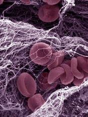
Credit: Kevin MacKenzie
An investigational anticoagulant reversal agent known as PER977 can restore hemostasis following edoxaban administration, according to a phase 1/2 trial.
The study included 80 healthy subjects who received PER977 or placebo 3 hours after they received a dose of edoxaban.
PER977 decreased anticoagulation to within 10% of baseline levels within 10 to 30 minutes of administration, an effect that took 12 to 15 hours in placebo-treated subjects.
Jack Ansell, MD, of Hofstra North Shore–LIJ School of Medicine in Hempstead, New York, and his colleagues reported these results in a letter to NEJM. The research was funded by Perosphere, the company developing PER977.
PER977 is a small molecule that can bind to unfractionated heparin, low-molecular-weight heparin, edoxaban, rivaroxaban, apixaban, and dabigatran. In preclinical studies, PER977 reversed anticoagulation with each of the new oral anticoagulants.
Dr Ansell and his colleagues wanted to determine the safety and efficacy of PER977 when given to healthy subjects after edoxaban. So the team enrolled 80 subjects and gave them placebo or single, escalating doses of PER977 (5 mg to 300 mg) 3 hours after a 60 mg dose of edoxaban.
After edoxaban administration, the mean whole-blood clotting time (WBCT) increased 37% from baseline. In placebo-treated subjects, it took 12 to 15 hours for the mean WBCT to return to within 10% above the baseline level.
In subjects who received PER977 at 100 mg to 300 mg, it took 10 minutes or less to reach the same level. And the WBCT remained within 10% above or below the baseline value for 24 hours, with no rebound and no infusions needed.
The researchers also assessed clots while measuring WBCT to determine the mean fibrin-fiber diameter. They found that edoxaban significantly reduced the mean fibrin-fiber diameter relative to baseline, from about 250 nm to about 125 nm (P<0.001).
But the mean fibrin-fiber diameter was restored to normal 30 minutes after subjects received 100 mg to 300 mg of PER977.
In addition, the researchers saw no evidence of procoagulant activity after PER977 administration, as assessed by levels of D-dimer, prothrombin fragment 1.2, and tissue factor pathway inhibitor.
The team noted a few adverse events that may have been related to PER977, including transient, mild perioral and facial flushing, dysgeusia, and moderate headache.
One subject experienced a moderate muscle cramp and elevation in creatinine phosphokinase levels, but the researchers believe this was not related to PER977.
“[T]he fact that PER977 was shown to be safe, well tolerated, and, most importantly, effective in reversing edoxaban, one of the new factor Xa oral anticoagulants, is a major step forward in developing a readily available and simple-to-use reversal agent for the new oral anticoagulants,” Dr Ansell said.
Credit: Kevin MacKenzie
An investigational anticoagulant reversal agent known as PER977 can restore hemostasis following edoxaban administration, according to a phase 1/2 trial.
The study included 80 healthy subjects who received PER977 or placebo 3 hours after they received a dose of edoxaban.
PER977 decreased anticoagulation to within 10% of baseline levels within 10 to 30 minutes of administration, an effect that took 12 to 15 hours in placebo-treated subjects.
Jack Ansell, MD, of Hofstra North Shore–LIJ School of Medicine in Hempstead, New York, and his colleagues reported these results in a letter to NEJM. The research was funded by Perosphere, the company developing PER977.
PER977 is a small molecule that can bind to unfractionated heparin, low-molecular-weight heparin, edoxaban, rivaroxaban, apixaban, and dabigatran. In preclinical studies, PER977 reversed anticoagulation with each of the new oral anticoagulants.
Dr Ansell and his colleagues wanted to determine the safety and efficacy of PER977 when given to healthy subjects after edoxaban. So the team enrolled 80 subjects and gave them placebo or single, escalating doses of PER977 (5 mg to 300 mg) 3 hours after a 60 mg dose of edoxaban.
After edoxaban administration, the mean whole-blood clotting time (WBCT) increased 37% from baseline. In placebo-treated subjects, it took 12 to 15 hours for the mean WBCT to return to within 10% above the baseline level.
In subjects who received PER977 at 100 mg to 300 mg, it took 10 minutes or less to reach the same level. And the WBCT remained within 10% above or below the baseline value for 24 hours, with no rebound and no infusions needed.
The researchers also assessed clots while measuring WBCT to determine the mean fibrin-fiber diameter. They found that edoxaban significantly reduced the mean fibrin-fiber diameter relative to baseline, from about 250 nm to about 125 nm (P<0.001).
But the mean fibrin-fiber diameter was restored to normal 30 minutes after subjects received 100 mg to 300 mg of PER977.
In addition, the researchers saw no evidence of procoagulant activity after PER977 administration, as assessed by levels of D-dimer, prothrombin fragment 1.2, and tissue factor pathway inhibitor.
The team noted a few adverse events that may have been related to PER977, including transient, mild perioral and facial flushing, dysgeusia, and moderate headache.
One subject experienced a moderate muscle cramp and elevation in creatinine phosphokinase levels, but the researchers believe this was not related to PER977.
“[T]he fact that PER977 was shown to be safe, well tolerated, and, most importantly, effective in reversing edoxaban, one of the new factor Xa oral anticoagulants, is a major step forward in developing a readily available and simple-to-use reversal agent for the new oral anticoagulants,” Dr Ansell said.
Credit: Kevin MacKenzie
An investigational anticoagulant reversal agent known as PER977 can restore hemostasis following edoxaban administration, according to a phase 1/2 trial.
The study included 80 healthy subjects who received PER977 or placebo 3 hours after they received a dose of edoxaban.
PER977 decreased anticoagulation to within 10% of baseline levels within 10 to 30 minutes of administration, an effect that took 12 to 15 hours in placebo-treated subjects.
Jack Ansell, MD, of Hofstra North Shore–LIJ School of Medicine in Hempstead, New York, and his colleagues reported these results in a letter to NEJM. The research was funded by Perosphere, the company developing PER977.
PER977 is a small molecule that can bind to unfractionated heparin, low-molecular-weight heparin, edoxaban, rivaroxaban, apixaban, and dabigatran. In preclinical studies, PER977 reversed anticoagulation with each of the new oral anticoagulants.
Dr Ansell and his colleagues wanted to determine the safety and efficacy of PER977 when given to healthy subjects after edoxaban. So the team enrolled 80 subjects and gave them placebo or single, escalating doses of PER977 (5 mg to 300 mg) 3 hours after a 60 mg dose of edoxaban.
After edoxaban administration, the mean whole-blood clotting time (WBCT) increased 37% from baseline. In placebo-treated subjects, it took 12 to 15 hours for the mean WBCT to return to within 10% above the baseline level.
In subjects who received PER977 at 100 mg to 300 mg, it took 10 minutes or less to reach the same level. And the WBCT remained within 10% above or below the baseline value for 24 hours, with no rebound and no infusions needed.
The researchers also assessed clots while measuring WBCT to determine the mean fibrin-fiber diameter. They found that edoxaban significantly reduced the mean fibrin-fiber diameter relative to baseline, from about 250 nm to about 125 nm (P<0.001).
But the mean fibrin-fiber diameter was restored to normal 30 minutes after subjects received 100 mg to 300 mg of PER977.
In addition, the researchers saw no evidence of procoagulant activity after PER977 administration, as assessed by levels of D-dimer, prothrombin fragment 1.2, and tissue factor pathway inhibitor.
The team noted a few adverse events that may have been related to PER977, including transient, mild perioral and facial flushing, dysgeusia, and moderate headache.
One subject experienced a moderate muscle cramp and elevation in creatinine phosphokinase levels, but the researchers believe this was not related to PER977.
“[T]he fact that PER977 was shown to be safe, well tolerated, and, most importantly, effective in reversing edoxaban, one of the new factor Xa oral anticoagulants, is a major step forward in developing a readily available and simple-to-use reversal agent for the new oral anticoagulants,” Dr Ansell said.
Aspartame, sweetened drinks don’t increase risk of NHL

beverages at the supermarket
Consuming aspartame and drinking sweetened beverages do not increase a person’s risk of developing non-Hodgkin lymphoma (NHL), according to research published in the Journal of Nutrition.
Investigators analyzed information from more than 100,000 men and women in the US.
The results suggested that neither aspartame intake nor the consumption of sugar-sweetened or artificially sweetened beverages were associated with an increased risk of NHL.
Marjorie L. McCullough, SCD, RD, of the American Cancer Society in Atlanta, Georgia, and her colleagues conducted this research, analyzing data from the nutrition cohort of the Cancer Prevention Study II, an assessment of cancer incidence
and mortality in the US.
Study subjects first completed a questionnaire in 1992, noting information related to diet and other lifestyle factors. They completed follow-up questionnaires in 1999 and 2003, which included questions related to the consumption of sugar-sweetened and artificially sweetened beverages, as well as tabletop sweeteners containing aspartame.
Among the 100,442 adult men and women who provided information on diet and lifestyle factors in 1999, there were 1196 NHL cases verified during a 10-year follow-up period.
The investigators assessed the risk of NHL associated with sweetened beverage and aspartame consumption, adjusted for the subjects’ smoking status, body mass index, and history of diabetes.
The analysis revealed that, in women and men combined, there was no association between NHL risk and the consumption of 1 or more servings (355 mL) of artificially sweetened beverages. Compared to nondrinkers, subjects who drank artificially sweetened beverages had a risk ratio (RR) of 0.92 (P=0.14).
Similarly, there was no association between NHL risk and sugar-sweetened beverages. Compared to nondrinkers, the RR for sugar-sweetened beverage drinkers was 1.10 (P=0.62).
Furthermore, subjects’ overall aspartame intake, which was estimated from artificially sweetened carbonated beverage consumption and the use of aspartame packets, was not associated with NHL risk.
The RR was 1.02 (P=0.69) for the top quintile (which had a median aspartame intake of 145 mg per day) vs the bottom quintile (which had a median aspartame intake of 0 mg per day).
The investigators also found that associations between disease and sweetened beverage consumption or aspartame intake were generally null for specific NHL subtypes, including multiple myeloma, diffuse large B-cell lymphoma, chronic lymphocytic leukemia/small lymphocytic lymphoma, follicular lymphoma, and other B-cell lymphomas.
“The study supports the decades of research that have continued to find that aspartame is safe for use in foods and beverages,” said Haley Stevens, PhD, President of the Calorie Control Council. “It also supports the conclusions of the National Cancer Institute, who have determined that aspartame does not increase a person’s risk of developing cancer.”
beverages at the supermarket
Consuming aspartame and drinking sweetened beverages do not increase a person’s risk of developing non-Hodgkin lymphoma (NHL), according to research published in the Journal of Nutrition.
Investigators analyzed information from more than 100,000 men and women in the US.
The results suggested that neither aspartame intake nor the consumption of sugar-sweetened or artificially sweetened beverages were associated with an increased risk of NHL.
Marjorie L. McCullough, SCD, RD, of the American Cancer Society in Atlanta, Georgia, and her colleagues conducted this research, analyzing data from the nutrition cohort of the Cancer Prevention Study II, an assessment of cancer incidence
and mortality in the US.
Study subjects first completed a questionnaire in 1992, noting information related to diet and other lifestyle factors. They completed follow-up questionnaires in 1999 and 2003, which included questions related to the consumption of sugar-sweetened and artificially sweetened beverages, as well as tabletop sweeteners containing aspartame.
Among the 100,442 adult men and women who provided information on diet and lifestyle factors in 1999, there were 1196 NHL cases verified during a 10-year follow-up period.
The investigators assessed the risk of NHL associated with sweetened beverage and aspartame consumption, adjusted for the subjects’ smoking status, body mass index, and history of diabetes.
The analysis revealed that, in women and men combined, there was no association between NHL risk and the consumption of 1 or more servings (355 mL) of artificially sweetened beverages. Compared to nondrinkers, subjects who drank artificially sweetened beverages had a risk ratio (RR) of 0.92 (P=0.14).
Similarly, there was no association between NHL risk and sugar-sweetened beverages. Compared to nondrinkers, the RR for sugar-sweetened beverage drinkers was 1.10 (P=0.62).
Furthermore, subjects’ overall aspartame intake, which was estimated from artificially sweetened carbonated beverage consumption and the use of aspartame packets, was not associated with NHL risk.
The RR was 1.02 (P=0.69) for the top quintile (which had a median aspartame intake of 145 mg per day) vs the bottom quintile (which had a median aspartame intake of 0 mg per day).
The investigators also found that associations between disease and sweetened beverage consumption or aspartame intake were generally null for specific NHL subtypes, including multiple myeloma, diffuse large B-cell lymphoma, chronic lymphocytic leukemia/small lymphocytic lymphoma, follicular lymphoma, and other B-cell lymphomas.
“The study supports the decades of research that have continued to find that aspartame is safe for use in foods and beverages,” said Haley Stevens, PhD, President of the Calorie Control Council. “It also supports the conclusions of the National Cancer Institute, who have determined that aspartame does not increase a person’s risk of developing cancer.”
beverages at the supermarket
Consuming aspartame and drinking sweetened beverages do not increase a person’s risk of developing non-Hodgkin lymphoma (NHL), according to research published in the Journal of Nutrition.
Investigators analyzed information from more than 100,000 men and women in the US.
The results suggested that neither aspartame intake nor the consumption of sugar-sweetened or artificially sweetened beverages were associated with an increased risk of NHL.
Marjorie L. McCullough, SCD, RD, of the American Cancer Society in Atlanta, Georgia, and her colleagues conducted this research, analyzing data from the nutrition cohort of the Cancer Prevention Study II, an assessment of cancer incidence
and mortality in the US.
Study subjects first completed a questionnaire in 1992, noting information related to diet and other lifestyle factors. They completed follow-up questionnaires in 1999 and 2003, which included questions related to the consumption of sugar-sweetened and artificially sweetened beverages, as well as tabletop sweeteners containing aspartame.
Among the 100,442 adult men and women who provided information on diet and lifestyle factors in 1999, there were 1196 NHL cases verified during a 10-year follow-up period.
The investigators assessed the risk of NHL associated with sweetened beverage and aspartame consumption, adjusted for the subjects’ smoking status, body mass index, and history of diabetes.
The analysis revealed that, in women and men combined, there was no association between NHL risk and the consumption of 1 or more servings (355 mL) of artificially sweetened beverages. Compared to nondrinkers, subjects who drank artificially sweetened beverages had a risk ratio (RR) of 0.92 (P=0.14).
Similarly, there was no association between NHL risk and sugar-sweetened beverages. Compared to nondrinkers, the RR for sugar-sweetened beverage drinkers was 1.10 (P=0.62).
Furthermore, subjects’ overall aspartame intake, which was estimated from artificially sweetened carbonated beverage consumption and the use of aspartame packets, was not associated with NHL risk.
The RR was 1.02 (P=0.69) for the top quintile (which had a median aspartame intake of 145 mg per day) vs the bottom quintile (which had a median aspartame intake of 0 mg per day).
The investigators also found that associations between disease and sweetened beverage consumption or aspartame intake were generally null for specific NHL subtypes, including multiple myeloma, diffuse large B-cell lymphoma, chronic lymphocytic leukemia/small lymphocytic lymphoma, follicular lymphoma, and other B-cell lymphomas.
“The study supports the decades of research that have continued to find that aspartame is safe for use in foods and beverages,” said Haley Stevens, PhD, President of the Calorie Control Council. “It also supports the conclusions of the National Cancer Institute, who have determined that aspartame does not increase a person’s risk of developing cancer.”
Study reveals gap in patient blood management
PHILADELPHIA—Healthcare professionals may not be using blood management interventions in a majority of patients receiving red blood cell (RBC) transfusions, a large study suggests.
The research showed that 72 US hospitals have made strides in reducing the use of RBCs in patients undergoing orthopedic and cardiac surgery.
And smaller reductions have occurred in patients with gastrointestinal bleeding, obstetric patients, and those receiving bone marrow transplants or inpatient chemotherapy.
However, more than 60% of the transfusions studied were given to patients who did not belong to any of the aforementioned groups.
“So when we target our [patient blood management] interventions to these kind of surgical procedures, in fact, we’re looking at only about 40% of the red cell use, and 60% goes to a myriad of other patients,” said Robert L. Thurer, MD, of Haemonetics in Braintree, Massachusetts.
He added that the groups of specific patient populations within that 60% were so small—“2% of patients here and 3% of patients there”—that it was too difficult to examine them individually in this study.
Dr Thurer presented this research at the AABB Annual Meeting 2014 (abstract S65-030K).
He and his colleagues wanted to determine which specific groups of patients have been most affected by patient blood management, understand further opportunities for decreasing transfusion use, and project future blood needs.
So the researchers analyzed data from 3,946,428 inpatients at 72 US hospitals, comparing the use of RBC transfusions in 2009/2010 to use in 2013.
In 2009/2010, there were 1,378,581 patients admitted to the hospitals, the RBC transfusion rate was 11.5.%, and the utilization (total units/total patients) was 0.41. In 2013, there were 861,804 patients, the transfusion rate was 10%, and the utilization was 0.34.
So from 2009/2010 to 2013, there was a 13% reduction in transfusion rate and a 17% reduction in utilization.
The greatest decrease was in orthopedic surgery patients. In those undergoing hip and knee joint replacement surgery, there was a 45% reduction in transfusion rate and a 43% reduction in utilization. In patients with a hip fracture, there was a 20% decrease in transfusion rate and a 20% decrease in utilization.
There was a smaller, though still sizable, reduction in transfusion use among patients undergoing cardiac surgery—a 15% decrease in transfusion rate and an 18% decrease in utilization.
“My personal thoughts about this is that it represents, certainly, lower transfusion triggers, which are becoming more widely adopted, [and] the use of antifibrinolytic drugs, particularly in orthopedic surgeries,” Dr Thurer said.
“We like to think that comprehensive coagulation testing and better matching of coagulation abnormalities contributes to this. Perhaps correction of preoperative anemia [contributes] for elective patients. And, clearly, surgical techniques have evolved, and, as they do, blood loss goes down.”
Reductions in RBC use were also seen in patients with gastrointestinal bleeding, where there was a 3% decrease in transfusion rate and a 13% decrease in utilization.
“For gastrointestinal bleeding, I think the lower transfusion triggers [have made an impact], but there’s also . . . more interest in timely interventions to stop bleeding,” Dr Thurer said.
“So rather than the gastroenterologist saying, ‘Correct the hematocrit and the coagulation factors, and I’ll stop the bleeding in the morning,’ we’re seeing now much more interest in very prompt endoscopy to stop bleeding. And as you know, the way to stop giving transfusions is to stop the patient from bleeding.”
Obstetric patients saw a 5% reduction in transfusion rate and an 8% reduction in utilization. And patients undergoing bone marrow transplant or inpatient chemotherapy saw a 6% decrease in transfusion rate and an 8% decrease in utilization.
Other transplant patients actually saw an increase in RBC transfusions. In liver transplant recipients, there was a 2.2% increase in transfusion rate and a 6% increase in utilization. And in kidney transplant recipients, there was a 0.2% increase in transfusion rate and a 19% increase in utilization.
However, Dr Thurer noted that the majority of RBC transfusions are administered to patients outside of these groups. In 2013, 60.6% of transfusions went to patients who did not fit into any of the aforementioned categories.
“So clearly,” he concluded, “further studies are needed to determine whether these reductions that we’ve seen in some areas can be implemented in a wider variety of patients.”
PHILADELPHIA—Healthcare professionals may not be using blood management interventions in a majority of patients receiving red blood cell (RBC) transfusions, a large study suggests.
The research showed that 72 US hospitals have made strides in reducing the use of RBCs in patients undergoing orthopedic and cardiac surgery.
And smaller reductions have occurred in patients with gastrointestinal bleeding, obstetric patients, and those receiving bone marrow transplants or inpatient chemotherapy.
However, more than 60% of the transfusions studied were given to patients who did not belong to any of the aforementioned groups.
“So when we target our [patient blood management] interventions to these kind of surgical procedures, in fact, we’re looking at only about 40% of the red cell use, and 60% goes to a myriad of other patients,” said Robert L. Thurer, MD, of Haemonetics in Braintree, Massachusetts.
He added that the groups of specific patient populations within that 60% were so small—“2% of patients here and 3% of patients there”—that it was too difficult to examine them individually in this study.
Dr Thurer presented this research at the AABB Annual Meeting 2014 (abstract S65-030K).
He and his colleagues wanted to determine which specific groups of patients have been most affected by patient blood management, understand further opportunities for decreasing transfusion use, and project future blood needs.
So the researchers analyzed data from 3,946,428 inpatients at 72 US hospitals, comparing the use of RBC transfusions in 2009/2010 to use in 2013.
In 2009/2010, there were 1,378,581 patients admitted to the hospitals, the RBC transfusion rate was 11.5.%, and the utilization (total units/total patients) was 0.41. In 2013, there were 861,804 patients, the transfusion rate was 10%, and the utilization was 0.34.
So from 2009/2010 to 2013, there was a 13% reduction in transfusion rate and a 17% reduction in utilization.
The greatest decrease was in orthopedic surgery patients. In those undergoing hip and knee joint replacement surgery, there was a 45% reduction in transfusion rate and a 43% reduction in utilization. In patients with a hip fracture, there was a 20% decrease in transfusion rate and a 20% decrease in utilization.
There was a smaller, though still sizable, reduction in transfusion use among patients undergoing cardiac surgery—a 15% decrease in transfusion rate and an 18% decrease in utilization.
“My personal thoughts about this is that it represents, certainly, lower transfusion triggers, which are becoming more widely adopted, [and] the use of antifibrinolytic drugs, particularly in orthopedic surgeries,” Dr Thurer said.
“We like to think that comprehensive coagulation testing and better matching of coagulation abnormalities contributes to this. Perhaps correction of preoperative anemia [contributes] for elective patients. And, clearly, surgical techniques have evolved, and, as they do, blood loss goes down.”
Reductions in RBC use were also seen in patients with gastrointestinal bleeding, where there was a 3% decrease in transfusion rate and a 13% decrease in utilization.
“For gastrointestinal bleeding, I think the lower transfusion triggers [have made an impact], but there’s also . . . more interest in timely interventions to stop bleeding,” Dr Thurer said.
“So rather than the gastroenterologist saying, ‘Correct the hematocrit and the coagulation factors, and I’ll stop the bleeding in the morning,’ we’re seeing now much more interest in very prompt endoscopy to stop bleeding. And as you know, the way to stop giving transfusions is to stop the patient from bleeding.”
Obstetric patients saw a 5% reduction in transfusion rate and an 8% reduction in utilization. And patients undergoing bone marrow transplant or inpatient chemotherapy saw a 6% decrease in transfusion rate and an 8% decrease in utilization.
Other transplant patients actually saw an increase in RBC transfusions. In liver transplant recipients, there was a 2.2% increase in transfusion rate and a 6% increase in utilization. And in kidney transplant recipients, there was a 0.2% increase in transfusion rate and a 19% increase in utilization.
However, Dr Thurer noted that the majority of RBC transfusions are administered to patients outside of these groups. In 2013, 60.6% of transfusions went to patients who did not fit into any of the aforementioned categories.
“So clearly,” he concluded, “further studies are needed to determine whether these reductions that we’ve seen in some areas can be implemented in a wider variety of patients.”
PHILADELPHIA—Healthcare professionals may not be using blood management interventions in a majority of patients receiving red blood cell (RBC) transfusions, a large study suggests.
The research showed that 72 US hospitals have made strides in reducing the use of RBCs in patients undergoing orthopedic and cardiac surgery.
And smaller reductions have occurred in patients with gastrointestinal bleeding, obstetric patients, and those receiving bone marrow transplants or inpatient chemotherapy.
However, more than 60% of the transfusions studied were given to patients who did not belong to any of the aforementioned groups.
“So when we target our [patient blood management] interventions to these kind of surgical procedures, in fact, we’re looking at only about 40% of the red cell use, and 60% goes to a myriad of other patients,” said Robert L. Thurer, MD, of Haemonetics in Braintree, Massachusetts.
He added that the groups of specific patient populations within that 60% were so small—“2% of patients here and 3% of patients there”—that it was too difficult to examine them individually in this study.
Dr Thurer presented this research at the AABB Annual Meeting 2014 (abstract S65-030K).
He and his colleagues wanted to determine which specific groups of patients have been most affected by patient blood management, understand further opportunities for decreasing transfusion use, and project future blood needs.
So the researchers analyzed data from 3,946,428 inpatients at 72 US hospitals, comparing the use of RBC transfusions in 2009/2010 to use in 2013.
In 2009/2010, there were 1,378,581 patients admitted to the hospitals, the RBC transfusion rate was 11.5.%, and the utilization (total units/total patients) was 0.41. In 2013, there were 861,804 patients, the transfusion rate was 10%, and the utilization was 0.34.
So from 2009/2010 to 2013, there was a 13% reduction in transfusion rate and a 17% reduction in utilization.
The greatest decrease was in orthopedic surgery patients. In those undergoing hip and knee joint replacement surgery, there was a 45% reduction in transfusion rate and a 43% reduction in utilization. In patients with a hip fracture, there was a 20% decrease in transfusion rate and a 20% decrease in utilization.
There was a smaller, though still sizable, reduction in transfusion use among patients undergoing cardiac surgery—a 15% decrease in transfusion rate and an 18% decrease in utilization.
“My personal thoughts about this is that it represents, certainly, lower transfusion triggers, which are becoming more widely adopted, [and] the use of antifibrinolytic drugs, particularly in orthopedic surgeries,” Dr Thurer said.
“We like to think that comprehensive coagulation testing and better matching of coagulation abnormalities contributes to this. Perhaps correction of preoperative anemia [contributes] for elective patients. And, clearly, surgical techniques have evolved, and, as they do, blood loss goes down.”
Reductions in RBC use were also seen in patients with gastrointestinal bleeding, where there was a 3% decrease in transfusion rate and a 13% decrease in utilization.
“For gastrointestinal bleeding, I think the lower transfusion triggers [have made an impact], but there’s also . . . more interest in timely interventions to stop bleeding,” Dr Thurer said.
“So rather than the gastroenterologist saying, ‘Correct the hematocrit and the coagulation factors, and I’ll stop the bleeding in the morning,’ we’re seeing now much more interest in very prompt endoscopy to stop bleeding. And as you know, the way to stop giving transfusions is to stop the patient from bleeding.”
Obstetric patients saw a 5% reduction in transfusion rate and an 8% reduction in utilization. And patients undergoing bone marrow transplant or inpatient chemotherapy saw a 6% decrease in transfusion rate and an 8% decrease in utilization.
Other transplant patients actually saw an increase in RBC transfusions. In liver transplant recipients, there was a 2.2% increase in transfusion rate and a 6% increase in utilization. And in kidney transplant recipients, there was a 0.2% increase in transfusion rate and a 19% increase in utilization.
However, Dr Thurer noted that the majority of RBC transfusions are administered to patients outside of these groups. In 2013, 60.6% of transfusions went to patients who did not fit into any of the aforementioned categories.
“So clearly,” he concluded, “further studies are needed to determine whether these reductions that we’ve seen in some areas can be implemented in a wider variety of patients.”