User login
Breast cancer drug may also work in MCL, myeloma
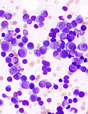
showing multiple myeloma
NEW YORK—Targeting the cell cycle with cyclin-dependent kinase (CDK) inhibitors may be an effective strategy to treat lymphoma and myeloma, according to a presentation at Lymphoma & Myeloma 2015.
Palbociclib, an inhibitor of CDK4 and CDK6, received accelerated approval from the US Food and Drug Administration to treat advanced breast cancer.
Now, it is showing promise in mantle cell lymphoma (MCL) and multiple myeloma (MM) as well.
CDK family members are important regulators of cell-cycle progression. Dysregulation of CDK4 and CDK6 is one of the most common genomic aberrations in human cancer, including myeloma, lymphoma, leukemia, breast cancer, metastatic lung adenocarcinoma, and glioblastoma.
MCL, which accounts for 6% of non-Hodgkin lymphomas, has an overall poor prognosis, with most patients eventually becoming resistant to drugs. MCL expresses cyclin D1 as a consequence of the t(11;14) translocation and overexpresses CDK4.
“So this is a perfect disease for the development of targeting CDK4,” said Selina Chen-Kiang, PhD, of Weill Cornell Medical College in New York, New York, who presented this information at the meeting.
CDK4 and CDK6 signaling occur at the beginning of the cell cycle and bring the cell from the resting state into early G2.
“If we could control that,” Dr Chen-Kiang explained, “we reasoned that we could control the DNA replication and cell division” and increase tumor-specific cell death.
Palbociclib (PD0332991; Ibrance), an orally bioavailable, selective CDK4/CDK6 inhibitor, induces early G1 arrest. It is reversible and low in toxicity, according to Dr Chen-Kiang.
Currently, it’s being tested in phase 1 trials in MCL with ibrutinib and in MM with lenalidomide-dexamethasone. Phase 1 trials have also been completed at Cornell with palbociclib as a single agent in MCL, with bortezomib in MCL, and with bortezomib-dexamethasone in MM.
Palbociclib in MCL
Investigators conducted a phase 1, single-agent study of palbociclib to determine whether it could be tolerated in humans.
“CDK4/CDK6 is expressed in every cell,” Dr Chen-Kiang noted, “and you are targeting 2 proteins that are needed for every cell.”
Seventeen patients received 125 mg of palbociclib per day for 21 of 28 days.
“And surprisingly,” Dr Chen-Kiang said, “it’s very well tolerated.”
The most common adverse events were neutropenia, fatigue, and diarrhea.
“And even more surprising,” she added “is that we actually had a complete response (CR) [and] 2 partial responses, in addition to 5 stable diseases.”
The investigators hypothesized that blocking the cell cycle in G1 creates an imbalance in gene expression.
They then conducted a trial of palbociclib plus bortezomib in 17 patients with recurrent MCL. Patients received palbociclib on days 1–12 and low-dose bortezomib on days 8, 11, 15, and 18.
The palbociclib dose ranged from 75 mg to 125 mg, and the bortezomib dose ranged from 1.0 mg/m2 to 1.3 mg/m2.
Eleven patients experienced a reduction in tumor volume, the majority at the 125-mg dose.
Using whole-exome and whole-transcriptome sequencing of serial biopsies, investigators determined that CDK4/CDK6 inhibition induces early G1 arrest in MCL cells in both responders and non-responders initially.
This may occur because the cell cycle is perfectly controlled, and there’s no mutation in CDK4 in any of the patients.
Investigators attempted to identify genes that could differentiate sensitivity from resistance to CDK4 targeting. They found that a very small number of genes display opposite regulation in prolonged early G1 arrest in responding versus non-responding patients.
These genes are involved in metabolism and redox homeostasis and include a gene called PIK3IP1, an inhibitor of PI3 kinase.
In earlier analyses, the investigators had discovered a relapse-specific C481S mutation at the ibrutinib binding site of Bruton’s tyrosine kinase (BTK) in MCL cells. The mutation occurred at progression following a durable response to ibrutinib.
The mutation is absent, however, in patients with transient ibrutinib responses or in primary resistance.
The team observed that early cell-cycle arrest by CDK4 inhibition reprograms MCL cells for killing by ibrutinib. This occurs through inhibition of BTK and AKT (protein kinase B).
So the investigators undertook to study palbociclib in combination with ibrutinib, “with extraordinary results,” Dr Chen-Kiang said.
The trial is ongoing and, at the moment, has a 60% CR rate with durable responses.
“We’re very happy with this,” Dr Chen-Kiang said.
Palbociclib in MM
In addition to the phase 1/2 study of palbociclib with bortezomib and dexamethasone, investigators are also pursuing palbociclib in combination with lenalidomide and dexamethasone, which Dr Chen-Kiang briefly elaborated upon.
Lenalidomide rarely produces a complete remission on its own, she explained, so the team decided to study palbociclib in combination with immunomodulatory drugs in MM (NCT02030483).
The strategy is to prime the cell cycle with palbociclib and then use lenalidomide and dexamethasone to increase efficacy.
Twenty patients have received this combination thus far, and investigators found that palbociclib enhances the activity of lenalidomide in the killing of primary bone marrow myeloma cells.
Lenalidomide reduces the MEIS2/CRBN ratio in these cells. The drug changes the ratio, reduces the blocker, and allows the CRBN to work.
“The whole principle here is to control the gene coupling to the cell cycle and induce imbalance in gene expression,” Dr Chen-Kiang said. “And this weakens the tumor cells.”
Two-thirds of the samples respond to palboclib, she said, and those patients go on to be treated with lenalidomide or pomalidomide.
One third will not respond, but their in vivo clinical response and the ex vivo responders’ purified cells mimic one another.
“Now, this is very exciting to us,” she said.
The combination study with lenalidomide and dexamethasone is currently underway.
Palbociclib is being developed by Pfizer. ![]()
RIC can improve HSCT outcomes in older AML patients
![]()
Photo by Chad McNeeley
Results of a phase 2 study suggest that reduced-intensity conditioning (RIC) may allow older patients with acute myeloid leukemia (AML) to remain in long-term remission after allogeneic hematopoietic stem cell transplant (HSCT).
The study included patients age 60 and older who were in their first complete remission (CR1) at transplant.
Two years later, the probability of overall survival was 48%, and the probability of disease-free survival was 42%.
“This new data offers strong support against using biological age as a limiting factor for stem cell transplantation in AML patients who are otherwise well positioned to tolerate and achieve long-term remission with this approach,” said Steven Devine, MD, of The Ohio State University in Columbus.
He and his colleagues shared the data in the Journal of Clinical Oncology.
The researchers wanted to determine whether RIC could improve long-term remission rates after HSCT for older patients with AML.
So the team studied 114 patients with a median age of 65 (range, 60-74) who were treated at 21 US hospitals between November 2004 and November 2011. Most patients were male (62%), and most had intermediate cytogenetics (70%).
All of the patients were in CR1 according to International Working Group criteria. The CR had to be achieved after no more than 2 cycles of induction chemotherapy, and patients were required to undergo HSCT within 6 months of the initial documentation of morphologic CR.
The median time from CR1 documentation to HSCT was 85 days (range, 9-184), and the median time from AML diagnosis to HSCT was 138 days (range, 61-265).
All patients received RIC (fludarabine followed by busulfan) prior to HSCT, which essentially cut treatment strength by half compared to traditional high-dose conditioning. The patients received tacrolimus and methotrexate as graft-versus-host disease (GVHD) prophylaxis.
Forty-eight percent of patients underwent HSCT from a matched, related donor, and 52% had a matched, unrelated donor.
At 2 years, disease-free survival was 42%, and overall survival was 48%. Among patients with unrelated donors, disease-free survival was 40%, and overall survival was 50%.
The cumulative incidence of relapse was 44%, and non-relapse mortality was 15%. Twenty-eight percent of patients had chronic GVHD, and 9.6% had grade 2-4 acute GVHD.
“Close to half of the patients treated in this study achieved long-term, cancer-free survival after 2 years,” Dr Devine noted. “These outcomes are similar to what we would expect to see in younger patients and appear to be better results than those that can be achieved with conventional chemotherapy-based approaches typically used in AML patients over 60.” ![]()
HSC self-renewal depends on surroundings

in the bone marrow
Scientists say that, using a model of the hematopoietic system, they have determined which signaling pathways play an essential role in the self-renewal of hematopoietic stem cells (HSCs).
They found that a particularly important role in this process is the interactive communication with surrounding tissue cells in the bone marrow.
Robert Oostendorp, PhD, of Klinikum Rechts der Isar der Technischen Universität München in Munich, Germany, and his colleagues described these findings in Stem Cell Reports.
The team noted that, in steady-state conditions, HSCs are maintained as slow-dividing clones of quiescent cells. However, when stress occurs, such as an accident that leads to substantial blood loss or the defense against a pathogen requires more blood cells in the course of an infection, HSCs are activated.
In response, the entire hematopoietic system switches from “standby” mode into a state of alert. The activated HSCs generate new blood cells to counteract the blood loss or combat the pathogen. At the same time, self-renewal keeps the stem cell pool replenished.
This switch is accompanied by a complex communication process between the HSCs and tissue cells—an area that had not previously been examined in depth.
“In our study, we set out to establish which tissue signals are important to stem cell maintenance and functionality, and which HSC signals influence the microenvironment,” Dr Oostendorp said.
He and his colleagues used mixed cultures of tissue cells and HSCs to investigate how these cell types interact.
The scientists analyzed factors that are upregulated or downregulated in the interplay between tissue cells and HSCs. And they linked these findings with the signaling pathways described in existing literature.
The team then consolidated this information in a bioinformatics computer model. And they conducted extensive cell experiments to confirm the computer-generated signaling pathway model.
“The outcome was very interesting indeed,” Dr Oostendorp said. “The entire system operates in a feedback loop. In ‘alert’ mode, the stem cells first influence the behavior of the tissue cells, which, in turn, impact on the stem cells, triggering the self-renewal step.”
In alert mode, HSCs emit signaling substances, which induce tissue cells to release the connective tissue growth factor (CTGF) messenger. This is essential to maintain the HSCs through self-renewal. In the absence of CTGF, HSCs age and cannot replenish the stem cell pool.
“Our findings could prove significant in treating leukemia,” Dr Oostendorp noted. “In this condition, the stem cells are hyperactive, and their division is unchecked. Leukemic blood cells are in a constant state of alert, so we would expect a similar interplay with the tissue cells.”
To date, however, the focus here has been limited to stem cells as the actual source of the defect.
“Given what we know now about feedback loops, it would be important to integrate the surrounding cells in therapeutic approaches too, since they exert a strong influence on stem cell division,” Dr Oostendorp concluded. ![]()
Experiment reveals new method of RBC production
Photo courtesy of
Josh Barney/University of
Virginia Health System
An unexpected result of a lab experiment has led researchers to a new way to trigger red blood cell (RBC) production.
They found that engaging a stress receptor on type 1 conventional dendritic cells can induce stress erythropoiesis in mice.
The team believes that, eventually, this method could be used to turn on RBC production in humans when necessary, perhaps to replace a blood transfusion or as a stop-gap measure when a transfusion is delayed.
Thomas J. Braciale, MD, PhD, of the University of Virginia in Charlottesville, and his colleagues conducted this research and reported the results in The Journal of Clinical Investigation.
The team was not investigating RBC production when they made their discovery. They were looking into the role of dendritic cells in the lungs.
Dendritic cells have traditionally been thought to be sensors of infection and inflammation, but a lab test involving the flu virus produced an unexpected effect in mice that ultimately revealed a new aspect to the cells’ function.
The researchers injected mice with the flu virus and an αCD24 monoclonal antibody, which resulted in splenomegaly. The team was baffled at this outcome, so they repeated the experiment, only to get the same results.
“We did it again, and I didn’t believe it, and we did it again, and I didn’t believe it,” Dr Braciale recalled. “I asked whether you needed flu to infect the mice when you injected this antibody. So the postdoc did the experiment, and he just injected the antibody without flu-injecting the mice—giant spleens. After much consultation, after talking with my colleagues in pathology, we decided we were inducing stress erythropoiesis.”
Specifically, the researchers found that engaging CD24 on type 1 conventional dendritic cells upregulates expression of the Kit ligand stem cell factor, which results in Kit-mediated proliferative expansion of early erythroid progenitors and transient reticulocytosis.
“In a very basic way, what we’ve discovered is that the process of regulating stress in the body is mediated—certainly in part, at least—by these dendritic cells,” Dr Braciale explained. “And stress can be a variety of different stresses.”
“It doesn’t have to be infection. It doesn’t have to be inflammation. It can be anemia. It can be hemorrhage. And these cells act to initiate this response that, until this report, there’s been really no evidence that [dendritic] cells ever participate in making red blood cells.”
More work is needed before this approach to RBC production can be tested in humans. However, Dr Braciale is optimistic, based on the findings so far.
“We’re very excited to see where this goes,” he said. “We know that the same things can be done in humans in the following sense. There are mice called humanized mice. These are mice that are engineered so they have a human blood system. And if you inject these mice with this antibody, they’ll make red blood cells.” ![]()
EC expands indication for azacitidine in AML

The European Commission (EC) has expanded the approved indication for azacitidine for injection (Vidaza) in acute myeloid leukemia (AML).
Now, the drug is approved to treat AML patients age 65 and older who are ineligible for hematopoietic stem cell transplant (HSCT) and have more than 30% myeloblasts according to the WHO classification.
Previously, HSCT-ineligible elderly AML patients could only receive azacitidine if they had less than 30% blasts.
Because this new indication for azacitidine is thought to bring significant clinical benefit in comparison with existing therapies, the drug will receive extended market protection in all its indications for an additional year throughout the European Economic Area.
In addition to the aforementioned AML indications, azacitidine is approved in the European Economic Area to treat HSCT-ineligible adults with intermediate-2- and high-risk myelodysplastic syndromes and HSCT-ineligible adults who have chronic myelomonocytic leukemia and 10%-29% marrow blasts without myeloproliferative disorder.
Azacitidine is marketed as Vidaza by Celgene.
AML-001 trial
The EC’s recommendation to expand the indication of azacitidine in AML was based on data from the AML-001 trial. This randomized study included patients age 65 and older with newly diagnosed or secondary AML with greater than 30% blasts.
Patients were pre-selected to receive 1 of 3 regimens per investigator’s choice. This included intensive chemotherapy (standard 7+3 regimen), low-dose cytarabine (20 mg subcutaneously twice a day for 10 days of each 28-day cycle), or best supportive care only.
Patients were then randomized to receive either azacitidine (75 mg/m2/day subcutaneously for 7 days of each 28-day cycle, n=241) or their predetermined conventional care regimen (CCR, n=247).
Median overall survival, the study’s primary endpoint, was 10.4 months for patients receiving azacitidine and 6.5 months for patients receiving CCR (hazard ratio=0.85, P=0.1009).
One-year survival rates with azacitidine and CCR were 46.5% and 34.2%, respectively.
Grade 3/4 anemia occurred in 16% of patients who received azacitidine, 5% who received best supportive care, 23% who received low-dose cytarabine, and 14% who received intensive chemotherapy.
Grade 3/4 neutropenia occurred in 26%, 5%, 25%, and 33% of patients, respectively. Grade 3/4 febrile neutropenia occurred in 28%, 28%, 30%, and 31%, respectively. And grade 3/4 thrombocytopenia occurred in 24%, 5%, 28%, and 21%, respectively. ![]()
France to lift lifetime ban on MSM blood donors
Photo by Михаило Јовановић
France is planning to lift its lifetime ban on blood donations from men who have sex with men (MSM), and the change is set to take effect next year.
French officials say that, beginning in Spring 2016, an MSM will be able to donate whole blood in France if he has not had sex with another man in the last 12
months.
An MSM will be allowed to donate plasma if he has been abstinent or had only 1 sexual partner in the last 4 months.
Experts will analyze the impact of this policy change for about a year. If there has been no increase in health risks, the policy may be relaxed further, and MSM donors may be treated the same as non-MSM donors.
Non-MSM individuals in France are deferred from donating blood if they have had more than 1 sexual partner in the last 4 months.
Studies recently conducted in Canada and the UK have suggested that lifting restrictions on MSM blood donors does not
increase the risk of sexually transmitted infections affecting the blood
supply.
France’s lifetime ban on MSM blood donation was enacted in 1983 in an attempt to counter the spread of HIV/AIDS.
Hundreds of people died in France in the 1980s after HIV-tainted blood was distributed by the French National Blood Transfusion Center. HIV-positive blood donations were exported as well, which led to hundreds of additional deaths in other countries.
Bans lifting worldwide
In lifting its lifetime ban on MSM blood donors, France is following the lead of other countries within and outside of Europe.
England, Scotland, Wales, Sweden, and the Netherlands have all lifted their bans on MSM blood donors and changed to a 12-month deferral period. Australia, Japan, and New Zealand also have 12-month deferral periods for MSM blood donors.
South Africa has a 6-month deferral period for MSMs, while Italy and Spain have no policy specific to MSM blood donors. All donors are screened for high-risk sexual practices, and deferrals are made accordingly.
The US and Canada are currently considering adopting a 12-month deferral policy for MSM blood donors. The lifetime ban is still in place in the US, but Canada has a 5-year deferral period for MSMs. ![]()
Survey: 3 in 10 MSMs don’t comply with UK blood donor policy
Photo by Marja Helander
ANAHEIM—A survey of UK blood donors suggests that as many as 30% of donors who are men who have sex with men (MSM) may not be compliant with the MSM blood donor policy.
The UK’s policy requires that MSMs do not donate blood if they have engaged in sexual activity with another male in the last 12 months.
But the survey indicates that as many as 3 in 10 MSMs are disregarding this policy.
The research also suggests that MSMs who do not comply with the policy engage in riskier sexual behavior than non-MSM male blood donors.
However, the researchers found no increase in the number of sexually transmitted infections present in the blood supply since the donation policy for MSMs changed from a lifetime ban to a 12-month deferral period.
The infections evaluated include human immunodeficiency virus (HIV), hepatitis C virus (HCV), hepatitis B virus (HBV), and syphilis.
The researchers also emphasized that the prevalence of HIV-positive blood donations in the UK remains low.
Katie Davidson, of Public Health England in London, presented these findings at the 2015 AABB Annual Meeting (abstract S36-030E*).
She noted that, in 2011, the blood services of England, Wales, and Scotland changed the blood donor policy for MSMs from a lifetime ban to a 12-month deferral since last male-to-male sex.
Before this policy change took effect, the blood services estimated that the change would mean 2679 MSMs would be newly eligible to donate blood (0.7% of male donors), and 8 of these donors would have HIV. So there would be a 0.5% increase in HIV risk.
“But what was clear was that these predictions in terms of HIV risk would be very dependent upon compliance,” Davidson said. “And what we mean by compliance is that a donor understands the rule, applies it correctly, and discloses any relevant information when they’re asked.”
To investigate donor behavior and compliance, Davidson and her colleagues conducted a large-scale, anonymous, web-based survey of blood donors.
Each month for 1 year (2013), all eligible new blood donors and at least an equal number of repeat blood donors in the UK were invited, via email, to complete an online questionnaire asking about their sexual history and compliance with the 12-month deferral policy for MSM (if applicable).
The researchers also looked at UK surveillance data on infections (HIV, HBV, HCV, and syphilis) in new and repeat blood donors over 6 years, comparing the incidence of infections before and after the policy change took effect (3 years pre- and post-change).
Donation and compliance
Among 65,439 survey respondents, 22,776 (35%) were male, and 242 (1%) were MSMs. Among the MSMs, 73 reported male-to-male sex within the last 12 months (non-compliance), and 181 said it had been more than 12 months since their last male-to-male sexual encounter.
The researchers adjusted these proportions for differences among the respondents and the donor population and extrapolated the data to the whole UK donor population.
The team estimated that, among 488,523 UK donors, there would be 5471 MSMs. Of the MSM donors, 3713 would be eligible under the new policy, and 1759 would be non-compliant.
So MSM compliance with the 12-month deferral policy would be 99.7% among all male donors but 70.4% of the MSM population.
“So 3 in every 10 MSMs donating blood in the UK shouldn’t be, [according to the estimates],” Davidson said.
The survey asked non-compliant MSM donors to provide their reasons for non-compliance, and many gave more than 1 reason.
“The reasons seemed to be associated, mostly, with self-assessment of their own risk [of transmitting infection] to be low,” Davidson said. “So that was based on the fact that they were in a monogamous relationship, they used condoms, practiced safe sex, or had regular [sexual health] screenings.”
However, there were some donors who regarded the policy as unimportant or said they didn’t agree with it. And there were some donors who didn’t declare their sexual behavior because they knew they wouldn’t be allowed to donate if they did.
Sexual behavior
Among all male respondents who reported having sex within the last 12 months, MSMs were more likely than men who had only female sexual partners to report having sex with more than 1 partner. Fifty percent of MSMs had more than 1 sexual partner in the last 12 months, as did 9.1% of male donors with only female sexual partners.
Ten percent of MSMs reported paying for sex, as did 0.3% of non-MSMs. None of the MSMs reported having a partner who was HIV-positive, and less than 0.1% of non-MSMs said they had an HIV-positive partner.
Eleven percent of MSMs said they had a history of sexually transmitted infection, as did less than 0.1% of non-MSMs.
“So among the responders, there was very low numbers who reported a high-risk partner in the last 12 months,” Davidson noted. “But there was some suggestion, among these low numbers, that this was more common in the MSMs than the non-MSMs.”
She also acknowledged that some donors were unsure about whether they had a high-risk partner in the last 12 months.
Infections
The UK surveillance data on infections encompassed HIV, HBV, HCV, and syphilis.
In all, 3,667,408 blood donations from males were tested for infection in the 3 years prior to the MSM donor policy change, and 3,066,076 donations were tested in the 3 years after the change was implemented.
There were 428 donors who reported having an infection risk before the change and 268 who did so after. There were 577 donors who actually had an infection before the change and 434 who did after. And there were 32 infected MSM donors before the change and 34 after.
“So the number of male donors fell post-change by approximately 20%, [and] the total number of infected donors . . . fell by almost 30%,” Davidson noted.
“However, the number of MSM infected donors marginally increased, [and] the proportion of male infected donors who were MSMs, among all of those who reported a risk, increased from 7% [32/428] to 13% [34/268]. So there seems to be some impact [on infection] from MSMs, but the numbers are very small, and these differences are not significant.”
Predictions and HIV infection
Finally, the researchers compared their predictions from before the MSM blood donor policy change to the actual data after the change. This comparison assumed that the absolute number of compliant MSMs did not change after the policy changed.
In 2007, the group predicted there would be about 2 million blood donations, including 2679 from MSMs. In reality, in 2014, there were 1.9 million blood donations, including 3126 from MSMs.
The researchers predicted the number of HIV-positive donations would be 30, including 8 from MSMs. In reality, in 2014, there were 13 HIV-positive donations, including 1 from an MSM.
So the predicted HIV prevalence per 100,000 donations was 1.4, and the actual HIV prevalence was 0.7. The predicted HIV incidence per 100,000 person-years was 0.9, and the actual HIV incidence was 0.7.
The predicted HIV risk was 0.022 per 100,000, and the actual HIV risk was 0.016 per 100,000.
“So the estimated risk of HIV post-change remains very low,” Davidson noted, adding that she and her colleagues will continue to monitor the impact of the policy change. ![]()
*Data in the abstract differ from data presented at the meeting.
Photo by Marja Helander
ANAHEIM—A survey of UK blood donors suggests that as many as 30% of donors who are men who have sex with men (MSM) may not be compliant with the MSM blood donor policy.
The UK’s policy requires that MSMs do not donate blood if they have engaged in sexual activity with another male in the last 12 months.
But the survey indicates that as many as 3 in 10 MSMs are disregarding this policy.
The research also suggests that MSMs who do not comply with the policy engage in riskier sexual behavior than non-MSM male blood donors.
However, the researchers found no increase in the number of sexually transmitted infections present in the blood supply since the donation policy for MSMs changed from a lifetime ban to a 12-month deferral period.
The infections evaluated include human immunodeficiency virus (HIV), hepatitis C virus (HCV), hepatitis B virus (HBV), and syphilis.
The researchers also emphasized that the prevalence of HIV-positive blood donations in the UK remains low.
Katie Davidson, of Public Health England in London, presented these findings at the 2015 AABB Annual Meeting (abstract S36-030E*).
She noted that, in 2011, the blood services of England, Wales, and Scotland changed the blood donor policy for MSMs from a lifetime ban to a 12-month deferral since last male-to-male sex.
Before this policy change took effect, the blood services estimated that the change would mean 2679 MSMs would be newly eligible to donate blood (0.7% of male donors), and 8 of these donors would have HIV. So there would be a 0.5% increase in HIV risk.
“But what was clear was that these predictions in terms of HIV risk would be very dependent upon compliance,” Davidson said. “And what we mean by compliance is that a donor understands the rule, applies it correctly, and discloses any relevant information when they’re asked.”
To investigate donor behavior and compliance, Davidson and her colleagues conducted a large-scale, anonymous, web-based survey of blood donors.
Each month for 1 year (2013), all eligible new blood donors and at least an equal number of repeat blood donors in the UK were invited, via email, to complete an online questionnaire asking about their sexual history and compliance with the 12-month deferral policy for MSM (if applicable).
The researchers also looked at UK surveillance data on infections (HIV, HBV, HCV, and syphilis) in new and repeat blood donors over 6 years, comparing the incidence of infections before and after the policy change took effect (3 years pre- and post-change).
Donation and compliance
Among 65,439 survey respondents, 22,776 (35%) were male, and 242 (1%) were MSMs. Among the MSMs, 73 reported male-to-male sex within the last 12 months (non-compliance), and 181 said it had been more than 12 months since their last male-to-male sexual encounter.
The researchers adjusted these proportions for differences among the respondents and the donor population and extrapolated the data to the whole UK donor population.
The team estimated that, among 488,523 UK donors, there would be 5471 MSMs. Of the MSM donors, 3713 would be eligible under the new policy, and 1759 would be non-compliant.
So MSM compliance with the 12-month deferral policy would be 99.7% among all male donors but 70.4% of the MSM population.
“So 3 in every 10 MSMs donating blood in the UK shouldn’t be, [according to the estimates],” Davidson said.
The survey asked non-compliant MSM donors to provide their reasons for non-compliance, and many gave more than 1 reason.
“The reasons seemed to be associated, mostly, with self-assessment of their own risk [of transmitting infection] to be low,” Davidson said. “So that was based on the fact that they were in a monogamous relationship, they used condoms, practiced safe sex, or had regular [sexual health] screenings.”
However, there were some donors who regarded the policy as unimportant or said they didn’t agree with it. And there were some donors who didn’t declare their sexual behavior because they knew they wouldn’t be allowed to donate if they did.
Sexual behavior
Among all male respondents who reported having sex within the last 12 months, MSMs were more likely than men who had only female sexual partners to report having sex with more than 1 partner. Fifty percent of MSMs had more than 1 sexual partner in the last 12 months, as did 9.1% of male donors with only female sexual partners.
Ten percent of MSMs reported paying for sex, as did 0.3% of non-MSMs. None of the MSMs reported having a partner who was HIV-positive, and less than 0.1% of non-MSMs said they had an HIV-positive partner.
Eleven percent of MSMs said they had a history of sexually transmitted infection, as did less than 0.1% of non-MSMs.
“So among the responders, there was very low numbers who reported a high-risk partner in the last 12 months,” Davidson noted. “But there was some suggestion, among these low numbers, that this was more common in the MSMs than the non-MSMs.”
She also acknowledged that some donors were unsure about whether they had a high-risk partner in the last 12 months.
Infections
The UK surveillance data on infections encompassed HIV, HBV, HCV, and syphilis.
In all, 3,667,408 blood donations from males were tested for infection in the 3 years prior to the MSM donor policy change, and 3,066,076 donations were tested in the 3 years after the change was implemented.
There were 428 donors who reported having an infection risk before the change and 268 who did so after. There were 577 donors who actually had an infection before the change and 434 who did after. And there were 32 infected MSM donors before the change and 34 after.
“So the number of male donors fell post-change by approximately 20%, [and] the total number of infected donors . . . fell by almost 30%,” Davidson noted.
“However, the number of MSM infected donors marginally increased, [and] the proportion of male infected donors who were MSMs, among all of those who reported a risk, increased from 7% [32/428] to 13% [34/268]. So there seems to be some impact [on infection] from MSMs, but the numbers are very small, and these differences are not significant.”
Predictions and HIV infection
Finally, the researchers compared their predictions from before the MSM blood donor policy change to the actual data after the change. This comparison assumed that the absolute number of compliant MSMs did not change after the policy changed.
In 2007, the group predicted there would be about 2 million blood donations, including 2679 from MSMs. In reality, in 2014, there were 1.9 million blood donations, including 3126 from MSMs.
The researchers predicted the number of HIV-positive donations would be 30, including 8 from MSMs. In reality, in 2014, there were 13 HIV-positive donations, including 1 from an MSM.
So the predicted HIV prevalence per 100,000 donations was 1.4, and the actual HIV prevalence was 0.7. The predicted HIV incidence per 100,000 person-years was 0.9, and the actual HIV incidence was 0.7.
The predicted HIV risk was 0.022 per 100,000, and the actual HIV risk was 0.016 per 100,000.
“So the estimated risk of HIV post-change remains very low,” Davidson noted, adding that she and her colleagues will continue to monitor the impact of the policy change.
*Data in the abstract differ from data presented at the meeting.
Duo solves mystery regarding FVIII
Turner and Joel Moake
Photo from Rice University
A pair of researchers solved a long-standing mystery about where factor VIII (FVIII) is made, stored, and deployed in the body, according to an article published in PLOS ONE.
The duo found that FVIII is synthesized in glomerular microvascular endothelial cells (GMVECs) and human umbilical vein endothelial cells (HUVECs).
FVIII and von Willebrand factor (VWF) are stored in Weibel-Palade bodies (WPBs) in both of these cell types. And stimulated GMVECs and HUVECs secrete cell-anchored, ultra-large VWF strings with FVIII bound to them.
The researchers said these findings may lead to improved therapy for hemophilia A.
“Great clinical advances have been made over the past 50 years, in spite of our lack of understanding of where FVIII was made and stored,” said study author Joel Moake, MD, of Rice University in Houston, Texas.
“Understanding how the body makes, stores, and deploys the protein will be increasingly important in the future as physicians look to develop gene therapies that could free patients from a reliance on regular injections of FVIII.”
The FVIII research in Dr Moake’s lab began in early 2014 and was based on a hunch by lead author Nancy Turner, who specializes in the study of endothelial cells.
“Endothelial cells are the gate-controllers for the blood system, and, to me, they make all the exciting proteins,” she said. “They’re right on the surface next to the blood, and they constantly interact with the plasma. Similar to circulating cells, they produce their own defensive proteins to fight infections.”
Though Turner had not previously studied FVIII, she had done extensive experiments on VWF, which is made in endothelial cells and stored in WPBs. And VWF and FVIII are often bound together.
Based on her prior work, Turner was intrigued by a pair of 2014 studies that examined FVIII in mouse endothelial cells and showed that FVIII was produced in the endothelial cells within the liver, and not in the liver cells.
“They stopped short of saying that FVIII was stored in endothelial cells, but they suggested the possibility,” she said. “One of the papers was very elegant, and I liked it a lot, but it made a statement that really bothered me.”
The paper explained that FVIII had been found in a half-dozen types of endothelial cell types, but never HUVECs, the type of endothelial cell found in large veins.
“HUVECs are the generic human endothelial cells that [researchers] use the first time they do anything,” Turner said. “They’re cheap. They’re easy to work with, and they’ve been the model for endothelial cells for, I don’t know, at least 50 years.”
“So far, everything I’ve ever looked at in endothelial cells has been consistent. The different types might have different amounts of something, but they’re very similar. There hasn’t been anything that was wholly different from one type to another. Not yet. So I thought, ‘OK, fine. I’m already doing gene-expression experiments. I might as well just throw FVIII in there and see.”
The experiments she was conducting involved both HUVECs and GMVECs, which are found in the smallest capillaries of the kidney. Turner was conducting an extensive analysis to see how protein production differed in the 2 varieties.
She examined the cells for FVIII messenger RNA and found that it was present, which meant it was possible the cells were making the protein. She then ordered a fluorescent antibody designed to detect FVIII.
“I did the experiment and looked, and not only was it there, but it was bright, easy to see, and it worked perfectly the first time,” Turner said. “I thought, ‘What have people been doing for 20 or 30 years? Why couldn’t they see this?’ And then I thought, ‘This was too easy. No one is going to believe me.’”
So Turner set about running controls to rule out mistakes. Were the antibodies interfering with one another? Was there any contamination? Was the microscope working correctly? Could the reading be a false-positive, an inadvertent result of another reaction she hadn’t expected?
“No. 1, I had to convince myself,” she said. “I am always skeptical if something is too easy. I have reviewed many, many papers, and I can always find what people do wrong.”
Once she had convinced herself, she and Dr Moake had to convince the paper’s referees. As she’d expected, they were skeptical. She said the bulk of the work over the past year involved doing a number of controls to remove any doubts about the findings.
In the end, the research confirmed that FVIII was made in both HUVECs and GMVECs. Moreover, Turner found that FVIII, like VWF, is both stored and secreted from WPBs.
Dr Moake said the discovery has clear implications for any future treatments that aim to repair genetic defects in patients with hemophilia A.
“Now that we recognize that FVIII is normally synthesized in endothelial cells and stored in Weibel-Palade bodies,” Dr Moake said, “those become the precise, most effective physiological targets for gene delivery.”
A new category of cytopenias

New research suggests that clonality may underlie the cytopenias observed in patients with idiopathic cytopenias of undetermined significance (ICUS).
So researchers say these patients should be classified as having clonal cytopenias of undetermined significance (CCUS).
The team believes that recognition of CCUS as a clinical entity would help identify it as a formally defined diagnostic group and could aid future studies.
“We don’t know to what extent patients who have low blood counts and mutations are at increased risk of developing an overt malignancy,” said Rafael Bejar, MD, PhD, of the UCSD Moores Cancer Center in La Jolla, California.
“We hope that by defining CCUS, future studies will follow these patients to learn what these mutations mean for their future, as their genetically abnormal cells may represent early stages of subsequent blood cancers.”
Dr Bejar and his colleagues defined CCUS in Blood. Their research was funded, in part, by Genoptix Medical Laboratory, and several study authors are employees of Genoptix.
The researchers prospectively examined bone marrow samples from 144 patients with unexplained cytopenias. Seventeen percent of these patients were diagnosed with myelodysplastic syndromes (MDS), 15% with ICUS and some evidence of dysplasia, and 69% with ICUS and no dysplasia.
The team then sequenced patient samples looking for mutations in 22 frequently mutated myeloid malignancy genes. They identified somatic mutations in 71% of MDS patients, 62% of patients with ICUS and some dysplasia, and 20% of ICUS patients with no dysplasia.
Overall, 35% of ICUS patients carried a somatic mutation or chromosomal abnormality indicative of clonal hematopoiesis.
To build upon these findings, the researchers performed a retrospective analysis of data from 91 patients with lower-risk MDS and 249 patients with ICUS.
The team identified mutations in 79% of patients with MDS, 45% of patients with ICUS and dysplasia, and 17% of patients with ICUS and no dysplasia.
The researchers said the spectrum of mutated genes was similar among the 3 groups. The exception was SF3B1, which was rarely mutated in patients without dysplasia.
The team added that variant allele fractions were comparable between clonal ICUS (CCUS) and MDS, and the same was true for the patients’ mean age and blood counts. But CCUS was a more frequent diagnosis than MDS.
Dr Bejar and his colleagues said larger, carefully controlled studies will be needed to confirm the findings of this research.
Autophagy works in nucleus to ward off cancer

Image by Sarah Pfau
Autophagy works in the cell nucleus to guard against the start of cancer, according to research published in Nature.
The study showed, for the first time, that autophagy is used to digest nuclear material in mammalian cells.
“We found that the molecular machinery of autophagy guides the degradation of components of the nuclear lamina in mammals,” said study author Shelley Berger, PhD, of the University of Pennsylvania in Philadelphia.
The nuclear lamina is a network of protein filaments lining the inside of the membrane of the nucleus. It provides mechanical support to the nucleus and regulates gene expression by making some areas of the genome less or more available to be transcribed into messenger RNA.
Previous studies showed that the autophagy protein LC3 can be found in the nucleus, but it was not clear why, as the protein was thought to be functional in the cytoplasm. Study author Zhixun Dou, PhD, came to the Berger lab with this in mind.
At the same time, Peter Adams, PhD, from the University of Glasgow in Scotland, published a study on the breakdown of the nuclear lamina in which he observed a peculiar protrusion, or blebbing, of the nuclear envelope into the cytoplasm. These blebs contained DNA, nuclear lamina proteins, and chromatin.
This evidence led the Berger and Adams labs to work together to find out what was going on.
Using biochemical and sequencing methods, Dr Dou found that laminB1, a key component of the nuclear lamina, and LC3 were contacting each other in the same places on chromatin.
In fact, LC3 and laminB1 are physically bound to each other. LC3 directly interacts with lamin B1 and binds to lamin-associated domains on chromatin.
Autophagy in cancer and aging
The investigators found that, in response to cellular stress that can cause cancer, LC3, chromatin, and laminB1 migrate from the nucleus—via the nuclear blebs—into the cytoplasm and are eventually targeted for disposal.
This breakdown of laminB1 and other nuclear material leads to senescence. The Berger and Adams labs have been studying senescence in conjunction with cancer for quite some time. One way human cells protect themselves from becoming cancerous is to accelerate aging via senescence so the cells can no longer replicate.
The team showed that when a cell’s DNA is damaged or an oncogene is activated (both of which can cause cancer), a normal cell triggers the digestion of nuclear lamina by autophagy, which promotes senescence. Inhibiting this digestion of nuclear material weakens the senescence program and leads to cancerous growth of cells.
“The nucleus is the headquarters of a cell,” Dr Dou said. “When a cell receives a danger alarm, amazingly, it deliberately messes up its headquarters, with the consequence that many functions are completely stopped for the cell. Our study suggests this new function of autophagy is a guarding mechanism that protects cells from becoming cancerous.”
The investigators noted that senescence is associated with normal aging, and senescent cells accumulate in aged tissues, which impair the normal functions of the tissue and contribute to age-related diseases. While autophagy in the nucleus is able to restrain cancer, this machinery is improperly turned on during normal aging.
“There is a short-term ‘tactical’ advantage, but a long-term ‘strategic’ defeat,” Dr Berger explained. “This mechanism makes a normal cell, even without cancer stress, get old much faster, and in a detrimental way.”
In support of this notion, the investigators found that, in late-middle-aged normal cells, blocking the autophagy-driven breakdown of the nuclear lamina can make cells live 60% longer. In fact, the team said the age extension is equivalent to a 70-year-old person living to over 110 years old.
Looking toward the future, the investigators reason that specific manipulation of the nuclear digestion by autophagy holds promise to intervene in age-related diseases.
The team showed that blocking a peptide, which inhibits LC3-laminB1 interaction, is able to slow cell aging. The implications are that a small molecule could be made to stop the long-term “strategic defeat” of the senescence pathway, and to treat age-related diseases, especially those related to chronic inflammation.
Such a molecule might also be able to ameliorate the side effects of chemotherapy or radiation therapy in cancer patients. Drs Dou and Berger are actively investigating this possibility.