User login
EZH2 inhibitor can produce durable responses

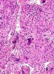
ORLANDO, FL—Updated results of a phase 1 study suggest the EZH2 inhibitor tazemetostat (EPZ-6438) can produce durable responses in patients with advanced non-Hodgkin lymphoma (NHL).
The drug has demonstrated activity against diffuse large B-cell lymphoma (DLBCL), follicular lymphoma (FL), and marginal zone lymphoma (MZL).
The overall response rate among NHL patients in this study was 56%, and 1 patient has maintained a response for more than 21 months.
In addition, the drug’s safety profile is “still acceptable,” according to Vincent Ribrag, MD, of Institut Gustave Roussy in Villejuif, France.
Dr Ribrag presented the results of this study at the 2015 ASH Annual Meeting (abstract 473*). The research, which was previously presented at the 13th International Conference on Malignant Lymphoma, was sponsored by Epizyme, Inc., the company developing tazemetostat.
The trial has enrolled 58 patients, 21 with relapsed or refractory B-cell NHL and 37 with advanced solid tumors. The NHL cohort includes 5 patients with germinal center B-cell (GCB) DLBCL, 6 cases of non-GCB DLBCL, 3 DLBCL cases of an undetermined subtype, 6 patients with FL, and 1 case of MZL.
At baseline, the NHL patients had a median age of 63 (range, 24-84) and were heavily pretreated. Eighty-five percent of patients had received 3 or more prior therapies, and 33% had received 5 or more. Thirty-eight percent of patients had undergone an autologous transplant, and 57% had received radiotherapy.
The patients received tazemetostat twice daily at a range of doses. For the dose-escalation portion of the trial, they received 100 mg, 200 mg, 400 mg, 800 mg, or 1600 mg. For the dose-expansion phase, they received 800 mg or 1600 mg.
The researchers are now conducting a drug-drug interaction substudy in which patients receive 800 mg of tazemetostat twice daily and a food-effect substudy in which patients receive the drug at 400 mg twice daily.
Dr Ribrag said the recommended phase 2 dose of tazemetostat is 800 mg twice daily.
Safety
At the data cutoff point (November 7, 2015), 55 patients—20 with NHL and 35 with solid tumors—were evaluable for safety.
Treatment-related adverse events in these patients included asthenia (n=13), nausea (n=8), thrombocytopenia (n=7), dysgeusia (n=5), vomiting (n=5), dry skin (n=4), decreased appetite (n=4), diarrhea (n=4), muscle spasms (n=3), neutropenia (n=3), anemia (n=3), night sweats (n=3), hypertension (n=2), constipation (n=2), peripheral edema (n=2), hypophosphatemia (n=1), anxiety (n=1), depression (n=1), abdominal pain (n=1), and hepatocellular injury (n=1).
There were 4 grade 3 or higher adverse events that were considered treatment-related, including nausea, hypertension, neutropenia, and hepatocellular injury.
Efficacy
Sixteen of the NHL patients were evaluable for efficacy. Nine patients responded to treatment, 2 with complete responses (CRs) and 7 with partial responses (PRs).
Five of the 10 DLBCL patients responded, 4 with PRs and 1 with a CR. Three of the 5 FL patients responded, 2 with PRs and 1 with a CR. The patient with MZL achieved a PR.
Four responders remain on study—2 with DLBCL and 2 with FL.
One DLBCL patient with an EZH2 mutation (Y646H) had relapsed after or was refractory to 6 previous treatment regimens. This patient achieved a PR after 16 weeks of tazemetostat. The patient is still in PR at week 44 and remains on study.
Based on these results, Epizyme is currently enrolling patients in a phase 2 study of tazemetostat monotherapy. The trial is open to patients with DLBCL or FL in France, Australia, and the UK. ![]()
*Data in the abstract differ from the presentation.
BM fibrosis grade may impact OS in PMF
ORLANDO, FL—Having a higher grade of bone marrow (BM) fibrosis may confer inferior overall survival (OS) in patients with primary myelofibrosis (PMF), according to a retrospective study.
Investigators found that having a fibrosis grade of 2 or higher at diagnosis was associated with “unique clinical and molecular variables” that suggested a more aggressive disease phenotype.
And the median OS was significantly shorter in patients with higher grades of fibrosis.
However, when the investigators divided patients according to their International Prognostic Scoring System (IPSS) risk group, having a fibrosis grade of 2 or higher was only significantly associated with reduced OS among patients in the low-risk or intermediate-1-risk categories.
Paola Guglielmelli, MD, PhD, of the University of Florence in Italy, presented these findings at the 2015 ASH Annual Meeting (abstract 351*).
Dr Guglielmelli noted that the prognostic significance of BM fibrosis grade in PMF has been debated. So she and her colleagues set out to analyze the prognostic impact of fibrosis in 540 PMF patients from 6 Italian centers belonging to AGIMM (AIRC-Gruppo Italiano Malattie Mieloproliferative).
BM biopsies were obtained at diagnosis and evaluated by local pathologists according to 2008 World Health Organization criteria. The European consensus scoring system was used to grade fibrosis on a scale of MF-0 to MF-3.
Fifty patients were classified as MF-0 (9.3%), 180 were MF-1 (33.3%), 196 were MF-2 (36.3%), and 114 were MF-3 (21.1%).
Patients in the MF-2 and MF-3 groups were significantly more likely to have constitutional symptoms (P<0.0001), splenomegaly ≥10 cm from left costal margin (P<0.0001), a peripheral blast count ≥1% (P<0.0001), a greater risk of anemia (P<0.0001) or thrombocytopenia (P=0.001), and belong to the intermediate-2 or high-risk IPSS categories (P<0.0001).
In addition, patients in the MF-2 and MF-3 groups were significantly more likely to qualify as high-molecular-risk (HMR), which was defined as having at least 1 mutation in ASXL1, EZH2, SRSF2, or IDH1/2 (P<0.0001). The frequency of HMR patients increased progressively according to fibrosis grade: MF-0 (16%), MF-1 (25.6%), MF-2 (33.7%), and MF-3 (44.7%).
Patients with 2 or more HMR mutated genes were preferentially MF-2 or MF-3. None of the MF-0 patients fell into this category, compared to 4.4% for MF-1, 10.2% for MF-2, and 10.5% for MF-3 (P<0.0001).
Survival
The median OS was significantly shorter in patients with higher BM fibrosis grades (P<0.0001). The median OS was 7.2 years in the MF-3 group (hazard ratio [HR]=8.7), 6.7 years in the MF-2 group (HR=7.3), 14.7 years in the MF-1 group (HR=3.9), and not reached in the MF-0 group (reference).
In multivariable analysis, having a BM fibrosis grade of 2 or greater was significantly associated with reduced OS (HR=3.8, P=0.01).
Other variables significantly associated with reduced OS were being in the intermediate-1 (HR=2.9, P<0.0001), intermedicate-2 (HR=10.0, P<0.0001), or high-risk IPSS categories (HR=9.7, P<0.0001); having CALR type 2 mutation (HR=3.4, P=0.010), JAK2/MPL mutation (HR=2.4, P=0.003), or being triple-negative (HR=4.5, P<0.0001); being classified as HMR (HR=2.4, P<0.0001); and having 2 or more HMR mutations (HR=4.3, P=0.009).
Dr Guglielmelli and her colleagues also assessed the impact of BM fibrosis grade according to IPSS risk score.
They found that, for patients in the low/intermediate-1-risk categories, the median OS was not reached in the MF-0 group, was 22.8 years in the MF-1 group (HR=3.9), and was 15.4 years in the MF-2 and -3 groups combined (HR=7.4, P=0.001).
In the intermediate-2/high-risk categories, the median OS was 11 years for the MF-0 group, 3.6 years for the MF-1 group (HR=2.2), and 3.6 years in the MF-2 and -3 groups (HR=2.7, P=0.28).
Dr Guglielmelli therefore concluded that BM fibrosis grade might help refine prognostic stratification for PMF patients in the lower-risk IPSS categories. However, she noted that this study had limitations, and the results should be confirmed with prospective research. ![]()
*Data in the abstract differ from the presentation.
Tool may provide new insight into pediatric cancers

and Xin Zhou, PhD
Photo by Peter Barta/St. Jude
Children’s Research Hospital
Researchers say they have developed a tool that may advance our understanding of the mutations that drive pediatric cancers.
The tool, called ProteinPaint, is a web application that allows the user to visualize genetic lesions and RNA expression in pediatric cancers.
ProteinPaint’s infographics let users see all mutations in individual genes and their corresponding proteins, including detailed information about mutation type, frequency in cancer subtype, and location in the protein domain.
That information provides clues about how a change might contribute to cancer’s start, progression, or relapse.
Jinghui Zhang, PhD, of St. Jude Children’s Research Hospital in Memphis, Tennessee, and her colleagues described ProteinPaint in a letter to Nature Genetics.
ProteinPaint currently integrates information from 5 studies, but Dr Zhang and her colleagues said the data will be updated as new studies are published.
ProteinPaint now includes information on almost 27,500 mutations discovered in more than 1000 pediatric patients with 21 cancer subtypes. The application also includes RNA-sequencing data from 928 pediatric tumors belonging to 36 different subtypes.
Xin Zhou, PhD, also of St. Jude, developed ProteinPaint’s infographics to display the genomic information in an interactive format. A click of the mouse gives users additional details about the mutations listed, including the pediatric cancer subtype where the change has been validated and a link to the publication.
“ProteinPaint’s focus on pediatric cancer and presentation of mutations at the gene level complements existing cancer genome data portals,” Dr Zhang said. “For St. Jude, the application is the foundation for developing a global reference database for information about pediatric cancer.”
Dr Zhou added that the ProteinPaint software has the potential to help researchers studying other disorders, including sickle cell disease, that involve a mutation that affects protein function.
ProteinPaint is available at no cost to academic researchers who are free to use the tool to analyze their own data. The application also lets researchers compare information about pediatric and adult cancer genomes by providing a parallel view of data from COSMIC, the world’s largest database of somatic mutations, primarily from adult cancer.
ProteinPaint has already been used to study the role played by germline mutations in pediatric cancers. That research was published in NEJM in November.
More information about ProteinPaint is available on the St. Jude PeCan Data Portal. ![]()
Identifying druggable proteins

Photo by Darren Baker
A computer model that employs techniques used to analyze social networks could aid the development of new cancer treatments, according to researchers.
The model analyzes the unique behaviors of cancer-causing proteins, spotting what makes them different from normal proteins and mapping out molecular targets for drugs that could potentially be developed to treat cancers.
The researchers described this model in PLOS Computational Biology.
Bissan Al-Lazikani, PhD, of The Institute of Cancer Research in London, UK, and her colleagues compared proteins to members of an enormous social network, mapping the ways they interact. This allowed the team to predict which proteins might be most effectively targeted with drugs.
Cancer-causing proteins that have already been successfully targeted tended to have particular “social” characteristics that differed from non-cancer proteins. “Hub-like” proteins that were shown to “communicate” with lots of other proteins were more likely to cause cancers.
The researchers said this suggests that previously unexplored cancer proteins with similar characteristics could make good drug targets.
“Our study is the first to identify the rules of social behavior of cancer proteins and use it to predict new targets for potential cancer drugs,” Dr Al-Lazikani said.
“It shows that cancer drug targets behave very differently from normal proteins and often have a complex web of social interactions. Finding new targets is one of the most important steps in drug discovery, but it can be a lengthy, expensive process.”
“The map that we’ve made will help researchers design better new drugs, more quickly, saving time and money. It also sheds light on how resistance to treatments may occur and, in just a few years, could help doctors choose the best drug combinations to suit individual patients.”
All of the researchers’ target predictions are available on the canSAR website. The underlying data and tools are also available on the site. ![]()
Frailty in HSCT population not dependent on age

Photo by Peter Griffin
ORLANDO, FL—Frailty after hematopoietic stem cell transplant (HSCT), while associated with higher mortality, is not necessarily a function of age, according to investigators studying the impact of frailty on transplant outcomes.
Instead, other factors, such as increasing time from transplant, employment status, medical leave or disability, and limitations of social activities, were significantly associated with higher odds of frailty.
The investigators prospectively studied 96 HSCT recipients, age 40 and older, to determine the prevalence of frailty in HSCT populations and its impact on outcomes, including early post-transplant non-relapse mortality (NRM).
Mukta Arora, MD, of the University of Minnesota in Minneapolis, reported the findings at the 2015 ASH Annual Meeting (abstract 388*).
The investigators defined frailty as the presence of 3 or more of the following criteria: low grip strength, exhaustion, slowed walking speed, low physical activity, and unintentional weight loss. They defined pre-frailty as having 1 or 2 of these characteristics.
The investigators conducted multi-domain geriatric assessments of patients prior to HSCT and after transplant at 100 days, 6 months, and 1 year. The assessment included function, comorbidity, cognition, psychological state, social activity/support, nutritional status, and demographic, transplant, and disease-related information.
Forty-eight patients were in the younger age group (40–59), and 48 were in the older age group (60–74). All had undergone HSCT between February 2014 and April 2015.
Patient demographics
Patients in the younger group were a median age of 54 (range, 40–59) at transplant. Sixty-five percent were male, 58% had an autologous transplant, and 79% received myeloablative conditioning.
Patients in the older group were a median age of 65 (range, 60–73) at transplant. Fifty-four percent were male, 46% had an autologous transplant, and 46% had myeloablative conditioning.
The difference between the older and younger groups in their conditioning regimen was significant (P<0.01).
The groups were comparable in terms of the HSCT comorbidity index but were significantly different in employment status (P<0.01).
“As expected,” Dr Arora said, “there were more patients who were retired in the older group.”
In the younger group, 31% were employed, 3% retired, 56% on medical leave or disabled, and 10% unemployed.
In the older group, 6% were employed, 62% retired, 28% on medical leave or disabled, and 4% unemployed.
“There was no difference in the social activity and social support scores between the 2 groups,” Dr Arora observed.
Frailty assessments
In the younger group, at baseline, the prevalence of pre-frailty was 47%, and the prevalence of frailty was 11%. At 6 months after HSCT, the prevalence of pre-frailty was 45%, and the prevalence of frailty was 41% (P<0.01).
In the older group, at baseline, the prevalence of pre-frailty was 42%, and the prevalence of frailty was 6%. At 6 months, the prevalence of pre-frailty was 44%, and the prevalence of frailty was 38% (P<0.01).
The investigators then estimated the predictors of frailty.
Significant predictors of frailty included time since HSCT (odds ratio [OR]=3.7, 95% CI: 1.9-7.2, P<0.01), employment status (retired: OR=7.3, 95% CI 1.2 – 46.2, P=0.03), on medical leave or disabled (OR=11.2, 95% CI: 1.8 – 67.7, P=0.01), limitations in social activities (OR=1.04, 95% CI: 1.01 – 1.08, P=0.01), and baseline pre-frailty (OR=3.1, 95% CI: 2.3 – 45.5, P<0.01).
Allogeneic transplant was associated with higher odds of frailty than autologous (OR=3.1, 95% CI: 0.9 – 10.2), although it did not reach significance (P=0.06).
Investigators next estimated the impact of frailty or pre-frailty on NRM and identified a trend toward increased NRM in frail patients.
The 46 patients classified as not frail at baseline had a 7% cumulative incidence of NRM (P=0.07). The 42 patients classified as pre-frail had a 23% cumulative incidence of NRM, while the 8 patients classified as frail at baseline had a 28% cumulative incidence of NRM.
“So, to conclude, in this early study, frailty was noted in 8% and pre-frailty in 44% of the transplant population prior to transplant, and was not dependent on age,” Dr Arora said. “Frailty is a transitional state and appears to reflect a dynamic progression from robustness to functional decline with time since [HSCT].”
Because frailty is associated with higher mortality, the investigators believe vulnerable populations should be identified and their need for specific interventions defined.
This research was funded by the Leukemia & Lymphoma Society. ![]()
*Data in the abstract differ from the presentation.
Combo deepens responses and improves PFS in MM

Photo courtesy of the
University of Navarra
ORLANDO, FL—The addition of panobinostat to bortezomib-dexamethasone therapy in relapsed or refractory multiple myeloma (MM) patients can double the rate of deep responses and prolong progression-free survival (PFS), according to an updated analysis of data from the PANORMA 1 trial.
Panobinostat, a pan-deacetylase inhibitor, was the first agent of its class to produce a statistically significant and clinically meaningful increase in the median PFS of patients with relapsed/refractory MM in a phase 3 trial, noted Jesús F. San-Miguel, MD, PhD, of the University of Navarra in Pamplona, Spain.
Dr San-Miguel presented results of the updated analysis at the 2015 ASH Annual Meeting (abstract 4230).
In the PANORAMA 1 trial, patients receiving panobinostat plus bortezomib and dexamethasone had a significantly prolonged median PFS of 12 months versus 8.1 months in patients treated with placebo-bortezomib-dexamethasone.
A subgroup analysis showed that the PFS benefit was improved in patients with previous exposure to bortezomib and immunomodulatory drugs (IMiDs). The addition of panobinostat to bortezomib-dexamethasone also led to a significant increase in high-quality responses.
With their analysis, Dr San-Miguel and his colleagues set out to determine the effect of responses on clinical outcomes of patients treated in PANORAMA 1, including those with prior exposure to bortezomib and IMiDs.
The researchers conducted a landmark analysis at 12, 18, and 24 weeks to assess the median PFS in patients who achieved a complete response (CR)/near complete response (nCR) or partial response (PR).
“For the total study population, the rates of high-quality responses [CR/nCR rate] were significantly higher in the panobinostat-bortezomib-dexamethasone arm [28%] than in the control arm [16%],” Dr San-Miguel said.
Among the subgroup with prior exposure to bortezomib and IMiDs, the CR/nCR rate was also higher in the triple-drug arm (22.3%) than in the 2-drug arm (9.9%).
Among patients who took panobinostat-bortezomib-dexamethasone, the duration of response was twice as long in those who achieved CR/nCR (18.4 months) as in those who achieved a PR (9 months).
The median PFS at 12 weeks for patients who received panobinostat-bortezomib-dexamethasone was increased in patients achieving high-quality responses: 16.5 months for nCR as compared to 10.3 months for PR.
The subgroup of patients with prior exposure to bortezomib and IMiDs who achieved deeper responses also demonstrated longer PFS at 12 weeks: a median of 13.7 months for nCR and 8.1 months for PR.
“In both the overall study population and the subgroup of patients with prior exposure to bortezomib and IMiDs, a 2-fold increase in deep responses was achieved with panobinostat-bortezomib-dexamethasone compared with placebo-bortezomib-dexamethasone,” Dr San-Miguel said. “In both groups, deep responses were associated with a prolonged PFS and a longer duration of response.”
He noted that the magnitude of benefit at each time point appeared greater among patients who received the triple-drug combination.
“These data further support achievement of deeper responses as a treatment goal and a robust and consistent benefit of panobinostat in the phase 3 study in patients with relapsed/refractory multiple myeloma, including those with prior exposure to bortezomib and IMiDs,” Dr San-Miguel said.
The PANORAMA 1 trial was sponsored by Novartis, the company developing panobinostat. Three researchers involved in the current analysis are employees of Novartis, and other researchers reported having relationships (receiving research funding, consulting, etc.) with a range of other pharmaceutical companies. ![]()
Factors predict low accrual in cancer clinical trials

for a clinical trial
Photo by Esther Dyson
Twelve factors may predict low patient accrual in cancer clinical trials, according to research published in JNCI.
Many studies have been conducted to investigate the perceived barriers to clinical trial accrual from the patient or provider perspective.
However, researchers have rarely taken a trial-level view and investigated why certain trials are able to accrue patients faster than expected while others fail to attract even a fraction of the intended number of participants.
Caroline S. Bennette, PhD, of the University of Washington in Seattle, and her colleagues conducted their study to do just that.
They analyzed information on 787 phase 2/3 clinical trials sponsored by the National Clinical Trials Network (NCTN; formerly the Cooperative Group Program) launched between 2000 and 2011.
After excluding trials that closed because of toxicity or interim results, the researchers found that 145 (18%) NCTN trials closed with low accrual or were accruing at less than 50% of target accrual 3 years or more after opening.
The team identified potential risk factors from the literature and interviews with clinical trial experts and found multiple trial-level factors that were associated with poor accrual to NCTN trials, such as increased competition for patients from currently ongoing trials, planning to enroll a higher proportion of the available patient population, and not evaluating a new investigational agent or targeted therapy.
The researchers then developed a multivariable prediction model of low accrual using 12 trial-level risk factors. The team said these factors had good agreement between predicted and observed risks of low accrual in a preliminary validation using 46 trials opened between 2012 and 2013.
Those 12 risk factors are:
- The number of competing trials per 10,000 eligible patients per year (odds ratio [OR]=1.88)
- Phase 3 vs phase 2 trial (OR=1.86)
- Enrollment as percentage of eligible population for targeted therapy (OR=0.57)
- Enrollment as percentage of eligible population for radiation therapy (OR=1.81)
- Annual incidence of clinical condition(s) per 10,000 (OR=0.99)
- Tissue sample required to assess eligibility (OR=1.26)
- Investigational new drug (OR=0.34)
- Metastatic setting (OR=1.46)
- Sample size per 100 (OR=0.95)
- More than one condition evaluated (OR=1.98)
- Common solid cancer (prostate, breast, lung, or colon) vs liquid or rare solid cancers (OR=2.32)
- Interaction term (phase 3 x investigational new drug, OR=2.47).
The researchers concluded that systematically considering the overall influence of these risk factors could aid in the design and prioritization of future clinical trials. ![]()
for a clinical trial
Photo by Esther Dyson
Twelve factors may predict low patient accrual in cancer clinical trials, according to research published in JNCI.
Many studies have been conducted to investigate the perceived barriers to clinical trial accrual from the patient or provider perspective.
However, researchers have rarely taken a trial-level view and investigated why certain trials are able to accrue patients faster than expected while others fail to attract even a fraction of the intended number of participants.
Caroline S. Bennette, PhD, of the University of Washington in Seattle, and her colleagues conducted their study to do just that.
They analyzed information on 787 phase 2/3 clinical trials sponsored by the National Clinical Trials Network (NCTN; formerly the Cooperative Group Program) launched between 2000 and 2011.
After excluding trials that closed because of toxicity or interim results, the researchers found that 145 (18%) NCTN trials closed with low accrual or were accruing at less than 50% of target accrual 3 years or more after opening.
The team identified potential risk factors from the literature and interviews with clinical trial experts and found multiple trial-level factors that were associated with poor accrual to NCTN trials, such as increased competition for patients from currently ongoing trials, planning to enroll a higher proportion of the available patient population, and not evaluating a new investigational agent or targeted therapy.
The researchers then developed a multivariable prediction model of low accrual using 12 trial-level risk factors. The team said these factors had good agreement between predicted and observed risks of low accrual in a preliminary validation using 46 trials opened between 2012 and 2013.
Those 12 risk factors are:
- The number of competing trials per 10,000 eligible patients per year (odds ratio [OR]=1.88)
- Phase 3 vs phase 2 trial (OR=1.86)
- Enrollment as percentage of eligible population for targeted therapy (OR=0.57)
- Enrollment as percentage of eligible population for radiation therapy (OR=1.81)
- Annual incidence of clinical condition(s) per 10,000 (OR=0.99)
- Tissue sample required to assess eligibility (OR=1.26)
- Investigational new drug (OR=0.34)
- Metastatic setting (OR=1.46)
- Sample size per 100 (OR=0.95)
- More than one condition evaluated (OR=1.98)
- Common solid cancer (prostate, breast, lung, or colon) vs liquid or rare solid cancers (OR=2.32)
- Interaction term (phase 3 x investigational new drug, OR=2.47).
The researchers concluded that systematically considering the overall influence of these risk factors could aid in the design and prioritization of future clinical trials.
Why it’s hard to develop immunity against malaria

Diana Hansen, PhD
Photo courtesy of the Walter
and Eliza Hall Institute
Results of preclinical research appear to explain how the malaria parasite Plasmodium falciparum causes an inflammatory reaction that sabotages the body’s ability to protect itself from malaria.
Researchers found evidence to suggest that the same inflammatory molecules that drive the immune response in clinical and severe malaria also prevent the body from developing protective antibodies against the parasite.
They said this discovery opens up the possibility of improving new or existing malaria vaccines by boosting immune cells needed for long-lasting immunity.
Diana Hansen, PhD, of the Walter and Eliza Hall Institute of Medical Research in Parkville, Victoria, Australia, and her colleagues described this work in Cell Reports.
Dr Hansen said this is the first time scientists have pinpointed why the immune system fails to develop immunity during malaria infection.
“With many infections, a single exposure to the pathogen is enough to induce production of antibodies that will protect you for the rest of your life,” she explained. “However, with malaria, it can take up to 20 years for someone to build up sufficient immunity to be protected.”
The fact that the body is not good at developing long-lasting immunity to the parasite has hampered vaccine development, Dr Hansen added.
“This was complicated by the fact that we didn’t know whether it was the malaria parasite itself or the inflammatory reaction to malaria that was actually inhibiting the ability to develop protective immunity,” she said.
“We have now shown that it was a double-edged sword. The strong inflammatory reaction that accompanies and, in fact, drives severe clinical malaria is also responsible for silencing the key immune cells needed for long-term protection against the parasite.”
Dr Hansen and her colleagues conducted experiments in mouse models of malaria and found that inflammatory molecules released by the body to fight the infection were preventing protective antibodies from being made.
“Specialized immune cells called helper T cells join forces with B cells to generate these protective antibodies,” said Axel Kallies, PhD, of the Walter and Eliza Hall Institute of Medical Research.
“However, we showed that, during malaria infection, critical inflammatory molecules actually arrest development of helper T cells, and, therefore, the B cells don’t get the necessary instructions to make antibodies.”
Specifically, the researchers found that severe malaria infection inhibited the establishment of germinal centers in the spleens of the mice. And malaria infection induced high frequencies of T-follicular-helper cell precursors but resulted in impaired T-follicular-helper cell differentiation.
Precursor T-follicular-helper cells induced during infection had low levels of PD-1 and CXCR5 and co-expressed Th1-associated molecules such as T-bet and CXCR3.
However, when the researchers blocked the inflammatory cytokines TNF and IFN-γ or deleted T-bet, they were able to restore T-follicular-helper cell differentiation and germinal center responses to infection.
Dr Hansen said these findings could lead to new avenues in the search for effective malaria vaccines.
“This research opens the door to therapeutic approaches to accelerate development of protective immunity to malaria and improve efficacy of malaria vaccines,” she said.
“Until now, malaria vaccines have had disappointing results. We can now see a way of improving these responses by tailoring or augmenting the vaccine to boost development of helper T cells that will enable the body to make protective antibodies that target the malaria parasites.”
Drug disappoints in phase 3 HSCT trial
The antiviral drug brincidofovir did not meet the primary endpoint of the phase 3 SUPPRESS trial, according to the drug’s developer, Chimerix.
Brincidofovir did not prevent clinically significant cytomegalovirus (CMV) infection through week 24 after hematopoietic stem cell transplant (HSCT).
However, the president and CEO of Chimerix said the company still believes there is a “viable path forward” for the drug.
Brincidofovir is an oral nucleotide analog that has shown in vitro antiviral activity against all 5 families of DNA viruses that affect humans, including herpes viruses and adenovirus.
The SUPPRESS trial, which was initiated in August 2013 and fully enrolled in June 2015, was informed by a successful phase 2 trial conducted in HSCT recipients.
The SUPPRESS trial enrolled and treated 452 adults who received allogeneic HSCTs from more than 40 transplant centers in the US, Canada, and Europe.
Subjects received twice-weekly brincidofovir or placebo (2:1 ratio) from the early post-transplant period through week 14 post-transplant, the period of highest risk for viral infections.
All patients in the trial were CMV-seropositive, placing them at high risk of CMV infection. The most common indications leading to HSCT were acute myelogenous leukemia (43% of patients), myelodysplasia (17%), non-Hodgkin lymphoma (10%), and acute lymphocytic leukemia (9%).
During the on-treatment period through week 14 after HSCT, fewer patients in the brincidofovir arm had a CMV infection, which was consistent with results from the phase 2 study of the drug.
However, during the 10 weeks off treatment from week 14 to week 24, there was an increase in CMV infections in the brincidofovir arm compared to the control arm. And there was a non-statistically significant increase in mortality in the brincidofovir arm compared to the control arm.
Preliminary analysis suggests the failure in preventing CMV infections and the increased mortality in the brincidofovir arm were driven by confirmed cases of graft-versus-host-disease (GVHD), which resulted in a significantly higher use of corticosteroids than in the control arm.
Both GVHD and the use of corticosteroids are risk factors for “late” CMV infection that occurs after the discontinuation of the antiviral in HSCT recipients.
The rate of study drug discontinuation for gastrointestinal events was less than 10% in this study, which is comparable to that observed in the phase 2 trial of brincidofovir in a similar HSCT population.
“While we are clearly disappointed in the top-line results from SUPPRESS, we remain committed to better understanding the full data set as we consider potential paths forward for brincidofovir,” said M. Michelle Berrey, MD, president and CEO of Chimerix.
“We will be evaluating the subgroups of patients within SUPPRESS, such as T-cell-depleted transplant recipients who have a lower risk of GVHD, to better understand these results and inform our next steps,” said W. Garrett Nichols, MD, Chimerix’s chief medical officer.
“We are reaching out to investigators and other experts to help us assess the complete data set to understand what may have caused the results of the SUPPRESS trial to differ substantially from those seen in the phase 2 study. Additionally, we are in communication with the US Food and Drug Administration and other regulatory bodies and will share any updates on the brincidofovir clinical program when we can.”
“With data currently in hand, we believe that brincidofovir may ultimately demonstrate a positive risk-benefit profile for the treatment of adenovirus and smallpox, as well as use in other populations in need of a novel compound for DNA viral infections.”
Chimerix plans to continue the programs testing brincidofovir in serious adenovirus infections and in smallpox. Pending the availability of complete data from SUPPRESS, including secondary endpoints in other dsDNA viral infections, Chimerix has elected to pause further enrollment in the phase 3 SUSTAIN and SURPASS trials in kidney transplant recipients.
A full analysis of the SUPPRESS trial results is ongoing. The data are scheduled to be presented at the BMT Tandem Meetings in Honolulu, Hawaii, in February.
Study reveals germline variants in AML, other cancers

A study published in Nature Communications has shed light on the hereditary elements of 12 cancer types.
Investigators looked for rare germline mutations in genes known to be associated with cancer and found the frequency of these mutations varied widely, from 4% in the acute myeloid leukemia (AML) cases studied to 19% in cases of ovarian cancer.
The team’s analysis also revealed an unexpected inherited component to stomach cancer and provided some clarity on the consequences of certain mutations in the BRCA1 and BRCA2 genes.
Li Ding, PhD, of Washington University School of Medicine in St Louis, Missouri, and her colleagues conducted this study, analyzing genetic information from more than 4000 cancer cases included in The Cancer Genome Atlas project.
“In general, we have known that ovarian and breast cancers have a significant inherited component, and others, such as acute myeloid leukemia and lung cancer, have a much smaller inherited genetic contribution,” Dr Ding said. “But this is the first time, on a large scale, that we’ve been able to pinpoint gene culprits or even the actual mutations responsible for cancer susceptibility.”
To help tease out cancer’s inherited components, Dr Ding and her colleagues looked for germline truncations in 114 genes known to be associated with cancer.
“We looked for germline mutations in the tumor, but it was not enough for the mutations simply to be present,” Dr Ding said. “They needed to be enriched in the tumor—present at higher frequency. If a mutation is present in the germline and amplified in the tumor, there is a high likelihood it is playing a role in the cancer.”
The investigators found germline truncations in all 12 cancer types analyzed, but the mutations occurred in varying frequencies depending on the cancer.
The percentage of tumors with truncations in the germline was 4% for AML and glioblastoma; 5% for kidney cancer; 7% for lung adenocarcinoma and endometrial cancer; 8% for head and neck cancer, glioma, lung squamous cell carcinoma, and prostate cancer; 9% for breast cancer; 11% for stomach cancer; and 19% for ovarian cancer.
“We also found a significant number of germline truncations in the BRCA1 and BRCA2 genes present in tumor types other than breast cancer, including stomach and prostate cancers, for example,” Dr Ding said. “This suggests we should pay attention to the potential involvement of these 2 genes in other cancer types.”
The investigators said they identified 13 cancer genes with significant enrichment of rare truncations. Some of these were associated with specific cancers—for example, RAD51C in AML, PALB2 in stomach cancer, and MSH6 in endometrial cancer.
And the team observed significant, tumor-specific loss of heterozygosity in 9 genes—ATM, BAP1, BRCA1/2, BRIP1, FANCM, PALB2, and RAD51C/D.
Dr Ding said more research is needed to confirm these results before they can be used to advise patients making healthcare decisions.
“Our strategy of investigating germline-tumor interactions provides a good way to prioritize important mutations that we should focus on,” she said. “For the information to eventually be used in the clinic, we will need to perform this type of analysis on even larger numbers of patients.”