User login
Method makes gene editing more efficient, team says

Image by Spencer Phillips
Researchers say they have made an improvement in CRISPR-Cas9 technology that enables a success rate of 60% when replacing a short stretch of DNA
with another.
They say this technique could be especially useful when trying to repair genetic mutations that cause hereditary diseases, such as sickle cell disease.
The technique allows researchers to patch an abnormal section of DNA with the normal sequence and potentially correct the defect.
“The exciting thing about CRISPR-Cas9 is the promise of fixing genes in place in our genome, but the efficiency for that can be very low,” said Jacob Corn, PhD, of the University of California, Berkeley.
“If you think of gene editing as a word processor, we know how to cut, but we need a more efficient way to paste and glue a new piece of DNA where we make the cut.”
Dr Corn and his colleagues described their more efficient technique in Nature Biotechnology.
“In cases where you want to change very small regions of DNA, up to 30 base pairs, this technique would be extremely effective,” said study author Christopher Richardson, PhD, of the University of California, Berkeley.
Dr Richardson invented the new approach after finding that the Cas9 protein, which does the actual DNA cutting, remains attached to the chromosome for up to 6 hours, long after it has sliced through the double-stranded DNA.
Dr Richardson looked closely at the Cas9 protein bound to the 2 strands of DNA and discovered that while the protein hangs onto 3 of the cut ends, 1 of the ends remains free.
When Cas9 cuts DNA, repair systems in the cell can grab a piece of complementary DNA, called a template, to repair the cut. Researchers can add templates containing changes that alter existing sequences in the genome.
Dr Richardson reasoned that bringing the substitute template directly to the site of the cut would improve the patching efficiency. So he constructed a piece of DNA that matches the free DNA end and carries the genetic sequence to be inserted at the other end.
The technique allowed successful repair of a mutation with up to 60% efficiency.
“Our data indicate that Cas9 breaks could be different, at a molecular level, from breaks generated by other targeted nucleases, such as TALENS and zinc-finger nucleases, which suggests that strategies like the ones we are using can give you more efficient repair of Cas9 breaks,” Dr Richardson said.
The researchers also showed that variants of the Cas9 protein that bind DNA but do not cut can also paste a new DNA sequence at the binding site, possibly by forming a “bubble” structure on the target DNA that also acts to attract the repair template.
Gene editing using Cas9 without genome cutting could be safer than typical gene editing by removing the danger of off-target cutting in the genome, Dr Corn said. ![]()
Generic drugs often out of reach, experts say

Photo courtesy of the CDC
An article published in Blood suggests pharmaceutical companies use several strategies to keep affordable generic drugs from the US market.
“The timely availability of affordable generic drugs is the difference between life or death for patients with cancer and other diseases who cannot afford brand-name pharmaceuticals, the majority of which are priced at monopoly levels and protected by 20-year patents,” said lead author Hagop Kantarjian, MD, of The University of Texas MD Anderson Cancer Center in Houston.
“Unfortunately, these sorely needed generics are increasingly out of reach. As we sought to understand what keeps these affordable drugs from the market, we identified several specific strategies that pharmaceutical companies use to extend their patents and eliminate competition.”
Dr Kantarjian and his colleagues assert that pharmaceutical companies use a variety of strategies to delay, prevent, and suppress the timely availability of affordable generic drugs.
Among them, the authors detail “pay-for-delay,” in which the company that owns the patent pays a generic company to delay entry into the market. The Federal Trade Commission estimates that the pay-for-delay settlements cost taxpayers, insurance companies, and consumers approximately $3.5 billion per year.
In other cases detailed in the article, the patent-holder deters competition by creating its own version of drugs at generic prices.
While this practice may reduce costs for consumers by 4% to 8% in the short-term, the authors suggest that companies often use the authorized generics as a bargaining chip in “pay-for-delay” deals, pledging not to release their own drugs in return for the true generic company promising to delay market entry.
Other strategies the authors discuss include investing heavily in advertising the brand-name drug (often spending more on marketing than on research and development) and lobbying for laws that prevent patients from importing cheaper generics from other countries, which the authors write can cost as little as 20% to 50% of US prices.
The authors also say some drug companies buy out competitors and then increase the price of a newly acquired generic drug by several fold overnight.
In addition, the authors describe a strategy they call “product hopping,” which involves switching the market for a drug to a reformulated “new and improved” version with a slightly different tablet or capsule dose that offers no therapeutic advantage over the original but has a later-expiring patent.
The company then heavily advertises the new brand-name drug in an effort to convince patients and physicians to switch.
As a result, when the generic version of the original becomes available, pharmacists cannot substitute it for the new branded version because state laws allow substitution only if certain characteristics, such as dosing, remain the same.
In recognition of the harm and expense the authors suggest these strategies impart on both patients and the economy, they propose several solutions that would support timely access to affordable generic drugs.
These include allowing Medicare to negotiate drug prices, monitoring and penalizing pay-for-delay deals, allowing transportation of pharmaceuticals across borders for individual use, and challenging weak patents.
“Each day, in my clinic, I see leukemia patients who are harmed because they cannot afford their treatment, some risking death because they cannot pay for the medicine keeping them alive,” Dr Kantarjian said.
“Overall, these strategies demonstrate that the trend of high brand-name drug prices has recently infected generic drugs, as companies value profit at the expense of long-term utility to society. We must be vigilant in recognizing these strategies and advocating for solutions that will allow companies to accomplish their dual mission: make reasonable profits and help save and/or improve patients’ lives.” ![]()
Study reveals how Tregs protect themselves
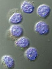
Image by Kathryn Iacono
Researchers say they have discovered how regulatory T cells (Tregs) remain intact and functional during activation.
The team found that once Tregs are activated, they are protected by autophagy, which maintains metabolic balance.
Hongbo Chi, PhD, of St. Jude Children’s Hospital in Memphis, Tennessee, and his colleagues described this discovery in Nature Immunology.
Until this study, no one knew how Tregs maintained themselves when activated.
“Regulatory T cells are very specialized cells that require activation to perform their function in curtailing undesirable immune responses,” Dr Chi explained. “But this activation is a double-edged sword, in that this very activation can destabilize them. They need to modulate this activation, or they will lose their stability and many of them will die. That could damage immune function.”
Dr Chi and his colleagues performed imaging studies in activated Tregs and found that autophagy was functional in the cells.
In experiments with mice, the researchers deleted Atg7 or Atg5, genes whose functions are necessary for autophagy in Tregs.
The mice showed key characteristics of Treg malfunction, including inflammatory and autoimmune disorders. The mice also more readily cleared tumors from their bodies, due to activated immune systems.
Dr Chi said that eliminating autophagy also affected the fate of Tregs.
“Once those T cells lack autophagy activity, they tend to undergo excessive cell death,” he said. “But even for the remaining surviving cells, they tend to be overly activated and lose their identity because they start to behave like non-regulatory T cells. That is why loss of autophagy in regulatory T cells produces a 2-fold effect on both survival and stability.”
Detailed analysis also revealed how the elimination of autophagy affected the basic energy-producing metabolic pathways of Tregs, compromising their function.
Dr Chi said this new understanding of autophagy’s role in Tregs could enable a 2-fold approach to immune therapy for cancers. Namely, by strengthening tumor-associated immune responses, targeting Treg autophagy could act in synergy with strategies that block autophagy in tumor cells.
In this study, the researchers used a transplanted colon cancer cell line. In further studies, they plan to explore the role of autophagy in immune reactions toward other tumor cell types to determine whether such therapies might be effective in a broad range of cancers.
The team also hopes to gain a better understanding of the detailed biochemical mechanisms regulating how autophagy connects to the cell’s metabolic pathways. ![]()
Malaria test granted CE mark

Image by Peter H. Seeberger
A malaria test called illumigene® Malaria has received the CE mark, which suggests it meets European health and safety standards.
illumigene Malaria is a molecular test that involves the use of loop-mediated isothermal amplification (LAMP) technology.
According to the test’s developers, it provides results in under an hour, doesn’t require a high level technical expertise, and is up to 80,000 times more sensitive than conventional malaria tests.
illumigene Malaria was developed by Meridian Bioscience, Inc., with technical assistance from the US Centers for Disease Control and Prevention (CDC) and Cheikh Anta Diop University in Dakar, Senegal.
“illumigene Malaria has the potential to change current practices,” said Daouda NDIAYE, PharmD, PhD, of Cheikh Anta Diop University.
“Faster and more accurate diagnosis is vital in the fight against malaria. Earlier diagnosis enables the correct treatment to be prescribed, which leads to better clinical outcomes for the person with malaria and keeps malaria treatments for the right people.”
“Because of submicroscopic parasitemia carriage among the populations, a robust, sensitive, and field-community-deployable screening tool is needed to track the malaria reservoir in pre-elimination regions. illumigene Malaria shows this capacity.”
illumigene Malaria uses LAMP technology to amplify DNA and detect the presence of the malaria parasite.
LAMP technology is isothermal and can be used at room temperature without the need to heat reagents or the material being tested, unlike the rapid diagnostic tests currently used in malaria, which use polymerase chain reaction technology. illumigene Malaria does not require refrigeration.
Meridian Bioscience has worked with experts at the CDC and the Cheikh Anta Diop University of Dakar during the development of illumigene Malaria and collaborated with these organizations to design clinical trials.
According to Meridian, data from more than 200 patients in Senegal validated the performance of illumigene Malaria. The test demonstrated 100% sensitivity and detected infected patients that were missed by conventional testing methods.
Meridian said illumigene Malaria will be distributed in the European, Middle Eastern, and African regions by Meridian Bioscience Europe and in additional international markets by the company’s global distribution network. ![]()
Compounds could treat leukemia, lymphoma
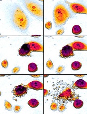
apoptosis in cancer cells
Preclinical research suggests a new class of small-molecule inhibitors could potentially treat leukemias and lymphomas.
Researchers identified compounds targeting the Mdm2–MdmX RING–RING interaction as a new class of E3 ligase inhibitors.
Experiments showed that these compounds, dubbed MMRis, can induce apoptosis in leukemia and lymphoma cells.
The researchers detailed these experiments in Cell Death and Disease.
“We are excited about the unique activities of these compounds and will continue to focus our research efforts on development of their clinical potential,” said study author Xinjiang Wang, PhD, of Roswell Park Cancer Institute in Buffalo, New York.
“These compounds kill cancer cells. [They don’t] just stop cancer cell growth temporarily. These types of agents offer the promise of therapeutic benefit.”
Dr Wang and his colleagues found that MMRis specifically inhibit Mdm2–MdmX E3 ligase activity toward Mdm2 and p53 substrates.
The team said MMRis have an advantage over p53-activating agents that are currently in use as cancer therapies.
That is because MMRis activate the pro-apoptotic function of the p53 pathway, whereas current p53-activating agents temporarily prevent cancer growth but don’t damage existing cancer cells or prevent cancer growth long-term.
The researchers found that 2 MMRi compounds—MMRi6 and its analog, MMRi64—can disrupt Mdm2–MdmX interactions and activate p53 in vitro.
And MMRi64 selectively induces the apoptotic arm of the p53 pathway in leukemia and lymphoma cells.
The team noted that, unlike Nutlin3a, MMRi64 only induces expression of the pro-apoptotic gene PUMA, with minimal induction of the growth-arresting gene p21. However, combining MMRi64 and Nutlin3a produces a synergistic apoptotic effect.
“This study opens a new area for anticancer drug development,” Dr Wang said. “MMRi compounds also can be used as a tool for better understanding the anti-death mechanisms developed by cancer cells.”
“We are moving the research of MMRi compounds forward using both preclinical models and human cancer cell lines. Our hope is that further development of clinically useful MMRi will eventually provide a new treatment option for cancer patients.” ![]()
Leukemia death rates expected to fall in EU

receiving chemotherapy
Photo by Rhoda Baer
Projections for 2016 suggest that leukemia deaths are on the decline in the European Union (EU), particularly for children and young adults.
Leukemia death rates are expected to fall 19% in men and 16% in women in 2016, when compared to 2007 data.
Rates are projected to fall 38% in boys and 20% in girls ages 0 to 14, 26% in men and 22% in women ages 15 to 44, and 19% in men and women ages 45 to 69.
Researchers said improvements in management, multidrug chemotherapy, immunotherapies, stem cell transplants, radiotherapy, and targeted therapies have all contributed to improvements in survival for leukemia patients.
However, some leukemias remain hard to treat, particularly those that are more common in adults and the elderly.
“Predictions of death rates from leukemia are complicated by the fact that leukemias are a varied collection of blood cancers, with some being more treatable than others,” said study author Carlo La Vecchia, MD, of the University of Milan in Italy.
“However, the important falls in overall death rates from this group of diseases are very encouraging and are a testament to the hard work of researchers and clinicians in developing and implementing better diagnosis and treatments.”
Dr Vecchia and his colleagues reported the results of this research in Annals of Oncology.
The researchers looked at cancer deaths in the EU as a whole and in the 6 largest countries—France, Germany, Italy, Poland, Spain, and the UK.
The team assessed mortality for all cancers and looked at individual data for cancers of the stomach, intestines, pancreas, lung, prostate, breast, and uterus (including cervix), as well as leukemias.
All cancers
The researchers’ projections suggest that total cancer mortality rates will decrease for both sexes in 2016, despite a rise in the absolute number of deaths due to the aging population. The team said the median age of the total EU population was 41 in 2011 and will be 42 in 2016.
So a total of 1,359,500 EU citizens are expected to die of cancer in 2016—753,600 men and 605,900 women. This is compared to 1,314,787 cancer deaths in 2011—734,259 men and 580,528 women.
The age-standardized mortality rates for 2016 are 133.5 per 100,000 men and 85.2 per 100,000 women, compared to 144.6 per 100,000 men and 88.1 per 100,000 women in 2011.
So that’s a 7.7% fall in cancer death rates for men and a 3.3% fall for women from 2011 through 2016, despite a 3.3% increase in the absolute number of cancer deaths.
Leukemias
The researchers predict that, in 2016, age-standardized leukemia mortality rates will be 3.95 per 100,000 men and 2.46 per 100,000 women.
In comparison, the age-standardized mortality rates for 2000-2004 were 5.23 for men and 3.20 for women. The rates for 2005-2009 were 4.85 for men and 2.92 for women.
For males ages 0 to 14, the age-standardized mortality rates are 1.10 for 2000-2004, 0.87 for 2005-2009, and 0.54 for 2016. For females in this age group, the rates are 0.85 for 2000-2004, 0.69 for 2005-2009, and 0.55 for 2016.
For males ages 15 to 44, the age-standardized mortality rates are 1.50 for 2000-2004, 1.28 for 2005-2009, and 0.95 for 2016. For females, the rates are 1.07 for 2000-2004, 0.92 for 2005-2009, and 0.72 for 2016.
For males ages 45 to 69, the age-standardized mortality rates are 9.19 for 2000-2004, 8.39 for 2005-2009, and 6.79 for 2016. For females, the rates are 5.74 for 2000-2004, 5.12 for 2005-2009, and 4.16 for 2016. ![]()
ISTH releases new clinical core curriculum

Image by Kevin MacKenzie
The International Society on Thrombosis and Haemostasis (ISTH) has announced its new international clinical core curriculum on thrombosis and hemostasis.
It is the first framework of its kind to define the minimum standards for a medical doctor to attain a level of proficiency to practice independently as a specialist in the field.
The core curriculum appears in the January 2016 issue of the Journal of Thrombosis and Haemostasis.
“Internationally, there is wide variation in how clinicians who work in thrombosis and hemostasis reach their final destination as recognized specialists,” said Claire McLintock, MD, of Auckland City Hospital in New Zealand.
“The ISTH clinical core curriculum serves to provide a framework for training of specialists in this field and to promote harmonization of training internationally.”
Dr McLintock noted that, prior to the development of the ISTH core curriculum, there was no international consensus on what constituted the requirements for a specialist in thrombosis and hemostasis in terms of clinical competencies.
In 2013, ISTH identified the need for an international clinical core curriculum and developed a working group consisting of specialists from around the world to address and draft a list of competencies.
ISTH members and the global thrombosis and hemostasis community were surveyed to rate the importance of the proposed competencies. The survey garnered more than 640 responses with broad geographical representation, which determined the ultimate framework for the new curriculum.
“We are pleased to release this exciting new resource for the global thrombosis and hemostasis community,” said Nigel Key, MD, of the UNC Hemophilia and Thrombosis Center in Chapel Hill, North Carolina.
“Our intent is for the core curriculum to serve as a common reference point for national and regional thrombosis and hemostasis societies to create or revise their own clinical training programs. Additionally, it can potentially be used as a framework for continuous professional development, maintenance of competence, and to inform future ISTH educational offerings.” ![]()
Image by Kevin MacKenzie
The International Society on Thrombosis and Haemostasis (ISTH) has announced its new international clinical core curriculum on thrombosis and hemostasis.
It is the first framework of its kind to define the minimum standards for a medical doctor to attain a level of proficiency to practice independently as a specialist in the field.
The core curriculum appears in the January 2016 issue of the Journal of Thrombosis and Haemostasis.
“Internationally, there is wide variation in how clinicians who work in thrombosis and hemostasis reach their final destination as recognized specialists,” said Claire McLintock, MD, of Auckland City Hospital in New Zealand.
“The ISTH clinical core curriculum serves to provide a framework for training of specialists in this field and to promote harmonization of training internationally.”
Dr McLintock noted that, prior to the development of the ISTH core curriculum, there was no international consensus on what constituted the requirements for a specialist in thrombosis and hemostasis in terms of clinical competencies.
In 2013, ISTH identified the need for an international clinical core curriculum and developed a working group consisting of specialists from around the world to address and draft a list of competencies.
ISTH members and the global thrombosis and hemostasis community were surveyed to rate the importance of the proposed competencies. The survey garnered more than 640 responses with broad geographical representation, which determined the ultimate framework for the new curriculum.
“We are pleased to release this exciting new resource for the global thrombosis and hemostasis community,” said Nigel Key, MD, of the UNC Hemophilia and Thrombosis Center in Chapel Hill, North Carolina.
“Our intent is for the core curriculum to serve as a common reference point for national and regional thrombosis and hemostasis societies to create or revise their own clinical training programs. Additionally, it can potentially be used as a framework for continuous professional development, maintenance of competence, and to inform future ISTH educational offerings.”
NHSBT offers more precise blood typing

Photo by Juan D. Alfonso
NHS Blood and Transplant (NHSBT) has announced an initiative to provide more detailed blood-group typing for patients with hemoglobinopathies, with the goal of enabling better-matched and potentially safer transfusions.
The typing will detect Rh variant blood groups, which need to be considered when planning transfusions.
Previously, typing to this level was only possible through reference laboratories using complex genotyping methods.
NHSBT is offering the testing at no extra cost to hospitals in England until the end of June 2016.
The initiative involves routinely testing for the RHD and RHCE variants most commonly found in patients with hemoglobinopathies. NHSBT will also test genes for the blood groups K, k, Kpa, Kpb, Jsa, Jsb, Jka, Jkb, Fya, Fyb, Fy (GATA), M, N, S, s, U, Doa, and Dob.
Unlike older methods, this testing can be performed in patients who have recently received blood.
NHSBT said this initiative will enable the creation of a database of genotyped blood details for patients with hemoglobinopathies.
Extended blood type information and fast access to the database could potentially enable safer blood transfusions for these patients, who may need numerous transfusions during their lifetime and may move between hospitals.
NHSBT said it has received more than 2500 blood samples thus far. The results are processed centrally by NHSBT at the International Blood Group Reference Laboratory in Filton and are securely stored.
Patients’ test results will be accessible to the teams involved in the patients’ care.
“Patients taking part can now potentially receive more finely matched blood if we know not just their blood group but whether they have a variant Rh type,” said Sara Trompeter, MB ChB, a consultant hematologist for NHSBT.
“And there will also be greater safety and likelihood of getting matched blood in an emergency, as their records will be held centrally and can be accessed by blood banks in local hospitals. We would urge all patients with hemoglobin disorders such as sickle cell disease or thalassemia to speak to their medical or nursing team about providing a blood sample to NHS Blood and Transplant via their local transfusion laboratories to be genotyped.”
Additional information on this initiative can be found at www.nhsbt.nhs.uk/extendedbloodgrouptesting.
Race plays role in Hodgkin lymphoma outcomes in kids

Photo courtesy of Sylvester
Comprehensive Cancer Center
In a retrospective study, young African American patients with Hodgkin lymphoma (HL) had inferior long-term overall survival when compared to their Hispanic and white peers.
Hispanic and white patients had similar rates of overall survival, but Hispanic males had inferior disease-specific survival compared to white males.
The study, published in Pediatric Blood & Cancer, is the largest yet on racial and ethnic disparity in the pediatric HL population in the US.
“Little was known about the association between race, ethnicity, and survival in the pediatric Hodgkin lymphoma population,” said Joseph Panoff, MD, of Sylvester Comprehensive Cancer Center at the University of Miami in Florida.
“Our study showed that African American children and teenagers had worse overall survival than whites and Hispanics at 25 years after diagnosis. We also found that Hispanic males had inferior disease-specific survival compared to white males.”
Dr Panoff and his colleagues analyzed data from more than 7800 patients listed in the Florida Cancer Data System (FCDS) and the National Institutes of Health’s Surveillance, Epidemiology, and End Results Program (SEER).
The patients were 0.1 to 21 years of age (average, 17 years) and were diagnosed with HL from 1981 to 2010.
In the FCDS cohort, which was significantly smaller than the SEER cohort (1778 vs 6027), African Americans had a 33% overall survival rate at 25 years, compared to 44.7% for Hispanics and 49.2% for whites (P=0.0005).
In a multivariate analysis, African American race was associated with inferior overall survival. The hazard ratio was 1.81 (P=0.0003).
Patients in the FCDS cohort had worse overall survival than patients in the SEER cohort, indicating that patients treated in Florida have worse outcomes when compared to the rest of the nation.
In the SEER cohort, the overall survival rate at 25 years was 74.2% for African Americans and 82% for both Hispanic and white patients (P=0.0005). Disease-specific survival rates at 25 years were 85.7% for African Americans, 88.1% for Hispanics, and 90.8% for whites (P=0.0002).
The researchers noted that Hispanic males had inferior disease-specific survival when compared to white males—84.8% and 90.6%, respectively (P=0.0478).
And Hispanic race was a predictor of inferior disease-specific survival in multivariate analysis. The hazard ratio was 1.238 (P<0.0001).
“Clearly, racial and ethnic disparities persist in the pediatric Hodgkin lymphoma population, despite modern treatment, particularly in Florida,” Dr Panoff noted. “The underlying causes of these disparities are complex and need further explanation.”
As a next step, Dr Panoff suggests identifying flaws in the diagnostic and treatment process with regard to African American and Hispanic patients.
“It is important to identify sociocultural factors and health behaviors that negatively affect overall survival in African American patients and disease-free survival in Hispanic males,” he said. “The fact that the entire Florida cohort seems to have worse overall survival than patients in the rest of the country is a new finding that requires further research.”
Team uncovers new info on hemangioblasts

Research published in PNAS has shed new light on the mechanism by which hemangioblasts become blood cells.
Hemangioblasts, which give rise to both hematopoietic and endothelial progenitors, have been identified in the embryos of chickens, mice, fish, and humans. It has also become clear that the cells are present in adult organisms.
However, the mechanism by which hemangioblasts differentiate into blood cells and vascular endothelia has remained a mystery in many aspects.
With that in mind, Makoto Kobayashi, PhD, of the University of Tsukuba in Japan, and his colleagues studied hemangioblasts in zebrafish.
The investigators looked for zebrafish mutants with defects in the hemangioblast expression of Gata1, which is not expressed in endothelial progenitors.
This revealed a mutant with downregulation of hematopoietic genes and upregulation of endothelial genes.
The team then identified the gene responsible for this mutant—LSD1. Additional experiments showed that LSD1 silences Etv2, a critical regulator of hemangioblast development.
The investigators said these results indicate that epigenetic silencing of Etv2 by LSD1 may be a significant event required for hemangioblasts to initiate hematopoietic differentiation.