User login
Team explains CD8 Treg dysfunction
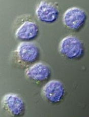
Image by Kathryn T. Iacono
Research published in The Journal of Clinical Investigation provides new insights regarding CD8 regulatory T cells (Tregs).
Investigators found that, in young, healthy individuals, CD8 Tregs suppress the activation and expansion of CD4 T cells.
However, older individuals and patients with a rare form of vasculitis exhibit CD8 Treg dysfunction, which is tied to a drop in production of an enzyme called NADPH oxidase 2 (NOX2).
Cornelia Weyand, MD, of Stanford University Medical Center in California, and her colleagues conducted this research.
First, the team found that CD8 Tregs preferentially take up residence in lymph nodes, the spleen, and other regions where there are huge “armies” of CD4 T cells. This proximity puts the CD8 Tregs in a position to stamp out CD4 T-cell activation early on.
Further experiments demonstrated that CD8 Tregs manufacture copious amounts of NOX2, which they package into tiny membrane-bound packets and transfer to the surfaces of abutting CD4 T cells.
These NOX2-laden packets are then taken up by the CD4 T cells. Inside their new home, the enzymes produce large volumes of highly reactive signaling substances that dial down CD4 T cells’ activation and proliferation.
The investigators noted that contact between CD8 Tregs and CD4 T cells in the early stages of activation shuts down the CD4 T cells’ activity and reduces their proliferation by half or more, even several days after the CD8 Tregs have been removed. Transferring NOX2 alone onto activated CD4 T cells also produces this effect.
Next, the team analyzed blood samples from healthy individuals and observed that CD8 Tregs were only about half as common in blood from people age 60 and older as in blood from 20- to 30-year-olds.
Both CD8 Treg numbers and their ability to suppress CD4 T-cell proliferation declined with advancing age. Experiments traced this to a drop in NOX2 production by older donors’ CD8 Tregs.
The investigators also discovered CD8 Treg failure in giant-cell arteritis (GCA). The team compared blood from GCA patients to blood from age-matched healthy control subjects and from patients with 2 other autoimmune diseases—psoriatic arthritis and small-vessel vasculitis. This revealed a severe deficit among GCA patients in NOX2-producing CD8 Tregs.
“This tells us that the deficit in NOX2-producing CD8 Tregs is specific to GCA, not just driving or driven by inflammation,” Dr Weyand said. “That’s good news for our patients who have this disease, which has been an enigma. Now we know something about what’s causing it.”
The discovery of NOX2 on the surface of CD8 Tregs—but not on other T-cell types—makes them much easier to identify and count, Dr Weyand added.
She and her colleagues are taking advantage of the new-found biomarker to tally CD8 Tregs in patients with age-associated disorders now understood to be driven by chronic inflammation to see if CD8 Treg deficits underlie some of these conditions’ pathology and whether they may be amenable to potential NOX2-restoring treatments. ![]()
mAb can reduce CSCs in newly diagnosed MM

Photo by Linda Bartlett
NEW ORLEANS—A small study suggests that treatment with lenalidomide and dexamethasone (len-dex) prompts an increase in cancer stem cells (CSCs) for patients with newly diagnosed multiple myeloma (MM).
However, adding an anti-CD19 monoclonal antibody (mAb) to the regimen can reduce CSCs.
Most patients who received the mAb, MEDI-551, experienced a decrease in CSCs, but the cells rebounded after the patients stopped receiving MEDI-551.
And those patients who did not see a decrease in CSCs progressed. However, some patients are still in response and remain on treatment with len-dex.
The investigators believe these early results suggest prolonged treatment with len-dex and MED-551 may be safe and clinically beneficial for MM patients.
The results were presented at the 2016 AACR Annual Meeting (abstract CT102).
The study included 17 patients with newly diagnosed MM. They had a median age of 65 (range, 34-73). Most had ISS stage I (n=11), 2 had stage II, and 4 had stage III. Seven patients had t(4;14).
“We chose to carry out this clinical trial in newly diagnosed patients because our original data showed that CD19 was almost always expressed by myeloma stem cells in these patients, whereas we don’t know if that is the case in more advanced patients,” said investigator William Matsui, MD, of Johns Hopkins University School of Medicine in Baltimore, Maryland.
The patients received 28-day cycles of len-dex (len at 25 mg PO, days 1-21, and dex at 40 mg PO, weekly). Patients received MEDI-551 (at 4 mg/kg IV) in cycle 3 (day 1, 8) and cycle 4 (day 1). Responding patients continued on len-dex.
The investigators measured MM CSCs by quantifying the growth of MM colonies (CFU-MM) from marrow aspirates at baseline and at the end of cycles 2 and 4.
The team quantified peripheral blood CSCs by flow cytometry (CD19+CD27+ALDH+) at baseline and the end of cycles 2, 4, 5, and 7.
“We wanted to see if these 2 assays gave similar results, and, in this clinical trial, they were almost identical,” said investigator Carol Ann Huff, MD, also of Johns Hopkins.
“Since it is much easier to draw blood than bone marrow from our patients, we think that we can primarily use blood to track multiple myeloma stem cells in the future.”
Response
Two patients did not receive MEDI-551 due to progressive disease and noncompliance. So the investigators assessed responses in 15 patients.
After cycle 2 (len-dex alone), there were 3 very good partial responses (VGPRs), 10 partial responses (PRs), 1 molecular response, and 1 case of stable disease.
After cycle 4 (len-dex plus MEDI-551), there were 6 VGPRs, 8 PRs, and 1 molecular response.
Ten patients who completed treatment with MED-551 remain on len-dex. At the end of cycle 7, there was 1 complete response, 8 VGPRs, and 1 PR.
CFU-MM
When compared to baseline, bone-marrow-derived CFU-MM increased a median of 2.5-fold (range, 0.4-7.4) after cycle 2 but decreased a median of 0.48-fold (range, 0.14-0.85) in 14 patients after cycle 4.
The investigators compared these results to 5 newly diagnosed MM patients who only received standard treatment with len-dex.
In these patients, CFU-MM increased a median of 9.3-fold (range, 4-14) at a median of 4 months (range, 2-4). This is in spite of the fact that all of these patients had a PR or better.
Circulating CSCs
Compared to baseline, circulating MM CSCs increased a median of 1.6-fold (range, 0.4-8.6) in 14 patients after cycle 2 but decreased a median of 0.6-fold (range, 0.01-7.4) in 13 patients after cycle 4.
At the end of cycle 5, MM CSCs had increased in 4 of the 10 patients who were still on len-dex. By the end of cycle 7, MM CSCs had increased in 8 of the patients.
Circulating MM CSCs increased by the end of cycle 4 in 2 patients, and both had progressed by end of cycle 7.
Safety and next steps
The investigators said there were no serious adverse events in this trial, but 2 patients experienced grade 2 infusion reactions after the first MEDI-551 dose.
The team plans to conduct further studies to assess the long-term impact of MED-551 in MM patients and determine how the mAb might work in combination with other treatments, particularly transplant.
“In other studies at Johns Hopkins, we have found that antibody therapies can work much better after a bone marrow transplant, especially allogeneic transplants,” Dr Matsui said.
Funding and drugs for this study were provided by MedImmune Inc., the developers of MEDI-551. Drs Huff and Matsui served as a paid scientific advisory board member and a consultant to MedImmune Inc., respectively. ![]()
Antibody shows activity against ALL, CLL

Photo by Aaron Logan
NEW ORLEANS—Preclinical data suggest IMMU-114, a humanized anti-HLA-DR IgG4 antibody, is active against acute and chronic leukemias.
In a mouse model of chronic lymphocytic leukemia (CLL), IMMU-114 significantly prolonged survival when compared to rituximab.
IMMU-114 also produced a significant survival benefit in a mouse model of acute lymphoblastic leukemia (ALL) that is refractory to doxorubicin.
These results were presented at the 2016 AACR Annual Meeting (abstract 587). The research was carried out by employees of Immunomedics, Inc., the company developing IMMU-114.
The researchers generated a mouse model of human CLL by growing the cell line JVM-3 in SCID mice. The team noted that this model has similar HLA-DR and CD20 expression.
So they compared the efficacy of IMMU-114 and the anti-CD20 antibody rituximab in these mice and found that, at all doses tested, IMMU-114 significantly improved the median survival time (MST).
When both drugs were given at 50 µg, the MST was 42 days with IMMU-114 and 19 days with rituximab (P<0.0001).
When both drugs were given at 100 µg, the MSTs were 54 days and 18 days, respectively (P=0.017). And when both drugs were given at 200 µg, the MSTs were 46 days and 18 days, respectively (P<0.0001).
In control mice that received only saline, the MST was 14 days.
In in vitro experiments with the cell line JVM-3, IMMU-114 and the BTK inhibitor ibrutinib exhibited synergy fighting against the CLL cells. When given with the PI3K inhibitor idelalisib, IMMU-114 produced an additive effect.
In a doxorubicin-refractory mouse model of ALL (MN 60), IMMU-114 provided a significant survival benefit over doxorubicin and saline controls.
The MSTs were 21 days with saline, 23 days with doxorubicin, 39 days with IMMU-114 at 25 µg (P<0.0001), and 42.5 days with IMMU-114 at 50 µg (P<0.0001).
The researchers said IMMU-114 was well tolerated in these experiments, as evidenced by no significant weight loss in the mice. ![]()
Model used to estimate CSCs in CML
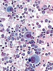
Image from UC San Diego
Scientists say they have developed a model that can be used to calculate the proportion of cancer stem cells (CSCs) present over the course of treatment.
The model is designed to enable estimation of CSC fractions from longitudinal measurements of tumor burden.
The scientists tested the model in patients with chronic myeloid leukemia (CML) and found evidence to suggest the proportion of CSCs increases
substantially during extended treatment.
The team believes the model could eventually be used to help doctors predict tumor development and help them select suitable treatments for cancer patients.
“Cancer stem cells not only promote the growth of a tumor, they can also be resistant to radiotherapy and chemotherapy,” said Philipp Altrock, PhD, of the Dana Farber Cancer Institute in Boston, Massachusetts.
“If we can estimate the number of cancer stem cells at diagnosis and over the course of treatment, the treatment can be tailored accordingly.”
Dr Altrock and his colleagues discussed this possibility in Cancer Research.
The team first explained that their model incorporates tumor dynamics and tumor burden information. They said tumor expansion and regression curves can be leveraged to estimate the proportion of CSCs in individual patients at baseline and during therapy.
To test their model, the scientists used 2 independent cohorts of CML patients. The team evaluated the growth and decline of CML over the course of treatment with the tyrosine kinase inhibitor imatinib.
Based on the change of disease burden during treatment, the model calculated the proportion of CSCs.
Results suggested the proportion of CSCs in CML patients increases 100-fold after a year of treatment with imatinib. And that proportion continues to increase up to 1000-fold after 5 years of treatment.
The scientists noted that this model is parameter-free, so it can be applied to different types of cancer. However, they said further development is required before the model can be used in clinical practice. ![]()
Drug granted breakthrough designation for cHL

Photo courtesy of Merck
The US Food and Drug Administration (FDA) has granted breakthrough therapy designation for pembrolizumab (Keytruda) to treat patients with relapsed or refractory classical Hodgkin lymphoma (cHL).
Pembrolizumab is a monoclonal antibody that binds to the PD-1 receptor and blocks its interaction with PD-L1 and PD-L2, releasing PD-1 pathway-mediated inhibition of the immune response, including the antitumor immune response.
The FDA’s breakthrough therapy designation is intended to expedite the development and review of new therapies for serious or life threatening conditions, which have shown encouraging early clinical results demonstrating substantial improvement on a clinically significant endpoint over available therapies.
The breakthrough designation for pembrolizumab in cHL is based on data from the phase 1b KEYNOTE-013 trial and the phase 2 KEYNOTE-087 trial.
Findings from the KEYNOTE-013 study were presented at ASH 2014 (in patients with cHL) and ASH 2015 (in primary mediastinal large B-cell lymphoma).
Data from KEYNOTE-087 will be presented at an upcoming medical meeting, according to Merck, the company developing pembrolizumab.
Pembrolizumab also has breakthrough designation from the FDA as a treatment for advanced melanoma, advanced non-small cell lung cancer, and advanced colorectal cancer.
The drug is already FDA-approved to treat melanoma and non-small cell lung cancer. Pembrolizumab is administered at a dose of 2 mg/kg as an intravenous infusion over 30 minutes every 3 weeks for the approved indications. ![]()
Hybrid drug could treat resistant malaria

Photo by James Gathany
A newly developed hybrid drug can treat malaria that is resistant to other therapies, according to preclinical research.
With previous work, researchers found that chemoreversal agents can re-sensitize resistant malaria parasites to the antimalarial agent chloroquine.
For the current study, the team created hybrid compounds that combine chloroquine and a chemoreversal agent.
One of these compounds, 35, proved particularly active, killing malaria parasites that were resistant to chloroquine and/or artemisinin.
Compound 35 was significantly more effective than chloroquine at killing these resistant strains, which included Hb3 (P<0.001), Dd2 (P<0.001), ARS-233 (P<0.001), ARS-272 (P<0.01), NHP-04559 (P<0.01), NHP04773 (P<0.001), and 7G8 (P<0.01).
In addition, the researchers said compound 35 has a “good therapeutic window” and “favorable drug-like properties,” but they are continuing to refine the compound to make it more effective.
The team noted that malaria drugs and chemoreversal agents have been used to treat drug-resistant malaria before. But this is the first time a hybrid of chloroquine and a newly discovered chemoreversal factor has been used in a single novel molecule for this purpose.
The researchers said a single therapy has several advantages over combination therapy. Besides being more convenient to take, it has less risk of drug-drug interactions, may be better absorbed and distributed in the body, and could result in slower development of new resistant strains of malaria.
Kevin S. W. Tan, PhD, of the National University of Singapore, and his colleagues described this research in Antimicrobial Agents and Chemotherapy. ![]()
Patients may be uninformed about risks of warfarin

ATHENS—A study of patients taking warfarin suggests many do not fully understand the risks associated with the drug.
Researchers asked patients to complete a questionnaire on warfarin use and found that, on average, patients answered 64% of the questions correctly.
The patients tended to be the least informed about food and drug interactions and which side effects necessitate a call or visit to the doctor.
Kjersti Oterhals, RN, PhD, of Haukeland University Hospital in Bergen, Norway, and her colleagues presented these findings at EuroHeartCare 2016 (abstract 36).
“The stroke and bleeding complications from warfarin can be fatal,” Dr Oterhals noted. “Worldwide, warfarin causes the most deaths from drug-related side effects. Patients need to know what foods and drugs have an impact on how warfarin works and what to do if they have symptoms of an overdose or underdose.”
Dr Oterhals and her colleagues evaluated warfarin knowledge in 404 patients with aortic stenosis. The patients’ mean age was 68, and 70% were male.
Nearly two-thirds of the patients (63%) were taking warfarin because they had a mechanical valve, and 24% were taking the drug because they had atrial fibrillation. The remaining patients were taking the drug for unknown reasons (6%) or “other” reasons (7%).
The patients received a postal questionnaire with 28 multiple-choice questions about warfarin. On average, patients answered 18 of the 28 questions correctly. However, 22% of patients answered less than half of the questions correctly.
The questions that were most often answered incorrectly were those concerning food and drug interactions and when to call or see a doctor.
For example, patients were asked which of the following foods would interfere with warfarin: celery, carrots, coleslaw, or green beans. Only 25% correctly said coleslaw. Most patients answered green beans.
“Patients often think green vegetables have the most vitamin K, but that’s not true,” Dr Oterhals said. “Brassica vegetables such as cabbage, broccoli, and cauliflower are rich sources.”
“Patients do not have to avoid these foods, but they should eat an equal amount every week because the vitamin K will decrease their INR and put them at increased risk of thrombosis or embolism. Patients who like to eat a lot of vitamin K-containing foods can take a higher warfarin dosage, but they need to be consistent.”
Dr Oterhals and her colleagues also found that 80% of patients knew they should go directly to the emergency room if they had nose bleeding that would not stop. However, only 45% of patients correctly said diarrhea for more than one day necessitates a visit to the doctor.
The study also showed that increased age was associated with a decrease in correct answers.
“We can only speculate why,” Dr Oterhals said. “Younger people tend to seek out information about how to manage their disease, while the older generation wants the doctor to tell them what to do.”
“Motivated patients should be offered an INR testing kit so that they can monitor their levels and adjust the warfarin dose themselves, just as patients with diabetes who use insulin do. It enables patients to travel and try new foods without having to find a clinic to get tested. Patients tell me that hot weather increases their INR, while another found out while in Asia that nori decreased his INR.”
“Warfarin is a life-saving drug but can be deadly if not used carefully. Health professionals have a responsibility to educate patients, but, unfortunately, even cardiac nurses do not know enough. There is an urgent need to improve health professionals’ warfarin knowledge so they can educate patients.” ![]()
ATHENS—A study of patients taking warfarin suggests many do not fully understand the risks associated with the drug.
Researchers asked patients to complete a questionnaire on warfarin use and found that, on average, patients answered 64% of the questions correctly.
The patients tended to be the least informed about food and drug interactions and which side effects necessitate a call or visit to the doctor.
Kjersti Oterhals, RN, PhD, of Haukeland University Hospital in Bergen, Norway, and her colleagues presented these findings at EuroHeartCare 2016 (abstract 36).
“The stroke and bleeding complications from warfarin can be fatal,” Dr Oterhals noted. “Worldwide, warfarin causes the most deaths from drug-related side effects. Patients need to know what foods and drugs have an impact on how warfarin works and what to do if they have symptoms of an overdose or underdose.”
Dr Oterhals and her colleagues evaluated warfarin knowledge in 404 patients with aortic stenosis. The patients’ mean age was 68, and 70% were male.
Nearly two-thirds of the patients (63%) were taking warfarin because they had a mechanical valve, and 24% were taking the drug because they had atrial fibrillation. The remaining patients were taking the drug for unknown reasons (6%) or “other” reasons (7%).
The patients received a postal questionnaire with 28 multiple-choice questions about warfarin. On average, patients answered 18 of the 28 questions correctly. However, 22% of patients answered less than half of the questions correctly.
The questions that were most often answered incorrectly were those concerning food and drug interactions and when to call or see a doctor.
For example, patients were asked which of the following foods would interfere with warfarin: celery, carrots, coleslaw, or green beans. Only 25% correctly said coleslaw. Most patients answered green beans.
“Patients often think green vegetables have the most vitamin K, but that’s not true,” Dr Oterhals said. “Brassica vegetables such as cabbage, broccoli, and cauliflower are rich sources.”
“Patients do not have to avoid these foods, but they should eat an equal amount every week because the vitamin K will decrease their INR and put them at increased risk of thrombosis or embolism. Patients who like to eat a lot of vitamin K-containing foods can take a higher warfarin dosage, but they need to be consistent.”
Dr Oterhals and her colleagues also found that 80% of patients knew they should go directly to the emergency room if they had nose bleeding that would not stop. However, only 45% of patients correctly said diarrhea for more than one day necessitates a visit to the doctor.
The study also showed that increased age was associated with a decrease in correct answers.
“We can only speculate why,” Dr Oterhals said. “Younger people tend to seek out information about how to manage their disease, while the older generation wants the doctor to tell them what to do.”
“Motivated patients should be offered an INR testing kit so that they can monitor their levels and adjust the warfarin dose themselves, just as patients with diabetes who use insulin do. It enables patients to travel and try new foods without having to find a clinic to get tested. Patients tell me that hot weather increases their INR, while another found out while in Asia that nori decreased his INR.”
“Warfarin is a life-saving drug but can be deadly if not used carefully. Health professionals have a responsibility to educate patients, but, unfortunately, even cardiac nurses do not know enough. There is an urgent need to improve health professionals’ warfarin knowledge so they can educate patients.”
Patients with ASD may have lower cancer risk

Photo by Darren Baker
New research suggests that patients diagnosed with an autism spectrum disorder (ASD) have a higher burden of mutations in oncogenes but lower rates of cancer than the rest of the population.
Investigators analyzed large, publicly available genomic databases of patients with ASD and found that, compared to a set of control subjects, ASD patients had significantly higher rates of DNA variation in oncogenes.
The team followed up this result with an analysis of the University of Iowa Hospitals and Clinics’ electronic medical record and discovered that ASD patients were also significantly less likely to have a co-occurring diagnosis of cancer.
“It’s a very provocative result that makes sense on one level and is extremely perplexing on another,” said Benjamin Darbro, MD, PhD, of the University of Iowa Carver College of Medicine in Iowa City.
Dr Darbro and his colleagues discussed the result, and the research that led to it, in a paper published in PLOS ONE.
The investigators used exome sequencing data from the ARRA Autism Sequencing Collaboration and compared that data to a control cohort from the Exome Variant Server database.
This revealed that rare, coding variants within oncogenes were greatly enriched in the ARRA ASD cohort. By comparison, variants were not significantly enriched in tumor suppressor genes.
To ensure the genetic differences were not technical artifacts but actually bona fide differences in genetic architecture in ASD, the investigators ran numerous controls.
As expected, they found that individuals with ASD had many more DNA variations in genes previously associated with autism, epilepsy, and intellectual disability compared to control individuals.
However, there was no difference between the ASD and control groups when it came to genes involved in other, unrelated conditions such as skeletal dysplasia, retinitis pigmentosa, dilated cardiomyopathy, and non-syndromic hearing loss.
The investigators then turned their attention to the electronic medical record at the University of Iowa Hospitals and Clinics and conducted a retrospective case-control analysis comparing 1837 patients with ASD to 9336 patients with any other diagnosis, and determined what proportion of each group carried a cancer diagnosis.
The team found that, for children and adults with ASD, there appeared to be a protective effect against cancer. The cancer incidence was 1.3% for patients with ASD and 3.9% for controls.
The protective effect was evident in both males and females with ASD, but it was strongest for the youngest group of patients and decreased with age. For ASD patients who were under 14 years of age, the odds of having cancer were reduced by 94% compared to controls.
When the investigators determined the rates of other systemic diseases—such as high blood pressure and diabetes—in the ASD population, they found no relationship.
Furthermore, the team found no relationship with cancer when they examined the rates of other common conditions such as esophageal reflux, allergic rhinitis, atopic dermatitis, and short stature. They said this demonstrated that the inverse relationship observed between ASD and cancer is not due to a technical artifact.
Short transfusion delays can increase risk of death
![]()
Photo courtesy of UAB Hospital
Even a short delay in the administration of packed red blood cells (pRBCs) can increase the risk of death for some traumatically injured patients, according to research published in the Journal of Trauma and Acute Care Surgery.
The study showed that a delay of 10 minutes was associated with a higher risk of death among patients who required pRBCs early.
However, a 10-minute delay did not increase the risk of death for the entire study cohort.
For this study, researchers tracked trauma patients taken from the scene of their injury to the University of Cincinnati Medical Center by a helicopter service known as Air Care. The service carries 2 units of pRBCs for protocol-driven prehospital transfusion.
“Air Care is the only helicopter in the area to carry blood (and plasma), so we had the research platform to study how early blood transfusions impact outcomes,” said study author Elizabeth Powell, MD, of the University of Cincinnati Medical Center in Ohio.
Dr Powell and her colleagues studied 94 patients who had received at least 1 unit of pRBCs within 24 hours of arriving at the hospital. Ninety-three percent of patients (n=87) were Caucasian, 70% (n=66) were male, and they had a mean age of 43.
Ninety-four percent of patients (n=88) had sustained blunt force injuries, and 33% (n=31) died within 30 days of hospital arrival.
Thirty-three percent of patients (n=31) received a transfusion during transport, 54% (n=51) were transfused within an hour of hospital arrival, and 13% (n=12) were transfused after the first hour but within 24 hours of hospital arrival.
When considering all 94 patients together, the researchers found that a 10-minute increase in time to pRBC administration did not significantly affect the odds of death, even when adjusting for injury severity. The odds ratio was 1.00 (P=0.575).
However, among the 82 patients who received their first pRBC transfusion during transport or within an hour of hospital arrival, each 10 minute increase in time to transfusion increased the odds of death. When the researchers controlled for Trauma Injury Severity Score, the odds ratio was 1.27 (P=0.044).
“Delays in the time to blood transfusion are associated with increased chances of dying,” Dr Powell said. “Shortening the time to transfusion, including having blood available in the prehospital setting, may improve outcomes.”
Antimalarial resistance can’t be passed on, team says

in mosquito gut
Image by Antoine Nicot
and Jacques Denoyelle
Parasites that develop resistance to the antimalarial drug atovaquone cannot pass this resistance on to their offspring, a new study suggests.
Researchers found that malaria parasites develop resistance to atovaquone via mutations in the mitochondrial cytochrome b complex.
However, these mutations also prevent female parasites from reproducing, so the resistance cannot be passed on to future generations.
Geoff McFadden, PhD, of the University of Melbourne in Victoria, Australia, and his colleagues reported these findings in Science.
“These results are very exciting because the spread of drug resistance is currently destroying our ability to control malaria,” Dr McFadden said.
“We now understand the particular genetic mutation that gave rise to drug resistance in some malaria parasite populations and how it eventually kills them in the mosquito, providing new targets for the development of drugs. So the development of drug resistance may not be a major problem if the resistance cannot spread, meaning the drug atovaquone could be more widely used in malaria control.”
To conduct this study, Dr McFadden and his colleagues analyzed 3 atovaquone-resistant strains of Plasmodium berghei, a malaria parasite that infects rodents. Each strain contained a different mutation in cytochrome b.
The researchers found that 2 of the mutations resulted in developmental defects in the parasite zygotes, and the third mutation resulted in complete infertility in the parasites due to severely impaired female germ cells.
Cross breeding parasites with and without these mutations showed that the mutations are not passed on to offspring. From 44 separate transmission attempts involving 750 mosquito bites, transmission of atovaquone resistance was only observed once, and this mutant was unable to transmit further, despite 7 attempts.
The researchers said it appears that atovaquone-resistant mutations severely impair the lifecycle of the parasites when they are living in mosquito hosts, so these mutations cannot be passed on.
In the human malaria parasite Plasmodium falciparum, the researchers identified similar mutations that impaired the ability of the parasites to infect mosquitos, as well as the number of oocysts produced when infection did occur.