User login
AVAHO
div[contains(@class, 'header__large-screen')]
div[contains(@class, 'read-next-article')]
div[contains(@class, 'nav-primary')]
nav[contains(@class, 'nav-primary')]
section[contains(@class, 'footer-nav-section-wrapper')]
footer[@id='footer']
div[contains(@class, 'main-prefix')]
section[contains(@class, 'nav-hidden')]
div[contains(@class, 'ce-card-content')]
nav[contains(@class, 'nav-ce-stack')]

Survival Rates of Black and White Veterans With Metastatic Castration-Resistant Prostate Cancer
Rationale: Understand the survival outcomes of black and white Veterans with metastatic castration-resistant prostate cancer (mCRPC).
Background: Black men have a higher incidence of prostate cancer and are more likely to be diagnosed at an earlier age, have a more aggressive disease at presentation and experience worse clinical outcome than white men. Evidence has shown that at diagnosis race is associated with disease progression, however, few studies have examined whether race is associated with survival in men once they reach an advanced stage of the disease. This study examines the survival outcomes among black and white men with mCRPC treated in the Veterans Health Administration (VHA).
Methods: Patient information from the Veterans Affairs (VA) Central Cancer Registry and the VA Corporate Data Warehouse was used to identify patients who were diagnosed with prostate cancer and later developed mCRPC, defined as: (1) Radiologic evidence of metastasis obtained from radiology reports using a natural language processing algorithm; (2) Evidence of rising prostate-specific antigen (PSA); and (3) Evidence of ongoing androgen deprivation consisting of a serum testosterone level of 50 ng/dL. Patient demographics, disease characteristics and treatment practices, and survival outcomes were extracted.
Results: From 2006 to 2015, 120,374 patients were diagnosed and treated with prostate cancer in the VHA; with 3,637 developing mCRPC. At diagnosis 2,429 (67%) were white, 1,066 (29%) were black, and 142 (4%) were reported as other. Compared to white men, black men were younger (66 vs 69 years) and had a higher PSA (92 vs 41 ng/mL), although there were no differences in disease characteristics
(Gleason score and stage) and early treatments (radiation prostatectomy, surgical orchiectomy, castration by agonists, castration by agonists/androgen deprivation). There were no significant differences between black and white men with mCRPC and their overall survival on both univariable (HR, .95; P = .203) or multivariable (HR, 1.0; P = .971) analyses.
Conclusions: Consistent with prior reports, black men are more likely to develop mCRPC than white men, although once black men progress to advanced disease, a multivariable analysis suggests that race is not associated with overall survival.
Rationale: Understand the survival outcomes of black and white Veterans with metastatic castration-resistant prostate cancer (mCRPC).
Background: Black men have a higher incidence of prostate cancer and are more likely to be diagnosed at an earlier age, have a more aggressive disease at presentation and experience worse clinical outcome than white men. Evidence has shown that at diagnosis race is associated with disease progression, however, few studies have examined whether race is associated with survival in men once they reach an advanced stage of the disease. This study examines the survival outcomes among black and white men with mCRPC treated in the Veterans Health Administration (VHA).
Methods: Patient information from the Veterans Affairs (VA) Central Cancer Registry and the VA Corporate Data Warehouse was used to identify patients who were diagnosed with prostate cancer and later developed mCRPC, defined as: (1) Radiologic evidence of metastasis obtained from radiology reports using a natural language processing algorithm; (2) Evidence of rising prostate-specific antigen (PSA); and (3) Evidence of ongoing androgen deprivation consisting of a serum testosterone level of 50 ng/dL. Patient demographics, disease characteristics and treatment practices, and survival outcomes were extracted.
Results: From 2006 to 2015, 120,374 patients were diagnosed and treated with prostate cancer in the VHA; with 3,637 developing mCRPC. At diagnosis 2,429 (67%) were white, 1,066 (29%) were black, and 142 (4%) were reported as other. Compared to white men, black men were younger (66 vs 69 years) and had a higher PSA (92 vs 41 ng/mL), although there were no differences in disease characteristics
(Gleason score and stage) and early treatments (radiation prostatectomy, surgical orchiectomy, castration by agonists, castration by agonists/androgen deprivation). There were no significant differences between black and white men with mCRPC and their overall survival on both univariable (HR, .95; P = .203) or multivariable (HR, 1.0; P = .971) analyses.
Conclusions: Consistent with prior reports, black men are more likely to develop mCRPC than white men, although once black men progress to advanced disease, a multivariable analysis suggests that race is not associated with overall survival.
Rationale: Understand the survival outcomes of black and white Veterans with metastatic castration-resistant prostate cancer (mCRPC).
Background: Black men have a higher incidence of prostate cancer and are more likely to be diagnosed at an earlier age, have a more aggressive disease at presentation and experience worse clinical outcome than white men. Evidence has shown that at diagnosis race is associated with disease progression, however, few studies have examined whether race is associated with survival in men once they reach an advanced stage of the disease. This study examines the survival outcomes among black and white men with mCRPC treated in the Veterans Health Administration (VHA).
Methods: Patient information from the Veterans Affairs (VA) Central Cancer Registry and the VA Corporate Data Warehouse was used to identify patients who were diagnosed with prostate cancer and later developed mCRPC, defined as: (1) Radiologic evidence of metastasis obtained from radiology reports using a natural language processing algorithm; (2) Evidence of rising prostate-specific antigen (PSA); and (3) Evidence of ongoing androgen deprivation consisting of a serum testosterone level of 50 ng/dL. Patient demographics, disease characteristics and treatment practices, and survival outcomes were extracted.
Results: From 2006 to 2015, 120,374 patients were diagnosed and treated with prostate cancer in the VHA; with 3,637 developing mCRPC. At diagnosis 2,429 (67%) were white, 1,066 (29%) were black, and 142 (4%) were reported as other. Compared to white men, black men were younger (66 vs 69 years) and had a higher PSA (92 vs 41 ng/mL), although there were no differences in disease characteristics
(Gleason score and stage) and early treatments (radiation prostatectomy, surgical orchiectomy, castration by agonists, castration by agonists/androgen deprivation). There were no significant differences between black and white men with mCRPC and their overall survival on both univariable (HR, .95; P = .203) or multivariable (HR, 1.0; P = .971) analyses.
Conclusions: Consistent with prior reports, black men are more likely to develop mCRPC than white men, although once black men progress to advanced disease, a multivariable analysis suggests that race is not associated with overall survival.
Screening Cancer Survivors for Bone Density Loss: An Analysis of Prevalence and Risk Factors in the Veteran Population
Purpose: Describe the prevalence of bone density loss and risk factors for osteoporosis in Veterans seen in a Cancer Survivorship Clinic. Assess the value of identified risk factors in predicting abnormal bone density.
Background: Cancer survivors are at risk for bone density loss secondary to side effects of cancer treatment. The prevalence of osteoporosis is not well defined in this patient population and guidelines regarding screening for osteoporosis, particularly in men, are sparse.
Methods: Veterans seen in the VA Puget Sound Cancer Survivorship Clinic are screened routinely for risk factors for bone density loss as identified in medical literature including medications, treatment sequelae and other factors. Demographics, medical history and DEXA scan results were abstracted from CPRS.
Results: There were 226 Cancer Survivorship Clinic visits between 2/27/15 and 5/18/18. The average age was 64.7 (range 28-87 years) and 91% were male. The most common cancer site was lung (28%), followed by head and neck (18%), prostate (15%), and colorectal (11%).
Thirty-five (15%) Veterans had a prior DEXA scan and 21 (9%) had a diagnosis of osteopenia or osteoporosis. Risk factors for bone density loss were identified for 77 Veterans (34%) and DEXA scans were recommended. DEXA scans ordered by the survivorship nurse practitioner had a higher
rate of completion than when recommended to the primary care provider, 23 of 36 (64%) were completed versus 7 of 42 (17%).
Among the 29 Veterans for whom a DEXA was completed, 13 (43%) had normal bone density, 14 (47%) had osteopenia, and 3 (10%) had osteoporosis.
Risk factors most strongly associated with a finding of decreased bone density were low body weight (100%), chemotherapy (67%), and radiographic osteopenia (67%). Height loss (50%) and hypogonadism (50%) were also associated with abnormal findings.
Implications: At least 17% of Veterans seen in Cancer Survivorship Clinic had abnormal bone density. This number likely is an underestimation as DEXA scans were not completed for half of those with risk factors for osteoporosis. Using specific chemotherapy exposures, radiographic osteopenia, hypogonadism, steroids and self-reported height loss as risk factors appears to be reasonable. Ordering the DEXA scan as part of the survivorship visit may increase compliance.
Purpose: Describe the prevalence of bone density loss and risk factors for osteoporosis in Veterans seen in a Cancer Survivorship Clinic. Assess the value of identified risk factors in predicting abnormal bone density.
Background: Cancer survivors are at risk for bone density loss secondary to side effects of cancer treatment. The prevalence of osteoporosis is not well defined in this patient population and guidelines regarding screening for osteoporosis, particularly in men, are sparse.
Methods: Veterans seen in the VA Puget Sound Cancer Survivorship Clinic are screened routinely for risk factors for bone density loss as identified in medical literature including medications, treatment sequelae and other factors. Demographics, medical history and DEXA scan results were abstracted from CPRS.
Results: There were 226 Cancer Survivorship Clinic visits between 2/27/15 and 5/18/18. The average age was 64.7 (range 28-87 years) and 91% were male. The most common cancer site was lung (28%), followed by head and neck (18%), prostate (15%), and colorectal (11%).
Thirty-five (15%) Veterans had a prior DEXA scan and 21 (9%) had a diagnosis of osteopenia or osteoporosis. Risk factors for bone density loss were identified for 77 Veterans (34%) and DEXA scans were recommended. DEXA scans ordered by the survivorship nurse practitioner had a higher
rate of completion than when recommended to the primary care provider, 23 of 36 (64%) were completed versus 7 of 42 (17%).
Among the 29 Veterans for whom a DEXA was completed, 13 (43%) had normal bone density, 14 (47%) had osteopenia, and 3 (10%) had osteoporosis.
Risk factors most strongly associated with a finding of decreased bone density were low body weight (100%), chemotherapy (67%), and radiographic osteopenia (67%). Height loss (50%) and hypogonadism (50%) were also associated with abnormal findings.
Implications: At least 17% of Veterans seen in Cancer Survivorship Clinic had abnormal bone density. This number likely is an underestimation as DEXA scans were not completed for half of those with risk factors for osteoporosis. Using specific chemotherapy exposures, radiographic osteopenia, hypogonadism, steroids and self-reported height loss as risk factors appears to be reasonable. Ordering the DEXA scan as part of the survivorship visit may increase compliance.
Purpose: Describe the prevalence of bone density loss and risk factors for osteoporosis in Veterans seen in a Cancer Survivorship Clinic. Assess the value of identified risk factors in predicting abnormal bone density.
Background: Cancer survivors are at risk for bone density loss secondary to side effects of cancer treatment. The prevalence of osteoporosis is not well defined in this patient population and guidelines regarding screening for osteoporosis, particularly in men, are sparse.
Methods: Veterans seen in the VA Puget Sound Cancer Survivorship Clinic are screened routinely for risk factors for bone density loss as identified in medical literature including medications, treatment sequelae and other factors. Demographics, medical history and DEXA scan results were abstracted from CPRS.
Results: There were 226 Cancer Survivorship Clinic visits between 2/27/15 and 5/18/18. The average age was 64.7 (range 28-87 years) and 91% were male. The most common cancer site was lung (28%), followed by head and neck (18%), prostate (15%), and colorectal (11%).
Thirty-five (15%) Veterans had a prior DEXA scan and 21 (9%) had a diagnosis of osteopenia or osteoporosis. Risk factors for bone density loss were identified for 77 Veterans (34%) and DEXA scans were recommended. DEXA scans ordered by the survivorship nurse practitioner had a higher
rate of completion than when recommended to the primary care provider, 23 of 36 (64%) were completed versus 7 of 42 (17%).
Among the 29 Veterans for whom a DEXA was completed, 13 (43%) had normal bone density, 14 (47%) had osteopenia, and 3 (10%) had osteoporosis.
Risk factors most strongly associated with a finding of decreased bone density were low body weight (100%), chemotherapy (67%), and radiographic osteopenia (67%). Height loss (50%) and hypogonadism (50%) were also associated with abnormal findings.
Implications: At least 17% of Veterans seen in Cancer Survivorship Clinic had abnormal bone density. This number likely is an underestimation as DEXA scans were not completed for half of those with risk factors for osteoporosis. Using specific chemotherapy exposures, radiographic osteopenia, hypogonadism, steroids and self-reported height loss as risk factors appears to be reasonable. Ordering the DEXA scan as part of the survivorship visit may increase compliance.
Concurrent Capecitabine and Radiation to Treat End Stage Renal Disease Patients on Dialysis With Locally Advanced Unresectable Gastro-Intestinal Malignancies: A Veteran Population Experience
Background: Capecitabine is an oral precursor of 5-FU (5' deoxy-5-fluoridine), a commonly prescribed chemotherapeutic agent to treat gastrointestinal and breast cancers. Capecitabine is currently contraindicated in patients with severe renal failure with Glomerular filtration rate <
30 ml/min. Literature review shows limited evidence in safety and effectiveness of using capecitabine in patients undergoing hemodialysis.
Case Report 1: A 75-year-old old male with a history of end stage renal disease on hemodialysis, was diagnosed with a 10 cm duodenal mass on CT scan when presented with three months history of abdominal pain and 45 lb weight loss. esophagogastroduodenoscopy and biopsy confirmed
adenocarcinoma of duodenum/ampulla. Patient was deemed to be a high-risk candidate for Whipple’s procedure. The case was discussed in multidisciplinary tumor board and the patient was offered concurrent chemotherapy and radiation with capecitabine 500 mg BID. Posttreatment
CT scans suggested 60% shrinkage in tumor size. Patient was continued on capecitabine 300 mg BID two weeks on and one week off with continued response noted on restaging CT scans.
Case Report 2: A 76-year-old male with end stage renal disease on hemodialysis complained of bleeding per rectum for over 2 years. A colonoscopy showed a circumferential mass at 15 cm from anal verge, and biopsy was consistent with rectal adeno carcinoma. PET/CT scan confirmed primary lesion in rectum as well as abnormal retroperitoneal and left iliac adenopathy with high FDG uptake. EUS staged disease at uT3N0Mx. Given significant pain and bleeding patient was offered palliative radiation along with low dose capecitabine 500 mg BID. Two months after concurrent chemotherapy and radiation, restaging scans showed 50% shrinkage in primary tumor. The patient opted to continue treatment with capecitabine and completed two more cycles of 300 mg BID two weeks on and one week off. A repeat CT scan showed near complete resolution of rectal mass and lymphadenopathy.
Conclusions: Capecitabine is converted to active form 5-FU in liver by thymidine phosphorylase. Over 95% of the drug is excreted in urine. In the original phase II trial utilizing capecitabine at 1250 mg/m2 BID, four patients with GFR < 30 ml/min noted to have grade 3-4 toxicities. Jhaveri et al. in their retrospective analysis showed 12 patients tolerated reduced doses.
Background: Capecitabine is an oral precursor of 5-FU (5' deoxy-5-fluoridine), a commonly prescribed chemotherapeutic agent to treat gastrointestinal and breast cancers. Capecitabine is currently contraindicated in patients with severe renal failure with Glomerular filtration rate <
30 ml/min. Literature review shows limited evidence in safety and effectiveness of using capecitabine in patients undergoing hemodialysis.
Case Report 1: A 75-year-old old male with a history of end stage renal disease on hemodialysis, was diagnosed with a 10 cm duodenal mass on CT scan when presented with three months history of abdominal pain and 45 lb weight loss. esophagogastroduodenoscopy and biopsy confirmed
adenocarcinoma of duodenum/ampulla. Patient was deemed to be a high-risk candidate for Whipple’s procedure. The case was discussed in multidisciplinary tumor board and the patient was offered concurrent chemotherapy and radiation with capecitabine 500 mg BID. Posttreatment
CT scans suggested 60% shrinkage in tumor size. Patient was continued on capecitabine 300 mg BID two weeks on and one week off with continued response noted on restaging CT scans.
Case Report 2: A 76-year-old male with end stage renal disease on hemodialysis complained of bleeding per rectum for over 2 years. A colonoscopy showed a circumferential mass at 15 cm from anal verge, and biopsy was consistent with rectal adeno carcinoma. PET/CT scan confirmed primary lesion in rectum as well as abnormal retroperitoneal and left iliac adenopathy with high FDG uptake. EUS staged disease at uT3N0Mx. Given significant pain and bleeding patient was offered palliative radiation along with low dose capecitabine 500 mg BID. Two months after concurrent chemotherapy and radiation, restaging scans showed 50% shrinkage in primary tumor. The patient opted to continue treatment with capecitabine and completed two more cycles of 300 mg BID two weeks on and one week off. A repeat CT scan showed near complete resolution of rectal mass and lymphadenopathy.
Conclusions: Capecitabine is converted to active form 5-FU in liver by thymidine phosphorylase. Over 95% of the drug is excreted in urine. In the original phase II trial utilizing capecitabine at 1250 mg/m2 BID, four patients with GFR < 30 ml/min noted to have grade 3-4 toxicities. Jhaveri et al. in their retrospective analysis showed 12 patients tolerated reduced doses.
Background: Capecitabine is an oral precursor of 5-FU (5' deoxy-5-fluoridine), a commonly prescribed chemotherapeutic agent to treat gastrointestinal and breast cancers. Capecitabine is currently contraindicated in patients with severe renal failure with Glomerular filtration rate <
30 ml/min. Literature review shows limited evidence in safety and effectiveness of using capecitabine in patients undergoing hemodialysis.
Case Report 1: A 75-year-old old male with a history of end stage renal disease on hemodialysis, was diagnosed with a 10 cm duodenal mass on CT scan when presented with three months history of abdominal pain and 45 lb weight loss. esophagogastroduodenoscopy and biopsy confirmed
adenocarcinoma of duodenum/ampulla. Patient was deemed to be a high-risk candidate for Whipple’s procedure. The case was discussed in multidisciplinary tumor board and the patient was offered concurrent chemotherapy and radiation with capecitabine 500 mg BID. Posttreatment
CT scans suggested 60% shrinkage in tumor size. Patient was continued on capecitabine 300 mg BID two weeks on and one week off with continued response noted on restaging CT scans.
Case Report 2: A 76-year-old male with end stage renal disease on hemodialysis complained of bleeding per rectum for over 2 years. A colonoscopy showed a circumferential mass at 15 cm from anal verge, and biopsy was consistent with rectal adeno carcinoma. PET/CT scan confirmed primary lesion in rectum as well as abnormal retroperitoneal and left iliac adenopathy with high FDG uptake. EUS staged disease at uT3N0Mx. Given significant pain and bleeding patient was offered palliative radiation along with low dose capecitabine 500 mg BID. Two months after concurrent chemotherapy and radiation, restaging scans showed 50% shrinkage in primary tumor. The patient opted to continue treatment with capecitabine and completed two more cycles of 300 mg BID two weeks on and one week off. A repeat CT scan showed near complete resolution of rectal mass and lymphadenopathy.
Conclusions: Capecitabine is converted to active form 5-FU in liver by thymidine phosphorylase. Over 95% of the drug is excreted in urine. In the original phase II trial utilizing capecitabine at 1250 mg/m2 BID, four patients with GFR < 30 ml/min noted to have grade 3-4 toxicities. Jhaveri et al. in their retrospective analysis showed 12 patients tolerated reduced doses.
Association of Prostate-Specific Antigen (PSA) Trajectories With Mortality in Veterans With Non-Metastatic Castration-Resistant Prostate Cancer (nmCRPC)
Background: There are nearly 40,000 patients with nmCRPC in the US. This study aims to describe the characteristics of Veterans with nmCRPC and assess the impact of PSA trajectory, baseline PSA and development of metastases on OS.
Methods: Veterans Health Administration electronic health record data (01/2007-08/2017) were used to conduct a retrospective longitudinal study in Veterans with nmCRPC. Patients with castrate testosterone levels ( < 50 ng/dL) or continuous androgen deprivation therapy, a 25% rise in PSA from nadir and absolute increase of 2 ng/mL (index), and a PC diagnosis 12 months before index were selected. Patients were excluded if they had < 12 months of enrollment before or < 6 months after the index date, evidence of metastatic disease 12 months before index (defined based on diagnoses for metastatic disease, use of chemotherapy, immunotherapy, radiopharmaceuticals, or oral mCRPC therapy), or evidence of participation in a clinical trial (defined based on diagnosis codes or prescription for experimental therapies). PSA was measured in the 12 months before index, at index, and 6 months after index. Patients with similar PSA trajectories (PSA patterns over time) were grouped using group-based trajectory analysis (GBTA). OS was assessed with a Cox proportional hazards model adjusted for baseline characteristics, PSA trajectory group, and a timevarying indicator for metastases.
Results: 13,552 Veterans formed the study population. Mean (SD) age was 76.4 (9.4) years and most were white (66%). During follow up (mean [SD]=36 [26] months) there were 6,552 (48%) deaths; median OS was 47 months. GBTA resulted in 4 PSA trajectory groups. The Cox model showed that PSA trajectory group was a strong predictor of OS; compared to Group 1 (n = 213), patients in the other groups were more likely to die (Group 2 [n = 6,558]: HR=1.602, P < .001; Group 3 [n = 5,323]: HR = 1.716, P < .001; Group 4 [n = 1,458]: HR = 2.005, P < .001). Higher log PSA at baseline increased risk of death (HR = 1.092, P < .001). Moreover, compared to patients without metastases, patients who eventually developed metastases were more likely to die (HR: 3.661, P < .001).
Conclusions: In this study of Veterans with nmCRPC, higher baseline PSA, PSA trajectory group, and development of metastases were associated with increased risk of death.
Background: There are nearly 40,000 patients with nmCRPC in the US. This study aims to describe the characteristics of Veterans with nmCRPC and assess the impact of PSA trajectory, baseline PSA and development of metastases on OS.
Methods: Veterans Health Administration electronic health record data (01/2007-08/2017) were used to conduct a retrospective longitudinal study in Veterans with nmCRPC. Patients with castrate testosterone levels ( < 50 ng/dL) or continuous androgen deprivation therapy, a 25% rise in PSA from nadir and absolute increase of 2 ng/mL (index), and a PC diagnosis 12 months before index were selected. Patients were excluded if they had < 12 months of enrollment before or < 6 months after the index date, evidence of metastatic disease 12 months before index (defined based on diagnoses for metastatic disease, use of chemotherapy, immunotherapy, radiopharmaceuticals, or oral mCRPC therapy), or evidence of participation in a clinical trial (defined based on diagnosis codes or prescription for experimental therapies). PSA was measured in the 12 months before index, at index, and 6 months after index. Patients with similar PSA trajectories (PSA patterns over time) were grouped using group-based trajectory analysis (GBTA). OS was assessed with a Cox proportional hazards model adjusted for baseline characteristics, PSA trajectory group, and a timevarying indicator for metastases.
Results: 13,552 Veterans formed the study population. Mean (SD) age was 76.4 (9.4) years and most were white (66%). During follow up (mean [SD]=36 [26] months) there were 6,552 (48%) deaths; median OS was 47 months. GBTA resulted in 4 PSA trajectory groups. The Cox model showed that PSA trajectory group was a strong predictor of OS; compared to Group 1 (n = 213), patients in the other groups were more likely to die (Group 2 [n = 6,558]: HR=1.602, P < .001; Group 3 [n = 5,323]: HR = 1.716, P < .001; Group 4 [n = 1,458]: HR = 2.005, P < .001). Higher log PSA at baseline increased risk of death (HR = 1.092, P < .001). Moreover, compared to patients without metastases, patients who eventually developed metastases were more likely to die (HR: 3.661, P < .001).
Conclusions: In this study of Veterans with nmCRPC, higher baseline PSA, PSA trajectory group, and development of metastases were associated with increased risk of death.
Background: There are nearly 40,000 patients with nmCRPC in the US. This study aims to describe the characteristics of Veterans with nmCRPC and assess the impact of PSA trajectory, baseline PSA and development of metastases on OS.
Methods: Veterans Health Administration electronic health record data (01/2007-08/2017) were used to conduct a retrospective longitudinal study in Veterans with nmCRPC. Patients with castrate testosterone levels ( < 50 ng/dL) or continuous androgen deprivation therapy, a 25% rise in PSA from nadir and absolute increase of 2 ng/mL (index), and a PC diagnosis 12 months before index were selected. Patients were excluded if they had < 12 months of enrollment before or < 6 months after the index date, evidence of metastatic disease 12 months before index (defined based on diagnoses for metastatic disease, use of chemotherapy, immunotherapy, radiopharmaceuticals, or oral mCRPC therapy), or evidence of participation in a clinical trial (defined based on diagnosis codes or prescription for experimental therapies). PSA was measured in the 12 months before index, at index, and 6 months after index. Patients with similar PSA trajectories (PSA patterns over time) were grouped using group-based trajectory analysis (GBTA). OS was assessed with a Cox proportional hazards model adjusted for baseline characteristics, PSA trajectory group, and a timevarying indicator for metastases.
Results: 13,552 Veterans formed the study population. Mean (SD) age was 76.4 (9.4) years and most were white (66%). During follow up (mean [SD]=36 [26] months) there were 6,552 (48%) deaths; median OS was 47 months. GBTA resulted in 4 PSA trajectory groups. The Cox model showed that PSA trajectory group was a strong predictor of OS; compared to Group 1 (n = 213), patients in the other groups were more likely to die (Group 2 [n = 6,558]: HR=1.602, P < .001; Group 3 [n = 5,323]: HR = 1.716, P < .001; Group 4 [n = 1,458]: HR = 2.005, P < .001). Higher log PSA at baseline increased risk of death (HR = 1.092, P < .001). Moreover, compared to patients without metastases, patients who eventually developed metastases were more likely to die (HR: 3.661, P < .001).
Conclusions: In this study of Veterans with nmCRPC, higher baseline PSA, PSA trajectory group, and development of metastases were associated with increased risk of death.
Aromatherapy Evidence-Based Project On Inpatient Cancer/Palliative Care Unit
Purpose: An evidenced-based project to introduce aromatherapy with the use of essential oils for oncology/palliative care patients to improve symptoms associated with cancer treatment and end-of-life.
Background: Aromatherapy reduces the cancer patients’ complications, such as sleep disorders, nausea, vomiting, pain, anxiety, and depression (Keyhanmehr et al, 2018). Lavender, peppermint, and orange are common essential oils that can help support patients with cancer who experience
insomnia, nausea, and anxiety (Reis and Jones, 2017). Palliative care research indicates that more conventional interventions, such as medications, do not always equate to a higher quality of life but often decrease the quality of life due to unnecessary interventions that cause unwanted side effects (Marchand, 2014). It is important to give patients as many choices as possible to treat their symptoms without creating new problems (Marchand, 2014).
Methods: A six-month trial was conducted on the inpatient cancer/palliative care unit and 104 patients filled out a comment card. During the trial, lavender, peppermint, frankincense, lemon, orange, and eucalyptus were used. Patients were educated on the benefits of aromatherapy and were offered samples of their choosing. Patients were given the choice of inhalation or the use of a diffuser.
Results: Of the 104 patients that filled out a comment card 97 stated they would use it again, and 51% reported an increase in well-being, 15.8% improved sleep, 10.5% reduced anxiety, 8.3% reduced depression, 7.5% improved congestion, 3.8% improved pain, and 3% reduced nausea. After the conclusion of the trial, aromatherapy is a permanent intervention for cancer and palliative care patients. The program also is being utilized in the outpatient oncology clinic, infusion clinic, and inpatient rehabilitation unit, and will eventually be offered to all departments providing patient care.
Conclusions: Aromatherapy is a viable intervention for improving various symptoms and is used effectively with patients with cancer primarily as supportive care (Aromatherapy and Essential Oils [PDQ®]: Health Professional Version, 2018). Oncology nurses can provide aromatherapy safely, inexpensively, and with minimal training, as an effective therapy in lessening many symptoms that cancer and end of life patients experience (Blackburn et al, 2017).
Purpose: An evidenced-based project to introduce aromatherapy with the use of essential oils for oncology/palliative care patients to improve symptoms associated with cancer treatment and end-of-life.
Background: Aromatherapy reduces the cancer patients’ complications, such as sleep disorders, nausea, vomiting, pain, anxiety, and depression (Keyhanmehr et al, 2018). Lavender, peppermint, and orange are common essential oils that can help support patients with cancer who experience
insomnia, nausea, and anxiety (Reis and Jones, 2017). Palliative care research indicates that more conventional interventions, such as medications, do not always equate to a higher quality of life but often decrease the quality of life due to unnecessary interventions that cause unwanted side effects (Marchand, 2014). It is important to give patients as many choices as possible to treat their symptoms without creating new problems (Marchand, 2014).
Methods: A six-month trial was conducted on the inpatient cancer/palliative care unit and 104 patients filled out a comment card. During the trial, lavender, peppermint, frankincense, lemon, orange, and eucalyptus were used. Patients were educated on the benefits of aromatherapy and were offered samples of their choosing. Patients were given the choice of inhalation or the use of a diffuser.
Results: Of the 104 patients that filled out a comment card 97 stated they would use it again, and 51% reported an increase in well-being, 15.8% improved sleep, 10.5% reduced anxiety, 8.3% reduced depression, 7.5% improved congestion, 3.8% improved pain, and 3% reduced nausea. After the conclusion of the trial, aromatherapy is a permanent intervention for cancer and palliative care patients. The program also is being utilized in the outpatient oncology clinic, infusion clinic, and inpatient rehabilitation unit, and will eventually be offered to all departments providing patient care.
Conclusions: Aromatherapy is a viable intervention for improving various symptoms and is used effectively with patients with cancer primarily as supportive care (Aromatherapy and Essential Oils [PDQ®]: Health Professional Version, 2018). Oncology nurses can provide aromatherapy safely, inexpensively, and with minimal training, as an effective therapy in lessening many symptoms that cancer and end of life patients experience (Blackburn et al, 2017).
Purpose: An evidenced-based project to introduce aromatherapy with the use of essential oils for oncology/palliative care patients to improve symptoms associated with cancer treatment and end-of-life.
Background: Aromatherapy reduces the cancer patients’ complications, such as sleep disorders, nausea, vomiting, pain, anxiety, and depression (Keyhanmehr et al, 2018). Lavender, peppermint, and orange are common essential oils that can help support patients with cancer who experience
insomnia, nausea, and anxiety (Reis and Jones, 2017). Palliative care research indicates that more conventional interventions, such as medications, do not always equate to a higher quality of life but often decrease the quality of life due to unnecessary interventions that cause unwanted side effects (Marchand, 2014). It is important to give patients as many choices as possible to treat their symptoms without creating new problems (Marchand, 2014).
Methods: A six-month trial was conducted on the inpatient cancer/palliative care unit and 104 patients filled out a comment card. During the trial, lavender, peppermint, frankincense, lemon, orange, and eucalyptus were used. Patients were educated on the benefits of aromatherapy and were offered samples of their choosing. Patients were given the choice of inhalation or the use of a diffuser.
Results: Of the 104 patients that filled out a comment card 97 stated they would use it again, and 51% reported an increase in well-being, 15.8% improved sleep, 10.5% reduced anxiety, 8.3% reduced depression, 7.5% improved congestion, 3.8% improved pain, and 3% reduced nausea. After the conclusion of the trial, aromatherapy is a permanent intervention for cancer and palliative care patients. The program also is being utilized in the outpatient oncology clinic, infusion clinic, and inpatient rehabilitation unit, and will eventually be offered to all departments providing patient care.
Conclusions: Aromatherapy is a viable intervention for improving various symptoms and is used effectively with patients with cancer primarily as supportive care (Aromatherapy and Essential Oils [PDQ®]: Health Professional Version, 2018). Oncology nurses can provide aromatherapy safely, inexpensively, and with minimal training, as an effective therapy in lessening many symptoms that cancer and end of life patients experience (Blackburn et al, 2017).
Implementation of Chemo and Anti-Neoplastic Extractor Cycle and Regimen Estimator (Cancer Care) in Follicular Lymphoma Patients Treated in VA
Rationale: Successfully extract, identify, and validate therapeutics lines used in the treatment of Veterans with follicular lymphoma.
Background: With the adoption of electronic health record (EHR) systems, leveraging this data is becoming increasingly important for clinical and observational research, especially in oncology, where precision oncology has become central to the Cancer Moonshot Initiative. One of the greatest challenges in using EHR data is extracting a cancer patient’s treatment history. The difficulty lies in identifying treatment “lines,” which may include one or more drugs, with each drug dispensation often recorded in an unstructured format within the EHR. Our objective was to conceptualize, develop, and validate an algorithm that reconstructs a cancer treatment line history using single-agent EHR pharmacy data in a cohort of follicular lymphoma patients treated in the Veterans Health Administration (VHA).
Methods: The CANCER CARE algorithm recreates and formalizes the heuristic a clinician uses to identify treatment lines dispensed using two inputs: (1) National Comprehensive Cancer Network treatment guidelines and the recommended chemotherapy lines and their comprising
antineoplastic agents that are used in the treatment of the cancer of interest; and (2) Single-agent dispensation information retrieved from the VA Corporate Data Warehouse. The algorithm uses rules to map concordant dispensation agents to a treatment line while taking into account common
practice variations such as omitted agents during the start or middle of a treatment line. It also identifies the initiation of a new line based on a change in agents received or time gaps between treatments. The algorithm was validated by comparing a set of 100 treatment lines that were independently annotated by a clinician in a cohort of patients with follicular lymphoma to the algorithm output. Accuracy, sensitivity, and precision were measured.
Results: CANCER CARE had an accuracy of 96%. Accuracy, sensitivity and precision for most prevalent lines were: 98%, 97% and 100% (rituximab), respectively; and 99%, 100%, and 95% (RCHOP), respectively. Accuracy, sensitivity, and precision for RCVP and BR were all 100%.
Conclusions: Cancer treatment line identification from EHR pharmacy dispensation data using a rule-based approach is feasible with high accuracy and can be used in real-world studies of cancer patient treatment practices and outcomes.
Rationale: Successfully extract, identify, and validate therapeutics lines used in the treatment of Veterans with follicular lymphoma.
Background: With the adoption of electronic health record (EHR) systems, leveraging this data is becoming increasingly important for clinical and observational research, especially in oncology, where precision oncology has become central to the Cancer Moonshot Initiative. One of the greatest challenges in using EHR data is extracting a cancer patient’s treatment history. The difficulty lies in identifying treatment “lines,” which may include one or more drugs, with each drug dispensation often recorded in an unstructured format within the EHR. Our objective was to conceptualize, develop, and validate an algorithm that reconstructs a cancer treatment line history using single-agent EHR pharmacy data in a cohort of follicular lymphoma patients treated in the Veterans Health Administration (VHA).
Methods: The CANCER CARE algorithm recreates and formalizes the heuristic a clinician uses to identify treatment lines dispensed using two inputs: (1) National Comprehensive Cancer Network treatment guidelines and the recommended chemotherapy lines and their comprising
antineoplastic agents that are used in the treatment of the cancer of interest; and (2) Single-agent dispensation information retrieved from the VA Corporate Data Warehouse. The algorithm uses rules to map concordant dispensation agents to a treatment line while taking into account common
practice variations such as omitted agents during the start or middle of a treatment line. It also identifies the initiation of a new line based on a change in agents received or time gaps between treatments. The algorithm was validated by comparing a set of 100 treatment lines that were independently annotated by a clinician in a cohort of patients with follicular lymphoma to the algorithm output. Accuracy, sensitivity, and precision were measured.
Results: CANCER CARE had an accuracy of 96%. Accuracy, sensitivity and precision for most prevalent lines were: 98%, 97% and 100% (rituximab), respectively; and 99%, 100%, and 95% (RCHOP), respectively. Accuracy, sensitivity, and precision for RCVP and BR were all 100%.
Conclusions: Cancer treatment line identification from EHR pharmacy dispensation data using a rule-based approach is feasible with high accuracy and can be used in real-world studies of cancer patient treatment practices and outcomes.
Rationale: Successfully extract, identify, and validate therapeutics lines used in the treatment of Veterans with follicular lymphoma.
Background: With the adoption of electronic health record (EHR) systems, leveraging this data is becoming increasingly important for clinical and observational research, especially in oncology, where precision oncology has become central to the Cancer Moonshot Initiative. One of the greatest challenges in using EHR data is extracting a cancer patient’s treatment history. The difficulty lies in identifying treatment “lines,” which may include one or more drugs, with each drug dispensation often recorded in an unstructured format within the EHR. Our objective was to conceptualize, develop, and validate an algorithm that reconstructs a cancer treatment line history using single-agent EHR pharmacy data in a cohort of follicular lymphoma patients treated in the Veterans Health Administration (VHA).
Methods: The CANCER CARE algorithm recreates and formalizes the heuristic a clinician uses to identify treatment lines dispensed using two inputs: (1) National Comprehensive Cancer Network treatment guidelines and the recommended chemotherapy lines and their comprising
antineoplastic agents that are used in the treatment of the cancer of interest; and (2) Single-agent dispensation information retrieved from the VA Corporate Data Warehouse. The algorithm uses rules to map concordant dispensation agents to a treatment line while taking into account common
practice variations such as omitted agents during the start or middle of a treatment line. It also identifies the initiation of a new line based on a change in agents received or time gaps between treatments. The algorithm was validated by comparing a set of 100 treatment lines that were independently annotated by a clinician in a cohort of patients with follicular lymphoma to the algorithm output. Accuracy, sensitivity, and precision were measured.
Results: CANCER CARE had an accuracy of 96%. Accuracy, sensitivity and precision for most prevalent lines were: 98%, 97% and 100% (rituximab), respectively; and 99%, 100%, and 95% (RCHOP), respectively. Accuracy, sensitivity, and precision for RCVP and BR were all 100%.
Conclusions: Cancer treatment line identification from EHR pharmacy dispensation data using a rule-based approach is feasible with high accuracy and can be used in real-world studies of cancer patient treatment practices and outcomes.
Trends in Cancer Incidence and Survival in the Veterans Health Administration
Background: Cancer diagnoses in the Veterans Affairs (VA) Health Care System (HCS) account for approximately 3% of all US cancer diagnoses each year. Certain cancer types disproportionately affect veterans. Many factors contribute to changes in cancer incidence and survival among veterans, including screening guidelines and practices, treatment advances, as well as changing demographics of the veteran population and VA HCS users.
Purpose: The specific objectives of this analysis were to evaluate trends in cancer incidence and 5-year overall and cancer-specific survival among veterans.
Methods: We conducted a retrospective analysis of patients diagnosed with 15 select cancers between 2002 and 2014 that were identified in the VA Central Cancer Registry. Age-adjusted incidence rates were calculated based on the US 2000 population estimates and VHA user population. 5-year survival was calculated using the Kaplan-Meier method.
Results: Of the 15 selected cancers, overall decreases in incidence were noted for the following cancers: bladder, brain, colorectal, esophageal, head & neck, leukemia, lung, lymphoma, melanoma, and prostate. Most pronounced changes were observed for colorectal, lung, and prostate cancers. Relatively small net increases in incidence were observed for breast, kidney, liver, myeloma, and pancreas cancers. Among these 15 select cancers, the highest 5-year overall survival (OS) rates were observed for melanoma, prostate, and breast cancers (all > 70%), whereas the lowest OS rates were noted for pancreas, brain, esophagus, lung, and liver cancers (all 20%). Between 2002-2014, OS rates improved for all cancers except for the following that remained relatively stable: brain (11%), leukemia (47%), and melanoma (72%). OS rates improved the most for head & neck cancer (37% to 47%) and myeloma (32% to 40%).
Conclusions: For the 15 cancers evaluated in this report among veterans, between 2002-2014 most cancer incidence rates have decreased and survival rates for most cancers have improved over time.
Background: Cancer diagnoses in the Veterans Affairs (VA) Health Care System (HCS) account for approximately 3% of all US cancer diagnoses each year. Certain cancer types disproportionately affect veterans. Many factors contribute to changes in cancer incidence and survival among veterans, including screening guidelines and practices, treatment advances, as well as changing demographics of the veteran population and VA HCS users.
Purpose: The specific objectives of this analysis were to evaluate trends in cancer incidence and 5-year overall and cancer-specific survival among veterans.
Methods: We conducted a retrospective analysis of patients diagnosed with 15 select cancers between 2002 and 2014 that were identified in the VA Central Cancer Registry. Age-adjusted incidence rates were calculated based on the US 2000 population estimates and VHA user population. 5-year survival was calculated using the Kaplan-Meier method.
Results: Of the 15 selected cancers, overall decreases in incidence were noted for the following cancers: bladder, brain, colorectal, esophageal, head & neck, leukemia, lung, lymphoma, melanoma, and prostate. Most pronounced changes were observed for colorectal, lung, and prostate cancers. Relatively small net increases in incidence were observed for breast, kidney, liver, myeloma, and pancreas cancers. Among these 15 select cancers, the highest 5-year overall survival (OS) rates were observed for melanoma, prostate, and breast cancers (all > 70%), whereas the lowest OS rates were noted for pancreas, brain, esophagus, lung, and liver cancers (all 20%). Between 2002-2014, OS rates improved for all cancers except for the following that remained relatively stable: brain (11%), leukemia (47%), and melanoma (72%). OS rates improved the most for head & neck cancer (37% to 47%) and myeloma (32% to 40%).
Conclusions: For the 15 cancers evaluated in this report among veterans, between 2002-2014 most cancer incidence rates have decreased and survival rates for most cancers have improved over time.
Background: Cancer diagnoses in the Veterans Affairs (VA) Health Care System (HCS) account for approximately 3% of all US cancer diagnoses each year. Certain cancer types disproportionately affect veterans. Many factors contribute to changes in cancer incidence and survival among veterans, including screening guidelines and practices, treatment advances, as well as changing demographics of the veteran population and VA HCS users.
Purpose: The specific objectives of this analysis were to evaluate trends in cancer incidence and 5-year overall and cancer-specific survival among veterans.
Methods: We conducted a retrospective analysis of patients diagnosed with 15 select cancers between 2002 and 2014 that were identified in the VA Central Cancer Registry. Age-adjusted incidence rates were calculated based on the US 2000 population estimates and VHA user population. 5-year survival was calculated using the Kaplan-Meier method.
Results: Of the 15 selected cancers, overall decreases in incidence were noted for the following cancers: bladder, brain, colorectal, esophageal, head & neck, leukemia, lung, lymphoma, melanoma, and prostate. Most pronounced changes were observed for colorectal, lung, and prostate cancers. Relatively small net increases in incidence were observed for breast, kidney, liver, myeloma, and pancreas cancers. Among these 15 select cancers, the highest 5-year overall survival (OS) rates were observed for melanoma, prostate, and breast cancers (all > 70%), whereas the lowest OS rates were noted for pancreas, brain, esophagus, lung, and liver cancers (all 20%). Between 2002-2014, OS rates improved for all cancers except for the following that remained relatively stable: brain (11%), leukemia (47%), and melanoma (72%). OS rates improved the most for head & neck cancer (37% to 47%) and myeloma (32% to 40%).
Conclusions: For the 15 cancers evaluated in this report among veterans, between 2002-2014 most cancer incidence rates have decreased and survival rates for most cancers have improved over time.
Standardization of the Discharge Process for Inpatient Hematology and Oncology
Purpose/Rationale: To standardize the discharge process for the hematology/oncology inpatient service at Hines VA Hospital to improve the transition of care
Background: The landmark 1999 report from the Institute of Medicine, To Err is Human, identified the impact of medical error on mortality and morbidity. Medical errors tend to occur during transitions of care. At Hines VA Hospital, a multidisciplinary team delivers specialized care to veterans on the hematology/oncology service. However, resident physicians staffing the inpatient hematology/oncology service may be unfamiliar with the unique needs of the service and population. Currently there is no standardized discharge process in place. Prior studies have demonstrated improved outcomes following standardization of the discharge process for hematology patients. The authors aim to develop and implement a standardized discharge process to minimize risk for medical error.
Method/Approach: A multidisciplinary team of hematology and oncology staff was formed, including attending physicians, fellows, residents, advanced practice nurses, registered nurses, clinical pharmacists, and patient care coordinators, and several interviews were conducted. A standardized discharge process was developed in the form of guidelines and expectations. These include an explanation of unique features of the hematology/oncology service and expectations of medication reconciliation with emphasis placed on antiemetics, antimicrobial prophylaxis, and bowel regimen when appropriate, ambulatory hematology/oncology follow up within 1-2 weeks, primary care followup, communication with ambulatory hematology/oncology physician, written discharge instructions, and bedside teaching when appropriate. The standardized process will be taught to rotating resident physicians in the form of both online orientation and an in-person orientation. Outcome measures were identified including key components of medication reconciliation, time to hematology & oncology clinic visit, time to primary care visit, communication of discharge with outpatient hematology/oncology physician, and 30-day readmission rate.
Conclusions: All patients discharged during the twomonth period prior to and all patients discharged after the implementation of the standardized process will be reviewed; the above-mentioned variables will be recorded. Outcomes will be compared. Interim multidisciplinary team focus group meetings will be held every quarter to review and refine the process.
Purpose/Rationale: To standardize the discharge process for the hematology/oncology inpatient service at Hines VA Hospital to improve the transition of care
Background: The landmark 1999 report from the Institute of Medicine, To Err is Human, identified the impact of medical error on mortality and morbidity. Medical errors tend to occur during transitions of care. At Hines VA Hospital, a multidisciplinary team delivers specialized care to veterans on the hematology/oncology service. However, resident physicians staffing the inpatient hematology/oncology service may be unfamiliar with the unique needs of the service and population. Currently there is no standardized discharge process in place. Prior studies have demonstrated improved outcomes following standardization of the discharge process for hematology patients. The authors aim to develop and implement a standardized discharge process to minimize risk for medical error.
Method/Approach: A multidisciplinary team of hematology and oncology staff was formed, including attending physicians, fellows, residents, advanced practice nurses, registered nurses, clinical pharmacists, and patient care coordinators, and several interviews were conducted. A standardized discharge process was developed in the form of guidelines and expectations. These include an explanation of unique features of the hematology/oncology service and expectations of medication reconciliation with emphasis placed on antiemetics, antimicrobial prophylaxis, and bowel regimen when appropriate, ambulatory hematology/oncology follow up within 1-2 weeks, primary care followup, communication with ambulatory hematology/oncology physician, written discharge instructions, and bedside teaching when appropriate. The standardized process will be taught to rotating resident physicians in the form of both online orientation and an in-person orientation. Outcome measures were identified including key components of medication reconciliation, time to hematology & oncology clinic visit, time to primary care visit, communication of discharge with outpatient hematology/oncology physician, and 30-day readmission rate.
Conclusions: All patients discharged during the twomonth period prior to and all patients discharged after the implementation of the standardized process will be reviewed; the above-mentioned variables will be recorded. Outcomes will be compared. Interim multidisciplinary team focus group meetings will be held every quarter to review and refine the process.
Purpose/Rationale: To standardize the discharge process for the hematology/oncology inpatient service at Hines VA Hospital to improve the transition of care
Background: The landmark 1999 report from the Institute of Medicine, To Err is Human, identified the impact of medical error on mortality and morbidity. Medical errors tend to occur during transitions of care. At Hines VA Hospital, a multidisciplinary team delivers specialized care to veterans on the hematology/oncology service. However, resident physicians staffing the inpatient hematology/oncology service may be unfamiliar with the unique needs of the service and population. Currently there is no standardized discharge process in place. Prior studies have demonstrated improved outcomes following standardization of the discharge process for hematology patients. The authors aim to develop and implement a standardized discharge process to minimize risk for medical error.
Method/Approach: A multidisciplinary team of hematology and oncology staff was formed, including attending physicians, fellows, residents, advanced practice nurses, registered nurses, clinical pharmacists, and patient care coordinators, and several interviews were conducted. A standardized discharge process was developed in the form of guidelines and expectations. These include an explanation of unique features of the hematology/oncology service and expectations of medication reconciliation with emphasis placed on antiemetics, antimicrobial prophylaxis, and bowel regimen when appropriate, ambulatory hematology/oncology follow up within 1-2 weeks, primary care followup, communication with ambulatory hematology/oncology physician, written discharge instructions, and bedside teaching when appropriate. The standardized process will be taught to rotating resident physicians in the form of both online orientation and an in-person orientation. Outcome measures were identified including key components of medication reconciliation, time to hematology & oncology clinic visit, time to primary care visit, communication of discharge with outpatient hematology/oncology physician, and 30-day readmission rate.
Conclusions: All patients discharged during the twomonth period prior to and all patients discharged after the implementation of the standardized process will be reviewed; the above-mentioned variables will be recorded. Outcomes will be compared. Interim multidisciplinary team focus group meetings will be held every quarter to review and refine the process.
Genomic Medicine Service Uses Group Telehealth Appointments to Reduce Wait Times From 5 Months To ~1 Week
Purpose/Rationale: Genomic Medicine Service (GMS) fields 100+ consults weekly. Due to an increase in the number of consults received, without an equal increase in staffing, the wait time for a non-urgent appointment approached 6 months. We explored the use of Group Telehealth
appointments (GTAs) for individuals referred for a family history of breast cancer as one way to reduce these wait times.
Background: While oncology specializes in those with cancer, they are often asked to see unaffected individuals with a family history of cancer who need risk assessments, management recommendations, and/or genetic testing. Many of these are then referred to GMS.
GMS uses the VA telehealth infrastructure to provide genetic evaluation to 84 VAMCs. We typically schedule appointments for one-hour, with an inability to double book due to the limitations of multi-site telehealth. As risk assessment for unaffected individuals is not urgent, these Veterans were scheduled routinely. As GMS got busier, wait times for routine appointments approached 6 months.
Methods/Approach: As part of a Leadership Development Institute, one of the authors (RAR) conceived and implemented a process whereby we held GTAs for unaffected individuals with family histories of breast cancer for whom we would most likely recommend testing an affected
relative. Before the GTA, we mailed a Breast Cancer Risk Assessment (BCRA) form to collect personal/family history. Patients who complete the GTA and BCRA were sent letters that included risk assessments and testing and screening recommendations. 4 GTAs are held each month.
We recorded the number of patients scheduled, appointments attended, and BCRA forms returned. The presentation will review results of an initial 3-month period, during which we held 14 GTAs with 97 patients scheduled, 65 seen, and 58 who turned in BCRAs. We compared time
spent on patients, documentation, and risk assessment in GTAs with the time needed for individual visits for the same number of people. We modeled time saved under a range of assumptions.
Conclusions: Our GTAs were successful, allowing our providers to more efficiently use their time and reducing our wait times. We have expanded our GTAs to include non-breast cancers and reasons for referral.
Purpose/Rationale: Genomic Medicine Service (GMS) fields 100+ consults weekly. Due to an increase in the number of consults received, without an equal increase in staffing, the wait time for a non-urgent appointment approached 6 months. We explored the use of Group Telehealth
appointments (GTAs) for individuals referred for a family history of breast cancer as one way to reduce these wait times.
Background: While oncology specializes in those with cancer, they are often asked to see unaffected individuals with a family history of cancer who need risk assessments, management recommendations, and/or genetic testing. Many of these are then referred to GMS.
GMS uses the VA telehealth infrastructure to provide genetic evaluation to 84 VAMCs. We typically schedule appointments for one-hour, with an inability to double book due to the limitations of multi-site telehealth. As risk assessment for unaffected individuals is not urgent, these Veterans were scheduled routinely. As GMS got busier, wait times for routine appointments approached 6 months.
Methods/Approach: As part of a Leadership Development Institute, one of the authors (RAR) conceived and implemented a process whereby we held GTAs for unaffected individuals with family histories of breast cancer for whom we would most likely recommend testing an affected
relative. Before the GTA, we mailed a Breast Cancer Risk Assessment (BCRA) form to collect personal/family history. Patients who complete the GTA and BCRA were sent letters that included risk assessments and testing and screening recommendations. 4 GTAs are held each month.
We recorded the number of patients scheduled, appointments attended, and BCRA forms returned. The presentation will review results of an initial 3-month period, during which we held 14 GTAs with 97 patients scheduled, 65 seen, and 58 who turned in BCRAs. We compared time
spent on patients, documentation, and risk assessment in GTAs with the time needed for individual visits for the same number of people. We modeled time saved under a range of assumptions.
Conclusions: Our GTAs were successful, allowing our providers to more efficiently use their time and reducing our wait times. We have expanded our GTAs to include non-breast cancers and reasons for referral.
Purpose/Rationale: Genomic Medicine Service (GMS) fields 100+ consults weekly. Due to an increase in the number of consults received, without an equal increase in staffing, the wait time for a non-urgent appointment approached 6 months. We explored the use of Group Telehealth
appointments (GTAs) for individuals referred for a family history of breast cancer as one way to reduce these wait times.
Background: While oncology specializes in those with cancer, they are often asked to see unaffected individuals with a family history of cancer who need risk assessments, management recommendations, and/or genetic testing. Many of these are then referred to GMS.
GMS uses the VA telehealth infrastructure to provide genetic evaluation to 84 VAMCs. We typically schedule appointments for one-hour, with an inability to double book due to the limitations of multi-site telehealth. As risk assessment for unaffected individuals is not urgent, these Veterans were scheduled routinely. As GMS got busier, wait times for routine appointments approached 6 months.
Methods/Approach: As part of a Leadership Development Institute, one of the authors (RAR) conceived and implemented a process whereby we held GTAs for unaffected individuals with family histories of breast cancer for whom we would most likely recommend testing an affected
relative. Before the GTA, we mailed a Breast Cancer Risk Assessment (BCRA) form to collect personal/family history. Patients who complete the GTA and BCRA were sent letters that included risk assessments and testing and screening recommendations. 4 GTAs are held each month.
We recorded the number of patients scheduled, appointments attended, and BCRA forms returned. The presentation will review results of an initial 3-month period, during which we held 14 GTAs with 97 patients scheduled, 65 seen, and 58 who turned in BCRAs. We compared time
spent on patients, documentation, and risk assessment in GTAs with the time needed for individual visits for the same number of people. We modeled time saved under a range of assumptions.
Conclusions: Our GTAs were successful, allowing our providers to more efficiently use their time and reducing our wait times. We have expanded our GTAs to include non-breast cancers and reasons for referral.
STORM trial shows response in penta-refractory myeloma
Treatment with selinexor and low-dose dexamethasone can provide a “meaningful clinical benefit” in patients with penta-refractory multiple myeloma, according to the principal investigator of the STORM trial.
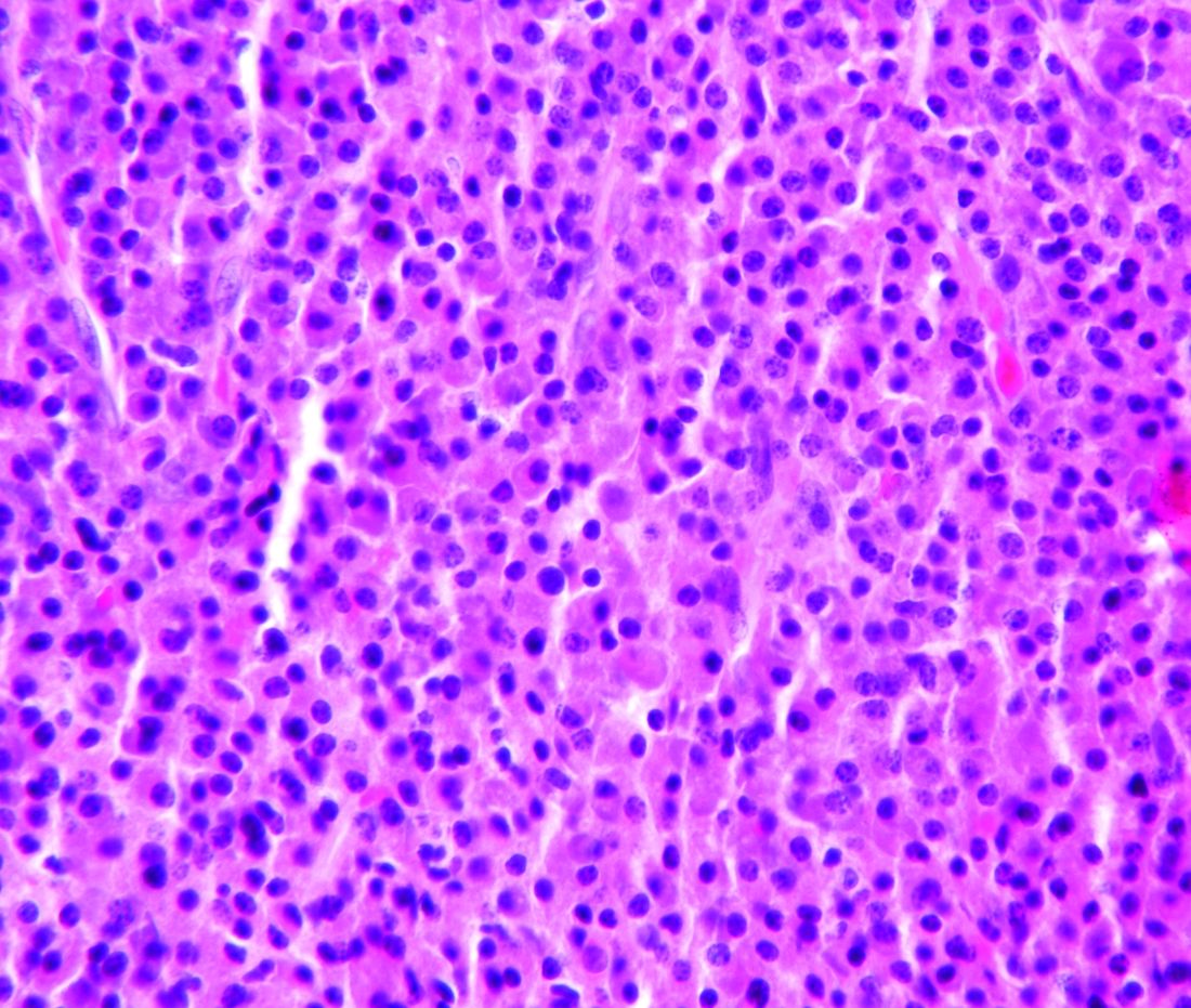
Updated results from this phase 2 trial showed that selinexor and low-dose dexamethasone produced an overall response rate of 26.2% and a clinical benefit rate of 39.3%. The median progression-free survival was 3.7 months and the median overall survival was 8.6 months.
The trial’s principal investigator, Sundar Jagannath, MBBS, of the Icahn School of Medicine at Mount Sinai, New York, presented these results at the annual meeting of the Society of Hematologic Oncology.
“The additional phase 2b clinical results… are very encouraging for the patients suffering from penta-refractory multiple myeloma and their families,” Dr. Jagannath said in a statement. “Of particular significance, for the nearly 40% of patients who had a minimal response or better, the median survival was 15.6 months, which provided the opportunity for a meaningful clinical benefit for patients on the STORM [Selinexor Treatment of Refractory Myeloma] study.”
STORM (NCT02336815) included 122 patients with penta-refractory multiple myeloma. They had previously received bortezomib, carfilzomib, lenalidomide, pomalidomide, daratumumab, alkylating agents, and glucocorticoids. Their disease was refractory to glucocorticoids, at least one proteasome inhibitor, at least one immunomodulatory drug, daratumumab, and their most recent therapy.
The patients had received a median of seven prior treatment regimens. Their median age was 65 years, a little more than half were men, and more than half had high-risk cytogenetics. Patients received oral selinexor at 80 mg twice weekly plus dexamethasone at 20 mg twice weekly until disease progression.Two patients (1.6%) achieved stringent complete responses. They also had minimal residual disease negativity, one at the level of 1 x 10⁻⁶ and one at 1 x 10⁻⁴.
Very good partial responses were seen in 4.9% of patients, 19.7% had partial responses, 13.1% had minimal responses (MRs), and 39.3% had stable disease. Progressive disease occurred in 13.1% of patients; 8.2% were not evaluable for response.
The overall response rate (partial response or better) was 26.2%, the clinical benefit rate (MR or better) was 39.3%, and the disease control rate (stable disease or better) was 78.7%.
The median duration of response was 4.4 months. The median progression-free survival was 3.7 months overall, 4.6 months in patients with an MR or better, and 1.1 months in patients who had progressive disease or were not evaluable.
The median overall survival was 8.6 months for the entire cohort. Overall survival was 15.6 months in patients with an MR or better and 1.7 months in patients who had progressive disease or were not evaluable (P less than .0001).
The “most important” grade 3/4 adverse events, according to Dr. Jagannath, were thrombocytopenia (53.7%), anemia (29.3%), fatigue (22.8%), hyponatremia (16.3%), nausea (9.8%), diarrhea (6.5%), anorexia (3.3%), and emesis (3.3%). A total of 23 patients (19.5%) discontinued treatment because of a related adverse.
This study was sponsored by Karyopharm Therapeutics. Dr. Jagannath reported relationships with Karyopharm, Janssen, Celgene, Amgen, and GlaxoSmithKline.
SOURCE: Jagannath S et al. SOHO 2018, Abstract MM-255
Treatment with selinexor and low-dose dexamethasone can provide a “meaningful clinical benefit” in patients with penta-refractory multiple myeloma, according to the principal investigator of the STORM trial.
Updated results from this phase 2 trial showed that selinexor and low-dose dexamethasone produced an overall response rate of 26.2% and a clinical benefit rate of 39.3%. The median progression-free survival was 3.7 months and the median overall survival was 8.6 months.
The trial’s principal investigator, Sundar Jagannath, MBBS, of the Icahn School of Medicine at Mount Sinai, New York, presented these results at the annual meeting of the Society of Hematologic Oncology.
“The additional phase 2b clinical results… are very encouraging for the patients suffering from penta-refractory multiple myeloma and their families,” Dr. Jagannath said in a statement. “Of particular significance, for the nearly 40% of patients who had a minimal response or better, the median survival was 15.6 months, which provided the opportunity for a meaningful clinical benefit for patients on the STORM [Selinexor Treatment of Refractory Myeloma] study.”
STORM (NCT02336815) included 122 patients with penta-refractory multiple myeloma. They had previously received bortezomib, carfilzomib, lenalidomide, pomalidomide, daratumumab, alkylating agents, and glucocorticoids. Their disease was refractory to glucocorticoids, at least one proteasome inhibitor, at least one immunomodulatory drug, daratumumab, and their most recent therapy.
The patients had received a median of seven prior treatment regimens. Their median age was 65 years, a little more than half were men, and more than half had high-risk cytogenetics. Patients received oral selinexor at 80 mg twice weekly plus dexamethasone at 20 mg twice weekly until disease progression.Two patients (1.6%) achieved stringent complete responses. They also had minimal residual disease negativity, one at the level of 1 x 10⁻⁶ and one at 1 x 10⁻⁴.
Very good partial responses were seen in 4.9% of patients, 19.7% had partial responses, 13.1% had minimal responses (MRs), and 39.3% had stable disease. Progressive disease occurred in 13.1% of patients; 8.2% were not evaluable for response.
The overall response rate (partial response or better) was 26.2%, the clinical benefit rate (MR or better) was 39.3%, and the disease control rate (stable disease or better) was 78.7%.
The median duration of response was 4.4 months. The median progression-free survival was 3.7 months overall, 4.6 months in patients with an MR or better, and 1.1 months in patients who had progressive disease or were not evaluable.
The median overall survival was 8.6 months for the entire cohort. Overall survival was 15.6 months in patients with an MR or better and 1.7 months in patients who had progressive disease or were not evaluable (P less than .0001).
The “most important” grade 3/4 adverse events, according to Dr. Jagannath, were thrombocytopenia (53.7%), anemia (29.3%), fatigue (22.8%), hyponatremia (16.3%), nausea (9.8%), diarrhea (6.5%), anorexia (3.3%), and emesis (3.3%). A total of 23 patients (19.5%) discontinued treatment because of a related adverse.
This study was sponsored by Karyopharm Therapeutics. Dr. Jagannath reported relationships with Karyopharm, Janssen, Celgene, Amgen, and GlaxoSmithKline.
SOURCE: Jagannath S et al. SOHO 2018, Abstract MM-255
Treatment with selinexor and low-dose dexamethasone can provide a “meaningful clinical benefit” in patients with penta-refractory multiple myeloma, according to the principal investigator of the STORM trial.
Updated results from this phase 2 trial showed that selinexor and low-dose dexamethasone produced an overall response rate of 26.2% and a clinical benefit rate of 39.3%. The median progression-free survival was 3.7 months and the median overall survival was 8.6 months.
The trial’s principal investigator, Sundar Jagannath, MBBS, of the Icahn School of Medicine at Mount Sinai, New York, presented these results at the annual meeting of the Society of Hematologic Oncology.
“The additional phase 2b clinical results… are very encouraging for the patients suffering from penta-refractory multiple myeloma and their families,” Dr. Jagannath said in a statement. “Of particular significance, for the nearly 40% of patients who had a minimal response or better, the median survival was 15.6 months, which provided the opportunity for a meaningful clinical benefit for patients on the STORM [Selinexor Treatment of Refractory Myeloma] study.”
STORM (NCT02336815) included 122 patients with penta-refractory multiple myeloma. They had previously received bortezomib, carfilzomib, lenalidomide, pomalidomide, daratumumab, alkylating agents, and glucocorticoids. Their disease was refractory to glucocorticoids, at least one proteasome inhibitor, at least one immunomodulatory drug, daratumumab, and their most recent therapy.
The patients had received a median of seven prior treatment regimens. Their median age was 65 years, a little more than half were men, and more than half had high-risk cytogenetics. Patients received oral selinexor at 80 mg twice weekly plus dexamethasone at 20 mg twice weekly until disease progression.Two patients (1.6%) achieved stringent complete responses. They also had minimal residual disease negativity, one at the level of 1 x 10⁻⁶ and one at 1 x 10⁻⁴.
Very good partial responses were seen in 4.9% of patients, 19.7% had partial responses, 13.1% had minimal responses (MRs), and 39.3% had stable disease. Progressive disease occurred in 13.1% of patients; 8.2% were not evaluable for response.
The overall response rate (partial response or better) was 26.2%, the clinical benefit rate (MR or better) was 39.3%, and the disease control rate (stable disease or better) was 78.7%.
The median duration of response was 4.4 months. The median progression-free survival was 3.7 months overall, 4.6 months in patients with an MR or better, and 1.1 months in patients who had progressive disease or were not evaluable.
The median overall survival was 8.6 months for the entire cohort. Overall survival was 15.6 months in patients with an MR or better and 1.7 months in patients who had progressive disease or were not evaluable (P less than .0001).
The “most important” grade 3/4 adverse events, according to Dr. Jagannath, were thrombocytopenia (53.7%), anemia (29.3%), fatigue (22.8%), hyponatremia (16.3%), nausea (9.8%), diarrhea (6.5%), anorexia (3.3%), and emesis (3.3%). A total of 23 patients (19.5%) discontinued treatment because of a related adverse.
This study was sponsored by Karyopharm Therapeutics. Dr. Jagannath reported relationships with Karyopharm, Janssen, Celgene, Amgen, and GlaxoSmithKline.
SOURCE: Jagannath S et al. SOHO 2018, Abstract MM-255
FROM SOHO 2018
Key clinical point:
Major finding: The overall response rate was 26.2% and the clinical benefit rate was 39.3%.
Study details: A phase 2 trial of 122 patients with penta-refractory multiple myeloma.
Disclosures: This study was sponsored by Karyopharm Therapeutics. Dr. Jagannath reported relationships with Karyopharm, Janssen, Celgene, Amgen, and GlaxoSmithKline.
Source: Jagannath S et al. SOHO 2018, Abstract MM-255.