Article Type
Changed
Fri, 12/01/2017 - 10:01
Display Headline
The Leser-Trélat sign

An 85-year-old woman presented with night sweats, dry cough, and an unintended 30-pound weight loss over the preceding 6 months. She also reported the sudden onset of “itchy moles” on her back.

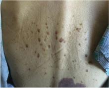
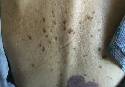
Multiple seborrheic keratoses on the patient’s upper back.
Figure 1. Multiple seborrheic keratoses on the patient’s upper back.
Physical examination revealed multiple brown papules with a “stuck-on” appearance on her upper back, consistent with seborrheic keratoses (Figure 1), raising concern for the Leser-Trélat sign.

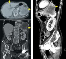
Computed tomography shows a left lung mass extending through the diaphragm and into the stomach with one metastatic lesion in the liver.
Figure 2. Computed tomography shows a left lung mass extending through the diaphragm and into the stomach with one metastatic lesion in the liver.
Computed tomography of the chest, abdomen, and pelvis revealed a left lower lobe lung mass extending through a diaphragmatic hernia and into the stomach and spleen and a metastatic lesion in the liver (Figure 2). A biopsy of the lung mass demonstrated squamous cell carcinoma of pulmonary origin.

KERATOSES AND MALIGNANCY

The Leser-Trélat sign is the sudden development of multiple pruritic seborrheic keratoses, often associated with malignancy.1–4 Roughly half of these associated malignancies are adenocarcinomas, most commonly of the stomach, breast, colon, or rectum. However, it can be seen in other malignancies, including lymphoma, leukemia, and squamous cell carcinoma, as in this case.

Eruption of seborrheic keratoses has also been observed with benign neoplasms, pregnancy, human immunodeficiency virus infections, and the use of adalimumab, which indicates that the Leser-Trélat sign is not very specific. Despite these concerns, the eruption of multiple seborrheic keratoses should continue to trigger the thought of an internal malignancy in the differential diagnosis.

References
  1. Ehst BD, Minzer-Conzetti K, Swerdlin A, Devere TS. Cutaneous manifestations of internal malignancy. Curr Probl Surg 2010; 47:384–445.
  2. Schwartz RA. Sign of Leser-Trélat. J Am Acad Dermatol 1996; 35:88–95.
  3. Ellis DL, Yates RA. Sign of Leser-Trélat. Clin Dermatol 1993; 11:141–148.
  4. Thiers BH, Sahn RE, Callen JP. Cutaneous manifestations of internal malignancy. CA Cancer J Clin 2009; 59:73–98.
Article PDF
Author and Disclosure Information

Christopher Sardon, MD, MPH
Flight Surgeon, Uniformed Services University of the Health Sciences, Tinker Air Force Base, OK

Timothy Dempsey MD, MPH
Department of Internal Medicine, University of California Davis, Sacramento, CA

Address: Christopher Sardon MD, MPH, Flight Surgeon, Tinker Air Force Base, 7050 Air Depot Blvd., Tinker AFB, OK 73145; sardonc@gmail.com

Issue
Cleveland Clinic Journal of Medicine - 84(12)
Publications
Topics
Page Number
918
Legacy Keywords
Leser-Trélat sign, lung cancer, moles, itchy, pruritic, keratosis, keratosis, computed tomography, Christopher Sardon, Timothy Dempsey
Sections
Author and Disclosure Information

Christopher Sardon, MD, MPH
Flight Surgeon, Uniformed Services University of the Health Sciences, Tinker Air Force Base, OK

Timothy Dempsey MD, MPH
Department of Internal Medicine, University of California Davis, Sacramento, CA

Address: Christopher Sardon MD, MPH, Flight Surgeon, Tinker Air Force Base, 7050 Air Depot Blvd., Tinker AFB, OK 73145; sardonc@gmail.com

Article PDF
Article PDF
Related Articles

An 85-year-old woman presented with night sweats, dry cough, and an unintended 30-pound weight loss over the preceding 6 months. She also reported the sudden onset of “itchy moles” on her back.

Multiple seborrheic keratoses on the patient’s upper back.
Figure 1. Multiple seborrheic keratoses on the patient’s upper back.
Physical examination revealed multiple brown papules with a “stuck-on” appearance on her upper back, consistent with seborrheic keratoses (Figure 1), raising concern for the Leser-Trélat sign.

Computed tomography shows a left lung mass extending through the diaphragm and into the stomach with one metastatic lesion in the liver.
Figure 2. Computed tomography shows a left lung mass extending through the diaphragm and into the stomach with one metastatic lesion in the liver.
Computed tomography of the chest, abdomen, and pelvis revealed a left lower lobe lung mass extending through a diaphragmatic hernia and into the stomach and spleen and a metastatic lesion in the liver (Figure 2). A biopsy of the lung mass demonstrated squamous cell carcinoma of pulmonary origin.

KERATOSES AND MALIGNANCY

The Leser-Trélat sign is the sudden development of multiple pruritic seborrheic keratoses, often associated with malignancy.1–4 Roughly half of these associated malignancies are adenocarcinomas, most commonly of the stomach, breast, colon, or rectum. However, it can be seen in other malignancies, including lymphoma, leukemia, and squamous cell carcinoma, as in this case.

Eruption of seborrheic keratoses has also been observed with benign neoplasms, pregnancy, human immunodeficiency virus infections, and the use of adalimumab, which indicates that the Leser-Trélat sign is not very specific. Despite these concerns, the eruption of multiple seborrheic keratoses should continue to trigger the thought of an internal malignancy in the differential diagnosis.

References
  1. Ehst BD, Minzer-Conzetti K, Swerdlin A, Devere TS. Cutaneous manifestations of internal malignancy. Curr Probl Surg 2010; 47:384–445.
  2. Schwartz RA. Sign of Leser-Trélat. J Am Acad Dermatol 1996; 35:88–95.
  3. Ellis DL, Yates RA. Sign of Leser-Trélat. Clin Dermatol 1993; 11:141–148.
  4. Thiers BH, Sahn RE, Callen JP. Cutaneous manifestations of internal malignancy. CA Cancer J Clin 2009; 59:73–98.
Issue
Cleveland Clinic Journal of Medicine - 84(12)
Issue
Cleveland Clinic Journal of Medicine - 84(12)
Page Number
918
Page Number
918
Publications
Publications
Topics
Article Type
Display Headline
The Leser-Trélat sign
Display Headline
The Leser-Trélat sign
Legacy Keywords
Leser-Trélat sign, lung cancer, moles, itchy, pruritic, keratosis, keratosis, computed tomography, Christopher Sardon, Timothy Dempsey
Legacy Keywords
Leser-Trélat sign, lung cancer, moles, itchy, pruritic, keratosis, keratosis, computed tomography, Christopher Sardon, Timothy Dempsey
Sections
Disallow All Ads
Content Gating
No Gating (article Unlocked/Free)
Alternative CME
Disqus Comments
Default
Article PDF Media